abrus agglutinin, a type ii ribosome inactivating protein
TRANSCRIPT
MOLECULAR CARCINOGENESIS
Abrus Agglutinin, a Type II Ribosome InactivatingProtein Inhibits Akt/PH Domain to Induce EndoplasmicReticulum Stress Mediated Autophagy-Dependent CellDeathPrashanta Kumar Panda,1 Birendra Behera,2 Biswa Ranjan Meher,3 Durgesh Nandini Das,1
Subhadip Mukhopadhyay,1 Niharika Sinha,1 Prajna Paramita Naik,1 Bibhas Roy,2 Joyjyoti Das,2
Subhankar Paul,4 Tapas K. Maiti,2 Rajesh Agarwal,5,6 and Sujit K. Bhutia1*1Department of Life Science, National Institute of Technology, Rourkela, Odisha, India2Department of Biotechnology, Indian Institute of Technology, Kharagpur, West Bengal, India3Department of Biochemistry, Indian Institute of Science, Bangalore, Karnataka, India4Department of Biotechnology and Medical Engineering, National Institute of Technology, Rourkela, Odisha, India5Department of Pharmaceutical Sciences, Skaggs School of Pharmacy and Pharmaceutical Sciences, Aurora, Colorado6University of Colorado Cancer Center, University of Colorado Denver, Aurora, Colorado
Abrus agglutinin (AGG), a type II ribosome-inactivating protein has been found to induce mitochondrial apoptosis. Inthe present study, we documented that AGG-mediated Akt dephosphorylation led to ER stress resulting the induction ofautophagy-dependent cell death through the canonical pathway in cervical cancer cells. Inhibition of autophagic deathwith 3-methyladenine (3-MA) and siRNA of Beclin-1 and ATG5 increased AGG-induced apoptosis. Further, inhibitingapoptosis by Z-DEVD-FMK and N-acetyl cysteine (NAC) increased autophagic cell death after AGG treatment, suggestingthat AGG simultaneously induced autophagic and apoptotic death in HeLa cells. Additionally, it observed thatAGG-induced autophagic cell death in Bax knock down (Bax-KD) and 5-FU resistant HeLa cells, confirming as an alternatecell killing pathway to apoptosis. At themolecular level, AGG-induced ER stress in PERK dependent pathway and inhibitionof ER stress by salubrinal, eIF2a phosphatase inhibitor as well as siPERK reduced autophagic death in the presence of AGG.Further, our in silico and colocalization study showed that AGG interactedwith pleckstrin homology (PH) domain of Akt tosuppress its phosphorylation and consequent downstream mTOR dephosphorylation in HeLa cells. We showed that Aktoverexpression could not augment GRP78 expression and reduced autophagic cell death by AGG as compared to pcDNAcontrol, indicating Akt modulation was the upstream signal during AGG's ER stress mediated autophagic cell death. Inconclusion,we established that AGG stimulated cell death by autophagymight be used as an alternative tumor suppressormechanism in human cervical cancer. © 2016 Wiley Periodicals, Inc.
Key words: Abrus agglutinin; autophagic cell death; apoptosis; ER stress; Akt; PH domain
INTRODUCTION
Autophagy is an evolutionary catabolic process inwhich stressed cells form cytoplasmic, double-layered,crescent-shaped membranes known as phagophores,which mature into complete autophagosomes. Theautophagosome engulfs damaged cytoplasmic organ-elles and long-lived proteins to provide cellular energyand building blocks for cellular biosynthesis [1,2]. Theautophagosome fuse with lysosome to form autolyso-someandcargoaredigestedby lysosomalhydrolases tometabolites and released back to the cytosol forrecycling. The autophagic process is regulated by theATG genes (autophagy-related genes) and the proteinsencoded by the autophagy related genes (ATG) arerequired for the regulation of autophagic vesicles.Initially, the autophagic pathway functions as anadaptive response to stress. However, in the face ofextreme or protracted stress, cells are committed toautophagic cell death; type II programmed cell death(PCD) [2,3].
Cervical cancer is the fourth leading cause of cancercell death and third most commonly diagnosed cancer
Abbreviations: AGG, Abrus agglutinin; ROS, reactive oxygenspecies; ER, endoplasmic reticulum; UPR, unfolded protein response;CHOP, C/EBP homologous protein; GRP94, glucose-regulated protein94; eIF2a, eukaryotic initiation factor 2 subunit a; ERAD, ER-associated ubiquitin/proteasome degradation; MD, molecular dy-namics; PDB, protein data bank; MM-PB/GBSA, molecular mechanicsPoisson–Boltzmann/generalized born surface area.Grant sponsor: Council of Scientific and Industrial Research (CSIR);
Grant number: 37(1608)/13/EMR-II; Grant sponsor: Department ofBiotechnology; Grant number: BT/PR1/5090/GBD/27/309/2011;Grant sponsor: Science and Engineering Research Board (SERB);Grant number: SR/SO/BB-0101/2012; Grant sponsor: NCI R01, UnitedStates; Grant number: CA195708*Correspondence to: Department of Life Science, National Institute
of Technology, Rourkela 769008, Odisha, India.Received 9 November 2015; Revised 26 April 2016; Accepted 13
May 2016DOI 10.1002/mc.22502Published online in Wiley Online Library
(wileyonlinelibrary.com).
� 2016 WILEY PERIODICALS, INC.
in women worldwide. Of these, nearly 85% of casesoccur in developing countries including India, andother parts of Asia due to inadequate access to screeningservices and lack of human papillomavirus (HPV)vaccination [4,5]. In the era of cancer therapy, apoptosisinduction in tumor cells is increasingly seen as primecandidates for the development of anticancer therapeu-tics for cervical cancer. However, development ofresistance phenomena to apoptosis and ineffectivenessof single treatment modality allow cancer cells tosurvive, consequently escape current cancer therapy.Therefore, novel therapeutic strategies are needed toenhance the effect of cancer therapy as well as addressthe emerging problem of drug resistance. Induction ofautophagic death, a type II programmed cell death,could be a potentially useful therapeutic approach inapoptosis resistant cancer cells and could complementas a multiple treatment approaches along with apopto-sis in apoptosis undergoing cancer cells [1,6]. Moreoverbecause cancer cells often display defective apoptoticpropensity, autophagy is considered a tumor suppressormechanism. As an alternative therapy for cancer,recently more efforts are made for the development ofnovelmolecules that specifically targets the autophagiccell death mechanism [7–9].
Abrus agglutinin (AGG) is one such prime candidatewhose autophagic attributes are being documented inthis work. AGG isolated from the seeds of A.precatorius is a hetero-tetrameric glycoprotein of134-kDa molecular weight, composed of two A andtwo B chains linked through disulfide bridges. AGGhas specificity towards [gal(b1!3)galNAc] and bel-ongs to type II ribosome inactivating protein family(RIP II) with a protein synthesis inhibitory concentra-tion (IC50) of 0.469mg/ml and a lethal dose (LD50)5mg/kg body weight in mice [10,11]. AGG containscytotoxic A chain having ribosomal RNA N-glycosidase activity which cleaves glycosidic bondat position A-4324 within the universally conserveda-sarcin loop of the 28S ribosomal RNA of eukaryoteswhile the B chain binds to carbohydrate moieties onthe cell surface and facilitates the internalization ofthe entire toxin into the cell [12]. Our groups havepreviously elucidated the anticancer effects of AGG inseveral tumor models at sublethal doses by directkilling of tumor cells through extrinsic and intrinsicapoptosis [13–15]. Along with direct antitumorpotential, AGGgenerates potent humoral and cellularimmune responses in normal as well as tumor-bearinganimals [16–18]. The adjuvant property of AGG isreported in oil emulsion and aqueous solution forpotentiating the systemic immune response [19].AGG activates splenocytes and induces production ofTh1 type of immune response. Further, AGG stim-ulates the innate effector arms like macrophage andnatural killer cells. Furthermore, heat denatured andtryptic digested AGG show potent antitumor as wellas immunomodulatory activity in normal as well astumor bearing mice [11,17,20,21].
Although the apoptotic potential of AGG has beenextensively investigated and well characterized, itsability to induce autophagy-dependent cell death inmammalian cells has not been documented. In thisreport, the study was designed to decipher the roleof AGG in autophagic death and discuss the possiblerole of autophagic death in relation with apoptosisin HeLa cells. Further, we examined that AGGinhibited Akt/PH domain to induce endoplasmicreticulum stress mediated autophagy-dependent celldeath.
MATERIALS AND METHODS
Reagents
40,6-Diamidino-2-phenylindole dihydrochloride(DAPI), Dihydrorhodamine 123, propidium iodide(PI), 3-[4,5-Dimethylthiazol-2-yl]-2,5-diphenyltetra-zolium (MTT), dimethylsulfoxide (DMSO), Caspaseinhibitor Z-DEVD-FMK, 3-methyl adenine (3 MA), N-Acetyl-L-cysteine (NAC), 5-fluorouracil (5-FU), andagarosewere purchased fromSigma–Aldrich (St. Louis,MO). Minimal essential medium (MEM), Fetal bovineserum (FBS) (sterile-filtered, South American origin),Dulbecco’s minimal essential medium (DMEM), anti-biotic-antimycotic (100�) solution, Lyso Tracker red,ER-Tracker Green, and Lipofectamine 20001 werepurchased from Invitrogen (Waltham,MA). Salubrinalobtained from Millipore (Billerica, MA).
Antibodies
LC3 (NB100-2220) fromNovus Biological (Littleton,CO); Phospho-mTOR (Ser2448) (2971), mTOR (2983),Beclin-1 (3738S), Atg5 (2630S), PARP (9542S), Akt(pan) (4691S), Phospho-Akt (Ser473) (4060S), Bax(2772BC), Phospho eIF2a (Ser 51) (9721S), GRP94(2104BC), CHOP (5554BC), and PERK (3192) fromCellSignaling Technologies (Danvers, MA); GRP78(610978), p62 (610832) from BD Biosciences (FranklinLakes,NJ); Phospho-PERK(Thr981) (sc-32577), si PERK(sc-36213), and ATF6 (sc-22799) were procured fromSantaCruz (Dallas, TX);b-actin (A5316)was purchasedfrom Sigma (St. Louis, MO).
Purification of AGG
Purification of AGG was carried out according topreviously reported method. Crude extract of Abrusprecatorius seed kernels were extracted with 30–90%ammonium sulfate precipitation followed by affinitychromatography using lactamyl Sephadex-G-100 affin-itycolumn.PurifiedAGGfromAbrusabrinwasobtainedperforming Sephadex-G-100 gel permeation chroma-tography using FPLC. Lectin activity of AGG wasanalyzed by Haemagglutination assay and purity ofAGGwascheckedbySDSandNativePAGEanalysis [10].
Cell Culture
Human cervical cancer cell linesHaCaT,HeLa, SiHa,and CaSki were obtained from the National Centre for
2 PANDA ET AL.
Molecular Carcinogenesis
Cell Science, Pune, India. HeLa, SiHa were cultured inmodified eagle medium (MEM) and supplementedwith antibiotic-antimitotic and 10% fetal bovineserum. CaSki cells were grown in RPMI 1640 mediumsupplemented with antibiotic-antimitotic and 10%fetal bovine serum. HaCaT (human keratinocyte cellline) were maintained in Dulbecco’s modified Eaglemedium (DMEM) containing similar supplements.After that, all cells were incubated at 378C in ahumidified 95% air, 5% CO2 incubator. The 5-FU-resistant HeLa cell line was achieved by continuousstepwise exposure to 5-FU with an initial concentra-tion of 10mM to final 100mM [7].
MTT Assay
Cells from the logarithmic phase were maintainedin culture after that they were counted in a hemocy-tometer using trypan blue solution. About 5�104
HeLa cells/ml was incubated with various concen-trations of AGG in a 96-well plate. The efficacy of AGGon the viability of various cancer cell lines wasdetermined using MTT dye reduction assay bydetermining the optical density at 595nm using amicro-plate reader spectrophotometer (Perkin-Elmer,Waltham, MA) [11].
Plasmids, Small Interfering RNA, and Transfection
HeLa cells were cultured in 60mm Petri plate andtransfected with an 80% confluency using Lipofect-amine 2000 reagent (Invitrogen) following manufac-turer’s protocol. Transfections were done in thepresence of human specific, GFP-LC3 (Addgeneplasmid No-11546), Akt (Addgene plasmid No-9008), pGFP-Akt-PH (Addgene plasmid No-18836),BAX knock down (KD), vector (Addgene plasmid No-16575) as well as with an empty backbone pcDNA(Addgene plasmid No-10792) used for mock transfec-tion. siRNA for Beclin-1 (sc29797), ATG5 (SC-41445),and PERK (sc-36213) were purchased from Santa CruzBiotechnology. HeLa cells were transfected withspecific siRNA by using Lipofectamine 2000, follow-ing the manufacturer’s instructions. After 48h oftransfection cells were treated with AGG and autoph-agy and apoptosis were studied.
Acridine Orange Staining
Quantification of acidic organelles was done byacridine orange staining. After treatmentwith variousdoses of AGG for 24h cells were stained with 10mMacridine orange at 378C in the dark for 15min andwashed twice with PBS. Images of acridine orangestaining were taken immediately using a fluorescencemicroscope (Olympus IX71, Tokyo, Japan) [22].
Transmission Electron Microscopy
For transmission electron microscopy (TEM), HeLacell populations were rinsed with 0.1 Sorensen’sbuffer (pH 7.5), fixed in 2.5% glutaraldehyde for1.5h, and subsequently dehydrated and embedded in
Spurr’s resin. The block was then sectioned into60-100-nm ultrathin sections and picked up oncopper grids. For routine analysis, ultrathin sectionswere stained with 2% uranyl acetate and lead citrate.Electron micrographs were obtained using a trans-mission electron microscope [23].
Measurement of Autophagy by GFP-LC3 Transfection
HeLa cells were transfected with pEGFP-LC3 (Addg-eneplasmid11546)usingLipofectamine2000 reagent1
(Gibco) according to the manufacturer’s instructions.The GFP-LC3-HeLa stable clone was generated usingG418 screening. HeLa cells were treated with differentdoses of AGG for 24h and analyzed by a confocal laserscanning microscope. The level of autophagy wasquantified by counting the mean number of punctadisplaying intensestaining,andaminimumof100GFP-LC3-transfected cells were counted.
Western Blot Analysis
HeLa cells were treated with AGG followed byextraction of proteins. Cell extracts in cell lysis bufferwere prepared, and equal amount of proteins wereresolved by SDS/PAGE, transferred to PVDFmembrane,and evaluated for LC3, Beclin-1, ATG5, p62, GRP78,GRP94, p-eIF2a, Akt, p-Akt, PARP, PERK, p-PERK, ATF6,Bax, actin protein level as described by Ref. [11].
Immunofluorescence Analysis
HeLa cells were treated with various doses of AGGfor 24h followed by fixation with 10% formaldehyde.Cell permeabilization was done in 0.1% Triton X 100which followed to blocking in 5%BSA. Following this,cells were incubated with primary antibodies p-eIF2a(1:500), CHOP (1:500). Following washing in PBSTcells were incubated with secondary antibodiesconjugated with the Alexa Flour. Imaging was doneusing a high-end fluorescence inverted microscope(Olympus IX-71) using Cell Sens Standard software.
Reactive Oxygen Species (ROS) Measurement
To detect reactive oxygen species (ROS), HeLa cellswere treated with AGG for 24h and incubated with2.5mg/ml Dihydrorhodamine123 (Dhr123) in PBS for30min in a CO2 incubator. Dhr123 is rapidly taken upby cells and is converted to rhodamine 123 in thepresence of ROS. HeLa cells were harvested andsuspended in PBS, and ROS generation was measuredby the fluorescence intensity (FL-1, 530nm) of 50000cells [11].
Colocalization Study by ER-Tracker, LysoTracker, and MitoTracker
Cells were treated with AGG for different timeintervals and stained with prewarmed ER-TrackerGreen (BODIPY1FLglibenclamide) (500nM) stainingsolution and were incubated for 20–30m at 378C.Glibenclamide attaches to the sulphonylurea recep-tors of ATP-sensitive Kþ channels which are generally
AGG INDUCES AUTOPHAGY-DEPENDENT CELL DEATH 3
Molecular Carcinogenesis
prominently present on ER. At the same time, cellswere stained with LysoTracker Red DND-99 (100nM)for 30m at 378C. LysoTracker probes are specific foracidic organelles. Likewise, mitochondrial probe likeMitoTracker Green (20nM) contains a mildly thiol-reactive chloromethyl moiety used for labelingmitochondria. Colocalization of ER and lysosomewas observed using ER- and Lyso-Tracker. Similarly,colocalization of mitochondria and lysosome wasdemonstrated for representing the selective occur-rence of ER-phagy by AGG. Colocalization wasmeasured applying JACoP plugin in single Z-stacksections of deconvoluted images.
Caspase-Glo 3/7 Assay
Caspase 3/7 activity in HeLa cells was measuredusing Caspase-Glo 3/7, Assay kits (Promega) accord-ing to the manufacturer’s instructions. Caspaseactivities were measured and expressed as relativeluciferase units.
RITC Labeling of AGG for Colocalization Study WithAkt-PH Domain
For colocalization study, AGG were labeled withRhodamine B isothiocyanate (RITC) dissolved inwater (1mg in 100ml water) and 1mg of AGGdissolved in 1ml of 100mM NaHCO3 buffer). Afterthat, the mixture was incubated for 4h in dark atroom temperature followed by treatment with 1MEthanolamine to inactivate the residual RITC. Thesolution was left in the dark for 2h and dialyzedagainst PBS for 48h and lyophilized. After 30min ofAGG (10mg/ml) treatment in GFP-Akt-PH domaintransfected HeLa cells, cells were washed and coloc-alization studywas performed in confocalmicroscopeand colocalization was measured by using JACoPplugin in single Z-stack sections of deconvolutedimages [11].
Modeling PH-AGG Complex Through Docking andMolecular Dynamics Simulation
The crystal structures of the pleckstrin homology(PH) domain and AGG were obtained from the PDBwith PDB ids: 2Q3N [12] for the AGG and 1UNQ [24]for the PHdomain. The docking algorithmwas carriedout by the ClusPro 2.0 protein–protein dockingserver. Finally, structure with the highest score wasconsidered for the MD simulation with the ff12SBforce field and TIP3P waters in AMBER12 pack-age [25–27]. The system was then minimized in fourphases and equilibrated in a total of 400ps. The PH-AGG complex trajectory was run for 15ns and wasused for the analysis. The binding free energies werecalculated using the MM-PB/GBSA method imple-mented in AMBER 12. The MMPBSA.py method inAmber12 was applied to calculate the binding freeenergy of the PH domain to the AGG. The MM-PB/GBSA method can be summarized as:
DGbind ¼ Gcomplex � Greceptor � Gligand
Thedetailedmaterials andmethods for in silico partwere described in the Supplementary Section.
Statistical Analysis
All the results were represented as the mean� SD.Experimental data were analyzed by Student’s t-test.The level of significance was regarded as P<0.05 forvalues obtained for treatment compared to control.The IC50 values of various cell lines after AGGtreatment were calculated by using the programGraphPad Prism 5 (GraphPad Software, San Diego,CA) to fit a variable slope-sigmoidal-dose-responsecurve.
RESULTS
AGG-Induced Autophagic Cell Death in CervicalCarcinoma
To investigate the role of AGG on growth andproliferation of cervical cancer cells, we performedcell viability assay. AGG was treated in variousconcentrations in several cervical cancer cell linesand the effective concentration at which cell growthinhibited by 50% (IC50) for HeLa, SiHa, and CaSkiare 7.2�1.2, 9�3, and 10.2�2.2mg/ml, respec-tively. However, we did not observe any significantgrowth inhibitory activity of AGG in normalkeratinocyte cell line (HaCaT) in comparison tocervical cancerous cell lines. This depicted theselective antitumor activity of AGG towards cervicalcancer cells (Figure 1a).In our initial experiment for detecting of the acidic
vesicles, we used the lysosomotropic agent acridineorange, a weak base that moves freely across biologicalmembranes when uncharged. The cytoplasm and thenucleus show dominant green fluorescence. Its proton-ated formaccumulates inacidic compartments,where itforms fluorescence bright red color aggregates. TheHeLa cells were incubated with different concentrationof AGG for 24h and acridine orange staining wasperformed to observe in a fluorescencemicroscope. Thedata showed that theacidic content as the red signalwasincreased in a dose depended way (Figure 1b and c).Similarly, the intracellular localization of LC3 inautophagic vacuoles induced by AGG was determinedby transient transfection of HeLa cells with a plasmidexpressing green fluorescent protein fused with LC3(GFP-LC3) followedbyAGGtreatment. In control,GFP-LC3 was found predominantly as diffuse green fluores-cence in the cytoplasm. However, in AGG treated cells,characteristic puncta fluorescent patterns were obs-erved, indicating the recruitment of GFP-LC3 duringautophagosome formation (Figure 1d, Upper panel).Moreover, the numbers of cells with GFP-LC3 punctaincreased significantly in a dose-dependent mannerafter 24h of AGG (Figure 1d, lower panel). We furtherverified AGG-induced autophagy in HeLa cells by
4 PANDA ET AL.
Molecular Carcinogenesis
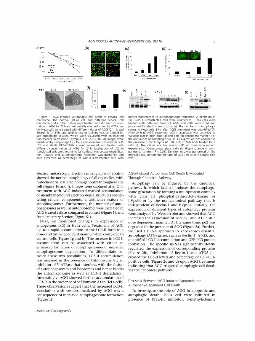
electron microscopy. Electron micrographs of controlshowed the normal morphology of all organelles, withmitochondria scattered homogenously throughout thecell (Figure 1e and f). Images were captured after 24htreatment with AGG indicated marked accumulationof membrane-bound electron dense structures seques-tering cellular components, a distinctive feature ofautophagosomes. Furthermore, the number of auto-phagosomes as well as autolysosomes were increased inAGG treated cells as compared to control (Figure 1f, andSupplementary Section, Figure S1).Next, we monitored changes in expression of
endogenous LC3 in HeLa cells. Treatment of AGGled to a rapid accumulation of the LC3-II form in adose- and time-dependentmannerwhen compared tocontrol cells (Figure 1g and h). The increase in LC3-IIaccumulation can be associated with either anenhanced formation of autophagosomes or impairedautophagosome degradation. To differentiate be-tween these two possibilities, LC3-II accumulationwas assessed in the presence of bafilomycin A1, aninhibitor of V-ATPase that interferes with the fusionof autophagosomes and lysosomes and hence blocksthe autophagosome as well as LC3-II degradation.Interestingly, AGG showed further accumulation ofLC3-II in the presence of bafilomycinA1 inHeLa cells.These observations suggest that the increased LC3-IIassociation with vesicles mediated by AGG was aconsequence of increased autophagosome formation(Figure 1i).
AGG-Induced Autophagic Cell Death Is MediatedThrough Canonical Pathway
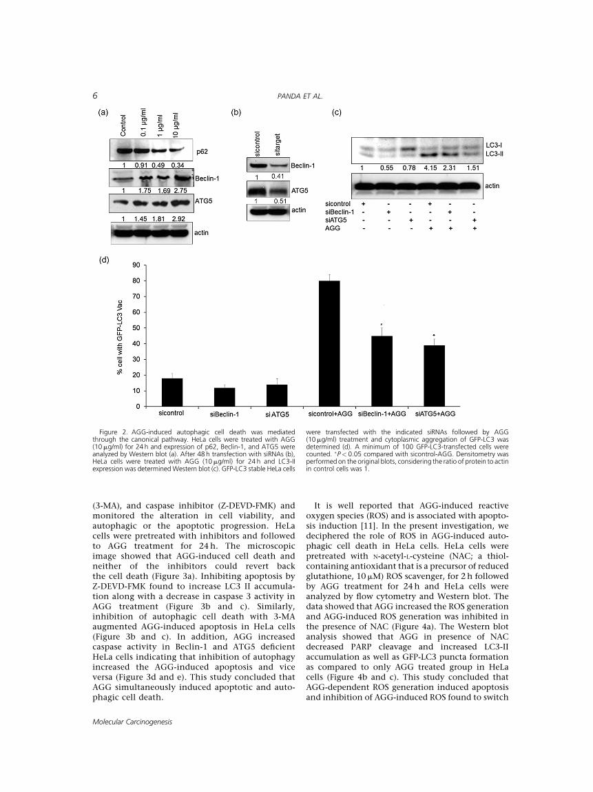
Autophagy can be induced by the canonicalpathway in which Beclin-1 induces the autophago-some generation by forming a multiprotein complexwith class III phosphatidylinositol-3-kinase orhVps34 or by the non-canonical pathway that isindependent of Beclin-1 and hVps34. Initially, theexpression of different types of autophagy proteinswere analyzed by Western blot and showed that AGGincreased the expression of Beclin-1 and ATG5 in adose dependent manner. At the same time, p62 wasdegraded in the presence of AGG (Figure 2a). Further,we used a siRNA approach to knockdown essentialautophagy (ATG) genes, such as Beclin-1, ATG5, andquantified LC3-II accumulation and GFP-LC3 punctaformation. The specific siRNAs significantly down-regulated the expression of corresponding proteins(Figure 2b). Inhibition of Beclin-1 and ATG5 de-creased the LC3-II levels and percentage of GFP-LC3-positive cells (Figure 2c and d) upon AGG treatmentindicating that AGG triggered autophagic cell deathvia the canonical pathway.
Crosstalk Between AGG-Induced Apoptosis andAutophagy-Dependent Cell Death
To investigate the role of AGG in apoptotic andautophagic death, HeLa cell were cultured inpresence of PI3K-III inhibitor, 3-methyladenine
Figure 1. AGG-induced autophagic cell death in cervical cellcarcinoma. The normal HaCaT cell and different cervical cellcarcinoma (HeLa, SiHa, CaSki) were treated with different concen-tration of AGG for 72 h and cell viability was performed by MTT assay(a). HeLa cells were treated with different doses of AGG (0.1, 1, and10mg/ml) for 24 h, and acridine orange staining was performed forlate autophagic vesicles, which were visualized with an invertedfluorescence microscope (Olympus IX71, 200�) (b). All images werequantified by using Image J (c). HeLa cells were transfected with GFP-LC3 and stable GFP-LC3-HeLa was generated and treated withdifferent concentration of AGG for 24 h, localization of LC3 intransfected cells were examined by confocal microscopy (magnifica-tion 1000�), and autophagosome formation was quantified anddata presented as percentage of GFP-LC3-transfected cells with
puncta fluorescence to autophagosome formation. A minimum of100 GFP-LC3-transfected cells were counted (d). HeLa cells weretreated with different doses of AGG and cells were fixed andprocessed for electron microscopy (e). The numbers of autophago-somes in HeLa cells 24 h after AGG treatment was quantified (f).After 24 h of AGG treatment, LC3-II expression was analyzed byWestern blot in both dose (g) and time (h) dependent manner. Forthe occurrence of autophagic flux, LC3-II expression was analyzed inthe presence of bafilomycin A1 (100 nM) in 24 h AGG treated HeLacells (i). The values are the means� SD of three independentexperiments. �Corresponds statistically significant change in com-parison to control (�P< 0.05). Densitometry was performed on theoriginal blots, considering the ratio of LC3-II to actin in control cellswas 1.
AGG INDUCES AUTOPHAGY-DEPENDENT CELL DEATH 5
Molecular Carcinogenesis
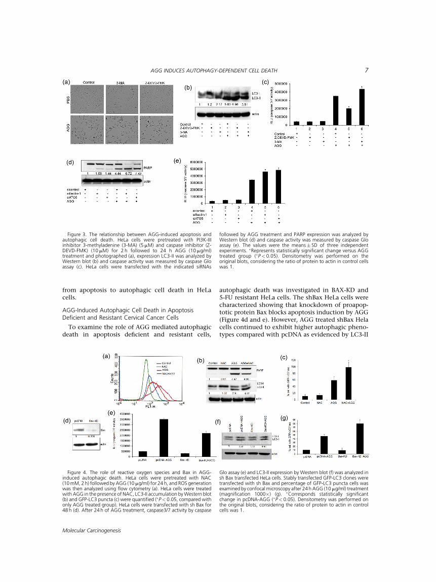
(3-MA), and caspase inhibitor (Z-DEVD-FMK) andmonitored the alteration in cell viability, andautophagic or the apoptotic progression. HeLacells were pretreated with inhibitors and followedto AGG treatment for 24 h. The microscopicimage showed that AGG-induced cell death andneither of the inhibitors could revert backthe cell death (Figure 3a). Inhibiting apoptosis byZ-DEVD-FMK found to increase LC3 II accumula-tion along with a decrease in caspase 3 activity inAGG treatment (Figure 3b and c). Similarly,inhibition of autophagic cell death with 3-MAaugmented AGG-induced apoptosis in HeLa cells(Figure 3b and c). In addition, AGG increasedcaspase activity in Beclin-1 and ATG5 deficientHeLa cells indicating that inhibition of autophagyincreased the AGG-induced apoptosis and viceversa (Figure 3d and e). This study concluded thatAGG simultaneously induced apoptotic and auto-phagic cell death.
It is well reported that AGG-induced reactiveoxygen species (ROS) and is associated with apopto-sis induction [11]. In the present investigation, wedeciphered the role of ROS in AGG-induced auto-phagic cell death in HeLa cells. HeLa cells werepretreated with N-acetyl-L-cysteine (NAC; a thiol-containing antioxidant that is a precursor of reducedglutathione, 10mM) ROS scavenger, for 2 h followedby AGG treatment for 24h and HeLa cells wereanalyzed by flow cytometry and Western blot. Thedata showed that AGG increased the ROS generationand AGG-induced ROS generation was inhibited inthe presence of NAC (Figure 4a). The Western blotanalysis showed that AGG in presence of NACdecreased PARP cleavage and increased LC3-IIaccumulation as well as GFP-LC3 puncta formationas compared to only AGG treated group in HeLacells (Figure 4b and c). This study concluded thatAGG-dependent ROS generation induced apoptosisand inhibition of AGG-induced ROS found to switch
Figure 2. AGG-induced autophagic cell death was mediatedthrough the canonical pathway. HeLa cells were treated with AGG(10mg/ml) for 24 h and expression of p62, Beclin-1, and ATG5 wereanalyzed by Western blot (a). After 48 h transfection with siRNAs (b),HeLa cells were treated with AGG (10mg/ml) for 24 h and LC3-IIexpression was determinedWestern blot (c). GFP-LC3 stable HeLa cells
were transfected with the indicated siRNAs followed by AGG(10mg/ml) treatment and cytoplasmic aggregation of GFP-LC3 wasdetermined (d). A minimum of 100 GFP-LC3-transfected cells werecounted. �P< 0.05 compared with sicontrol-AGG. Densitometry wasperformedon the original blots, considering the ratio of protein to actinin control cells was 1.
6 PANDA ET AL.
Molecular Carcinogenesis
from apoptosis to autophagic cell death in HeLacells.
AGG-Induced Autophagic Cell Death in ApoptosisDeficient and Resistant Cervical Cancer Cells
To examine the role of AGG mediated autophagicdeath in apoptosis deficient and resistant cells,
autophagic death was investigated in BAX-KD and5-FU resistant HeLa cells. The shBax HeLa cells werecharacterized showing that knockdown of proapop-totic protein Bax blocks apoptosis induction by AGG(Figure 4d and e). However, AGG treated shBax Helacells continued to exhibit higher autophagic pheno-types compared with pcDNA as evidenced by LC3-II
Figure 3. The relationship between AGG-induced apoptosis andautophagic cell death. HeLa cells were pretreated with PI3K-IIIinhibitor 3-methyladenine (3-MA) (5mM) and caspase inhibitor (Z-DEVD-FMK) (10mM) for 2 h followed to 24 h AGG (10mg/ml)treatment and photographed (a), expression LC3-II was analyzed byWestern blot (b) and caspase activity was measured by caspase Gloassay (c). HeLa cells were transfected with the indicated siRNAs
followed by AGG treatment and PARP expression was analyzed byWestern blot (d) and caspase activity was measured by caspase Gloassay (e). The values were the means� SD of three independentexperiments. �Represents statistically significant change versus AGGtreated group (�P< 0.05). Densitometry was performed on theoriginal blots, considering the ratio of protein to actin in control cellswas 1.
Figure 4. The role of reactive oxygen species and Bax in AGG-induced autophagic death. HeLa cells were pretreated with NAC(10mM, 2h) followed by AGG (10mg/ml) for 24 h, and ROS generationwas then analyzed using flow cytometry (a). HeLa cells were treatedwith AGG in the presence of NAC, LC3-II accumulation byWestern blot(b) and GFP-LC3 puncta (c) were quantified (�P< 0.05, compared withonly AGG treated group). HeLa cells were transfected with sh Bax for48 h (d). After 24 h of AGG treatment, caspase3/7 activity by caspase
Glo assay (e) and LC3-II expression by Western blot (f) was analyzed insh Bax transfected HeLa cells. Stably transfected GFP-LC3 clones weretransfected with sh Bax and percentage of GFP-LC3 puncta cells wasexamined by confocalmicroscopy after 24 h AGG (10mg/ml) treatment(magnification 1000�) (g). �Corresponds statistically significantchange in pcDNA-AGG (�P< 0.05). Densitometry was performed onthe original blots, considering the ratio of protein to actin in controlcells was 1.
AGG INDUCES AUTOPHAGY-DEPENDENT CELL DEATH 7
Molecular Carcinogenesis
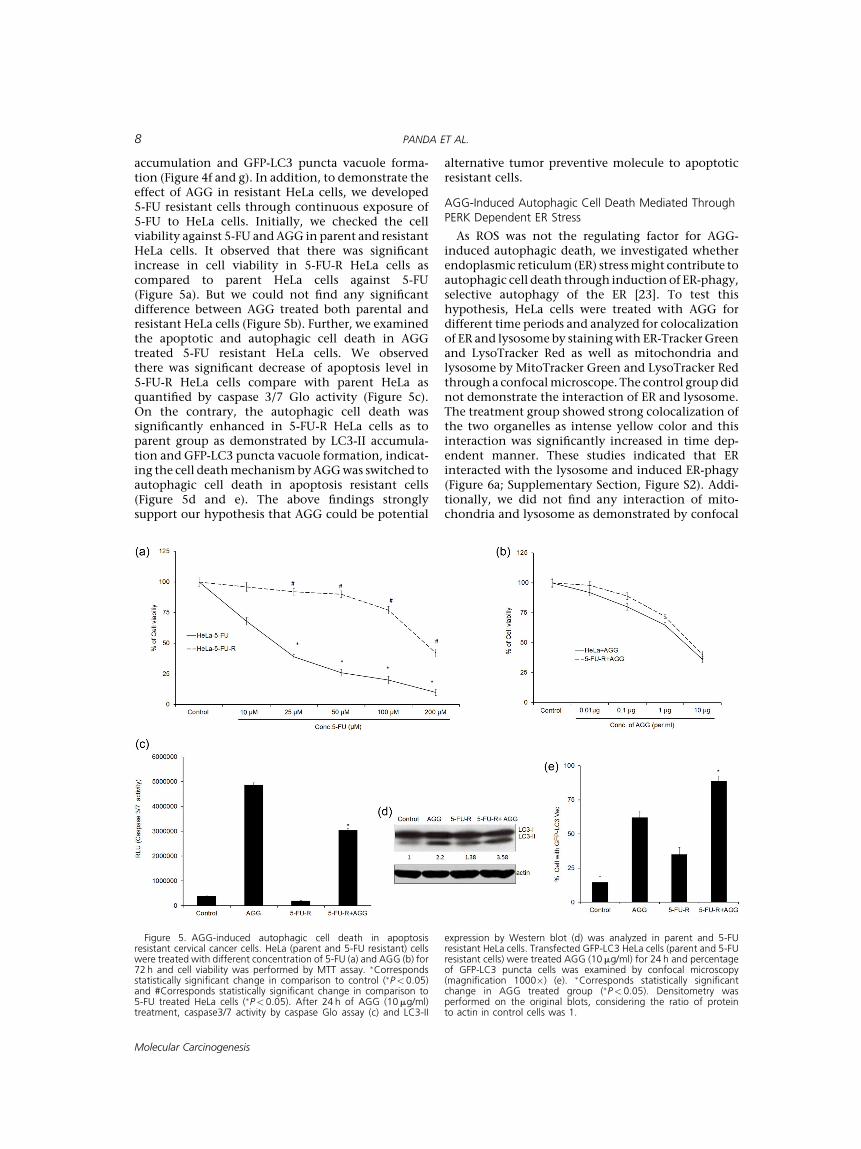
accumulation and GFP-LC3 puncta vacuole forma-tion (Figure 4f and g). In addition, to demonstrate theeffect of AGG in resistant HeLa cells, we developed5-FU resistant cells through continuous exposure of5-FU to HeLa cells. Initially, we checked the cellviability against 5-FU andAGG in parent and resistantHeLa cells. It observed that there was significantincrease in cell viability in 5-FU-R HeLa cells ascompared to parent HeLa cells against 5-FU(Figure 5a). But we could not find any significantdifference between AGG treated both parental andresistant HeLa cells (Figure 5b). Further, we examinedthe apoptotic and autophagic cell death in AGGtreated 5-FU resistant HeLa cells. We observedthere was significant decrease of apoptosis level in5-FU-R HeLa cells compare with parent HeLa asquantified by caspase 3/7 Glo activity (Figure 5c).On the contrary, the autophagic cell death wassignificantly enhanced in 5-FU-R HeLa cells as toparent group as demonstrated by LC3-II accumula-tion and GFP-LC3 puncta vacuole formation, indicat-ing the cell deathmechanismbyAGGwas switched toautophagic cell death in apoptosis resistant cells(Figure 5d and e). The above findings stronglysupport our hypothesis that AGG could be potential
alternative tumor preventive molecule to apoptoticresistant cells.
AGG-Induced Autophagic Cell Death Mediated ThroughPERK Dependent ER Stress
As ROS was not the regulating factor for AGG-induced autophagic death, we investigated whetherendoplasmic reticulum (ER) stressmight contribute toautophagic cell death through induction of ER-phagy,selective autophagy of the ER [23]. To test thishypothesis, HeLa cells were treated with AGG fordifferent time periods and analyzed for colocalizationof ER and lysosome by stainingwith ER-TrackerGreenand LysoTracker Red as well as mitochondria andlysosome by MitoTracker Green and LysoTracker Redthrough a confocalmicroscope. The control group didnot demonstrate the interaction of ER and lysosome.The treatment group showed strong colocalization ofthe two organelles as intense yellow color and thisinteraction was significantly increased in time dep-endent manner. These studies indicated that ERinteracted with the lysosome and induced ER-phagy(Figure 6a; Supplementary Section, Figure S2). Addi-tionally, we did not find any interaction of mito-chondria and lysosome as demonstrated by confocal
Figure 5. AGG-induced autophagic cell death in apoptosisresistant cervical cancer cells. HeLa (parent and 5-FU resistant) cellswere treated with different concentration of 5-FU (a) and AGG (b) for72 h and cell viability was performed by MTT assay. �Correspondsstatistically significant change in comparison to control (�P< 0.05)and #Corresponds statistically significant change in comparison to5-FU treated HeLa cells (�P< 0.05). After 24 h of AGG (10mg/ml)treatment, caspase3/7 activity by caspase Glo assay (c) and LC3-II
expression by Western blot (d) was analyzed in parent and 5-FUresistant HeLa cells. Transfected GFP-LC3 HeLa cells (parent and 5-FUresistant cells) were treated AGG (10mg/ml) for 24 h and percentageof GFP-LC3 puncta cells was examined by confocal microscopy(magnification 1000�) (e). �Corresponds statistically significantchange in AGG treated group (�P< 0.05). Densitometry wasperformed on the original blots, considering the ratio of proteinto actin in control cells was 1.
8 PANDA ET AL.
Molecular Carcinogenesis
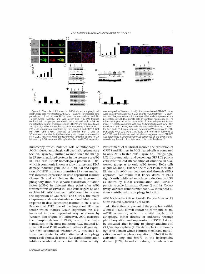
microscopy which nullified role of mitophagy inAGG-induced autophagic cell death (SupplementarySection, Figure S2). Further, wemonitored the changein ER stress regulated proteins in the presence of AGGin HeLa cells. C/EBP homologous protein (CHOP),which is commonly known as growth arrest-and DNAdamage inducible gene 153 (GADD153) and expres-sion of CHOP is the most sensitive ER stress marker,was increased expression in dose dependent manner(Figure 6b and c). Besides that, an increase inphosphorylation of eukaryotic translation initiationfactor (eIF2a) in different time point after AGGtreatment was observed in HeLa cells (Figure 6d ande). After 24h AGG treatment, AGG found to increasethe expression of GRP78/BiP and GRP94, the majorchaperones and central regulators of unfolded proteinresponse in dose dependent manner in HeLa cells.Besides that ATF6 one of the important ER stresssensor which induces CHOP expression was alsoincreased in dose dependent way as shown byWestern Blot (Figure 6f). Moreover, AGG increasedthe phosphorylation of PERK, one of the majortransducers of ER stress indicating AGG-induced ERstress followed PERK mediated pathway (Figure 6g).We next determined whether AGG mediated ERstress contribute to AGG stimulated autophagyusing a cell-permeable form eIF2a dephosphorylationinhibitor salubrinal, which inhibits eIF2a activity.
Pretreatment of salubrinal reduced the expression ofGRP78 and ER stress in AGG treated cells as comparedto only AGG treated cells (Figure 6h). Intriguingly,LC3-II accumulation and percentage GFP-LC3 punctacells were reduced after addition of salubrinal in AGGtreated group as to only AGG treated HeLa cells(Figure 6h and i). Further, the role of PERK mediatedER stress by AGG was demonstrated through siRNAapproach. We found that knock down of PERKsignificantly inhibited autophagy induction by AGGas shown by LC3-II accumulation and GFP-LC3puncta vacuole formation (Figure 6j and k). Collec-tively, our data demonstrate that AGG influenced ERstress contributed to autophagy induction.
AGG Mediated Inhibition of Akt/PH Domain Promoted ERStress-Induced Autophagic Cell Death
Akt, the active component of the phosphoinositide3-kinase (PI3K) is well-known to contribute to themTOR activation, which is a vital regulator ofautophagy, either directly or indirectly throughphosphorylation and suppression of TSC2. Akt canbe activated after binding to phosphatidylinositol(3,4,5)-trisphosphate (PIP3) via its pleckstrin homol-ogy (PH) domain which controls membrane translo-cation, as well as phosphorylation of Thr308 in theactivation loop and Ser473 in the hydrophobicdomain [1,28]. In order to study, the interactions
Figure 6. The role of ER stress in AGG-induced autophagic celldeath. HeLa cells were treated with AGG (10mg/ml) for indicated timeperiods and colocalization of ER and lysosome was analyzed with ER-Tracker Green (500nM) and LysoTracker Red (100 nM) throughconfocal microscopy (a). HeLa cells were treated with AGG forindicated time periods and expression of CHOP (b and c) and p-eIF2a (dand e) analyzed by immunofluorescence microscopy (Olympus IX71,200�, All images were quantified by using Image J) and GRP 78, GRP94, ATF6, and p-PERK, analyzed by Western blot (f and g).�Corresponds statistically significant change in comparison to control(�P< 0.05). HeLa cells were pretreated with salubrinal (5mM) for 2 hfollowed by AGG treatment (10mg/ml) for 24 h and LC3-II expression
was analyzed by Western blot (h). Stably transfected GFP-LC3 cloneswere treated with salubrinal (5mM) prior to AGG treatment (10mg/ml)and autophagosome formationwas quantified and data presented as apercentage of GFP-LC3 puncta cells by confocal microscopy (i). Thevalues are expressed as the mean� SD of three independent experi-ments (�P< 0.05, compared with only AGG treated group). After 48 htransfection with siPERK, HeLa cells were treated with AGG (10mg/ml)for 24 h and LC3-II expression was determined Western blot (j). GFP-LC3 stable HeLa cells were transfected with the siPERK followed byAGG (10mg/ml) treatment and cytoplasmic aggregation of GFP-LC3was determined (k). Densitometry was performed on the original blots,considering the ratio of protein to actin in control cells was 1.
AGG INDUCES AUTOPHAGY-DEPENDENT CELL DEATH 9
Molecular Carcinogenesis
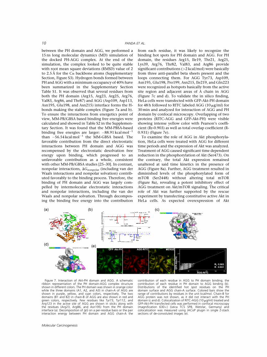
between the PH domain and AGG, we performed a15ns long molecular dynamics (MD) simulation ofthe docked PH-AGG complex. At the end of thesimulation, the complex looked to be quite stablewith root mean square deviations (RMSD) value of 2to 2.5 A for the Ca backbone atoms (SupplementarySection, Figure S3). Hydrogen bonds formed betweenPHandAGGwith aminimumoccupancy of 40%havebeen summarized in the Supplementary SectionTable S1. It was observed that several residues fromboth the PH domain (Arg15, Arg23, Arg25, Arg76,Val83, Arg86, and Thr87) and AGG (Asp109, Asp113,Asn195, Glu198, and Asn215) interface forms the H-bonds making the stable complex (Figure 7a and b).To ensure the interactions from energetics point ofview, MM-PB/GBSA based binding free energies werecalculated and showed in Table S2 in the Supplemen-tary Section. It was found that the MM-PBSA-basedbinding free energies are larger: �88.91 kcalmol�1
than �56.54 kcalmol�1 the MM-GBSA based. Thefavorable contribution from the direct electrostaticinteractions between PH domain and AGG wasrecompensed by the electrostatic desolvation freeenergy upon binding, which progressed to anunfavorable contribution as a whole, consistentwith other MM-PB/GBSA studies [25–30]. In contrast,nonpolar interactions, DGnonpolar (including van derWaals interactions and nonpolar solvation) contrib-uted favorably to the binding process. Therefore, thebinding of PH domain and AGG was largely com-pelled by intermolecular electrostatic interactionsand nonpolar interactions, including the van derWaals and nonpolar solvation. Through decompos-ing the binding free energy into the contribution
from each residue, it was likely to recognize thebinding hot spots for PH domain and AGG. For PHdomain, the residues Arg15, Ile19, Thr21, Arg25,Lys39, Arg76, Thr82, Val83, and Arg86 providesignificant contributions (>2 kcal/mol) were basicallyfrom three anti-parallel beta sheets present and theloops connecting them. For AGG Tyr73, Asp109,Asn195, Glu198, Pro199, Asn215, Ile219, and Gln223were recognized as hotspots basically from the activesite region and adjacent areas of A chain in AGG(Figure 7c and d). To validate the in silico finding,HeLa cells were transfected with GFP-Akt-PH domainfor 48h followed to RITC labeled AGG (10mg/ml) for30min and analyzed for interaction of AGG and PHdomain by confocal microscopy. Overlapping of twoproteins (RITC-AGG and GFP-Akt-PH) were visibleshowing intense yellow color with Pearson’s coeffi-cient (Rr-0.903) as well as total overlap coefficient (R-0.931) (Figure 7e).To examine the role of AGG in Akt phosphoryla-
tion, HeLa cells were treated with AGG for differenttime periods and the expression of Akt was analyzed.Treatment of AGG caused significant time dependentreduction in the phosphorylation of Akt (Ser473). Onthe contrary, the total Akt expression remainedunaltered at said time kinetics in the presence ofAGG (Figure 8a). Further, AGG treatment resulted indiminished levels of the phosphorylated form ofmTOR (Ser2448) without altering total mTOR(Figure 8a), revealing a potent inhibitory effect ofAGG treatment on Akt/mTOR signaling. The criticalrole of Akt was further supported by the rescueexperiment by transfecting constitutive active Akt inHeLa cells. As expected overexpression of Akt
Figure 7. Interaction of Akt-PH domain and AGG. A schematicribbon representation of the PH domain-AGG complex structureshown in different colors. The PH domain was shown in orange colorwhile the three domains (A1, A2, and A3) in chain-A of AGG areshown in purple, yellow, and cyan colors, respectively. The twodomains (B1 and B2) in chain-B of AGG are also shown in red andgreen colors, respectively. Few residues like Tyr73, Tyr112, andArg123 in the active site of AGG are shown in sticks along withthe residues (Arg23, Arg86, and Asn195) from the PH domaininterface (a). Decomposition of DG on a per-residue basis or the pairinteraction energy between PH domain and AGG chain-A the
contribution of each residue in AGG to PH domain binding; thecontribution of each residue in PH domain to AGG binding (b).Distributions of the identified hot spot residues on the PHdomain surface and AGG chain-A surface. Colored bars show therange of contributions by residues in the unit kcal/mol. Chain-B forAGG protein was not shown, as it did not interact with the PHdomain (c and d). Colocalization of RITC-AGG (10mg/ml) treated andGFP-Akt-PH transfected cells was performed in confocal microscopy(magnification 630�) (Leica TCS SP8, Wetzlar, Germany) andcolocalization was measured using JACoP plugin in single Z-stacksections of de-convoluted images (e).
10 PANDA ET AL.
Molecular Carcinogenesis
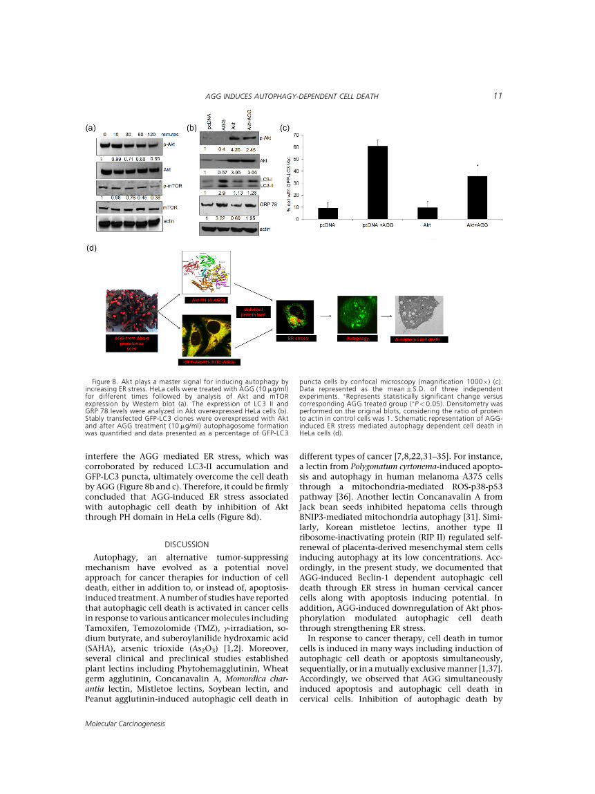
interfere the AGG mediated ER stress, which wascorroborated by reduced LC3-II accumulation andGFP-LC3 puncta, ultimately overcome the cell deathby AGG (Figure 8b and c). Therefore, it could be firmlyconcluded that AGG-induced ER stress associatedwith autophagic cell death by inhibition of Aktthrough PH domain in HeLa cells (Figure 8d).
DISCUSSION
Autophagy, an alternative tumor-suppressingmechanism have evolved as a potential novelapproach for cancer therapies for induction of celldeath, either in addition to, or instead of, apoptosis-induced treatment. Anumber of studies have reportedthat autophagic cell death is activated in cancer cellsin response to various anticancermolecules includingTamoxifen, Temozolomide (TMZ), g-irradiation, so-dium butyrate, and suberoylanilide hydroxamic acid(SAHA), arsenic trioxide (As2O3) [1,2]. Moreover,several clinical and preclinical studies establishedplant lectins including Phytohemagglutinin, Wheatgerm agglutinin, Concanavalin A, Momordica char-antia lectin, Mistletoe lectins, Soybean lectin, andPeanut agglutinin-induced autophagic cell death in
different types of cancer [7,8,22,31–35]. For instance,a lectin from Polygonatum cyrtonema-induced apopto-sis and autophagy in human melanoma A375 cellsthrough a mitochondria-mediated ROS-p38-p53pathway [36]. Another lectin Concanavalin A fromJack bean seeds inhibited hepatoma cells throughBNIP3-mediated mitochondria autophagy [31]. Simi-larly, Korean mistletoe lectins, another type IIribosome-inactivating protein (RIP II) regulated self-renewal of placenta-derived mesenchymal stem cellsinducing autophagy at its low concentrations. Acc-ordingly, in the present study, we documented thatAGG-induced Beclin-1 dependent autophagic celldeath through ER stress in human cervical cancercells along with apoptosis inducing potential. Inaddition, AGG-induced downregulation of Akt phos-phorylation modulated autophagic cell deaththrough strengthening ER stress.
In response to cancer therapy, cell death in tumorcells is induced in many ways including induction ofautophagic cell death or apoptosis simultaneously,sequentially, or in amutually exclusivemanner [1,37].Accordingly, we observed that AGG simultaneouslyinduced apoptosis and autophagic cell death incervical cells. Inhibition of autophagic death by
Figure 8. Akt plays a master signal for inducing autophagy byincreasing ER stress. HeLa cells were treated with AGG (10 mg/ml)for different times followed by analysis of Akt and mTORexpression by Western blot (a). The expression of LC3 II andGRP 78 levels were analyzed in Akt overexpressed HeLa cells (b).Stably transfected GFP-LC3 clones were overexpressed with Aktand after AGG treatment (10 mg/ml) autophagosome formationwas quantified and data presented as a percentage of GFP-LC3
puncta cells by confocal microscopy (magnification 1000�) (c).Data represented as the mean � S.D. of three independentexperiments. �Represents statistically significant change versuscorresponding AGG treated group (�P< 0.05). Densitometry wasperformed on the original blots, considering the ratio of proteinto actin in control cells was 1. Schematic representation of AGG-induced ER stress mediated autophagy dependent cell death inHeLa cells (d).
AGG INDUCES AUTOPHAGY-DEPENDENT CELL DEATH 11
Molecular Carcinogenesis
3-MA and knock down by siBeclin-1 and siATG5significantly switched the cell death to apoptosisinduced by AGG. In support, a previous study showedthat several anticancer drugs (SAHA) and plant lectinsinduced both autophagic death and apoptosis[1,38,39]. Further, AGG induced significantly enhan-ced autophagic cell death in shBax transfected cellsand 5-FU resistant cells indicating AGG could be veryeffective against apoptosis deficient and resistant cells.
Accumulating data indicate that ER stress promotesautophagy as an adaptive mechanism and uponpersistent stress it can switch into cell death mecha-nisms, the autophagic cell death [23,40]. Our studyshowed that AGG found to stimulate ER stressmarkersincludingGRP78,GRP94, CHOP, and eIF2aphosphor-ylation through PERK dependent pathway. In thisconnection, GRP78, a major chaperone is consideredto be the central regulator of UPRwhich acts as a novelobligatory component of autophagy in mammaliancells. For instance, knockdown of GRP78 inhibitedautophagosome formation, which was induced by ERstress or by nutrient starvation in HeLa cells [40].Similarly, stress dependent CHOP is important in thetranscriptional activation of genes involved in theformation, elongation, and function of the autopha-gosome [41]. Intriguingly, we demonstrated thatinhibition of ER stress with salubrinal and si PERkreduced AGG-induced autophagic cell death, suggest-ing a potential role of ER stress in AGG inducedautophagy. In support, it was demonstrated thatAbrusabrin-induced ER stress through stress kinases p38MAPK to regulate apoptosis in Jurkat cells [42].
Akt can mediate cell survival and growth and itsactivity is regulated by phosphorylation on tworegulatory residues, Thr308 in the activation loop ofthe catalytic domain and Ser473 in the regulatorydomain. Akt inhibits autophagy through mTORC1activation in response to growth factor stimula-tion [1,2,3]. The mutational Akt hyperactivationdiminishes autophagy during metabolic stress,whereas Akt inhibition by different types of antitumormolecules induces autophagic cell death. Our resultshowed that treatment of AGG caused significant timedependent reduction in the phosphorylation of Akt(Ser473) inHeLa cells and overexpression of Akt foundto suppress AGG-induced autophagic death. A previ-ous study showed that naturally occurring agentsincluding Concanavalin A and Plumbagin-induceautophagy by inhibiting the Akt/mTOR in cancercells [32,43]. For the first time, AGG demonstrated tobind with PH domain of Akt through the active siteregion and adjacent areas, located in the cleftmade bythree domains of A chain preserved among the type IIRIPs [12]. Apart from N-glycosidase enzymatic activity,our study documented an unknown function of Achain of AGG and AGG could be considered as non-lipid-based PH domain inhibitor which needs to bedeciphered indetail. Further, howdoesAchainofAGGinhibit PHdomain and influenceAkt phosphorylation
and translocation are not known. In our study,induction of ER stress through inhibition of Akt wasthe master signal for AGG-induced autophagic celldeath. Numerous studies were available where Akt-mTOR pathway was associated with the ERstress-induced induction of apoptosis as well asautophagy [44–46]. Recently it reported that resvera-trol a natural polyphenol triggers ER stress which leadsto autophagic cell death in prostate cancer cells viadown regulation of Akt-mTOR pathway [47]. Inconclusion, the present results establish that AGGstimulated cell death by autophagy through ER stressand Akt dephosphorylation by binding with PHdomain might be explored as an alternative tumorsuppressor mechanism in cervical carcinoma.
ACKNOWLEDGMENTS
Authors like to acknowledge CSIR, DBT, SERB andNCI R01 for financial support to carry out this projectand high performance computing facilities (ARJUNcluster) atMolecular Biophysics Unit, Indian Instituteof Science, Bangalore, India. PKP is obliged toDBT andMHRD (Govt of India) for providing fellowship. BRMis a Dr. D.S. Kothari Post-Doctoral Fellow (DSKPDF),UGC, India.
REFERENCES
1. Panda PK, Mukhopadhyay S, Das DN, Sinha N, Naik PP,Bhutia SK. Mechanism of autophagic regulation in carcino-genesis and cancer therapeutics. Semin Cell Dev Biol2015;39:43–55.
2. Bhutia SK, Mukhopadhyay S, Sinha N, et al. Autophagy:Cancer's friend or foe? Adv Cancer Res 2013;118:61–95.
3. Panda PK, Mukhopadhyay S, Sinha N, Das DN, Bhutia SK.Autophagy and apoptosis: Where do they meet? Apoptosis2014;19:555–566.
4. Jemal A, Bray F, Center MM, Ferlay J, Ward E, Forman D.Global cancer statistics. CA Cancer J Clin 2011;61:69–90.
5. SunejaG, BaconM, SmallW Jr, Ryu SY, Kitchener HC,GaffneyDK. The cervix cancer research network: Increasing access tocancer clinical trials in low- and middle-income countries.Front Oncol 2015;5:14.
6. Dalby KN, Tekedereli I, Lopez-Berestein G, Ozpolat B.Targeting the prodeath and prosurvival functions of autoph-agy as novel therapeutic strategies in cancer. Autophagy2010;6:322–329.
7. Liu Z, Luo Y, Zhou TT, Zhang WZ. Could plant lectins becomepromising anti-tumour drugs for causing autophagic celldeath? Cell Prolif 2013;46:509–515.
8. Fu LL, Zhou CC, Yao S, Yu JY, Liu B, Bao JK. Plant lectins:Targeting programmed cell death pathways as antitumoragents. Int J Biochem Cell Biol 2011;43:1442–1449.
9. Moustapha A, P�er�etout PA, Rainey NE, et al. Curcumininduces crosstalk between autophagy and apoptosis medi-ated by calcium release from the endoplasmic reticulum,lysosomal destabilization and mitochondrial events. CellDeath Discovery 2015;1:15017.
10. Hegde R, Maiti TK, Podder SK. Purification and characteriza-tion of three toxins and two agglutinins from Abrusprecatorius seed by using lactamyl-Sepharose affinity chro-matography. Anal Biochem 1991;194:101–109.
11. Bhutia SK, Mallick SK, Stevens SM, Prokai L, Vishwanatha JK,Maiti TK. Induction of mitochondria-dependent apoptosis byAbrus agglutinin derived peptides in human cervical cancercell. Toxicol In Vitro 2008;22:344–351.
12 PANDA ET AL.
Molecular Carcinogenesis
12. Bagaria A, Surendranath K, Ramagopal UA, Ramakumar S,Karande AA. Structure-function analysis and insights into thereduced toxicity ofAbrus precatorius agglutinin I in relation toabrin. J Biol Chem 2006;281:34465–34474.
13. Ghosh D, Maiti TK. Immunomodulatory and anti-tumoractivities of native and heat denatured Abrus agglutinin.Immunobiology 2007;212:589–599.
14. Mukhopadhyay S, Panda PK, Das DN, et al. Abrus agglutininsuppresses human hepatocellular carcinoma in vitro and invivo by inducing caspase-mediated cell death. Acta PharmacolSin 2014;35:814–824.
15. Bhutia SK, Behera B, Das DN, et al.Abrus agglutinin is a potentanti-proliferative and anti-angiogenic agent in human breastcancer. Int J Cancer 2016;139:457–466.
16. Ghosh D, Bhutia SK,Mallick SK, Banerjee I, Maiti TK. Stimulationof murine B and T lymphocytes by native and heat-denaturedAbrus agglutinin. Immunobiology 2009;214:227–234.
17. Tripathi S, Maiti TK. Stimulation of murine macrophages bynative and heat-denatured lectin from Abrus precatorius. IntImmunopharmacol 2003;3:375–381.
18. Tripathi S, Maiti TK. Immunomodulatory role of native andheat denatured agglutinin from Abrus precatorius. Int JBiochem Cell Biol 2005;37:451–462.
19. Tripathi S, Maiti TK. Efficiency of heat denatured lectins fromAbrus precatorius as immunoadjuvants. Food Agric Immunol2003;15:279–287.
20. Bhutia SK, Mallick SK, Maiti S, Maiti TK. Antitumor andproapoptotic effect of Abrus agglutinin derived peptide inDalton's lymphoma tumor model. Chem Biol Interact 2008;174:11–18.
21. Bhutia SK, Mallick SK, Maiti TK. In vitro immunostimulatoryproperties of Abrus lectins derived peptides in tumor bearingmice. Phytomedicine 2009;16:776–782.
22. Panda PK, Mukhopadhyay S, Behera B, et al. Antitumor effectof soybean lectin mediated through reactive oxygen species-dependent pathway. Life Sci 2014;11:27–35.
23. Salazar M, Carracedo A, Salanueva IJ, et al. Cannabinoidaction induces autophagy-mediated cell death throughstimulation of ER stress in human glioma cells. J Clin Invest2009;119:1359–1372.
24. Milburn CC, DeakM, Kelly SM, Price NC, Alessi DR, VanAaltenDM. Binding of phosphatidylinositol 3, 4, 5-trisphosphate tothe pleckstrin homology domain of protein kinase B induces aconformational change. Biochem J 2003; 375:531–538.
25. Meher BR, Wang Y. Interaction of I50V mutant and I50L/A71V double mutant HIV-protease with inhibitor TMC114(darunavir): Molecular dynamics simulation and binding freeenergy studies. J Phys Chem B 2012;116:1884–1900.
26. Meher BR,Wang Y. Binding of single walled carbon nanotubeto WT and mutant HIV-1 proteases: Analysis of flap dynamicsand binding mechanism. J Mol Graph Model 2012;38:430–445.
27. Meher BR, Wang Y. Exploring the drug resistance of V32I andM46L mutant HIV-1 protease to inhibitor TMC114: Flapdynamics and binding mechanism. J Mol Graph Model2015;56:60–73.
28. Mahadevan D, Powis G, Mash EA, et al. Discovery of a novelclass of AKT pleckstrin homology domain inhibitors. MolCancer Ther 2008;7:2621–2632.
29. Lafont V, Schaefer M, Stote RH, Altschu D, Dejaegere A.Protein–protein recognition and interaction hot spots in anantigen–antibody complex: free energy decomposition iden-tifies “efficient amino acids.” Proteins 2007;67:418–434.
30. Zoete V, Michielin O. Comparison between computationalalanine scanning and per-residue binding free energydecomposition for protein–protein association using MM-GBSA: Application to the TCR-p-MHC complex. Proteins2007;67:1026–1047.
31. Chang CP, Yang MC, Liu HS, Lin YS, Lei HY. Concanavalin Ainduces autophagy in hepatoma cells and has a therapeutic
effect in a murine in situ hepatoma model. Hepatology2007;45:286–296.
32. Roy B, Pattanaik AK, Das J, et al. Role of PI3 K/Akt/mTOR andMEK/ERK pathway in Concanavalin A induced autophagy inHeLa cells. Chem Biol Interact 2014;210:96–102.
33. Zhang CZ, Fang EF, Zhang HT, Liu LL, Yun JP. Momordicacharantia lectin exhibits antitumor activity towards hepato-cellular carcinoma. Invest New Drugs 2015;33:1–11.
34. Choi JH, Lyu SY, Lee HJ, Jung J, Park WB, Kim GJ. Koreanmistletoe lectin regulates selfrenewal of placenta-derivedmesenchymal stem cells via autophagic mechanisms. CellProlif 2012;45:420–429.
35. Mukhopadhyay S, Panda PK, Behera B, et al. In vitro and invivo antitumor effects of Peanut agglutinin through inductionof apoptotic and autophagic celldeath. Food Chem Toxicol2014;64:369–377.
36. Liu B, Cheng Y, Zhang B, Bian HJ, Bao JK. Polygonatumcyrtonema lectin induces apoptosis and autophagy in humanmelanoma A375 cells through a mitochondria-mediatedROS-p38-p53 pathway. Cancer Lett 2009;275:54–60.
37. Li H, Wang P, Sun Q, et al. Following cytochrome c release,autophagy is inhibited during chemotherapy-induced apo-ptosis by caspase 8-mediated cleavage of Beclin 1. Cancer Res2011;71:3625–3634.
38. Liu B,Wu JM, Li J, et al. Polygonatum cyrtonema lectin inducesmurine fibrosarcoma L929 cell apoptosis and autophagy viablocking Ras-Raf and PI3K-Akt signaling pathways. Biochimie2010;92:1934–1938.
39. Li C, Chen J, Lu B, et al. Molecular switch role of Akt inPolygonatum odoratum lectin-induced apoptosis and au-tophagy in human non-small cell lung cancer A549 cells. PLoSONE 2014;9:e101526.
40. Li J, Ni M, Lee B, Barron E, Hinton DR, Lee AS. The unfoldedprotein response regulator GRP78/BiP is required for endo-plasmic reticulum integrity and stress-induced autophagy inmammalian cells. Cell Death Differ 2008;15:1460–1471.
41. B'chir W, Maurin AC, Carraro V, et al. The eIF2a/ATF4pathway is essential for stress-induced autophagy geneexpression. Nucleic Acids Res 2013;41:7683–7699.
42. Mishra R, KarandeAA. Endoplasmic reticulum stress-mediatedactivation of p38 MAPK, Caspase-2 and Caspase-8 leads toabrin-induced apoptosis. PLoS ONE 2014;9:e92586.
43. Kuo PL, Hsu YL, Cho CY. Plumbagin induces G2-M arrest andautophagy by inhibiting the AKT/mammalian target ofrapamycin pathway in breast cancer cells. Mol Cancer Ther2006;5:3209–3221.
44. Jiang H, Sun J, Xu Q, et al. Marchantin M: A novel inhibitor ofproteasome induces autophagic cell death in prostate cancercells. Cell Death Dis 2013;4:e761.
45. Jung CH, Kim H, Ahn J, et al. Anthricin isolated fromAnthriscus sylvestris (L.) Hoffm. Inhibits the growth of breastcancer cells by inhibiting Akt/mTOR signaling, and itsapoptotic effects are enhanced by autophagy inhibition.Evid Based Complement Alternat Med 2013;2013:385219.
46. Yu KNChang SH, Park SJ, et al. Titaniumdioxide nanoparticlesinduce endoplasmic reticulum stress-mediated autophagiccell death via mitochondria-associated endoplasmic reticulummembrane disruption in normal lung cells. PLoS ONE2015;10:e0131208.
47. Selvaraj S, Sun Y, Sukumaran P, Singh BB. Resveratrolactivates autophagic cell death in prostate cancer cells viadownregulation of STIM1 and the mTOR pathway. MolCarcinog 2015; 55:818–831.
SUPPORTING INFORMATION
Additional supporting information may be found inthe online version of this article at the publisher’sweb-site.
AGG INDUCES AUTOPHAGY-DEPENDENT CELL DEATH 13
Molecular Carcinogenesis