analysis of squat and stoop dynamic liftings - muscle forces and internal spinal loads
TRANSCRIPT

ORIGINAL ARTICLE
Analysis of squat and stoop dynamic liftings: muscle forcesand internal spinal loads
Babak Bazrgari Æ Aboulfazl Shirazi-Adl ÆNavid Arjmand
Received: 6 March 2006 / Revised: 29 May 2006 / Accepted: 20 September 2006 / Published online: 14 November 2006� Springer-Verlag 2006
Abstract Despite the well-recognized role of lifting in
back injuries, the relative biomechanical merits of
squat versus stoop lifting remain controversial. In vivo
kinematics measurements and model studies are com-
bined to estimate trunk muscle forces and internal
spinal loads under dynamic squat and stoop lifts with
and without load in hands. Measurements were per-
formed on healthy subjects to collect segmental rota-
tions during lifts needed as input data in subsequent
model studies. The model accounted for nonlinear
properties of the ligamentous spine, wrapping of tho-
racic extensor muscles to take curved paths in flexion
and trunk dynamic characteristics (inertia and damp-
ing) while subject to measured kinematics and gravity/
external loads. A dynamic kinematics-driven approach
was employed accounting for the spinal synergy by
simultaneous consideration of passive structures and
muscle forces under given posture and loads. Results
satisfied kinematics and dynamic equilibrium condi-
tions at all levels and directions. Net moments, muscle
forces at different levels, passive (muscle or ligamen-
tous) forces and internal compression/shear forces
were larger in stoop lifts than in squat ones. These were
due to significantly larger thorax, lumbar and pelvis
rotations in stoop lifts. For the relatively slow lifting
tasks performed in this study with the lowering and
lifting phases each lasting ~2 s, the effect of inertia and
damping was not, in general, important. Moreover,
posterior shift in the position of the external load in
stoop lift reaching the same lever arm with respect to
the S1 as that in squat lift did not influence the con-
clusion of this study on the merits of squat lifts over
stoop ones. Results, for the tasks considered, advocate
squat lifting over stoop lifting as the technique of
choice in reducing net moments, muscle forces and
internal spinal loads (i.e., moment, compression and
shear force).
Keywords Muscle force � Finite element � Dynamic �Kinematics � Lifting technique
Introduction
Musculoskeletal impairments occur frequently and
have a substantial impact on the health and quality of
life of the population as well as on the health care
resources. Search for a safer lifting technique has at-
tracted considerable attention due to the high risk of
injury and low back pain (LBP) associated with fre-
quent lifting in industry. Compression force limits have
been recommended for safer manual material handling
(MMH) maneuvers based on the premise that exces-
sive compression loads could cause injury. Despite the
well-recognized role of lifting in low back injuries [4,
12, 17, 33, 53], the literature on safer lifting techniques
remains controversial [25, 48]. In search of optimal
lifting methods, squat lift (i.e., knee bent and back
straight) is generally considered to be safer than the
stoop lift (i.e., knee straight and back bent) in bringing
the load closer to the body and, hence, reducing the
extra demand on back muscles while counterbalancing
the moments of external loads. The importance of the
squat versus stoop lifting technique has, however, been
downplayed due to the lack of a clear biomechanical
B. Bazrgari � A. Shirazi-Adl (&) � N. ArjmandDepartment of Mechanical Engineering,Ecole Polytechnique, Montreal, QC, Canadae-mail: [email protected]
123
Eur Spine J (2007) 16:687–699
DOI 10.1007/s00586-006-0240-7

rationale for the promotion of either style [25, 48, 66].
Many workers, despite instruction to the contrary,
prefer the stoop lift due to its easier operation, lower
energy consumption in repetitive lifting tasks [38, 42]
and better balance [97]. Besides, it is known that squat
lift is not always possible due to the lift set up and load
size.
The advantages in preservation or flattening (i.e.,
flexing) of the lumbar lordosis during lifting tasks are
even less understood. Lifting has been categorized as
either squat or stoop often with no recording of
changes in the lumbar lordosis, which may influence
the risk of injury [68, 82]. The kyphotic lift (i.e.,
fully flexed lumbar spine) is recommended by some, as
it utilizes the passive posterior ligamentous system (i.e.,
posterior ligaments and lumbodorsal fascia) to their
maximum thus relieving the active extensor muscles
[36, 37]. In contrast, however, others advocate lordotic
and straight-back postures indicating that posterior
ligaments cannot effectively protect the spine and an
increase in erector spinae activities is beneficial in
increasing stability and reducing segmental shear for-
ces [22, 45, 47, 66, 100]. Moderate flexion has been
recommended by model [6, 91, 92] as well as experi-
mental studies [2] to reduce risk of failure under high
compressive forces. As the lumbar posture alters from
a lordotic one to a kyphotic one, the effectiveness of
erector spinae muscles in supporting the net moment
(due to smaller lever arms [50, 62, 99] and the anterior
shear force (due to changes in line of action [68]) de-
creases while the passive contribution of both extensor
muscles and the ligamentous spine increases [6, 8, 62].
Evidently, an improved assessment of various lifting
techniques and associated risk of tissue injuries de-
pends directly on a more accurate estimation of the
load partitioning in human trunk in dynamic lifting
conditions. The spinal loads are influenced not only by
the gravity, inertia and external loads, but also, more
importantly, by trunk muscle forces (due to their
smaller moment arms and their compensatory response
to stability demands and tissue injuries). Despite con-
flicting data in the literature, previous studies generally
indicated a decrease in trunk strength but an increase
in trunk moments, muscle coactivity, muscle forces and
spinal loads as the trunk movement was performed at a
faster rate [20, 27, 28, 39, 40, 41, 55, 69]. A number of
optimization and EMG driven models have been used
to estimate the muscle forces in various lifting condi-
tions. Many of these models do not properly account
for the nonlinear passive resistance and/or complex
geometry/loading/dynamics (i.e., inertia and damping)
of the spine [16, 26, 35, 93]. In addition, many are
simplified in not considering dynamic equilibrium
equations simultaneously in all directions and at all
levels.
Towards a more accurate estimation of muscle for-
ces and spinal loads in lifting tasks, an iterative hybrid
dynamic kinematics-based finite element approach is
introduced and applied in which a priori measured
kinematics of the spine along with nonlinear passive
properties are exploited. The kinematics-based ap-
proach that results in a synergistic solution of the ac-
tive/passive system has already been successfully
applied to isometric conditions in upright [30, 89] and
flexed postures [8]. The objectives of this work are,
hence, set as follows.
1. To extend kinematics-based approach to dynamic
conditions by accounting for the inertia and
damping in the solution of nonlinear transient
equations of motion over the lifting period,
2. To perform in vivo measurements of trunk kine-
matics, needed as input data in subsequent model
studies, on subjects performing sagittally symmet-
ric forward/backward lifting tasks with/without
loads in squat and stoop techniques,
3. To evaluate muscle forces (active/passive compo-
nents) as well as spinal loads at different levels
under loading and kinematics considered in vivo
while accounting for the curved path (i.e., wrap-
ping) of global extensor muscles,
4. To assess effects of dynamic characteristics of
trunk (inertia and damping) and positioning of the
external load on results.
Materials and methods
In vivo measurement
Fifteen healthy men with no recent back complications
volunteered for the study after signing an informed
consent form approved by the Institute de readaptation
de Montreal. Their mean (±SD) age, body height and
mass were 30 ± 6 years, 177 ± 7 cm and 74 ± 11 kg,
respectively. While bending slightly forward, light-
emitting diode, LED, markers were attached on the
skin at the tip of the T1, T5, T10, T12, L1, L3, L5 and
S1 spinous processes for evaluation of lumbar and
torso flexions. Three extra LED markers were placed
on the posterior/superior iliac spine and ilium (left/
right iliac crests) for the evaluation of pelvic rotation,
and one on the load to track the position of weights in
hands. A three-camera Optotrak system (NDI Inter-
national, Waterloo, ON, Canada) was employed to
collect 3D coordinates of LED markers.
688 Eur Spine J (2007) 16:687–699
123

Subjects were asked, with no instruction on lumbar
posture, to perform sagittally symmetric squat (knee
bent) and stoop (knee straight) lifts with and without
180 N weight placed on a bar in front at 20 cm height
from the floor. Each task lasted 4–5 s and started from
upright standing with no load in hands and ended again
in upright standing with or without load in hands. Two-
way analyses of variance (ANOVA) for repeated
measure factors were performed to study the effect of
different lifting techniques (2 levels: stoop and squat)
and load magnitude (2 levels: 0 and 180 N) on col-
lected kinematics data (Statistica, StaSoft, Tulsa, OK,
USA).
Thoracolumbar finite element model
A sagittally symmetric T1–S1 beam-rigid body model
made of six deformable beams to represent T12–S1
discs and seven rigid elements to represent T1–T12 (as
a single body) and lumbosacral (L1–S1) vertebrae was
used [6, 8, 30, 89]. The beams modeled the overall
nonlinear stiffness of T12–S1 motion segments (i.e.,
vertebrae, disc, facets and ligaments) at different
directions and levels. The nonlinear load-displacement
response under single and combined loads along with
flexion versus extension differences were represented
in this model based on numerical and measured results
of previous single- and multi-motion segment studies
[6, 73, 80, 85, 88, 104]. The trunk mass and mass mo-
ments of inertia were assigned at gravity centers at
different levels along the spine based on published data
for trunk segments [77, 78] and head/arms [105].
Connector elements parallel to deformable beams
were added to account for the intersegmental damping
using measured values [51, 63]; translational damp-
ing = 1,200 N s/m and angular damping = 1.2 N m s/
rad. For the cases with 180 N external load in hands,
the inertia of the load was computed using measured
kinematics and subsequently added as an external load.
Prescribed postures
Measured sagittal rotations at thorax (evaluated by the
change in inclination of the line attaching the T1
marker to the T12 one) and pelvic (evaluated by the
orientation of normal to the plane passing through the
three markers on the pelvis) of one subject were ap-
plied at the T12 and S1 vertebrae, respectively. The
total lumbar rotation, calculated as the difference be-
tween the foregoing two rotations, was subsequently
partitioned in between various segments based on
values reported in earlier investigations [3, 29, 34, 76,
79, 91, 104]. Relative proportions of ~7, 12, 15, 22, 27
and 17% were used to partition the lumbar rotation
between various motion segments from T12 to L5
levels, respectively.
Muscle model and muscle force calculation
A sagittally symmetric muscle architecture with 46 lo-
cal (attached to the lumbar vertebrae) and 10 global
(attached to the thoracic cage) muscles was used
(Fig. 1) [6, 8, 10, 94]. In order to accurately simulate
the curved path of global muscles (i.e., longissimus
thoracis pars thoracic and iliocostalis lumborum pars
thoracic) at flexion angles considered in this study,
these muscles were constrained to follow a curved path
whenever their distances from T12 to L5 vertebral
centers at undeformed configuration diminished be-
yond ~10% (i.e., to reach the limit values of 53, 53, 55,
56, 54 and 48 mm for the global longissimus and 58, 56,
56, 55, 52 and 45 mm for the global iliocostalis at T12–
L5 vertebrae, respectively). This wrapping mechanism,
similar to that formulated in our earlier works [86, 90,
92], was considered in order not to allow the line of
action of these muscles reach unrealistically close to
the vertebrae resulting in erroneous small lever arms at
different levels that occurs in larger flexions when
global muscles are simulated as straight lines between
their insertion points. During the analysis, when a
wrapping is detected at a vertebral level, the contact
force between the muscle and the corresponding ver-
tebra is evaluated using the equilibrium equation in the
Fig. 1 Representation of the model as well as global and localmusculatures in the sagittal and frontal planes. Fascicles on oneside are shown; ICpl iliocostalis lumborum pars lumborum, ICptiliocostalis lumborum pars thoracic, IP iliopsoas, LGpl longiss-imus thoracis pars lumborum, LGpt longissimus thoracis parsthoracic, MF multifidus, QL quadratus lumborum, IO internaloblique, EO external oblique and RA rectus abdominus
Eur Spine J (2007) 16:687–699 689
123
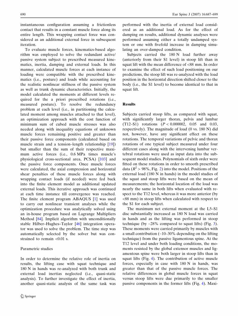
instantaneous configuration assuming a frictionless
contact that results in a constant muscle force along its
entire length. This wrapping contact force was con-
sidered as an additional external force in subsequent
iteration.
To evaluate muscle forces, kinematics-based algo-
rithm was employed to solve the redundant active/
passive system subject to prescribed measured kine-
matics, inertia, damping and external loads. In this
manner, calculated muscle forces at each instance of
loading were compatible with the prescribed kine-
matics (i.e., posture) and loads while accounting for
the realistic nonlinear stiffness of the passive system
as well as trunk dynamic characteristics. Initially, the
model calculated the moments at different levels re-
quired for the a priori prescribed rotations (i.e.,
measured posture). To resolve the redundancy
problem at each level (i.e., in partitioning the calcu-
lated moment among muscles attached to that level),
an optimization approach with the cost function of
minimum sum of cubed muscle stresses was also
needed along with inequality equations of unknown
muscle forces remaining positive and greater than
their passive force components (calculated based on
muscle strain and a tension–length relationship [19])
but smaller than the sum of their respective maxi-
mum active forces (i.e., 0.6 MPa times muscle’s
physiological cross-sectional area, PCSA) [103] and
the passive force components. Once muscle forces
were calculated, the axial compression and horizontal
shear penalties of these muscle forces along with
wrapping contact loads (if needed) were fed back
into the finite element model as additional updated
external loads. This iterative approach was continued
at each time instance till convergence was reached.
The finite element program ABAQUS [1] was used
to carry out nonlinear transient analyses while the
optimization procedure was analytically solved using
an in-house program based on Lagrange Multipliers
Method [84]. Implicit algorithm with unconditionally
stable Hilber–Hughes–Taylor [46] integration opera-
tor was used to solve the problem. The time step was
automatically selected by the solver but was con-
strained to remain <0.01 s.
Parametric studies
In order to determine the relative role of inertia on
results, the lifting case with squat technique and
180 N in hands was re-analyzed with both trunk and
external load inertias neglected (i.e., quasi-static
analysis). To further investigate the effect of inertia,
another quasi-static analysis of the same task was
performed with the inertia of external load consid-
ered as an additional load. As for the effect of
damping on results, additional dynamic analyses were
performed assuming either a totally undamped sys-
tem or one with fivefold increase in damping simu-
lating an over-damped condition.
Subjects carried the 180 N load further away
(anteriorly from their S1 level) in stoop lift than in
squat lift with the mean difference of <88 mm. In order
to examine the effect of such load positioning on our
predictions, the stoop lift was re-analyzed with the load
position in the horizontal direction shifted closer to the
body (i.e., the S1 level) to become identical to that in
squat lift.
Results
Subjects carried stoop lifts, as compared with squat,
with significantly larger thorax, pelvis and lumbar
(T12–S1) rotations (P < 0.000002, 0.05 and 0.03,
respectively). The magnitude of load (0 vs. 180 N) did
not, however, have any significant effect on these
rotations. The temporal variations of pelvic and thorax
rotations of one typical subject measured under four
different cases along with the intervening lumbar ver-
tebral rotations were used as input data into the sub-
sequent model studies. Polynomials of sixth order were
fitted on these rotations in order to smooth prescribed
data (R2 > 98%, Fig. 2) into the model. Positions of the
external load (180 N in hands) in the model studies of
the squat and stoop lifts were based on the mean of
measurements; the horizontal location of the load was
nearly the same in both lifts when evaluated with re-
spect to the T12 level, whereas it was more anterior (by
<88 mm) in stoop lifts when calculated with respect to
the S1 for each subject.
The maximum net external moment at the L5–S1
disc substantially increased as 180 N load was carried
in hands and as the lifting was performed in stoop
technique (by ~28% compared to squat lifts) (Fig. 3).
These moments were carried primarily by muscles with
a small contribution (~10–30% depending on the lifting
technique) from the passive ligamentous spine. At the
T12 level and under both loading conditions, the mo-
ments resisted by the global extensor muscles and lig-
amentous spine were both larger in stoop lifts than in
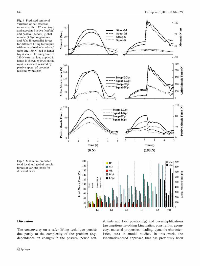
squat lifts (Fig. 4). The contribution of active muscle
forces, especially in case with 180 N in hands, was
greater than that of the passive muscle forces. The
relative differences in global muscle forces in squat
versus stoop lifts were due primarily to the smaller
passive components in the former lifts (Fig. 4). Maxi-
690 Eur Spine J (2007) 16:687–699
123

mum muscle forces at different local and global levels
(Fig. 5) were larger in stoop lifts than in squat lifts.
Internal local compression and shear forces at different
intervertebral disc levels were also greater in stoop lifts
than in squat lifts with maximum differences reaching
~800 N in compression and ~200 N in shear at the L5–
S1 level (Fig. 6; Table 1). Calculated shear forces
showed a dramatic increase from the L4–L5 level to
the L5–S1 in all cases. Due to larger intersegmental
lumbar rotations, passive segmental moments were
also larger in stoop lifts (Table 1).
Except for the time periods at the beginning and
end of tasks as well as immediately after lifting the
external load, results were almost the same for both
static and dynamic analyses over the entire duration
of motion (Fig. 7). Inclusion of the inertia of the
external load in the analysis was also found to have
negligible effects on results. Increasing the damping
at the motion segments did not change results, while
considerable fluctuations in response (±10 N m on
required thorax moment) were noted in the absence
of any damping in the model (Fig. 7). Closer posi-
tioning of the external load in stoop lift (by <88 mm
in order to arrive at the same relative lever arm with
respect to the S1 as that considered in squat lift)
reduced the total muscle forces as well as net mo-
ment and internal compression/anterior shear forces
at the L5–S1 level to values in between those pre-
dicted for squat and stoop lifts, i.e., nevertheless
remaining greater than those for the squat lift.
Fig. 2 Prescribed thorax (top) and pelvis (bottom) rotations inthe model for various cases based on in vivo measurements of atypical subject (smoothed by 6th order polynomials, R2 > 98%).The T12–S1 rotations are subsequently prescribed in the modelbased on the difference between these two rotations andproportions given in the text
Fig. 3 Predicted temporal variation of sagittal moments at theL5–S1 level for different cases (N m); net external moment (top),portion resisted by muscle forces (middle) and portion resistedby passive ligamentous spine (bottom). For the cases with load inhands, the sharp increase in moments is noted as the load reachesits maximum value of 180 N in 0.2 s duration
Eur Spine J (2007) 16:687–699 691
123
Discussion
The controversy on a safer lifting technique persists
due partly to the complexity of the problem (e.g.,
dependence on changes in the posture, pelvic con-
straints and load positioning) and oversimplifications
(assumptions involving kinematics, constraints, geom-
etry, material properties, loading, dynamic character-
istics, etc.) in model studies. In this work, the
kinematics-based approach that has previously been
Fig. 4 Predicted temporalvariation of net externalmoment at the T12 level (top)and associated active (middle)and passive (bottom) globalmuscle (LGpt longissimusand ICpt illiocostalis) forcesfor different lifting techniqueswithout any load in hands (leftside) and 180 N load in hands(right side). The rising time of180 N external load applied inhands is shown by lines on theright. S moment resisted bypassive spine, M momentresisted by muscles
Fig. 5 Maximum predictedtotal local and global muscleforces at various levels fordifferent cases
692 Eur Spine J (2007) 16:687–699
123

applied to isometric lifting conditions was extended to
predict muscle forces and internal spinal loads in dy-
namic stoop and squat lifts. For this purpose, parallel in
vivo studies were performed to collect kinematics re-
quired as input data into the model. The entire for-
ward–backward movements were carried out over 4–
5 s with either squat or stoop techniques but no
instructions on the lumbar posture.
Methodological issues
Evaluation of the segmental rotations from skin
markers is recognized to involve errors in the identifi-
cation of vertebral positions, skin movement relative to
the underlying vertebrae and deformability of verte-
brae themselves [14, 56, 60, 87, 106]. Due to these
inherent errors, the measurements were used to eval-
uate temporal variations of pelvic tilt and thorax
rotation while the intervening lumbar segmental rota-
tions were evaluated based on the partitioning of the
difference between foregoing measured rotations using
the relative values reported in the literature. The
assumption of rigid body motion at the T1–T12 seg-
ments (upper torso) in the model was justified, in
agreement with others [72, 96], by measuring nearly
equal rotations for lines attaching either the markers
T12–T5 or markers T12–T1. Changes in the relative
proportions used to partition the total T12–S1 rotation
among intervening segments would, as expected, alter
to some extent the net moment, passive ligamentous
resistance and muscle recruitments at these levels.
Moreover, although these proportions were assumed
constant during the entire lifting tasks, such may not
necessarily be true in vivo as the relative demand at
different levels could vary during lifting. These relative
ratios were taken from data obtained in static mea-
surements [29, 34, 76, 79], which have also been used in
previous dynamic studies [70, 82, 83] in order to eval-
uate the contribution of passive tissue in offsetting
external load. To prescribe measured rotations in the
model, kinematics data of one typical subject rather
than the mean of all subjects were considered. This was
done due mainly to noticeable variations in the dura-
tion of lowering/lifting phases in between subjects.
The transverse abdominal, latissimus dorsi, lumbo-
dorsal fascia, inter- and multisegmental muscles were
neglected, whereas the oblique abdominal muscles
were presented by straight single lines rather than
Fig. 6 Computed temporal variation of local compression (top)and anterior shear (bottom) forces at the L5–S1 level fordifferent cases. These forces are normal and tangential to thedisc mid-height planes
Table 1 Maximum internal loads in passive ligamentous spine for different cases at various levels; passive segmental moment, M(N m), local compression force, C (N), and local anterior shear force, S (N)
Disc level 0 N 180 N
Stoop Squat Stoop Squat
M C S M C S M C S M C S
T12–L1 20 926 226 17 902 187 18 2,416 384 14 2,315 222L1–L2 24 1,155 244 19 1,121 196 21 2,921 381 14 2,660 192L2–L3 24 1,445 184 18 1,374 135 19 3,383 244 12 2,922 85L3–L4 20 1,793 300 14 1,675 249 15 3,903 536 7 3,274 376L4–L5 20 2,162 258 14 1,989 227 19 4,518 502 9 3,704 425L5–S1 23 2,355 800 16 2,159 737 33 4,831 1,635 16 4,023 1,416
Maximum internal loads occur nearly at the time of maximum trunk flexion
Eur Spine J (2007) 16:687–699 693
123

curved sheets of muscle. Consideration of several fas-
cicles instead of just one for oblique muscles (EO and
IO) has influenced the estimated spinal loads signifi-
cantly in asymmetric lifting tasks but only slightly in
symmetric ones [21]. Indirect effect of the transverse
abdominal and latissimus dorsi muscles in unloading
the spine through lumbodorsal fascia have been re-
ported not being sizable during lifting tasks [11, 15, 61,
65, 70, 95]. Moreover, the likely mechanical effects of
the intra-abdominal pressure (IAP), neglected in this
study, have been found to depend on the posture and
the co-activity level of abdominal muscles [7]. While
local muscles were modeled as straight lines between
their respective insertion points, realistic muscle paths
were considered for global extensor muscles by wrap-
ping them over all T12–S1 vertebrae whenever in the
course of lifts their distance to associated vertebral
bodies reduced more than 10% of their initial dis-
tances. This allowed for a maximum of ~10% reduction
in muscle lever arms at different levels during flexion
which was chosen in accordance with published data in
the literature [50, 62, 99]. The wrapping of global
muscles occurred at all levels under larger flexion an-
gles and resulted in curved paths with realistic lever
arms at different levels. Had straight lines been as-
sumed for global muscles, much smaller lever arms
would have been generated resulting in greater muscle
forces and internal loads. The wrapping contact forces
(Table 2) remained relatively small compared with
muscle forces suggesting minor changes in lines of ac-
tion at wrapping points.
In the presence of nonlinearity in equations,
numerical integration using an unconditionally stable
implicit method was employed in this study. Minimum
sum of cubed muscle stresses, as the cost function used
in the optimization, has been recognized to agree
better with EMG data [5, 24, 49]. The convergence of
the nonlinear optimization solution on a global mini-
mum was assured in this study by solving the optimi-
zation problem analytically. For the sake of
comparison with EMG measurements of earlier stud-
ies, the computed muscle forces were partitioned, at
the post-processing phase of the analysis, into passive
and active components using a passive tension–length
relationship for all muscles [19]. Moreover, the maxi-
mum allowable muscle stress of 0.6 MPa was assumed
for all muscles neglecting the effect of activation level
on this value. It is important to emphasize that the
passive load–length relationship considered for mus-
cles in the current study have absolutely no bearing at
all on the predicted spinal loads and total muscle for-
ces. The rate-dependent viscoelastic properties of the
spinal segments, which could play a role at much
higher loading rates, [71, 101, 102] were not considered
in this study. Finally, in accordance with parallel in
vivo measurements, the response was limited to the
sagittal plane, thus neglecting out of plane motions.
Fig. 7 Predicted effect of changes in system dynamics charac-teristics on the net moment at the T12 level for the squat lift with180 N in hands; effect of consideration of trunk and load inertias(top) and of damping (bottom)
Table 2 Maximum wrapping contact forces for different cases atvarious levels (N)
Vertebralevel
0 N 180 N
Stoop Squat Stoop Squat
T12 0 0 0 0L1 40 27 60 32L2 55 39 113 66L3 62 43 97 44L4 80 55 168 89L5 99 70 251 138
Maximum wrapping contact forces occur nearly at the time ofmaximum trunk flexion
694 Eur Spine J (2007) 16:687–699
123

Effect of dynamic parameters
Generally, faster trunk movements have been associ-
ated with a decrease in trunk strength but increases in
trunk moments, muscle coactivity, muscle forces and
spinal loads [20, 27, 28, 39, 40, 41, 55, 69]. Inertia effects
of the trunk and external load have been indicated to
play a noticeable role at the onset of a lift with jerky
movements [55]. Our results showed a negligible effect
of inertia forces on trunk moment and spinal loads
except at three time intervals; the beginning and end of
the tasks as well as a short period after picking the load
up (Fig. 7) which agrees with earlier observations [47].
Apart from these periods, a quasi-static analysis would
yield sufficiently accurate results with no real need to
account for inertia forces which could be due to the
slow lifting performed by our subjects (i.e., lowering
and lifting periods each lasting ~2 s). Our results also
demonstrated that the inertia of the trunk, and not that
of the load, was the major factor for the observed
differences in these three time periods. In a different
lifting condition, however, the latter has been esti-
mated to be more important than the former [69]. The
computed net moment at the L5–S1 is noted to be in
good agreement with values reported in previous dy-
namic studies [18, 32, 52, 96].
Although recognized as an important parameter,
damping has been neglected in earlier biomechanical
model studies of dynamic lifting [52]. A fivefold in-
crease in the segmental damping value which was used
in the model based on earlier measurements did not
markedly alter predictions of this work, especially
away from the three time intervals indicated earlier
(Fig. 7). Introduction of damping appeared to primar-
ily smooth the temporal response by removing high-
frequency fluctuations (i.e., noise).
Effect of lifting techniques
The relative lumbar/pelvic rotations during lowering/
lifting phases showed greater contributions in all cases
from the pelvis than the lumbar spine (by as much as
twofold) and remained within the range of data re-
ported in the literature [31, 41, 64, 81]. Thorax and
pelvis rotations were both larger in stoop lifts com-
pared to those in squat lifts (Fig. 2) resulting in greater
lumbar (T12–S1) rotations in stoop lifts by 10.5� and
5.9� in cases with and without 180 N load in hands,
respectively. These additional flattenings of the lumbar
spine in stoop lifts increased the wrapping contact
forces (Table 2) and moment-carrying contribution of
passive ligamentous spine and trunk muscles. More-
over, despite identical lever arms considered (based on
measurements) for the external load of 180 N at the
T12 level, the net moments and hence muscle forces
and internal loads were all greater in stoop lifts than in
squat ones; e.g., maximum net moments of 200 N m
and 160 N m were predicted at the L5–S1 level for
stoop and squat lifting, respectively. Same trends were
also found in the absence of external loads or even
when the external load was shifted by <88 mm closer
to the body in the stoop lift in order to reach the same
lever arm with respect to the S1 as that considered in
the squat lift.
Therefore, results of this study appear to suggest the
squat lift as the safer lifting technique in reducing the
net moment, muscle forces and internal ligamentous
loads at all levels. It should be emphasized that the
relative merits of these lifting techniques depend not
only on the relative rotations at the thorax, pelvis and
lumbar spine but also on other factors such as position
of external loads, voluntary alterations in the lumbar
curvature and speed of movement. These could partly
be the reason why the literature remains yet incon-
clusive as some report smaller net moment and trunk
load in squat lifting [13, 43, 58, 83] while others indicate
otherwise [23, 28, 57, 59, 98]. The reduction in net
moment in squat lifts, under all cases with and without
external load, is due primarily to smaller pelvic and
lumbar (and hence thorax) rotations in this technique
resulting in much reduced net moments from the mass
of the upper body and the external load about the L5–
S1 (Fig. 8). Variations in the location of external loads
and rotations of pelvis and lumbar spine from a lift to
another, as expected in different studies, are important
and could substantially influence the results and sub-
sequent comparison of lifting techniques towards
identification of the optimal one. The biomechanical
advantages for the squat lifts in our study would be-
come even more apparent had a smaller lever arm for
the external load been considered in these lifts [9, 98].
In an earlier combined in vivo model study on the ef-
fect of changes in the lumbar curvature on trunk re-
sponse in isometric lifts with identical thorax rotations
[6], the maximum segmental shear/compression forces
and activity in extensor muscles occurred in the lor-
dotic posture while the maximum segmental flexion
moment occurred in the kyphotic posture. The kyph-
otic postures exploited primarily the passive ligamen-
tous/muscle force components while the active muscle
forces played more important role in lordotic postures.
The study advocated the free style posture or a posture
with moderate flexion as the posture of choice in static
lifting tasks when considering both internal spinal
loads and active/passive muscle forces. One must note
that in that study the thorax rotation remained nearly
Eur Spine J (2007) 16:687–699 695
123
the same irrespective of changes in the lumbar curva-
ture. In the current study, however, the thorax rota-
tions of 66.9� and 70� in stoop lifts, respectively, with
and without 180 N in hands were much greater than
corresponding rotations of 38.4� and 49.7� in squat lifts
(Fig. 8). Although we did not investigate the effect of
changes in lumbar curvature in dynamic stoop and/or
squat lifts, the conclusions of the previous isometric
study advocating a flattened lumbar spine and current
dynamic one advocating a squat lift (involving more
lordotic lumbar curvature) do not contradict each
other due to the crucial effect of posture (i.e., thorax
and pelvic rotations) on results. Earlier studies on the
effect of posture in lifting have suggested a lordotic
posture in increasing the extensor activity during the
early phases of the lift [22, 45, 100].
Results of previous works on extensor muscle
activities in stoop lifts usually demonstrate two peaks:
the first and smaller one occurring in lowering phase
while the second and larger one in lifting phase of the
tasks [44, 54, 67, 74, 75]. Our predictions on active
extensor muscle forces also show similar variations
during the tasks (Fig. 4). Due to the relatively small
flattening of the lumbar spine (T12–S1) considered in
the model (remaining <26�), no flexion relaxation was
observed which would otherwise have influenced the
results in the final periods of the lowering phase of the
study.
In conclusion, the current work while accounting for
nonlinear properties of the ligamentous spine, wrap-
ping of global extensor muscles, trunk dynamic char-
acteristics (inertia and damping) and in vivo measured
postures, calculated muscle forces and internal spinal
loads during squat and stoop lifts using a novel dy-
namic kinematics-based approach. The model ac-
counted for the spinal synergy by simultaneous
consideration of passive ligamentous structure and
muscle forces under given posture and loads. The
predictions, therefore, satisfied kinematics and dy-
namic equilibrium conditions at all levels and direc-
tions. Results, for the tasks considered, advocate squat
lifting over stoop lifting as the technique of choice in
reducing net moments, muscle forces and internal
spinal loads. These values remained greater, though to
a lesser extent, even when the lever arm of the external
load in stoop lift was reduced to become equal to that
in squat lift. These were due to significantly larger
thorax, lumbar and pelvis rotations in stoop lifts. Fur-
thermore, for the relatively slow lifts performed and
modeled in this work, dynamic characteristics of trunk
did not demonstrate significant effects on results.
References
1. ABAQUS CAE [Computer Program] (2004) United Statesof America, ABAQUS, Inc. 2004
2. Adams MA, McNally DS, Chinn H, Dolan P (1994) Postureand the compressive strength of the lumbar spine. ClinBiomech 9:5–14
3. Anderson CK, Chaffin DB, Herrin GD, Matthews LS(1985) A biomechanical model of the lumbosacral jointduring lifting activities. J Biomech 18:571–584
4. Andersson GB (1981) Epidemiologic aspects on low-backpain in industry. Spine 6:53–60
5. Arjmand N, Shirazi-Adl A (2005) Sensitivity of kinematics-based model predictions to optimization criteria in staticlifting tasks. Med Eng Phys 28:504–514
6. Arjmand N, Shirazi-Adl A (2005) Biomechanics of changesin lumbar posture in static lifting. Spine 30:2637–2648
7. Arjmand N, Shirazi-Adl A (2005) Role of intra-abdominalpressure in the unloading and stabilization of the humanspine during static lifting tasks. Eur Spine J 15:1265–1275
8. Arjmand N, Shirazi-Adl A (2006) Model and in vivo studieson human trunk load partitioning and stability in isometricforward flexions. J Biomech 39:510–521
9. Bendix T, Eid SE (1983) The distance between the load andthe body with three bi-manual lifting techniques. Appl Er-gon 14:185–192
10. Bogduk N, Macintosh JE, Pearcy MJ (1992) A universalmodel of the lumbar back muscles in the upright position.Spine 17:897–913
11. Bogduk N, Johnson G, Spalding D (1998) The morphologyand biomechanics of latissimus dorsi. Clin Biomech 13:377–385
12. Burdorf A, Sorock G (1997) Positive and negative evidenceof risk factors for back disorders. Scand J Work EnvironHealth 23:243–256
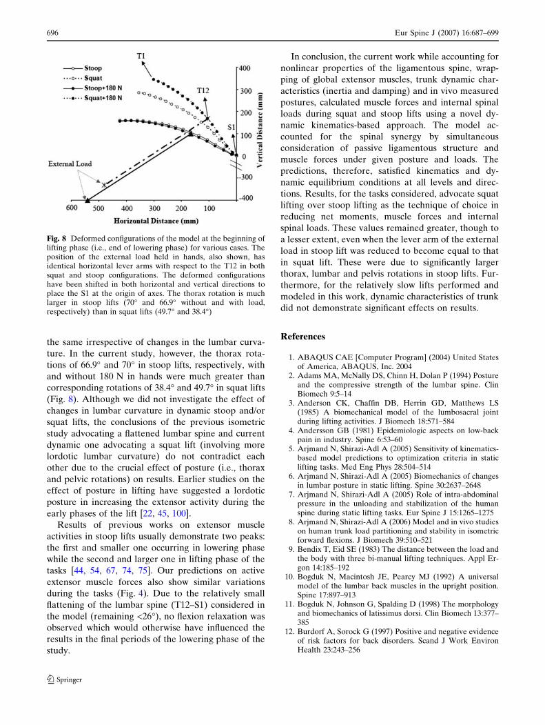
Fig. 8 Deformed configurations of the model at the beginning oflifting phase (i.e., end of lowering phase) for various cases. Theposition of the external load held in hands, also shown, hasidentical horizontal lever arms with respect to the T12 in bothsquat and stoop configurations. The deformed configurationshave been shifted in both horizontal and vertical directions toplace the S1 at the origin of axes. The thorax rotation is muchlarger in stoop lifts (70� and 66.9� without and with load,respectively) than in squat lifts (49.7� and 38.4�)
696 Eur Spine J (2007) 16:687–699
123
13. Buseck M, Schipplein OD, Andersson GB, Andriacchi TP(1988) Influence of dynamic factors and external loads onthe moment at the lumbar spine in lifting. Spine 13:918–921
14. Cappozzo A, Catani F, Leardini A, Benedetti MG, Croce UD(1996) Position and orientation in space of bones duringmovement: experimental artefacts. Clin Biomech 11:90–100
15. Cholewicki J, McGill SM, Norman RW (1991) Lumbarspine loads during the lifting of extremely heavy weights.Med Sci Sports Exerc 23:1179–1186
16. Cholewicki J, McGill S (1996) Mechanical stability of the invivo lumbar spine: implications for injury and chronic lowback pain. Clin Biomech 11:1–15
17. Damkot DK, Pope MH, Lord J, Frymoyer JW (1984) Therelationship between work history, work environment andlow-back pain in men. Spine 9:395–399
18. Davis KG, Marras WS, Waters TR (1998) Evaluation ofspinal loading during lowering and lifting. Clin Biomech13:141–152
19. Davis J, Kaufman KR, Lieber RL (2003) Correlation be-tween active and passive isometric force and intramuscularpressure in the isolated rabbit tibialis anterior muscle. JBiomech 36:505–512
20. Davis KG, Marras WS (2000) The effects of motion ontrunk biomechanics. Clin Biomech 15:703–717
21. Davis JR, Mirka GA (2000) Transverse-contour modelingof trunk muscle-distributed forces and spinal loads duringlifting and twisting. Spine 25:180–189
22. Delitto RS, Rose SJ, Apts DW (1987) Electromyographicanalysis of two techniques for squat lifting. Phys Ther67:1329–1334
23. van Dieen JH, Creemers M, Draisma I, Toussaint HM,Kingma I (1994) Repetitive lifting and spinal shrinkage,effects of age and lifting technique. Clin Biomech 9:367–374
24. van Dieen JH (1997) Are recruitment patterns of the trunkmusculature compatible with a synergy based on the max-imization of endurance? J Biomech 30:1095–1100
25. van Dieen JH, Hoozemans MJ, Toussaint HM (1999) Stoopor squat: a review of biomechanical studies on lifting tech-nique. Clin Biomech 14:685–696
26. Dietrich M, Kedzior K, Zagrajek T (1991) A biomechanicalmodel of the human spinal system. Inst Mech Eng Part [H]205(1):19–26
27. Dolan P, Earley M, Adams MA (1994) Bending and com-pressive stresses acting on the lumbar spine during liftingactivities. J Biomech 27:1237–1248
28. Dolan P, Kingma I, van Dieen J, de Looze MP, ToussaintHM, Baten CT, Adams MA (1999) Dynamic forces actingon the lumbar spine during manual handling. Can they beestimated using electromyographic techniques alone? Spine24:698–703
29. Dvorak J, Panjabi MM, Chang DG, Theiler R, Grob D(1991) Functional radiographic diagnosis of the lumbarspine. Flexion–extension and lateral bending. Spine 16:562–571
30. El-Rich M, Shirazi-Adl A, Arjmand N (2004) Muscleactivity, internal loads, and stability of the human spine instanding postures: combined model and in vivo studies.Spine 29:2633–2642
31. Esola MA, McClure PW, Fitzgerald GK, Siegler S (1996)Analysis of lumbar spine and hip motion during forwardbending in subjects with and without a history of low backpain. Spine 21:71–78
32. Fathallah FA, Marras WS, Parnianpour M (1998) Anassessment of complex spinal loads during dynamic liftingtasks. Spine 23:706–716
33. Ferguson S, Marras W (1997) A literature review of lowback disorder surveillance measures and risk factors. ClinBiomech 12:211–226
34. Frobin W, Brinckmann P, Leivseth G, Biggemann M, Re-ikeras O (1996) Precision measurement of segmental mo-tion from flexion–extension radiographs of the lumbarspine. Clin Biomech 11:457–465
35. Gardner-Morse M, Stokes IA, Laible JP (1995) Role ofmuscles in lumbar spine stability in maximum extensionefforts. J Orthop Res 13:802–808
36. Gracovetsky S, Farfan HF, Lamy C (1981) The mechanismof the lumbar spine. Spine 6:249–262
37. Gracovetsky S (1988) The spinal engine. Springer, BerlinHeidelberg New York
38. Grag A, Herrin GD (1979) Stoop or squat. A biomechan-ical and metabolic evaluation. AIIE Trans 11:293–302
39. Granata KP, Marras WS (1995) The influence of trunkmuscle coactivity on dynamic spinal loads. Spine 20:913–919
40. Granata KP, Marras WS (1995) An EMG-assisted model oftrunk loading during free-dynamic lifting. J Biomech28:1309–1317
41. Granata KP, Sanford AH (2000) Lumbar-pelvic coordina-tion is influenced by lifting task parameters. Spine 25:1413–1418
42. Hagen KB, Hallen J, Harms-Ringdahl K (1993) Physio-logical and subjective responses to maximal repetitive lift-ing employing stoop and squat technique. Eur J ApplPhysiol Occup Physiol 67:291–297
43. Hagen KB, Harms-Ringdahl K (1994) Ratings of perceivedthigh and back exertion in forest workers during repetitivelifting using squat and stoop techniques. Spine 19:2511–2517
44. Haig AJ, Weismann G, Haugh LD, Pope M, Grobler LJ(1993) Prospective evidence for change in paraspinal muscleactivity after herniated nucleus pulposus. Spine 18:926–930
45. Hart DL, Stobbe TJ, Jaraiedi M (1987) Effect of lumbarposture on lifting. Spine 12:138–145
46. Hilber HM, Hughes TJR, Taylor RL (1978) Collocation,dissipation and overshoot for time integration schemes instructural dynamics. Earthq Eng Struct Dyn 6:99–117
47. Holmes JA, Damaser MS, Lehman SL (1992) Erector spi-nae activation and movement dynamics about the lumbarspine in lordotic and kyphotic squat-lifting. Spine 17:327–334
48. Hsiang SM, Brogmus GE, Courtney TK (1997) Low backpain (LBP) and lifting technique—a review. Int J Ind Ergon19:59–74
49. Hughes RE, Chaffin DB, Lavender SA, Andersson GB(1994) Evaluation of muscle force prediction models of thelumbar trunk using surface electromyography. J Orthop Res12:689–698
50. Jorgensen MJ, Marras WS, Gupta P, Waters TR (2003)Effect of torso flexion on the lumbar torso extensor musclesagittal plane moment arms. Spine J 3:363–369
51. Kasra M, Shirazi-Adl A, Drouin G (1992) Dynamics ofhuman lumbar intervertebral joints. Experimental and fi-nite-element investigations. Spine 17:93–102
52. Kingma I, Baten CT, Dolan P, Toussaint HM, van DieenJH, de Looze MP, Adams MA (2001) Lumbar loadingduring lifting: a comparative study of three measurementtechniques. J Electromyogr Kinesiol 11:337–345
53. Kuiper JI, Burdorf A, Verbeek JHAM, Frings-DresenMHW, van der Beek AJ, Viikari-Juntura ERA (1999) Ep-idemiologic evidence on manual materials handling as a riskfactor for back disorders: a systematic review. Int J IndErgon 24:389–404
Eur Spine J (2007) 16:687–699 697
123

54. Lariviere C, Gagnon D, Loisel P (2000) The comparison oftrunk muscles EMG activation between subjects with andwithout chronic low back pain during flexion-extension andlateral bending tasks. J Electromyogr Kinesiol 10:79–91
55. Lariviere C, Gagnon D (1998) Comparison between twodynamic methods to estimate triaxial net reaction momentsat the L5/S1 joint during lifting. Clin Biomech 13:36–47
56. Lee YH, Chiou WK, Chen WJ, Lee MY, Lin YH (1995)Predictive model of intersegmental mobility of lumbarspine in the sagittal plane from skin markers. Clin Biomech10:413–420
57. Lindbeck L, Arborelius UP (1991) Inertial effects fromsingle body segments in dynamic analysis of lifting. Ergo-nomics 34:421–433
58. de Looze MP, Kingma I, Thunnissen W, van Wijk MJ,Toussaint HM (1994) The evaluation of a practical biome-chanical model estimating lumbar moments in occupationalactivities. Ergonomics 37:1495–1502
59. de Looze MP, Dolan P, Kingma I, Baten CTM (1998) Doesan asymmetric straddle-legged lifting movement reduce thelow-back load? Hum Mov Sci 17:243–259
60. Lundberg A (1996) On the use of bone and skin markers inkinematics research. Hum Mov Sci 15:411–422
61. Macintosh JE, Bogduk N, Gracovetsky S (1987) Biome-chanics of the thoracolumbar fascia. Clin Biomech 2:78–83
62. Macintosh J, Bogduk N, Pearcy M (1993) The effects offlexion on the geometry and actions of the lumbar erectorspinae. Spine 18:884–893
63. Markolf KL (1970) Stiffness and damping characteristics ofthoracolumbar spine. In: Proceedings of workshop on bio-engineering approaches to problems of the spine. Divisionof Research Grants, NIH, Bethesda, pp 87–143
64. McClure PW, Esola M, Schreier R, Siegler S (1997) Kine-matic analysis of lumbar and hip motion while rising from aforward, flexed position in patients with and without a his-tory of low back pain. Spine 22:552–558
65. McGill SM, Patt N, Norman RW (1988) Measurement ofthe trunk musculature of active males using CT scan radi-ography: implications for force and moment generatingcapacity about the L4/L5 joint. J Biomech 21:329–341
66. McGill SM (1997) The biomechanics of low back injury:implications on current practice in industry and the clinic. JBiomech 30:465–475
67. McGill SM, Yingling VR, Peach JP (1999) Three-dimen-sional kinematics and trunk muscle myoelectric activity inthe elderly spine—a database compared to young people.Clin Biomech 14:389–395
68. McGill SM, Hughson RL, Parks K (2000) Changes inlumbar lordosis modify the role of the extensor muscles.Clin Biomech 15:777–780
69. McGill SM, Norman RW (1985) Dynamically and staticallydetermined low back moments during lifting. J Biomech18:877–885
70. McGill SM, Norman RW (1986) Partitioning of the L4-L5dynamic moment into disc, ligamentous, and muscularcomponents during lifting. Spine 11:666–678
71. Neumann P, Keller TS, Ekstrom L, Hansson T (1994) Ef-fect of strain rate and bone mineral on the structuralproperties of the human anterior longitudinal ligament.Spine 19:205–211
72. Nussbaum MA, Chaffin DB (1996) Development andevaluation of a scalable and deformable geometric model ofthe human torso. Clin Biomech 11:25–34
73. Oxland TR, Lin RM, Panjabi MM (1992) Three-dimen-sional mechanical properties of the thoracolumbar junction.J Orthop Res 10:573–580
74. Paquet N, Malouin F, Richards CL (1994) Hip-spinemovement interaction and muscle activation patterns dur-ing sagittal trunk movements in low back pain patients.Spine 19:596–603
75. Peach JP, Sutarno CG, McGill SM (1998) Three-dimen-sional kinematics and trunk muscle myoelectric activity inthe young lumbar spine: a database. Arch Phys MedRehabil 79:663–669
76. Pearcy M, Portek I, Shepherd J (1984) Three-dimensionalx-ray analysis of normal movement in the lumbar spine.Spine 9:294–297
77. Pearsall DJ, Reid JG, Livingston LA (1996) Segmentalinertial parameters of the human trunk as determined fromcomputed tomography. Ann Biomed Eng 24:198–210
78. Pearsall DJ (1994) Segmental inertial properties of thehuman trunk as determined from computer tomographyand magnetic resonance imagery. Dissertation, Queen’sUniversity, Kingston
79. Plamondon A, Gagnon M, Maurais G (1988) Application ofa stereoradiographic method for the study of intervertebralmotion. Spine 13:1027–1132
80. Pop DG (2001)Analyse non lineaire par elements finis dusystem actif passif de la colonne vertebrale humaine. Dis-sertation, Genie mecanique, Ecole Polytechnique, Montreal
81. Porter JL, Wilkinson A (1997) Lumbar-hip flexion motion.A comparative study between asymptomatic and chroniclow back pain in 18- to 36-year-old men. Spine 22:1508–1514
82. Potvin JR, Norman RW, McGill SM (1991) Reduction inanterior shear forces on the L4/L5 disc by the lumbarmusculature. Clin Biomech 6:88–96
83. Potvin JR, McGill SM, Norman RW (1991) Trunk muscleand lumbar ligament contributions to dynamic lifts withvarying degrees of trunk flexion. Spine 16:1099–1107
84. Raikova RT, Prilutsky BI (2001) Sensitivity of predictedmuscle forces to parameters of the optimization-based hu-man leg model revealed by analytical and numerical anal-yses. J Biomech 34:1243–1255
85. Sadouk S (1998) Analyse mecanique par elements finis dusysteme actif passif de la colonne lombaire humaine. Dis-sertation, Genie mecanique, Ecole Polytechnique, Montreal
86. Shirazi-Adl A (1989) Nonlinear finite element analysis ofwrapping uniaxial elements. Comput Struct 32:119–123
87. Shirazi-Adl A (1994) Analysis of role of bone complianceon mechanics of a lumbar motion segment. J Biomech Eng116:408–412
88. Shirazi-Adl A, Sadouk S, Parnianpour M, Pop D, El-RichM (2002) Muscle force evaluation and the role of posture inhuman lumbar spine under compression. Eur Spine J11:519–526
89. Shirazi-Adl A, El-Rich M, Pop DG, Parnianpour M (2005)Spinal muscle forces, internal loads and stability in standingunder various postures and loads–application of kinematics-based algorithm. Eur Spine J 14:381–392
90. Shirazi-Adl A (2006) Analysis of large compression loadson lumbar spine in flexion and in torsion using a novelwrapping element. J Biomech 39:267–275
91. Shirazi-Adl A, Parnianpour M (1999) Effect of changes inlordosis on mechanics of the lumbar spine-lumbar curvaturein lifting. J Spinal Disord 12:436–447
92. Shirazi-Adl A, Parnianpour M (2000) Load-bearing andstress analysis of the human spine under a novel wrappingcompression loading. Clin Biomech 15:718–725
93. Stokes I, Gardner-Morse M (1995) Lumbar spine maximumefforts and muscle recruitment patterns predicted by amodel with multijoint muscles and joints with stiffness. JBiomech 28:173–186
698 Eur Spine J (2007) 16:687–699
123

94. Stokes IAF, Gardner-Morse M (1999) Quantitative anat-omy of the lumbar musculature. J Biomech 32:311–316
95. Tesh KM, Dunn JS, Evans JH (1987) The abdominalmuscles and vertebral stability. Spine 12:501–508
96. Toussaint HM, de Winter AF, de Haas Y, de Looze MP,Van Dieen JH, Kingma I (1995) Flexion relaxation duringlifting: implications for torque production by muscle activityand tissue strain at the lumbo-sacral joint. J Biomech28:199–210
97. Toussaint HM, Commissaris DA, Beek PJ (1997) Antici-patory postural adjustments in the back and leg lift. Med SciSports Exerc 29:1216–1224
98. Troup JD, Leskinen TP, Stalhammar HR, Kuorinka IA(1983) A comparison of intraabdominal pressure increases,hip torque, and lumbar vertebral compression in differentlifting techniques. Hum Factors 25:517–525
99. Tveit P, Daggfeldt K, Hetland S, Thorstensson A (1994)Erector spinae lever arm length variations with changes inspinal curvature. Spine 19:199–204
100. Vakos JP, Nitz AJ, Threlkeld AJ, Shapiro R, Horn T (1994)Electromyographic activity of selected trunk and hip mus-cles during a squat lift. Effect of varying the lumbar posture.Spine 19:687–695
101. Wang JL, Parnianpour M, Shirazi-Adl A, Engin AE (1998)The dynamic response of L(2)/L(3) motion segment incyclic axial compressive loading. Clin Biomech 13:S16–S25
102. Wang JL, Parnianpour M, Shirazi-Adl A, Engin AE (2000)Viscoelastic finite-element analysis of a lumbar motionsegment in combined compression and sagittal flexion. Ef-fect of loading rate. Spine 25:310–318
103. Winter DA (2005) Biomechanics and motor control of hu-man movement, 3rd edn. John Wiley, Hoboken
104. Yamamoto I, Panjabi MM, Crisco T, Oxland T (1989)Three-dimensional movements of the whole lumbar spineand lumbosacral joint. Spine 14:1256–1260
105. Zatsiorsky VM, Seluyanov VN (1983) The mass and inertiacharacteristics of the main segments of the human body. In:Matsui H, Kobayashi K (eds) Biomechanics. HumanKinetics Publishers, Champaign, pp 1152–1159
106. Zhang X, Xiong J (2003) Model-guided derivation of lum-bar vertebral kinematics in vivo reveals the difference be-tween external marker-defined and internal segmentalrotations. J Biomech 36:9–17
Eur Spine J (2007) 16:687–699 699
123