an efficient method for high quality rna extraction from...
TRANSCRIPT

Journal of Plant Sciences 2017; 5(2): 68-74
http://www.sciencepublishinggroup.com/j/jps
doi: 10.11648/j.jps.20170502.14
ISSN: 2331-0723 (Print); ISSN: 2331-0731 (Online)
An Efficient Method for High Quality RNA Extraction from Moringa oleifera
Preethi Praba Umesh1, Mohd Akram Ansari
1, 2, Ganapathi Sridevi
1, *
1Department of Plant Biotechnology, School of Biotechnology, Madurai Kamaraj University, Madurai, Tamil Nadu, India 2Division of Genetics and Plant Molecular Biology, CSIR-NBRI, Rana Pratap Marg, Lucknow, Uttar Pradesh, India
Email address:
[email protected] (P. P. Umesh), [email protected] (M. A. Ansari), [email protected] (G. Sridevi) *Corresponding author
To cite this article: Preethi Praba Umesh, Mohd Akram Ansari, Ganapathi Sridevi. An Efficient Method for High Quality RNA Extraction from Moringa
oleifera. Journal of Plant Sciences. Vol. 5, No. 2, 2017, pp. 68-74. doi: 10.11648/j.jps.20170502.14
Received: February 19, 2017; Accepted: February 28, 2017; Published: March 28, 2017
Abstract: Moringa oleifera, the Miracle Tree is rich in all nutrients and minerals. It has prominent distribution of secondary
metabolites like polysaccharides, phenols and mucilage, which makes extraction of RNA quite difficult. A high quality and
pure RNA is prerequisite for the study of high through put transcriptomics and functional genomics. A protocol for isolation of
highly qualitative and quantitative RNA from M. oleifera was optimized by comparing five different methods like Trizol,
Guanidine hydrochloride, combined Trizol and Guanidine hydrochloride, modified CTAB and hot phenol method. The
combined Guanidine hydrochloride and Trizol method gave good yield and pure RNA based on the absorbance A260 value 2.0.
The model plant Nicotiana tabacum served as a positive control in which the Trizol method yielded a good quality and
quantity RNA. The present study is a preliminary step for studying the function and expression pattern of the genes. This is the
first report on the comparison of different RNA extraction methods in M. oleifera to our knowledge.
Keywords: Moringa oleifera, Nicotiana tabacum, RNA Isolation, Trizol, Guanidine Hydrochloride, RT-PCR
1. Introduction
Moringa oleifera commonly known as the drumstick or
ben oil tree is nutritious and the leaves are rich source of
vitamin A, B, C and iron. It is locally known as sajna,
muringa, mullanggay, etc. It is a cultivated species of
monogeneric family, Moringaceae. It is grown mainly in
semiarid, tropical and subtropical areas. Being a fast growing
tree, it grows best in dry, sandy soil, and tolerates poor soil
[9]. It is native to the sub-himalayan tracts of north western
India and are spread in eastward India to the lower part of
china, South East Asia and the Philippines. They are spread
westward from India to Egypt, the horn of Africa, around to
the Mediterranean and West Indies in America. M. oleifera is
first described as a medicinal herb around 2000 BC ago. It is
very nutritious and has a variety of potential uses [21]. There
exists number of reports highlighting the nutritional qualities
of Moringa. The fresh leaves of M. oleifera contains 7 times
more of vitamin C than oranges, 4 times more vitamin A and
calcium than carrot and milk, 3 times more amount of
potassium of bananas, 2 times protein of yogurt and ¾ times
iron of spinach. The leaves are enriched with micronutrients
like phosphorus, zinc, copper, magnesium, etc. [7]. The dried
leaves are characterized with high nutritional content
compared to fresh leaves as it is 10 times more vitamin A of
carrots, ½ the vitamin C of oranges, 17 times the calcium of
milk, 15 times the potassium of bananas, 9 times the protein
of yogurt and 25 times the iron of spinach [6]. The flowers,
leaves, roots and bark are used as folk remedies for tumors,
abdominal discomfort, boils, conjunctivitis, high blood
pressure, hysteria and skin disease [8]. M. oleifera is a
promising food source especially for its nutrient and mineral
rich during the dry season when other foods are typically
scarce [5]. M. oleifera is not nitrogen fixing tree, but its
fruits, flowers and leaves contain 5 to 10% proteins on
average [4].
The genome size of Moringa was estimated to be 315 Mb
and a well annotated high-quality draft genome sequence has
been reported [20]. Moringa possesses a compact genome
and a number of gene families related to heat tolerance, stress
tolerance, high protein content, fast-growth, etc. have been

69 Preethi Praba Umesh et al.: An Efficient Method for High Quality RNA Extraction from Moringa oleifera
identified. Further, the role of micro RNAs from Moringa
that might contribute to medicinal properties was studied
[19]. There is paucity of information available about the
transcriptomics and almost completely absent regarding the
functional genomics aspects in Moringa. As a result there is a
prerequisite for high quality RNA for the functional
characterization of those genes and high throughput
transcriptomics.
Nucleic acid extraction from plants is diverse and varies
among individual plant species. Isolation and purification of
RNA from some of the plant species may be problematic due
to the presence of highly viscous polysaccharides and
secondary metabolites like phenols. The presence of
polyphenols, which are powerful oxidizing agents can reduce
the yield and purity of nucleic acid [13, 16]. It has also been
observed that phenolic compounds are readily oxidized to
form covalently linked quinones [13]. Polyvinylpyrrolidone
(PVP) plays a role in removing phenolic compounds and
secondary metabolites during nucleic acid preparations [2]
and it also prevents browning effect caused due to
polyphenols. PVP can strongly bind to the polyphenol
compounds by its CO-N=group [22]. RNA isolation from
plants like Arabidopsis, tobacco, tomato, potato or maize is
usually achieved by classical phenol/lithium chloride method
and/or guanidine based methods [3, 17, 18]. RNA extraction
from leaves and stem of spinach was done by lithium
chloride method, where the leaves yield good RNA than stem
[14]. Extraction methods also depend on the stages of plant
tissues. In Arabidopsis thaliana, RNA extraction from young
leaves was done by two different methods (trizol and
phenol:chloroform - lithium chloride method) where the later
gave high quality of RNA [1].
Our main objective is to obtain a good quality and quantity
of RNA for the functional genomics detailed transcriptomics
in M. oleifera. We compared five different methods of RNA
extraction from the leaves of M. oleifera (trizol, guanidine
hydrochloride, guanidine hydrochloride with trizol, modified
CTAB and hot phenol). The yield and purity of RNA was
evaluated using spectrophotometer, integrity by gel
electrophoresis and finally confirmed by RT-PCR analyses.
2. Materials and Methods
2.1. Plant Material
The leaves of field grown Moringa oleifera var. PKM 1
was taken for RNA analyses. Five different methods were
deployed for RNA extraction and evaluated.
a. TRIzol method
RNA extraction by Trizol method was performed as per
manufacturer’s instruction (TRI reagent SIGMA-ALDRICH,
USA). One gram of plant tissue (leaf) was ground into fine
powder with the help of liquid nitrogen and transferred into
15 ml polypropylene tubes. The powder is then resuspended
in 5 ml of trizol reagent and mixed by vortexing for 30 sec
followed by the addition of 1/5th
volume of chloroform. The
tube was vortexed approximately for 20 sec and incubated in
room temperature for 15 min. The contents were centrifuged
at 12,000 rpm for 15 min at 4 °C in order to separate DNA
and proteins. The supernatant with RNA phase was
transferred into a fresh tube and added equal amount of ice
cold isopropanol. The tubes were kept at room temperature
for 10 min for precipitation and centrifuged at 12,000 rpm for
30 min at 4 °C. After the precipitation of RNA the
supernatant was discarded and the pellet was washed with
75% ethanol. Finally the pellet was vacuum dried and
dissolved in 20 µl of sterile DEPC water (preheated at 65 °C)
and stored at -80 °C until use.
b. Guanidine hydrochloride with TRIzol method
One gram of leaf tissue was homogenized preliminarily
using liquid nitrogen and later with 5 ml of guanidine
hydrochloride buffer [6.5 M guanidine hydrochloride, 100
mM Tris-Cl (pH 8.0), 100 mM sodium acetate (pH 5.5), and
0.1 M β- mercaptoethanol (added after sterilization)]. Then
the slurry was transferred to the centrifuge tube, mixed
vigorously and incubated at room temperature for 10 min
undisturbed. The samples were centrifuged at 12,000 rpm for
10 min at 4 °C. To the supernatant 5 ml of trizol reagent and
2 ml of chloroform were added. The tube was incubated at
room temperature for 2 min, and centrifuged at 12,000 rpm
for 10 min at 4 °C. The aqueous phase was taken carefully
and transferred to fresh tube. Ice cold isopropanol was added
in 1:1 ratio to the above and incubated in ice for 30 min
followed by centrifugation at 12,000 rpm for 20 min at 4 °C.
The supernatant was discarded and the pellet was washed
using 5 ml of 75% ethanol, centrifuged at 12,000 rpm for 5
min at 4 °C. The pellet was vacuum dried and dissolved in 20
µl of sterile DEPC water (preheated at 65 °C) and stored at -
80 °C until use.
c. Modified CTAB method
The isolation of RNA was performed using CTAB method
with some modifications. One gram of plant tissue was
ground with liquid nitrogen into fine powder. Approximately,
200 mg of leaf powder was taken in a 2 ml eppendorf tube
followed by addition of 1 ml of 2% CTAB buffer [2% CTAB,
100 mM Tris-Cl (pH 8.0), 1.4 M NaCl, 2% PVP, 20 mM
EDTA (pH 8.0), β- mercaptoethanol (added after
sterilization)], mixed for 30 sec and incubated at 65 °C for 30
min. The contents of the tubes were inverted 4-5 times in
between the incubation. After incubation, 800 µl of
chloroform was added and mixed properly. Then the samples
were centrifuged at 10,000 rpm for 10 min at 4 °C. To the
aqueous phase, 800 µl of TE saturated phenol was added,
mixed for 30 sec and centrifuged at 10,000 rpm for 10 min at
4 °C. Again to the supernatant, added equal volume of
chloroform: isoamyl alcohol (24:1) mixed for 30 sec and
centrifuged at 10,000 rpm for 10 min at 4 °C. The
supernatant along with 1/3 volume of 8 M LiCl2 was kept at
4 °C overnight for precipitation of RNA. The samples were
then centrifuged at 10,000 rpm for 20 min 4 °C and the pellet
was washed with 95% ethanol and then with 75% ethanol by
centrifuging at 10,000 rpm for 5 min at 4 °C. The pellet was
air dried and dissolved in 20 µl of sterile DEPC water
(preheated at 65 °C) and stored at -80 °C until use.

Journal of Plant Sciences 2017; 5(2): 68-74 70
d. Hot phenol method
RNA extraction by hot phenol method was performed
according to the method of Pawlowski et al. [15]. One gram
of leaf tissue was ground into fine powder using liquid
nitrogen and transferred to the eppendorf tubes. Added 500
µl of preheated (90 °C) RNA extraction buffer (100 mM
lithium chloride, 1% SDS, 100 mM Tris-Cl, pH 9.0, 100 mM
EDTA) to the powder mixed and incubated at 65 °C water
bath for 30 min. The contents of the tubes were inverted 3-4
times once in every 10 min. Added 500 µl of Tris EDTA
saturated phenol, mixed and centrifuged at 12,000 rpm for 10
min at room temperature. The upper liquid layer was
transferred to the fresh tube, to which equal volume of
chloroform: isoamyl alcohol (24:1) was added, mixed and
centrifuged at 10,000 rpm for 5 min at room temperature
(performed twice). The upper liquid layer was transferred to
a fresh tube and 8 M LiCl2 was added to achieve a final
concentration to 2 M LiCl2 followed by overnight
precipitation at 4 °C. The tubes were centrifuged at 12,000
rpm for 10 min at 4 °C and the supernatant was discarded.
The pellet was washed with 75% ethanol, and centrifuged at
12,000 rpm for 5 min at 4 °C. The pellet was dissolved in
300 µl of 0.3 M sodium acetate (pH 5.2). Added equal
volume of chloroform: isoamyl alcohol (24:1) mixed gently
and centrifuged at 10,000 rpm for 5 min at 4 °C. The upper
aqueous layer was measured and added 95% ethanol, mixed
gently and centrifuged at 12,000 rpm for 10 min at 4 °C.
Finally, the pellet was again washed with 75% ethanol and
centrifuged at 12,000 rpm for 5 min at 4 °C. The pellet was
vacuum dried and dissolved in 20 µl of sterile DEPC water
(preheated at 65 °C) and stored at -80 °C until use.
e. Guanidine hydrochloride method
One gram of plant leaf tissue is homogenized in a
guanidine hydrochloride - buffer containing 6.5 M guanidine
hydrochloride, 100 mM Tris-Cl (pH 8.0), 100 mM sodium
acetate (pH 5.5), and 0.1 M β-mercaptoethanol (added after
sterilization) as described by Logemann et al. [12]. The
slurry was transferred to the centrifuge tube, mixed
vigorously and incubated at room temperature for 10 min
undisturbed. The samples were centrifuged at 12,000 rpm for
10 min at 4 °C. To the supernatant, 2 ml of chloroform was
added. The tube was incubated at room temperature for 2
min, and then centrifuged at 12,000 rpm for 10 min at 4 °C.
The aqueous phase was taken carefully and transferred to
fresh tube. Ice cold isopropanol was added in 1:1 ratio to the
above and incubated in ice for 30 min for precipitation of
RNA, followed by centrifugation at 12,000 rpm for 20 min at
4 °C. The supernatant was discarded and the pellet was
washed using 5 ml of 75% ethanol, centrifuged at 12,000 rpm
for 5 min at 4 °C. The pellet was air dried and dissolved in 20
µl of sterile DEPC water (preheated at 65 °C) and stored at -
80 °C until use.
2.2. RNA Estimation
The total RNA was estimated by measuring the optical
density at 260 nm and 280 nm in UV spectrophotometer. The
RNA sample, 2 µl was mixed in 998 µl of DEPC water,
estimated and calculated by using the formula: [1 OD at 260
nm is equal to 40 ng of RNA] [17].
Concentration of RNA = 40*A260*500
The total RNA was loaded on 1.2% formaldehyde agarose
gel and visualized under UV light to determine the integrity
of RNA.
2.3. DNase Treatment
Total RNA (30 µg) was taken and the reaction volume
made up to 50 µl with 1 µl of DNase enzyme, 40 µM Tris
(pH 7.5) 6 mM MgCl2 and DEPC water. The tubes were
gently tapped and incubated at 37 °C for 30 min. After
incubation, 200 µl RNase free DEPC water was added. To
that equal volume of phenol: chloroform (24:1) was added
and centrifuged at 12,000 rpm for 5 min at 4 °C (repeated
twice). The supernatant was measured, added 0.1 volume of
3 M sodium acetate and 2.5 volume 95% ethanol. The
samples were stored at -70 °C for 30 min and centrifuged at
4 °C, 12,000 rpm for 10 min. The supernatant was discarded
and the pellet was washed by 75% ethanol. The pellet was
vacuum dried and dissolved in 20 µl of sterile DEPC water
(preheated at 65 °C) and stored at -80 °C until use.
2.4. RT-PCR Analysis
Total RNA sample (M. oleifera) after treatment with DNase
I enzyme was subjected to RT-PCR following the instruction
manual of two step RT-PCR kit (TaKaRa, Kyoto, Japan). The
cDNA was synthesized following the manufacturer’s
instruction. After the cDNA synthesis, 5 µl of sample was
taken for PCR using actin primers: forward primer 5’
GCTATTCAGGCTGTCCTTTCCTTGATATG 3’ and reverse
primer 5’ CCGATATCAACATCACACTTCATAATG 3’. The
PCR conditions include initial denaturation at 98 °C for 30 sec,
followed by 30 cycles [98 °C - 10 sec, 50 °C - 30 sec, 72 °C -
1 min] and final extension at 72 °C for 5 min. Total RNA
from Nicotiana tabacum was used as positive control along
with appropriate PCR controls. The PCR products after
amplification were electrophoresed on 1.2% agarose gel.
3. Results and Discussion
A reliable quality of RNA is essential for downstream
applications such as RT-PCR, cDNA library construction,
gene expression studies, high through put transcriptomics etc.
The quality and quantity of RNA differs based on the method
of the isolation. Therefore the present study was attempted to
isolate a high quality and quantity RNA which can be used in
high through put transcriptomics.
A comparison study of total RNA extraction from the
leaves of Moringa oleifera was done by five different
methods: TRIzol method, guanidine hydrochloride method,
combination of guanidine hydrochloride and TRIzol method,
CTAB method with some modifications and hot phenol
method. There were lot of hindrances in obtaining good
quality of RNA from M. oleifera due to presence of high
mucilage content and other polysaccharides. The leaves of

71 Preethi Praba Umesh et al.: An Efficient Method for High Quality RNA Extraction from Moringa oleifera
tissue culture grown wild type Nicotiana tabacum
(Wisconsin38) served as a positive control for the
comparison of different methods of RNA extraction.
The total RNA extracted by five different methods was
analyzed for their yield, purity and integrity by three different
steps. The quantity of total RNA was determined by
measuring the absorbance in UV Spectrophotometer at 260
nm. The quantity of total RNA was comparable in both trizol
method and hot phenol method (292.8 and 291.2 µg/g fresh
weight). The yield of above two methods was higher
compared to the other three methods. Further the combined
guanidine hydrochloride and trizol method gave 212.0 µg/g
FW followed by modified CTAB method and guanidine
hydrochloride method (Table 1). The isolation of RNA was
difficult in plants containing polysaccharide, polyphenols and
mucilage. Plants like Hibiscus tiliaceus [23], Cinnamomum
tenuipilum [24] contains high polysaccharides, mucilages and
other secondary metabolites. RNA isolation from these plants
was performed by modified CTAB method. C. tenuipilum is
highly viscous and RNA extraction was successful by trizol
kit based method, Guanidine thiocyanate method and
Phenol/SDS method. The yield of RNA in C. tenuipilum was
20 µg/g but when tried with CTAB method with some
modification in centrifugation step the yield was 60-100
µg/g. Similar results with increased RNA yield (113.6 µg/g)
was obtained in our modified CTAB method of extraction too
(Table 1).the addition of 2% PVP and 2% PVPP together also
contributed for increased quality and quantity of RNA in the
above method while it is lesser in trizol, hot phenol and
combined trizol and GnHCl methods [23].
The quality of RNA was analyzed spectrophotometrically
by evaluating the A260/A280 ratio. The value range of 1.8 - 2.0
indicates the purity of isolated RNA. The combined GnHCl
and trizol, hot phenol, modified CTAB and trizol methods
gave a similar absorbance value as 1.6 - 1.7. N. tabacum
served as a positive control and gave highest quality of RNA
in trizol method compared to other methods of extraction
(Table 1).
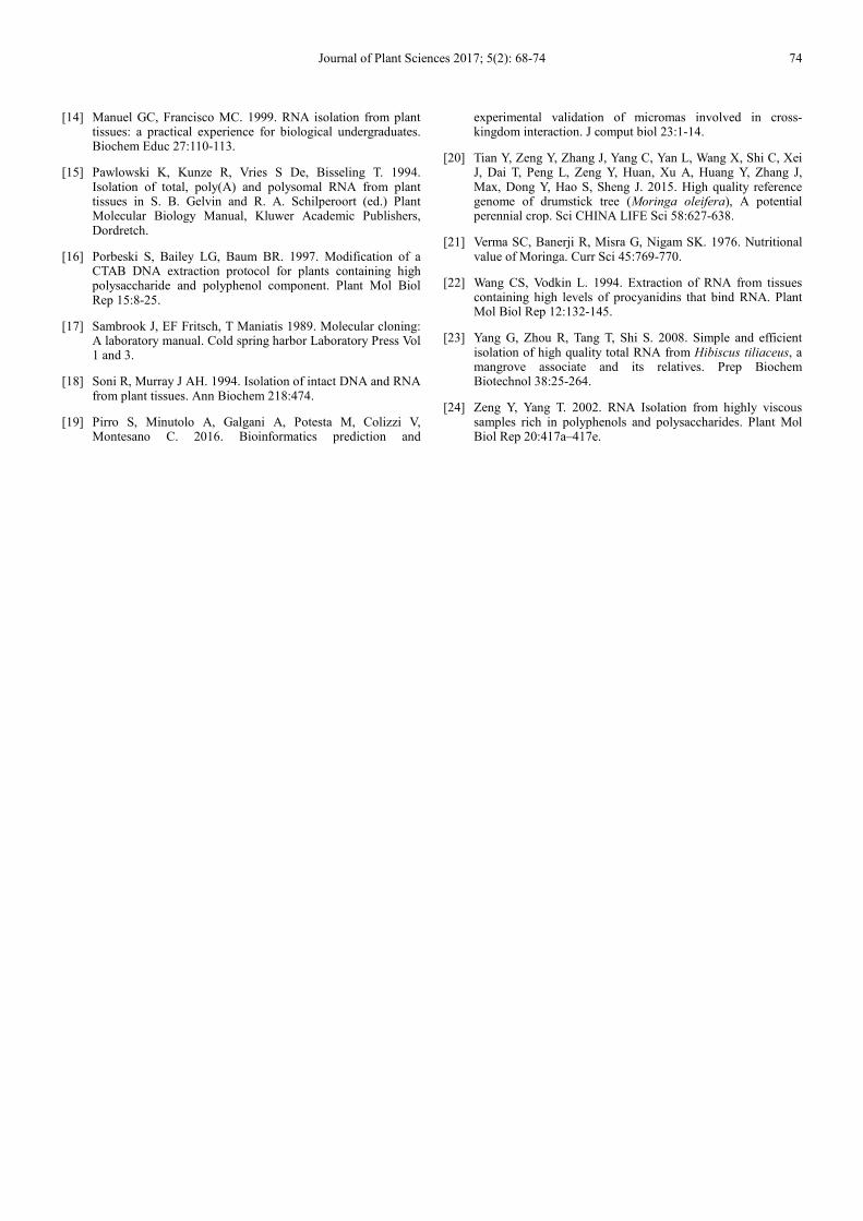
The RNA integrity was analyzed electrophoretically by
loading 5 µg of total RNA on to a 1.2% denaturing agarose
gel and electrophoresed. The RNA was visualized by staining
with ethidium bromide and observed under UV
transilluminator. All the four methods except guanidine
hydrochloride method showed good quality of bands which
revealed that the integrity of RNA is high in all the four
methods in terms of purity and quantity (Fig. 1).
DNase treatment was done to get rid of the trace amounts
of DNA. The purest RNA was achieved by combined
guanidine hydrochloride with trizol method with absorbance
value 2.0. The trizol method gave high quantity of total RNA
(20 µg) with low purity. Both modified CTAB method and
hot phenol method yielded high amount of RNA (18 µg and
16 µg) and similar purity in accordance with trizol method.
The least quality and quantity of RNA was achieved in
guanidine hydrochloride method whose purity was 1.3 only
(Table 2). One possible reason for poor quality or no bands
visualized in the guanidine hydrochloride method (Fig. 1)
may be due to the least purity of RNA with a ratio of 1 (Table
2).
Finally the cDNA was synthesized from the total RNA and
RT-PCR was performed using the tobacco actin primer that
yielded an amplified product of 500 bp in size. The RT-PCR
analysis with actin primer gave intact 500 bp amplification
(Fig. 2) in all the methods except hot phenol method. RT-
PCR performed with trizol method showed less amplification
intensity compared to the other methods. The other methods
such as modified CTAB method, and trizol method did have
comparable yield after DNase treatment (Table 2). On the
other hand the RT-PCR results were not comparable to the
combined method (Fig. 2). Trizol method was proven to be
the best method for the model plant, N. tabacum. This
method provided highest yield, pure and well integrated
RNA compared to all other methods.
RNA isolation protocols in each plant have to be
optimized [10]. A high quality and quantity of RNA is
mandatory for the construction of cDNA library from M.
oleifera to study the transcriptomics. The RNA extracted by
combined guanidine hydrochloride and trizol method gave
high quantity and quality RNA for M. oleifera. The
thiocyanate salt of guanidine is one of the constituent in
TRIzol and contributes for better cell disruption and
denaturation of proteins. The presence of phenol along with
thiocyanate salt and hydrochloric acid resulted in high quality
as well as higher quantity of pure RNA from Moringa.
Guanidine hydrochloride method resulted in poor quantity
and quality of RNA (Table 1 and 2), even though
amplification did occur with actin primers in RT-PCR.
Earlier reports revealed that the total RNA isolation with
guanidine hydrochloride method resulted in good yield [11,
12]. On the contrary, guanidine hydrochloride method in our
study resulted in poor quantity and quality of RNA (Table 1
and 2), even though amplification did occur with actin
primers in RT-PCR.
The hot phenol method of extraction did possess
hindrances due to the presence of high content of phenols and
viscosity. Earlier reports revealed that polysaccharides makes
nucleic acid viscous and the use of PVP in RNA extraction
buffer would be helpful to remove polyphenols [16]. The
spectrophotometer estimation of hot phenol method showed
good quantity and quality RNA compared to all the other
four methods (Table 1). The RNA isolated by this method
showed smear like pattern in 1.2% formaldehyde agarose gel
(Fig. 1), which indicates the degradation of RNA. Further,
this was confirmed by RT-PCR analyses, as the respective
lane did not show any amplification (Fig. 2). Therefore the
above results indicate that hot phenol method may not be a
suitable method for RNA extraction from M. oleifera.
Journal of Plant Sciences 2017; 5(2): 68-74 72
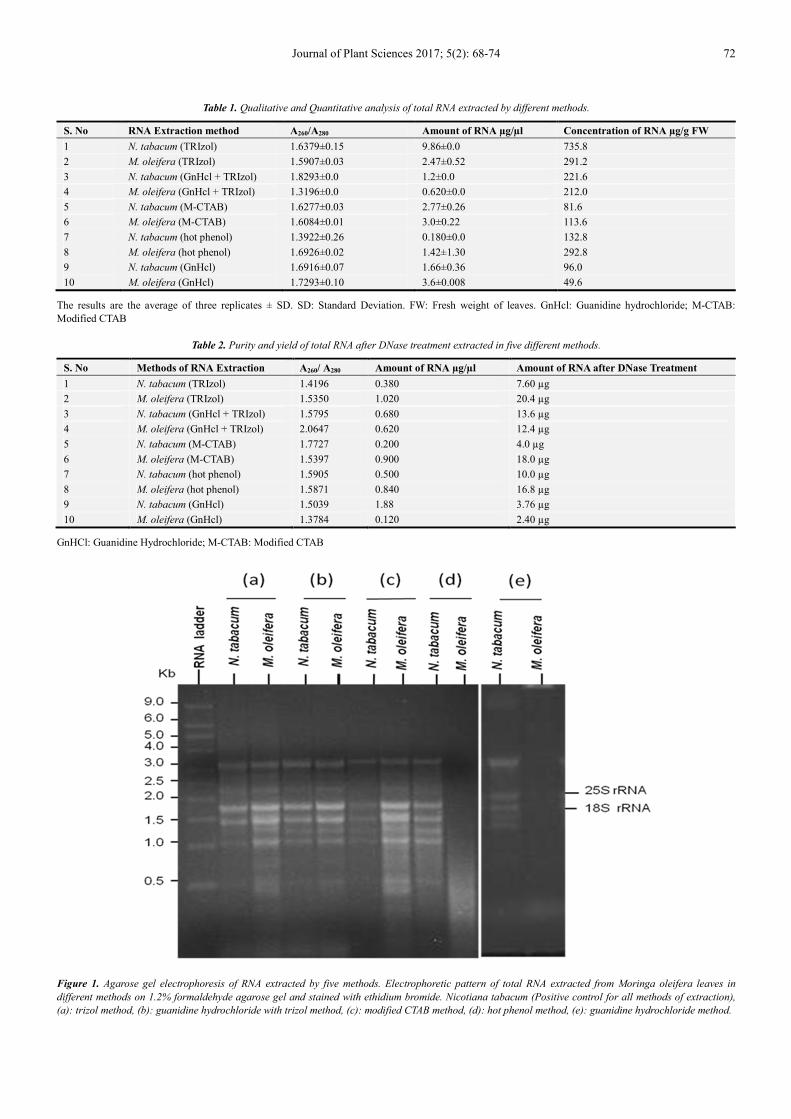
Table 1. Qualitative and Quantitative analysis of total RNA extracted by different methods.
S. No RNA Extraction method A260/A280 Amount of RNA µg/µl Concentration of RNA µg/g FW
1 N. tabacum (TRIzol) 1.6379±0.15 9.86±0.0 735.8
2 M. oleifera (TRIzol) 1.5907±0.03 2.47±0.52 291.2
3 N. tabacum (GnHcl + TRIzol) 1.8293±0.0 1.2±0.0 221.6
4 M. oleifera (GnHcl + TRIzol) 1.3196±0.0 0.620±0.0 212.0
5 N. tabacum (M-CTAB) 1.6277±0.03 2.77±0.26 81.6
6 M. oleifera (M-CTAB) 1.6084±0.01 3.0±0.22 113.6
7 N. tabacum (hot phenol) 1.3922±0.26 0.180±0.0 132.8
8 M. oleifera (hot phenol) 1.6926±0.02 1.42±1.30 292.8
9 N. tabacum (GnHcl) 1.6916±0.07 1.66±0.36 96.0
10 M. oleifera (GnHcl) 1.7293±0.10 3.6±0.008 49.6
The results are the average of three replicates ± SD. SD: Standard Deviation. FW: Fresh weight of leaves. GnHcl: Guanidine hydrochloride; M-CTAB:
Modified CTAB
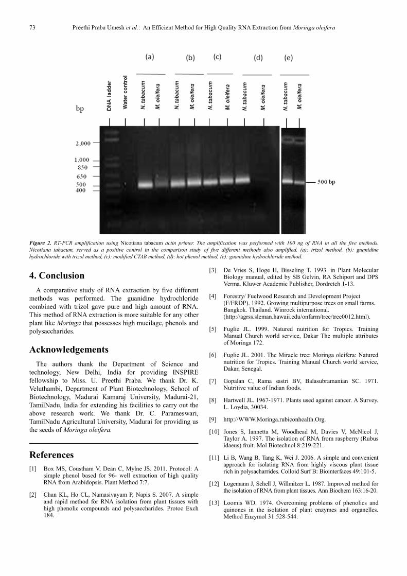
Table 2. Purity and yield of total RNA after DNase treatment extracted in five different methods.
S. No Methods of RNA Extraction A260/ A280 Amount of RNA µg/µl Amount of RNA after DNase Treatment
1 N. tabacum (TRIzol) 1.4196 0.380 7.60 µg
2 M. oleifera (TRIzol) 1.5350 1.020 20.4 µg
3 N. tabacum (GnHcl + TRIzol) 1.5795 0.680 13.6 µg
4 M. oleifera (GnHcl + TRIzol) 2.0647 0.620 12.4 µg
5 N. tabacum (M-CTAB) 1.7727 0.200 4.0 µg
6 M. oleifera (M-CTAB) 1.5397 0.900 18.0 µg
7 N. tabacum (hot phenol) 1.5905 0.500 10.0 µg
8 M. oleifera (hot phenol) 1.5871 0.840 16.8 µg
9 N. tabacum (GnHcl) 1.5039 1.88 3.76 µg
10 M. oleifera (GnHcl) 1.3784 0.120 2.40 µg
GnHCl: Guanidine Hydrochloride; M-CTAB: Modified CTAB
Figure 1. Agarose gel electrophoresis of RNA extracted by five methods. Electrophoretic pattern of total RNA extracted from Moringa oleifera leaves in
different methods on 1.2% formaldehyde agarose gel and stained with ethidium bromide. Nicotiana tabacum (Positive control for all methods of extraction),
(a): trizol method, (b): guanidine hydrochloride with trizol method, (c): modified CTAB method, (d): hot phenol method, (e): guanidine hydrochloride method.
73 Preethi Praba Umesh et al.: An Efficient Method for High Quality RNA Extraction from Moringa oleifera
Figure 2. RT-PCR amplification using Nicotiana tabacum actin primer. The amplification was performed with 100 ng of RNA in all the five methods.
Nicotiana tabacum, served as a positive control in the comparison study of five different methods also amplified. (a): trizol method, (b): guanidine
hydrochloride with trizol method, (c): modified CTAB method, (d): hot phenol method, (e): guanidine hydrochloride method.
4. Conclusion
A comparative study of RNA extraction by five different
methods was performed. The guanidine hydrochloride
combined with trizol gave pure and high amount of RNA.
This method of RNA extraction is more suitable for any other
plant like Moringa that possesses high mucilage, phenols and
polysaccharides.
Acknowledgements
The authors thank the Department of Science and
technology, New Delhi, India for providing INSPIRE
fellowship to Miss. U. Preethi Praba. We thank Dr. K.
Veluthambi, Department of Plant Biotechnology, School of
Biotechnology, Madurai Kamaraj University, Madurai-21,
TamilNadu, India for extending his facilities to carry out the
above research work. We thank Dr. C. Parameswari,
TamilNadu Agricultural University, Madurai for providing us
the seeds of Moringa oleifera.
References
[1] Box MS, Coustham V, Dean C, Mylne JS. 2011. Protocol: A simple phenol based for 96- well extraction of high quality RNA from Arabidopsis. Plant Method 7:7.
[2] Chan KL, Ho CL, Namasivayam P, Napis S. 2007. A simple and rapid method for RNA isolation from plant tissues with high phenolic compounds and polysaccharides. Protoc Exch 184.
[3] De Vries S, Hoge H, Bisseling T. 1993. in Plant Molecular Biology manual, edited by SB Gelvin, RA Schiport and DPS Verma. Kluwer Academic Publisher, Dordretch 1-13.
[4] Forestry/ Fuelwood Research and Development Project (F/FRDP). 1992. Growing multipurpose trees on small farms. Bangkok. Thailand. Winrock international. (http://agrss.sleman.hawaii.edu/onfarm/tree/tree0012.html).
[5] Fuglie JL. 1999. Natured nutrition for Tropics. Training Manual Church world service, Dakar The multiple attributes of Moringa 172.
[6] Fuglie JL. 2001. The Miracle tree: Moringa oleifera: Natured nutrition for Tropics. Training Manual Church world service, Dakar, Senegal.
[7] Gopalan C, Rama sastri BV, Balasubramanian SC. 1971. Nutritive value of Indian foods.
[8] Hartwell JL. 1967-1971. Plants used against cancer. A Survey. L. Loydia, 30034.
[9] http://WWW.Moringa.rubiconhealth.Org.
[10] Jones S, Iannetta M, Woodhead M, Davies V, McNicol J, Taylor A. 1997. The isolation of RNA from raspberry (Rubus idaeus) fruit. Mol Biotechnol 8:219-221.
[11] Li B, Wang B, Tang K, Wei J. 2006. A simple and convenient approach for isolating RNA from highly viscous plant tissue rich in polysacharrides. Colloid Surf B: Biointerfaces 49:101-5.
[12] Logemann J, Schell J, Willmitzer L. 1987. Improved method for the isolation of RNA from plant tissues. Ann Biochem 163:16-20.
[13] Loomis WD. 1974. Overcoming problems of phenolics and quinones in the isolation of plant enzymes and organelles. Method Enzymol 31:528-544.
Journal of Plant Sciences 2017; 5(2): 68-74 74
[14] Manuel GC, Francisco MC. 1999. RNA isolation from plant tissues: a practical experience for biological undergraduates. Biochem Educ 27:110-113.
[15] Pawlowski K, Kunze R, Vries S De, Bisseling T. 1994. Isolation of total, poly(A) and polysomal RNA from plant tissues in S. B. Gelvin and R. A. Schilperoort (ed.) Plant Molecular Biology Manual, Kluwer Academic Publishers, Dordretch.
[16] Porbeski S, Bailey LG, Baum BR. 1997. Modification of a CTAB DNA extraction protocol for plants containing high polysaccharide and polyphenol component. Plant Mol Biol Rep 15:8-25.
[17] Sambrook J, EF Fritsch, T Maniatis 1989. Molecular cloning: A laboratory manual. Cold spring harbor Laboratory Press Vol 1 and 3.
[18] Soni R, Murray J AH. 1994. Isolation of intact DNA and RNA from plant tissues. Ann Biochem 218:474.
[19] Pirro S, Minutolo A, Galgani A, Potesta M, Colizzi V, Montesano C. 2016. Bioinformatics prediction and
experimental validation of micromas involved in cross-kingdom interaction. J comput biol 23:1-14.
[20] Tian Y, Zeng Y, Zhang J, Yang C, Yan L, Wang X, Shi C, Xei J, Dai T, Peng L, Zeng Y, Huan, Xu A, Huang Y, Zhang J, Max, Dong Y, Hao S, Sheng J. 2015. High quality reference genome of drumstick tree (Moringa oleifera), A potential perennial crop. Sci CHINA LIFE Sci 58:627-638.
[21] Verma SC, Banerji R, Misra G, Nigam SK. 1976. Nutritional value of Moringa. Curr Sci 45:769-770.
[22] Wang CS, Vodkin L. 1994. Extraction of RNA from tissues containing high levels of procyanidins that bind RNA. Plant Mol Biol Rep 12:132-145.
[23] Yang G, Zhou R, Tang T, Shi S. 2008. Simple and efficient isolation of high quality total RNA from Hibiscus tiliaceus, a mangrove associate and its relatives. Prep Biochem Biotechnol 38:25-264.
[24] Zeng Y, Yang T. 2002. RNA Isolation from highly viscous samples rich in polyphenols and polysaccharides. Plant Mol Biol Rep 20:417a–417e.