efficient rna extraction protocol for …2)/32.pdfefficient rna extraction protocol for the wood...
TRANSCRIPT

Pak. J. Bot., 48(2): 661-672, 2016.
EFFICIENT RNA EXTRACTION PROTOCOL FOR THE WOOD MANGROVE SPECIES LAGUNCULARIA RACEMOSA SUITED FOR
NEXT-GENERATION RNA SEQUENCING
MAURICIO WOLF WILWERTH1, PRISCILLA DE BARROS ROSSETTO1*, FERNANDA REINERT2, RAQUEL SOARES PEIXOTO3 AND MÁRCIO ALVES-FERREIRA1
1Laboratório de Genética Molecular Vegetal, UFRJ, Av. Prof. Rodolpho Paulo Rocco, s/n, Bloco A CCS Cidade
Universitária, 21941-617, Rio de Janeiro, RJ, Brazil 2Laboratório de Ecofisiologia Vegetal, UFRJ, Av. Prof. Rodolpho Paulo Rocco, s/n, Bloco A CCS Cidade Universitária,
21941-617, Rio de Janeiro, RJ, Brazil 3Laboratório de Ecologia Microbiana Molecular, UFRJ, Av. Prof. Rodolpho Paulo Rocco, s/n, Bloco K CCS Cidade
Universitária, 21941-617, Rio de Janeiro, RJ, Brazil. *Corresponding author’s email: [email protected]; Phone - +55 21 39386381
Abstract
Mangrove flora and habitat have immeasurable importance in marine and coastal ecology as well as in the economy.
Despite their importance, they are constantly threatened by oil spill accidents and environmental contamination; therefore, it is crucial to understand the changes in gene expression to better predict toxicity in these plants. Among the species of Atlantic coast mangrove (Americas and Africa), Laguncularia racemosa, or white mangrove, is a conspicuous species. The wide distribution of L. racemosa in areas where marine oil exploration is rapidly increasing make it a candidate mangrove species model to uncover the impact of oil spills at the molecular level with the use of massive transcriptome sequencing. However, for this purpose, the RNA extraction protocol should ensure low levels of contaminants and structure integrity. In this study, eight RNA extraction methods were tested and analysed using downstream applications. The InviTrap Spin Plant RNA Mini Kit performed best with regard to purity and integrity. Moreover, the obtained RNA was submitted to cDNA synthesis and RT-PCR, successfully generating amplification products of the expected size. These results show the applicability of the RNA obtained here for downstream methodologies, such as the construction of cDNA libraries for the Illumina Hi-seq platform.
Key words: L. racemosa; RNA isolation; RT-PCR; Mangrove; Guanidine (iso) thiocyanate. Introduction
Mangrove forests are ecosystems of great importance, not only from a biological perspective but also economically and socially. Mangrove forests protect human communities against sea erosion and storms (Fosberg & Chapman, 1971; Dahdouh-Guebas et al., 2005; Barbier et al., 2008; Das & Vincent, 2009). The mangroves are also an essential habitat for a wide range of terrestrial and sea species (Robertson & Duke, 1987; Primavera, 1998; Kathiresan & Bingham, 2001; Luther & Greenberg, 2009) and is a source and sink of many nutrients and sediments for other marine habitats, such as algal communities and coral reefs (Dorenbosch et al., 2004; Duke et al., 2007). It constitutes the economic support of many coastal regions, having great importance in the fishery market (Costanza et al., 1997). Despite its importance, this ecosystem is constantly affected by anthropic degradation due to urbanisation, aquaculture and wood exploitation for coal production (Polidoro et al., 2010). Another increasing problem is the threat of oil spills, which affect mangrove forests more than any other environment (Hoff, 2010). To analyse the extent of stress damage, transcriptomic analysis has emerged as a crucial tool (Alkio et al., 2005; Öktem et al., 2008; Liu et al., 2009; Chen et al., 2011).
The purification of RNA free of contaminants and of high integrity is an essential step in gene expression analysis methodologies. There is a wide range of contaminants that may hamper the quality of RNA extraction methods, especially when sampling from extreme habitats. Mangrove flora are rich in phenolic and
secondary compounds, making RNA recovery a challenge (Sahebi et al., 2013). Few studies have been succeeded in purifying good quality RNA from mangrove species, such as Avicennia germinans and Rhizophora mangle (Gonzalez-Mendoza et al., 2008; Dassanayake et al., 2009; Rubio-Piña &Zapata-Pérez, 2011; Sahebi et al., 2013).
The aim of this study is to evaluate the performance of different methods of RNA isolation and purification in leaves and roots of Laguncularia racemosa grown in a greenhouse. RNA was extracted from plants grown under controlled conditions and under stress caused by exposure to oil. The extracted RNA will be used in the construction of cDNA libraries and in the performance of RNA-Seq experiments. This step is crucial for possible transcriptomic studies in this species and facilitates the elucidation of the expression profile of L. racemosa in response to oil spills. Materials and Methods Plant and growth conditions: Laguncularia racemosa propagules and mangrove sediment were collected in February 2011 from the Restinga da Marambaia, Rio de Janeiro, Brazil (23°3´27´´S 43°33´58´´W) (Fig. 1). The propagules were sown in moist filter paper, and the seedlings were planted directly in the collected sediment and grown in a greenhouse. After seven months, the plants were transferred to plastic pots (3 L) and grown for a further seven months (Fig. 2A). The plants were maintained under 50% natural sun light and at a temperature range of 22.7 to 38.3°C.

MAURICIO WOLF ET AL., 662
Fig. 1. Propagule harvest area. (A) Geographical outline of Rio de Janeiro with Restinga da Marambaia shown in detail (Google Earth); (B) Photo of the harvest area; (C) harvested propagules. Experimental conditions and treatments: For the oil exposure, 185 mL of Marine Fuel 380 (MF-380) was added to the pots (12 L m-2)– (Proffitt et al., 1995). The marine fuel MF-380 is a mixture of diesel and heavy fuel oil (Soares et al., 2006; Sodré et al., 2013). Prior to oil application, ten holes were made in the sediment with chopsticks to ensure oil penetration (Fig. 2B and 2C). After 12 hours, 48 hours and 17 days of oil exposure, samples were collected from the roots and leaves of plants watered with tap-water (control
samples – Fig. 2D) and plants exposed to MF-380 (tested samples – Fig. 2E). For each time point, three biological samples were collected. Root samples were composed of seven fragments from three distinct root regions (Supplementary Fig. 1). Leaf samples were composed of the third totally expanded leaf pair from the shoot apex. After the harvest, the root fragments were subjected to a series of washes and dried with a paper towel. The leaves and root samples were snap frozen in liquid N2 and stored at -80°C until use.

RNA EXTRACTION PROTOCOL FOR LAGUNCULARIA RACEMOSA 663
Fig. 2. Plant growth and experimental conditions. (A) Plants in definitive vases in the greenhouse 14 months after harvest; (B) Holes made in sediment to increase oil percolation; (C) Spilling oil on a vase plant; (D) Control plant and (E) Treated plant.

MAURICIO WOLF ET AL., 664
Supplementary Fig. 1. Sample harvesting. From five plants per treatment, the third totally expanded leaf pair was harvested. The roots were visually separated into three distinct areas, collecting an equal amount of material from each one. Each collected fragment was 3 cm in length and 1.5 mm in diameter. RNA extraction: The samples collected 12 hours after oil exposure were ground to a fine powder using liquid nitrogen and a mortar and pestle; 100 mg (or 200 mg for one method) of the material were collected in 2 mL centrifuge microtubes for RNA extraction, and the RNA samples obtained from four different plants were mixed to produce one sample. Eight different methods were tested (Supplementary Table 1), including the InviTrap Spin Plant RNA Mini kit with Lysis Solution RP (STRATEC Molecular, Berlin, BE, Germany), RNeasy Plant Mini Kit with buffer RLC (QIAGEN, Hilden, NW, Germany), Agilent Plant RNA Isolation Mini Kit (Agilent Technologies, Santa Clara, CA, USA), Plant RNA Reagent (Invitrogen Corporation, Carlsbad, CA, USA) and TRIzol Reagent (Life Technologies, Carlsbad, CA, USA) in addition to three protocols adapted from previous studies (Wang et al., 2007; Gonzalez-Mendoza et al., 2008; Rubio-Piña &Zapata-Pérez, 2011). For the commercial kits, the procedures were performed according to the recommendations of the manufacturer with 100 mg of tissue. For the literature-established protocols, the procedures were performed according to the authors' original recommended methods (Wang et al., 2007; Gonzalez-Mendoza et al., 2008). The Rubio-Piña and Zapata-Pérez (2011) method was originally designed for 200 mg of starting material. The work surfaces were cleaned with 0.1% SDS, the tips used were RNase-free and were purchased from Axygen (Corning Life Sciences, Union City, CA, USA), and the solutions were prepared with autoclaved DEPC-treated water. Analysis of RNA quality: RNA concentration and purity were determined using a NanoDrop™ Spectrophotometer ND-2000 (Thermo Scientific, Wilmington, DE, USA), and the integrity of the RNA was also assessed by 1.2% agarose gel electrophoresis and ethidium bromide staining. Gel-running apparatus was treated with 0.1% SDS and then washed with autoclaved DEPC-treated water. In addition, the RNA integrity and concentration were evaluated using an Agilent 2100 Bioanalyzer (Agilent Technologies, Santa Clara, CA, USA).
Reverse transcription (RT) and polymerase chain reaction (PCR): To evaluate the quality of the RNA obtained using the InviTrap Spin Plant Mini Kit for downstream applications, RNA was extracted from samples collected after 12 h, 48 h and 17 days of oil exposure. RNA samples obtained from three different plants were mixed to produce a single compound sample and were treated with DNase according to the manufacturer´s protocol (TURBO DNA-free Kit, Ambion-Life Technologies). cDNAs were synthesised by Superscript III (Life Technologies, Carlsbad, CA, USA) following the manufacturer’s instructions and stored at -20°C. Polymerase chain reactions were performed in microtubes with an Eppendorf Master Cycler Personal thermocycler. Reaction mixtures contained 5 μL of diluted cDNA (40 times), 0.2 μM of each primer, 50 μM of each dNTP, 1× PCR Buffer, 3 mM MgCl2, and 0.25 units of Platinum Taq DNA polymerase (Life Technologies, Carlsbad, CA, USA), in a total volume of 25 μL. The synthesized cDNA was used for PCR amplifications of ribulose-bisphosphate carboxylase large chain (rbcl) and maturase (MatR) which are sequences available on Mangrove Transcriptome Database (http://mangrove.illinois.edu/ transcriptome); and more two genes we have cloned, actin7 (ACT7), widely used as a reference gene (Jensen et al., 2013), and heat shock protein 90.3 (Hsp90.3), belong to a family know be responsive for almost all stresses (Al-Whaibi, 2011) (Table 1). The cycling program was as follows: initial denaturation at 94°C for 3 minutes; denaturation at 94°C for 30 seconds; annealing of primers for 30 seconds and extension at 72°C for 45 seconds, which was done 28 times and with an annealing temperature of 55°C for ACT7, 24 times and 50°C for rbcl; 28 times and 50°C for MatR, 30 times and 55°C for Hsp90.3; and an extension step of 5 minutes at 72°C. The expected lengths of the four fragments were 246 bp (ACT7), 499 bp (rbcl), 260 bp (MatR) and 548 (Hsp90.3). The PCR reactions were conducted with total RNA samples as the template under the same conditions to evaluate DNA contamination. Results RNA extraction from leaves and roots of Laguncularia racemosa: In total, eight methods for RNA isolation were tested in the four biological samples: control leaves, control roots, treated leaves and treated roots. A compilation of all assessed quality parameters is shown in Table 2. The concentration and purity of the isolated RNA was evaluated by spectrophotometry in the NanoDrop 2000 equipment .The InviTrap Spin Plant Mini Kit produced yields ranging from 24.7 to 46.5 µg/g for leaves and 66.8 to 102.6 µg/g for roots. The A260/280 ratios were higher than 1.7, and the A260/230 ratios ranged from 0.8 to 1.84. The extraction with the RNeasy Plant Mini kit from QIAGEN produced the lowest yields, ranging from 8.08 to 8.3 µg/g for leaves and 18.24 to 30.39 µg/g for roots. The A260/280 ratio was higher than 1.17, and the A260/230 ratio was very low, ranging from 0.33 to 0.5. The Agilent Kit performed poorly for leaves, yielding 4.5 to 9 µg/g for leaves and 25 to 30 µg/g for roots.

RNA EXTRACTION PROTOCOL FOR LAGUNCULARIA RACEMOSA 665

MAURICIO WOLF ET AL., 666
Table 2. Spectrophotometry analysis of RNA contaminants in the eight RNA isolation methods tested for leaves and roots of L. racemosa. We present a representative median value of Nano Drop spectrophotometer
ratios and concentrations (µg/g tissue) for the eight tested methods. InviTrap
Samples Purity (260/280) Purity (260/230) Total yield (µg/g tissue)
Leaves 1.89 0.63 24.7 Roots 2.01 0.84 66.8
Leaves from Treated Plants 1.83 0.93 46.5 Roots from Treated Plants 1.42 0.80 102.6
RNeasy Samples
Purity (260/280) Purity (260/230) Total yield (µg/g tissue) Leaves 1.22 0.33 8.61 Roots 1.40 0.50 30.4
Leaves from Treated Plants 1.17 0.35 8.1 Roots from Treated Plants 1.37 0.40 18.2
Agilent Samples
Purity (260/280) Purity (260/230) Total yield (µg/g tissue) Leaves 1.52 0.60 4.5 Roots 1.60 0.57 25.0
Leaves from Treated Plants 1.37 0.51 9.0 Roots from Treated Plants 1.60 0.55 30.0
Plant RNA Reagent Samples
Purity (260/280) Purity (260/230) Total yield (µg/g tissue) Leaves 1.44 0.51 256.6 Roots 1.70 0.50 194.1
Leaves from Treated Plants 1.32 0.46 466.2 Roots from Treated Plants 1.53 0.60 191.5
TRIzol Samples
Purity (260/280) Purity (260/230) Total yield (µg/g tissue) Leaves 0.88 0.32 295.7 Roots 1.30 0.43 60.8
Leaves from Treated Plants 0.93 0.38 249.2 Roots from Treated Plants 1.35 0.45 148.0
Wang et al. (2007) Samples
Purity (260/280) Purity (260/230) Total yield (µg/g tissue) Leaves 1.66 0.87 12.6 Roots 1.61 0.68 6.6
Leaves from Treated Plants 1.42 0.78 19.7 Roots from Treated Plants 1.35 0.70 13.4
Gonzalez-Mendonza et al. (2008) Samples
Purity (260/280) Purity (260/230) Total yield (µg/g tissue) Leaves 1.13 0.50 30.0 Roots 1.29 0.47 40.0
Leaves from Treated Plants 1.18 0.57 20.0 Roots from Treated Plants 1.32 0.54 74.4
Rubio-Piña & Zapata-Pérez (2011) Samples
Purity (260/280) Purity (260/230) Total yield (µg/g tissue) Leaves 1.57 0.85 144.9 Roots 1.44 0.76 38.0
Leaves from Treated Plants 1.25 0.62 183.0 Roots from Treated Plants 1.43 0.59 40.6
RNA EXTRACTION PROTOCOL FOR LAGUNCULARIA RACEMOSA 667
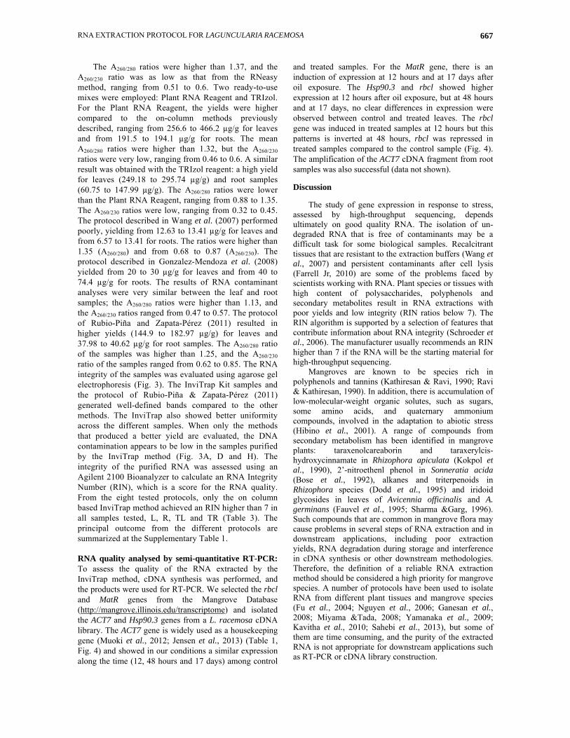
The A260/280 ratios were higher than 1.37, and the A260/230 ratio was as low as that from the RNeasy method, ranging from 0.51 to 0.6. Two ready-to-use mixes were employed: Plant RNA Reagent and TRIzol. For the Plant RNA Reagent, the yields were higher compared to the on-column methods previously described, ranging from 256.6 to 466.2 µg/g for leaves and from 191.5 to 194.1 µg/g for roots. The mean A260/280 ratios were higher than 1.32, but the A260/230 ratios were very low, ranging from 0.46 to 0.6. A similar result was obtained with the TRIzol reagent: a high yield for leaves (249.18 to 295.74 µg/g) and root samples (60.75 to 147.99 µg/g). The A260/280 ratios were lower than the Plant RNA Reagent, ranging from 0.88 to 1.35. The A260/230 ratios were low, ranging from 0.32 to 0.45. The protocol described in Wang et al. (2007) performed poorly, yielding from 12.63 to 13.41 µg/g for leaves and from 6.57 to 13.41 for roots. The ratios were higher than 1.35 (A260/280) and from 0.68 to 0.87 (A260/230). The protocol described in Gonzalez-Mendoza et al. (2008) yielded from 20 to 30 µg/g for leaves and from 40 to 74.4 µg/g for roots. The results of RNA contaminant analyses were very similar between the leaf and root samples; the A260/280 ratios were higher than 1.13, and the A260/230 ratios ranged from 0.47 to 0.57. The protocol of Rubio-Piña and Zapata-Pérez (2011) resulted in higher yields (144.9 to 182.97 µg/g) for leaves and 37.98 to 40.62 µg/g for root samples. The A260/280 ratio of the samples was higher than 1.25, and the A260/230 ratio of the samples ranged from 0.62 to 0.85. The RNA integrity of the samples was evaluated using agarose gel electrophoresis (Fig. 3). The InviTrap Kit samples and the protocol of Rubio-Piña & Zapata-Pérez (2011) generated well-defined bands compared to the other methods. The InviTrap also showed better uniformity across the different samples. When only the methods that produced a better yield are evaluated, the DNA contamination appears to be low in the samples purified by the InviTrap method (Fig. 3A, D and H). The integrity of the purified RNA was assessed using an Agilent 2100 Bioanalyzer to calculate an RNA Integrity Number (RIN), which is a score for the RNA quality. From the eight tested protocols, only the on column based InviTrap method achieved an RIN higher than 7 in all samples tested, L, R, TL and TR (Table 3). The principal outcome from the different protocols are summarized at the Supplementary Table 1. RNA quality analysed by semi-quantitative RT-PCR: To assess the quality of the RNA extracted by the InviTrap method, cDNA synthesis was performed, and the products were used for RT-PCR. We selected the rbcl and MatR genes from the Mangrove Database (http://mangrove.illinois.edu/transcriptome) and isolated the ACT7 and Hsp90.3 genes from a L. racemosa cDNA library. The ACT7 gene is widely used as a housekeeping gene (Muoki et al., 2012; Jensen et al., 2013) (Table 1, Fig. 4) and showed in our conditions a similar expression along the time (12, 48 hours and 17 days) among control
and treated samples. For the MatR gene, there is an induction of expression at 12 hours and at 17 days after oil exposure. The Hsp90.3 and rbcl showed higher expression at 12 hours after oil exposure, but at 48 hours and at 17 days, no clear differences in expression were observed between control and treated leaves. The rbcl gene was induced in treated samples at 12 hours but this patterns is inverted at 48 hours, rbcl was repressed in treated samples compared to the control sample (Fig. 4). The amplification of the ACT7 cDNA fragment from root samples was also successful (data not shown). Discussion
The study of gene expression in response to stress, assessed by high-throughput sequencing, depends ultimately on good quality RNA. The isolation of un-degraded RNA that is free of contaminants may be a difficult task for some biological samples. Recalcitrant tissues that are resistant to the extraction buffers (Wang et al., 2007) and persistent contaminants after cell lysis (Farrell Jr, 2010) are some of the problems faced by scientists working with RNA. Plant species or tissues with high content of polysaccharides, polyphenols and secondary metabolites result in RNA extractions with poor yields and low integrity (RIN ratios below 7). The RIN algorithm is supported by a selection of features that contribute information about RNA integrity (Schroeder et al., 2006). The manufacturer usually recommends an RIN higher than 7 if the RNA will be the starting material for high-throughput sequencing.
Mangroves are known to be species rich in polyphenols and tannins (Kathiresan & Ravi, 1990; Ravi & Kathiresan, 1990). In addition, there is accumulation of low-molecular-weight organic solutes, such as sugars, some amino acids, and quaternary ammonium compounds, involved in the adaptation to abiotic stress (Hibino et al., 2001). A range of compounds from secondary metabolism has been identified in mangrove plants: taraxenolcareaborin and taraxerylcis-hydroxycinnamate in Rhizophora apiculata (Kokpol et al., 1990), 2’-nitroethenl phenol in Sonneratia acida (Bose et al., 1992), alkanes and triterpenoids in Rhizophora species (Dodd et al., 1995) and iridoid glycosides in leaves of Avicennia officinalis and A. germinans (Fauvel et al., 1995; Sharma &Garg, 1996). Such compounds that are common in mangrove flora may cause problems in several steps of RNA extraction and in downstream applications, including poor extraction yields, RNA degradation during storage and interference in cDNA synthesis or other downstream methodologies. Therefore, the definition of a reliable RNA extraction method should be considered a high priority for mangrove species. A number of protocols have been used to isolate RNA from different plant tissues and mangrove species (Fu et al., 2004; Nguyen et al., 2006; Ganesan et al., 2008; Miyama &Tada, 2008; Yamanaka et al., 2009; Kavitha et al., 2010; Sahebi et al., 2013), but some of them are time consuming, and the purity of the extracted RNA is not appropriate for downstream applications such as RT-PCR or cDNA library construction.

MAURICIO WOLF ET AL., 668
Fig. 3. Visualization of the RNA profiles from eight different extraction methods. (A) InviTrap Spin Plant Mini Kit (STRATEC Molecular, Berlin, BE, Germany), (B) RNeasy Plant Mini Kit (QIAGEN, Hilden, NW, Germany), (C) Plant RNA Isolation Mini Kit (Agilent Technologies, Santa Clara, CA, USA), (D) Plant RNA Reagent (Invitrogen Corporation, Carlsbad, CA, USA), (E) TRIzol Reagent (Life Technologies, Carlsbad, CA, USA), (F) Wang et al. (2007), (G) Gonzalez-Mendoza et al. (2008), and (H) Rubio-Piña and Zapata-Pérez (2011). L. racemosa samples: 1-leaves, 2-roots, 3-leaves from treated plants and 4-roots from treated plants. The 1.2 % agarose gel was stained with ethidium bromide
The eight methodologies tested in this study are very diverse. We divided them into three groups according to their main differences: column-based RNA purification (InviTrap Spin Plant Mini Kit, RNeasy Plant Mini kit and Agilent Plant RNA Isolation Mini Kit), ready-to-use mixes (TRIzol and RNA Plant Reagent) and phenol–chloroform extraction in-house protocols previously employed for mangrove and wood species (Wang et al., 2007; Gonzalez-Mendoza et al., 2008;
Rubio-Piña &Zapata-Pérez, 2011). The column-based RNA purification kits are easy and fast, but are costly. In contrast, ready-to-use mixes and phenol–chloroform extraction are cheaper but more laborious and time-consuming. However, as shown here, one does not necessarily have the choice, because of the eight methods tested, only one was satisfactory.
In our hands, the InviTrap Spin Plant Mini Kit generated the best results, based on the uniformity of the

RNA EXTRACTION PROTOCOL FOR LAGUNCULARIA RACEMOSA 669
yields, the purity ratio values and RNA integrity. This spin column-based method generated the best values for the A260/280 (protein contamination) and A260/230 (carbohydrate and phenol contamination) ratios (Table 2). The InviTrap RP lysis buffer composition is not available from Startec Molecular company. Although, the user manual mentioned that the RP buffer is adapted to plant material with high phenol content and it does not contain guanidine (iso)thiocyanate what may cause solidification of the sample depending on the content and type of secondary metabolites in some plant materials. Among the other column-based RNA purification methods, only the RNAeasy method does not contain guanidine (iso)thiocynate in the extraction buffer. Although, it employs guanidine thiocyanate in the further step (RW1 buffer) (Supplementary Table 1). Therefore, this difference may account for part of the differences observed in integrity, purity and yields of total RNA between InviTrap and the other methods. The Plant RNA Isolation Mini Kit did not fulfil the recommended absorbance ratios. In addition, the yields of RNA from leaves were low. RNA isolation with the TRIzol and Plant RNA Reagent methods, both commercial reagents, resulted in contamination and degradation, respectively, of the samples. Therefore, we concluded that these methods are unsuitable for obtaining high-quality RNA from L. racemosa. It is well known that the cetyltrimethylammonium bromide (CTAB)-based methods are recommended for tissues with high polysaccharide, secondary metabolite content (Gasic et al., 2004; Yin et al., 2011; Muoki et al., 2012). However, the in-house methods used in this work that are based on CTAB, Wang et al. (2007) and Rubio-Piña & Zapata-Pérez (2011), did not performed well in L. racemosa as expected. In our hands, Rubio-Piña and Zapata-Pérez (2011) method performed better than with Wang et al. (2007) (Table 2), but the purity and RIN values were very low (Fig. 3, Table 3). In addition, Gonzalez-Mendoza et al. (2008), based on SDS instead of CTAB, showed the betters results in terms of integrity RNA (Fig. 3).
The InviTrap RNA quality confirmation based on the RT-PCR of ACT7, MatR, rbcl and Hsp90.3 suggests that the obtained RNA may be used for subsequent studies. The expression profile of the genes
tested indicates that MatR, Hsp90 and rbcl are induced after 12 hours of oil exposition. MatR is the only maturase-related ORF retained in the mtDNAs in angiosperms, it is highly conserved and is represented within all angiosperm lineages and is therefore expected to be essential for mitochondrial group II introns splicing (Schmitz-Linneweber et al., 2015). Up to now there is no direct evidence that MatR gene could be regulated by stresses. Among these genes tested, only Hsp90.3 has previously characterized in the response to abiotic stresses (Swindell et al., 2007). In Arabidopsis and other plant species, heat (Moon et al., 2014), low temperature (Bao et al., 2014), salinity (Faralli et al., 2015), drought (Yan et al., 2012) and heavy metals stress (Song et al., 2012) trigger expression of Hsp90. Our results indicate that rbcl expression increased in leaves by 12 hours. However, after 48 hours, the mRNA levels are lower than the control samples (Fig. 4). The increase in the amount of RuBisco can be beneficial for the survival of plants under harsh environmental conditions, because a positive relationship of leaf RuBisCo contents and net photosynthetic rate has been reported in most C3 plants (Makino, 2011). However, a decrease in RuBisCo expression has been reported in plants under conditions of heat stress and high ozone levels (Pelloux et al., 2001; Vu et al., 2001). Moreover, the decrease in the activity of RuBisCo, assayed based on enzyme activity or concentration changes, has been described for a range of stresses (Galmes et al., 2013).
The results described here will make it possible to evaluate differential expression at the genomic level in L. racemosa. Such studies will enable a precise understanding of L. racemosa transcriptome remodelling after oil exposure. To this end, we have already constructed full-length cDNA libraries with RNA isolated using the Invisorb method. Our preliminary analysis of the RNA-Seq results showed that we were successful in library construction and sequencing. Our analysis will provide breakthroughs and insights into mangrove physiology and the effects of oil spills on gene expression, leading to a better understanding of this vital habitat and the development of improved conservation strategies.
Table 3. RNA integrity number (RIN) assayed by Bioanalyzer. We present representative values of RIN assayed
using an Agilent 2100 Bioanalyzer (Agilent Technologies, Santa Clara, CA, USA) for the 8 tested methods and 4 samples: leaves (L), roots (R), leaves from treated plants (TL) and roots from treated plants (TR).
L R TL TR InviTrap Spin Plant Mini Kit (STRATEC) 7.7 7.4 8.5 8.2 RNeasy Plant Mini Kit (QIAGEN) 6.3 6.7 7.4 N/A Plant RNA Isolation Mini Kit (Agilent) N/A N/A 2.6 N/A Plant RNA Reagent (Invitrogen) 6.4 N/A N/A N/A TRIzol Reagent (Life Technologies) N/A N/A 2.4 2.4 Wang et al. (2007) 6.8 5.6 7.5 2.4 Gonzalez-Mendoza et al. (2008) 2.2 7.6 2.1 6.2 Rubio-Piña & Zapata-Pérez (2011) 5 1 1.2 1.3
MAURICIO WOLF ET AL., 670
Fig. 4. RT-PCR analysis with L. racemosa cDNA derived from total RNA from the InviTrap method. Amplification of ACT7 (246 bp), MatR (260 bp), rbcl (499 bp) and Hsp90.3 (548 bp) from leaves (L) and leaves from treated plants (TL) collected 12 hours, 48 hours and 17 days after oil treatment. The 1.2% agarose gel was stained with ethidium bromide. Acknowledgments
The author would like to thank the government agencies CAPES and CNPq for grants and Petrobras for financial support and for kindly providing the oil used in the experiment. References Al-Whaibi, M.H. 2011. Plant heat-shock proteins: A mini
review. J. King Saud University - Sci., 23: 139-150. Alkio, M., T.M. Tabuchi, X. Wang and A. Colon-Carmona.
2005. Stress responses to polycyclic aromatic hydrocarbons in Arabidopsis include growth inhibition and hypersensitive response-like symptoms. J. Exp. Bot., 56: 2983-2994.
Bao, F., X. Huang, C. Zhu, X. Zhang, X. Li and S. Yang. 2014. Arabidopsis HSP90 protein modulates RPP4-mediated temperature-dependent cell death and defense responses. New Phytologist, 202: 1320-1334.
Barbier, E.B., E.W. Koch, B.R. Silliman, S.D. Hacker, E. Wolanski, J. Primavera, E.F. Granek, S. Polasky, S. Aswani, L.A. Cramer, D.M. Stoms, C.J. Kennedy, D. Bael, C.V. Kappel, G.M. Perillo and D.J. Reed. 2008. Coastal ecosystem-based management with nonlinear ecological functions and values. Science, 319: 321-323.
Bose, A.K., Z. Urbanczyk-Lipkowska, G.V. Subbaraju and M.S. Manhas. 1992. An unusual secondary metabolite from an Indian mangrove plant, Sonneratia acida Linn F. in: Desai, B. N. (Ed.). Oceanography of the Indian Ocean, pp. 407-411.
Costanza, R., R. D'arge, R. De Groot, S. Farber, M. Grasso, B. Hannon, K. Limburg, S. Naeem, R. O'neill, J. Paruelo, R.G. Raskin, P. Sutton and M. Van Den Belt. 1997. The value of the world's ecosystem services and natural capital. Nature, 387: 253-260.
Chen, S., R. Zhou, Y. Huang, M. Zhang, G. Yang, C. Zhong and S. Shi. 2011. Transcriptome sequencing of a highly salt tolerant mangrove species Sonneratia alba using illumina platform. Mar. Genomics, 4: 129-136.
Dahdouh-Guebas, F., L.P. Jayatissa, D. Di Nitto, J.O. Bosire, D. Lo Seen and N. Koedam. 2005. How effective were mangroves as a defence against the recent tsunami? Curr. Biol., 15: R443-447.
Das, S. and J.R. Vincent. 2009. Mangroves protected villages and reduced death toll during Indian super cyclone. Proc. Natl. Acad. Sci. USA., 106: 7357-7360.
Dassanayake, M., J.S. Haas, H.J. Bohnert and J.M. Cheeseman. 2009. Shedding light on an extremophile lifestyle through transcriptomics. New Phytol., 183: 764-775.
Dodd, R.S., F. Fromard, Z.A. Rafii and F. Blasco. 1995. Biodiversity among West African Rhizophora: Foliar wax chemistry. Biochem. Sys. & Ecol., 23: 859-868.
Dorenbosch, M., M.C. Van Riel, I. Nagelkerken and G. Van Der Velde. 2004. The relationship of reef fish densities to the proximity of mangrove and seagrass nurseries. Estuarine, Coastal & Shelf Sci., 60: 37-48.
Duke, N.C., J.O. Meynecke, S. Dittmann, A.M. Ellison, K. Anger, U. Berger, S. Cannicci, K. Diele, K.C. Ewel, C.D. Field, N. Koedam, S.Y. Lee, C. Marchand, I. Nordhaus and F. Dahdouh-Guebas. 2007. A world without mangroves? Science, 317: 41-42.
Faralli, M., C. Lektemur, D. Rosellini and F. Gürel. 2015. Effects of heat shock and salinity on barley growth and stress-related gene transcription. Biologia Plantarum., 59: 537-546.
Farrell Jr, R.E. 2010. RNA methodologies - a laboratory guide for isolation and characterization. Elsevier, London, pp. 744.
Fauvel, M.T., A. Bousquet-Melou, C. Moulis, J. Gleye and S.R. Jensen. 1995. Iridoid glucosides from Avicennia germinans. Phytochem., 38: 893-894.
Fosberg, F.R. and V.J. Chapman. 1971. Mangroves v. Tidal waves. Biological Conservation, 4: 38-39.
Fu, X., S. Deng, G. Su, Q. Zeng and S. Shi. 2004. Isolating high-quality rna from mangroves without liquid nitrogen. Plant Mol. Bio. Rep., 22: 197-197.

RNA EXTRACTION PROTOCOL FOR LAGUNCULARIA RACEMOSA 671
Galmes, J., I. Aranjuelo, H. Medrano and J. Flexas. 2013. Variation in Rubisco content and activity under variable climatic factors. Photosyn. Res., 117: 73-90.
Ganesan, G., H.M. Sankararamasubramanian, J.M. Narayanan, K.R. Sivaprakash and A. Parida. 2008. Transcript level characterization of a cDNA encoding stress regulated NAC transcription factor in the mangrove plant Avicennia marina. Plant Physiol. & Biochem., 46: 928-934.
Gasic, K., A. Hernandez and S.S. Korban. 2004. RNA extraction from different apple tissues rich in polyphenols and polysaccharides for cDNA library construction. Plant Mol. Bio. Rep., 22: 437-438.
Gonzalez-Mendoza, D., A.Q. Moreno and O. Zapata-Pérez. 2008. An improved method for the isolation of total RNA from Avicennia germinans leaves. Verlag der Zeitschrift für Naturforschung, 63: 124-126.
Hibino, T., Y.L. Meng, Y. Kawamitsu, N. Uehara, N. Matsuda, Y. Tanaka, H. Ishikawa, S. Baba, T. Takabe, K. Wada, T. Ishii and T. Takabe. 2001. Molecular cloning and functional characterization of two kinds of betaine-aldehyde dehydrogenase in betaine-accumulating mangrove Avicennia marina (Forsk.) Vierh. Plant Mol. Bio., 45: 353-363.
Hoff, R. 2010. Oil spills in mangroves. In: U.S.D.o. Commerce (Editor), Planning & Response Considerations. National Oceanic and Atmospheric Administration, pp. 72.
Jensen, M.K., S. Lindemose, F. Masi, J.J. Reimer, M. Nielsen, V. Perera, C.T. Workman, F. Turck, M.R. Grant, J. Mundy, M. Petersen and K. Skriver. 2013. ATAF1 transcription factor directly regulates abscisic acid biosybthetic gene NCED3 in Arabidopsis thaliana. FEBS Open Bio., 3: 321-327.
Kathiresan, K. and B.L. Bingham. 2001. Biology of mangroves and mangrove ecosystems. Advances in Marine Biology, 40: 81-251.
Kathiresan, K. and A.V. Ravi. 1990. Seasonal changes in tannin content of mangrove leaves. Indian Forester, 116: 390-392.
Kavitha, K., S. George, G. Venkataraman and A. Parida. 2010. A salt-inducible chloroplastic monodehydroascorbate reductase from halophyte Avicennia marina confers salt stress tolerance on transgenic plants. Biochimie, 92: 1321-1329.
Kokpol, U., W. Chavarisi, V. Chittawong and D.H. Miles. 1990. Taraxeryl cis-p-hydroxycinnamate, a novel taraxeryl from Rhizophora apiculata. J. Nat. Products., 53: 953-955.
Liu, H., D. Weisman, Y.B. Ye, B. Cui, Y.H. Huang, A. Colón-Carmona and Z.H. Wang. 2009. An oxidative stress response to polycyclic aromatic hydrocarbon exposure is rapid and complex in Arabidopsis thaliana. Plant Science, 176: 375-382.
Luther, D.A. and R. Greenberg. 2009. Mangroves: A global perspective on the evolution and conservation of their terrestrial vertebrates. BioScience, 59: 602-612.
Makino, A. 2011. Photosynthesis, grain yield, and nitrogen utilization in rice and wheat. Plant Physiol., 155: 125-129.
Miyama, M. and Y. Tada. 2008. Transcriptional and physiological study of the response of burma mangrove (Bruguiera gymnorhiza) to salt and osmotic stress. Plant Mol. Bio., 68: 119-129.
Moon, J.C., D. Ham, S.G. Hwang, Y. Park, C. Lee and C. Jang. 2014. Molecular characterization of a heat inducible rice gene, OsHSP1, and implications for rice thermotolerance. Genes & Genomics, 36: 151-161.
Muoki, R.C., A. Paul, A. Kumari, K. Singh and S. Kumar. 2012. An improved protocol for the isolation of RNA from roots of tea (Camellia sinensis (L.) O. Kuntze). Mol. Biotechnol., 52: 82-88.
Nguyen, P., C.L. Ho, J. Harikrishna, M.V.L. Wong and R.A. Rahim. 2006. Generation and analysis of expressed sequence tags from the mangrove plant, Acanthus ebracteatus Vahl. Tree Genetics & Genomes, 2: 196-201.
Öktem, H.A., F. Eyidogan, F. Selçuk, M.T. Öz, J.A.T. Da Silva and M. Yücel. 2008. Revealing response of plants to biotic and abiotic stresses with microarray technology. Genes, Genomes and Genomics, 2: 14-48.
Pelloux, J., Y. Jolivet, V. Fontaine, J. Banvoy and P. Dizengremel. 2001. Changes in Rubisco and Rubisco activase gene expression and polypeptide content in Pinus halepensis M. subjected to ozone and drought. Plant Cell & Environ., 24: 123-131.
Polidoro, B.A., K.E. Carpenter, L. Collins, N.C. Duke, A.M. Ellison, J.C. Ellison, E.J. Farnsworth, E.S. Fernando, K. Kathiresan, N.E. Koedam, S.R. Livingstone, T. Miyagi, G.E. Moore, V. Ngoc Nam, J.E. Ong, J.H. Primavera, S.G. Salmo, Iii, J.C. Sanciangco, S. Sukardjo, Y. Wang and J.W.H. Yong. 2010. The loss of species: Mangrove extinction risk and geographic areas of global concern. PLoS ONE 5: e10095.
Primavera, J.H. 1998. Mangroves as nurseries: Shrimp populations in mangrove and non-mangrove habitats. Estuarine, Coastal and Shelf Science, 46: 457-464.
Proffitt, C.E., D.J. Devlin and M. Lindsey. 1995. Effects of oil on mangrove seedlings grown under different environmental conditions. Marine Poll. Bull., 30: 788-793.
Ravi, V. and K. Kathiresan. 1990. Seasonal variation in gallotannin from mangroves. Indian J. Mar. Sci., 19: 224-225. Robertson, A.I. and N.C. Duke. 1987. Mangroves as nursery sites: Comparisons of the abundance and species composition of fish and crustaceans in mangroves and other nearshore habitats in tropical Australia. Marine Biol., 96: 193-205.
Rubio-Piña, J.A. and O. Zapata-Pérez. 2011. Isolation of total rna from tissues rich in polyphenols and polysaccharides of mangrove plants. Elec. J. Biotechnol., DOI:10.2225/vol14-issue5-fulltext-10.
Sahebi, M., M.M. Hanafi, S.N.A. Abdullah, N. Nejat, M.Y. Rafii and P. Azizi. 2013. Extraction of total RNA from mangrove plants to identify different genes involved in its adaptability to the variety of stresses. Pak. J. Agri. Sci., 50: 529-536.
Schmitz-Linneweber, C., M.K. Lampe, L.D. Sultan and O. Ostersetzer-Biran. 2015. Organellar maturases: A window into the evolution of the spliceosome. Biochimica et Biophysica Acta (BBA) - Bioenergetics, 1847: 798-808.
Schroeder, A., O. Mueller, S. Stocker, R. Salowsky, M. Leiber, M. Gassmann, S. Lightfoot, W. Menzel, M. Granzow and T. Ragg. 2006. The RIN: An RNA integrity number for assigning integrity values to RNA measurements. BMC Mol. Biol., 7: DOI:10.1186/1471-2199-7-3.
Sharma, M. and H.S. Garg. 1996. Iridoid glycosides from Avicennia officinalis. Indian J. Chem., 35: 459-462.
Soares, M.L.G., C.M.G. Silva Jr, V.F. Cavalcanti, P.M.M. Almeida, A.S. Monteiro, F.O. Chaves, G.C.D. Estrada and B. Barbosa. 2006. Regeneração de floresta de mangue atingida por óleo na Baía de Guanabara (Rio de Janeiro, Brasil): Resultados de 5 anos de monitoramento Geochemica Brasiliensis, 20: 38-61.
Sodré, V., V.S. Caetano, R.M. Rocha, F.L. Carmo, L.O. Medici, R.S. Peixoto, A.S. Rosado and F. Reinert. 2013. Physiological aspects of mangrove (Laguncularia racemosa) grown in microcosms with oil-degrading bacteria and oil contaminated sediment. Environ. Poll., 172: 243-249.
MAURICIO WOLF ET AL., 672
Song, H.M., H.Z. Wang and X.B. Xu. 2012. Overexpression of AtHsp90.3 in Arabidopsis thaliana impairs plant tolerance to heavy metal stress. Biologia Plantarum, 56: 197-199.
Swindell, W., M. Huebner and A. Weber. 2007. Transcriptional profiling of Arabidopsis heat shock proteins and transcription factors reveals extensive overlap between heat and non-heat stress response pathways. BMC Genomics 8: DOI:10.1186/1471-2164-8-125.
Vu, J.C.V., R.W. Gesch, A.H. Pennanen, L.H. Allen, K.J. Boote and G. Bowes. 2001. Soybean photosynthesis, Rubisco and carbohydrate enzymes function at supraoptimal temperatures in elevated CO2. J. Plant Physiol., 158: 295-307.
Wang, X., W. Tian and Y. Li. 2007. Development of an efficient protocol of RNA isolation from recalcitrant tree tissues. Mol. Biotechnol., 38: 57-64.
Yamanaka, T., M. Miyama and Y. Tada. 2009. Transcriptome profiling of the mangrove plant Bruguiera gymnorhiza and identification of salt tolerance genes by Agrobacterium functional screening. Biosci. Biotechnol. & Biochem., 73: 304-310.
Yan, D.H., T. Fenning, S. Tang, X. Xia and W. Yin. 2012. Genome-wide transcriptional response of Populus euphratica to long-term drought stress. Plant Sci., 195: 24-35.
Yin, D., H. Liu, X. Zhang and D. Cui. 2011. A protocol for extraction of high-quality RNA and DNA from peanut plant tissues. Mol. Biotechnol., 49: 187-191.
(Received for publication 17 March 2015)