ameliorative effects of kolaviron, a biflavonoid fraction ...mj-med-u-tokai.com/pdf/410104.pdf ·...
TRANSCRIPT
―14―
Tokai J Exp Clin Med., Vol. 41, No. 1, pp. 14-21, 2016
Ameliorative Effects of Kolaviron, a Biflavonoid Fraction from Garcinia Kola Seed, on Hepato-renal Toxicity of Anti-tuberculosis Drugs
in Wistar Rats
Oluwatosin A. ADARAMOYE*1, Aderemi O. KEHINDE*2, 3, Adedoyin ADEFISAN*1, Olugbenga ADEYEMI*1, Idowu OYINLOLA*1 and Olubukola O. AKANNI*1
*1Department of Biochemistry *2Department of Medical Microbiology & Parasitology, Faculty of Basic Medical Sciences, College of Medicine, University of Ibadan
*3Department of Medical Microbiology & Parasitology, University College Hospital, Ibadan, Nigeria
(Received November 26, 2015; Accepted December 14, 2015)
Background: Tuberculosis (TB) is an infectious disease of international health priority. The combination of anti-TB drugs (4-Tabs)- isoniazid (INH), rifampicin (RIF), pyrazinamide (PZA) and ethambutol (ETB) are effective in the management of the disease, however, their toxic effect is a major concern. Purpose: The study was designed to evaluate the toxicity of anti-TB drugs in male Wistar rats and possible ameliorative effects of kolaviron (KV), a biflavonoid from Garcinia kola seeds. Methods: Twenty-eight rats were assigned into four groups; Group 1 (Control) received corn oil, Group 2 (4-Tabs) received therapeutic doses of INH (5 mg/kg), RIF (10 mg/kg), PZA (15 mg/kg) and ETB (15 mg/kg) in combination, Group 3 (4-Tabs + KV) received INH, RIF, PZA, ETB and KV (200 mg/kg) and Group 4 (KV) received KV (200 mg/kg) by oral gavage three times per week for 8 consecutive weeks.Results: Administration of 4-Tabs caused oxidative stress resulting in significant (p = 0.031, 0.027) increase in malondialdehyde levels in the liver and kidney of rats by 101% and 34%, respectively. Also, 4-Tabs caused significant (p = 0.023-0.035) elevation of serum alanine and aspartate aminotransferases by 41% and 48%, creatinine by 252% and total bilirubin by 89%, respectively. In contrast, hepatic and renal antioxidant indi-ces- reduced glutathione, glutathione peroxidase, glutathione-s-transferase and superoxide dismutase were significantly (p = 0.028-0.039) decreased in 4-Tabs-treated rats. Co-administration of KV with 4-Tabs signifi-cantly restored the antioxidant parameters and biochemical indices to near normal. Conclusion: These findings suggest that anti-TB drugs elicit oxidative damage in liver and kidney of rats while KV protects against the adverse effects via antioxidative mechanism.
Key words: Oxidative stress, anti-Tuberculosis, kolaviron, antioxidant enzymes
Oluwatosin A. ADARAMOYE, Department of Biochemistry, Faculty of Basic Medical Sciences, University of Ibadan, Ibadan, Oyo, State, NigeriaTel: +234-808-838-2846 Fax: +234-2-810-3043 Email: [email protected]
INTRODUCTION
Tuberculosis (TB) remains a major public-health problem in many low-income and middle-income countries [1]. Nigeria had the fourth highest rate of TB burden in the world, with an incidence of 0.37-0.55 million in 2008 [1]. Nigeria also has the highest num-ber of new TB cases in Africa, with about 300,000 cases recorded each year, resulting in approximately 30,000 deaths annually [2, 3]. Therefore, prompt anti-tuberculosis (anti-TB) treatment remains the most important and effective intervention for controlling spread, but adverse reactions from first-line anti-TB drugs are not uncommon. Rifampicin (RIF), isoniazid (INH), pyrazinamide (PZA) and ethambutol (ETB) are first-line drugs used for the treatment of TB. These drugs are not used solely but in combination pur-posely to avoid development of resistance or failure of treatment. Several adverse reactions of anti-TB drugs have been reported. The common toxic effects of these drugs are hepato- and nephrotoxicity [4, 5]. Recent studies indicate the existence of a strong correlation be-tween hepatic injury and oxidative stress in experimen-
tal animals treated with anti-TB drugs [6]. In addition, anti-TB drugs act as inducers of hepatic cytochrome P450 enzymes. For example, RIF is a potent inducer of CYP2D6 and CYP3A4, and INH induces CYP2E1 [7]. The induction of CYP450 enzymes promote drug dis-position, and may increase the development of multi-drug resistance [8]. A major isozyme of cytochrome P450 enzymes in bioactivation is CYP2E1, which is in-volved in hepatotoxicity of xenobiotics such as ethanol and acetaminophen [9, 10]. Inhibition of this isozyme by natural products or herbal drugs has been shown to be protective [11, 12]. Therefore, the quest for safe and potent candidate that can alleviate the adverse effects of anti-TB drugs is desirable.
Garcinia kola Heckel (Family; Guttiferae) is a herb grown in Nigeria with a characteristic astringent, bitter and resinous taste. Its seed, called “bitter kola” is eaten raw with the belief that it promotes longevity. Extracts of the plant are used in traditional African medicine for the treatment of laryngitis, cough and liver diseases [13]. Chemical investigations of the seed revealed the presence of Garcinia biflavanone (GB), xanthones, triterpenes and benzophenones [14]. The
O. A. ADARAMOYE et al. /Kolaviron Protects from Toxicity of Anti-TB Drugs
―15―
biflavanones are the most dominant in most Garcinia species [15]. Kolaviron (KV), the predominant con-stituent in Garcinia kola is a biflavonoid complex that has been reported to prevent hepatotoxicity mediated by CCl4, dimethyl nitrosamine, 2-acetylaminofluorene, D-galactosamine and aflatoxin-B1 [16-19]. Also, KV is known to elicit strong antioxidant activity in both in vivo and in vitro experimental models [20, 21]. In this study, we investigated the possible hepato- and nephro-protective effects of KV in rats treated with combina-tion of anti-TB drugs.
MATERIALS AND METHODS
Chemicals. Glutathione, hydrogen peroxide, 5,5’-dith-ios-bis-2-nitrobenzoic acid (DTNB) and epinephrine were purchased from Sigma Chemical Co., Saint Louis, MO USA. Trichloroacetic acid (TCA) and thiobarbituric acid (TBA) were purchased from British Drug House (BDH) Chemical Ltd., Poole, UK. Anti-TB drugs were purchased from a pharmaceutical store in Ibadan, Nigeria. Other chemicals were of analytical grade and purest quality available. Extraction of kolaviron (KV). Garcinia kola seeds were obtained commercially in Ibadan, Nigeria and certified at the herbarium in the Department of Botany, University of Ibadan, Nigeria, where a voucher specimen already exists (UI-00138/01). Three kilo-gram of peeled seeds was sliced, pulverized with an electric blender and air-dried in the laboratory (25-28°C). Extraction of KV was achieved by the methods of Cotterhill et al. [14] and Iwu et al. [22]. Briefly, powdered seeds were extracted with light petroleum ether (bp 40-60°C) in a soxhlet extractor. The defat-ted, dried marc was repacked and then extracted with methanol. The extract was concentrated and diluted to twice its volume with distilled water and extracted with ethyl acetate (6 × 250 ml). The concentrated ethyl acetate fraction was evaporated to dryness in rotary evaporator to completely remove the ethyl acetate. The residue gave a yellow solid known as kolaviron (KV) with a percentage yield of 5.6%. Animals. Adult male Wistar rats, weighing between 160-170 g were purchased from the animal house of the Department of Veterinary Physiology, Biochemistry and Pharmacology, University of Ibadan, Nigeria. The animals were kept in well ventilated cages at room temperature (28-30°C) and under controlled light cycles (12-h light/12-h dark). They were maintained on normal laboratory chow (Ladokun Feeds, Ibadan, Nigeria) and water ad libitum . Rat handling and treatments confirm to the guidelines of the National Institute of Health (NIH publication 85-23, 1985) for laboratory animal care and use. The study was ap-proved by the Animal Ethics Committee of the Faculty of Basic Medical Sciences, University of Ibadan. Study design. Twenty-eight adult male rats (Wistar strain) were randomly divided into four groups of seven animals each. Animals were given a period of 2 weeks for acclimatization before the experiment. The first group served as the control and was given corn oil. The second group received INH, RIF, PZA and ETB in combination, the third group received KV and anti-TB drugs (INH, RIF, PZA and ETB) and the fourth group received KV alone. KV was dissolved
in corn oil and administered at a dose of 200 mg/kg [23] while anti-TB drugs were dissolved in normal saline and therapeutic doses [INH (5 mg/kg), RIF (10 mg/kg), PZA (15 mg/kg) and ETB (15 mg/kg)] were given by oral gavage three times in a week for 8 consecutive weeks. At the end of treatment, rats were weighed and sacrificed under light ether anesthesia. Preparation of tissues. Liver and kidney were quick-ly removed and washed in ice-cold 1.15% KCl solution to remove blood stains, dried and weighed. The tissues were homogenized separately in 4 volumes of 50 mM phosphate buffer, pH 7.4 and centrifuged at 10,000 g for 15 min to obtain post-mitochondrial fraction (PMF). Procedures were carried out at 4°C. Preparation of serum. Blood was collected from the heart of the animals into plain centrifuge tubes and was allowed to stand for 1 h. Serum was prepared by centrifugation at 3,000 g for 15 min in a Beckman bench centrifuge. The clear supernatant was used for the estimation of serum enzymes and other biochemi-cal indices.
Biochemical assaysProtein determination. Serum, kidney and liver pro-tein levels were determined according to the method of Lowry et al. [24] using bovine serum albumin as standard. Total, conjugated and unconjugated bilirubin deter-mination. The bilirubin levels (total and direct) were assayed by the method of Rutkowski and Debaare [25], the method involved the reaction between bilirubin and diazotized sulfanilic acid in alkaline medium to form a blue-coloured complex, which was read spec-trophotometrically at 546 nm. The indirect bilirubin (unconjugated bilirubin) was obtained by subtracting the value of direct bilirubin (conjugated bilirubin) from total bilirubin.Alanine and aspartate aminotransferases determi-nation. Serum alanine aminotransferase (ALT) and aspartate aminotransferases (AST) activities were deter-mined using a combination of the methods of Mohun and Cook [26], and Reitman and Frankel [27]. Creatinine and blood urea nitrogen determination. Serum creatinine and blood urea nitrogen (BUN) levels were estimated by the methods of Jaffe [28], and Talke and Schubert [29], respectively. Assay of superoxide dismutase (SOD) activity. Activity of SOD was determined according to the method described by McCord and Fridovich [30]. The method was based on the inhibition of autoxidation of epinephrine (pH 10.2) at 30°C. The assay mixture contained 20 mL of the sample, and 2.5 mL of 0.05 M carbonate buffer (pH 10.2). This was allowed to equilibrate in the spectrophotometer. Freshly prepared 0.3 mL of 0.3 mM adrenaline was added and mixed by inversion. The increase in absorbance at 480 nm was monitored spectrophotometrically at 30 seconds intervals for 150 seconds. The specific activity of SOD was expressed in Units/mg protein. Assay of catalase activity. Catalase activity was de-termined by the method of Aebi [31]. The mixture of 2.4 mL of phosphate buffer (50 mM, pH 7.0), 10 mL of 19 mM of H2O2 and 50 mL of sample was allowed to run for 3 minutes at 30 seconds intervals. Next, the

O. A. ADARAMOYE et al. /Kolaviron Protects from Toxicity of Anti-TB Drugs
―16―
reaction was terminated by the addition of 2 mL of dichromate/acetic acid solution, followed by heating for 10 minutes in a boiling water bath. The solution was cooled at room temperature and, decrease in absorbance was measured in a spectrophotometer at 570 nm. Catalase activity was expressed as Units/ mg protein. Glutathione S-transferase (GST) activity. GST activ-ity was determined by the method of Habig et al. [32] using CDNB as a substrate. The reaction mixture con-tained 1.7 mL of 100 mmol/L of phosphate buffer (pH 6.5) and 0.1 mL of 30 mmol/L of CDNB. After pre incubating the reaction mixture at 37°C for 5 minutes, the reaction was started by the addition of 20 mL of samples, and absorbance was followed for 5 minutes at 340 nm. Reaction mixture without the enzyme served as a blank. Specific activity of GST was expressed as micromoles of GSH/CDNB conjugate formed per min per mg protein using an extinction coefficient of 9.61/mmol/cm. Assay of glutathione peroxidase (GPx) activity. GPx activity was determined by the method of Rotruck et al. [33]. The reaction mixture contained 500 mL of sodium phosphate buffer, 100 mL of 10.0 mM of sodi-um azide, 200 mL of 4.0 mM of reduced glutathione (GSH), 100 mL of 2.5 mM of H2O2 and 50 mL of the sample. This was made up to 2.0 mL with distilled wa-ter and incubated for 3 minutes at 37°C. The reaction was terminated by the addition of 0.5 mL of 10% TCA and centrifuged. The supernatant obtained was used for the determination of residual GSH content by the addition of 4.0 mL of disodium hydrogen phosphate (0.3 M) solution, and 1 mL of 5’, 5’-dithio-bis-2-nitro-benzoic acid (DTNB) reagent. The absorbance was measured in a spectrophotometer at 412 nm and GPx activity was expressed as micromoles/ mg protein. Reduced glutathione (GSH). GSH was determined ac-cording to Moron et al. [34]. Briefly, aliquot of liver or kidney homogenate was deproteinized by the addition of an equal volume of 4% sulfosalicylic acid, and the resulting solution was centrifuged at 10000 g for 15 minutes at 4°C. Supernatant (50 mL) was then added to 4.5 mL of DTNB. GSH was proportional to absor-bance at 412 nm. Values are expressed in micromoles/ g tissue. Lipid peroxidation (LPO) assay. LPO was deter-mined based on malondialdehyde (MDA) produced
after exposure to anti-TB drugs using the method of Buege and Aust [35]. In brief, 0.4 mL of the sample was mixed with 1.6 mL of Tris-KCl buffer containing 0.5 mL of 30% trichloroacetic acid (TCA). Subsequently, 0.5 mL of 0.75% thiobarbituric acid (TBA) was added to each tube in a water bath (80°C) for 45 minutes, cooled in ice and centrifuged at 3000 g. The absorbance of the clear supernatant was read in a spectrophotometer against a reference blank at 532 nm. LPO was expressed in micromole MDA formed/mg protein using a molar extinction coefficient of 1.56 x 10⁵ m-1 cm-1.Statistical analysis. The results were expressed as mean ± standard deviation of seven rats per group. Data were analysed using one-way ANOVA followed by the nonparametric Shirley-Williams post-hoc test. Values were considered statistically significant at p < 0.05.
RESULTS
Effects of KV on body weight and biochemical pa-rameters of rats treated with anti-TB drugs
In Table 1, rats treated with 4 -Tabs and (4 -Tabs + KV) had a significant decrease (p = 0.033, 0.041) in body weight-gain when compared to control. Furthermore, administration of 4-Tabs significantly (p = 0.028, 0.030) increased the relative weight of liver and kidney while co-treatment with KV reduced the relative weight of the tissues. Also, treatment with 4-Tabs caused significant (p = 0.031-0.043) increase in the levels of serum creatinine, total, conjugated and unconjugated bilirubin when compared to controls (Table 2). Specifically, the levels of creatinine, total, conjugated and unconjugated bilirubin increased by 252%, 89%, 227% and 50%, respectively in 4-Tabs treated rats. In addition, serum alanine and aspartate aminotransferases activities increased by 41% and 48%, respectively relative to controls in rats treated with 4-Tabs (Fig. 1). On the other hand, co-treatment with KV significantly reduced the 4-Tabs-induced in-crease in these biochemical indices.
Effects of KV on lipid peroxidation (LPO) and antioxidant parameters of rats treated with anti-TB drugs
The liver and kidney LPO levels were significantly increased in 4-Tabs treated rats by 42% and 123%, re-
Table 1 Changes in body weight and relative weight of organs of male Wistar rats given anti-tuberculosis drugs for 8 consecutive weeks.
Treatment Body weight (g) Weight of organs (g) Relative weight of organs
Initial Final Weightgain (g)
Liver Kidney Liver Kidney
Control 168.0 ± 4.5 276.3 ± 4.8 108.3 ± 4.1 5.4 ± 0.4 1.3 ± 0.2 2.0 ± 0.3 0.4 ± 0.03
4-Tabs 165.7 ± 5.3 222.0 ± 5.3 56.3 ± 2.3* 5.9 ± 0.2 1.3 ± 0.1 2.7 ± 0.2* 0.6 ± 0.02*
4-Tabs + KV 161.4 ± 4.1 234.7 ± 4.0 73.3 ± 3.7* 5.2 ± 0.2 1.2 ± 0.2 2.2 ± 0.3** 0.5 ± 0.01**
KV-only 164.1 ± 3.2 259.3 ± 5.3 95.2 ± 3.5 5.3 ± 0.4 1.3 ± 0.1 2.0 ± 0.2 0.5 ± 0.02
Values are mean ± S.D. of 7 animals per group*Significantly different from control (p < 0.05)**Significantly different from 4-Tabs (p < 0.05)
4-Tabs = INH, RIF, ETB and PZA, KV = Kolaviron
O. A. ADARAMOYE et al. /Kolaviron Protects from Toxicity of Anti-TB Drugs
―17―
spectively when compared with controls. The observed increase in LPO in these tissues were significantly (p = 0.022-0.047) reduced following co-treatment with KV (Fig. 2). The activities of kidney and liver GPx and levels of GSH were significantly decreased (p = 0.039-0.046) in 4-Tabs treated rats when compared to the control. Precisely, kidney GPx and GSH decreased by 53% and 46% while liver GPx and GSH decreased by
47% and 42%, respectively. Furthermore, co-treatment with KV significantly (p = 0.043-0.046) increased the activities of GPx and levels of GSH in the rats (Fig. 3 and 4). Furthermore, administration of 4-Tabs caused significant decrease (p = 0.036-0.041) in the activities of liver and kidney SOD and GST (Fig. 5, 6 and 7) while co-treatment with KV significantly (p = 0.039-0.047) attenuated the activities of GST and SOD in the
Table 2 Effects of KV on serum biochemical indices of male Wistar rats given anti tubercular drugs for 8 consecutive weeks.
Biochemical indices (µmol/L)
Treatment Creatinine Urea TB CB UB
Control 1.37 ± 0.3 26.23 ± 4.8 13.14 ± 2.0 2.92 ± 1.4 10.22 ± 3.4
4-Tabs 4.82 ± 0.7* 33.65 ± 3.5 24.86 ± 2.2* 9.54 ± 2.6* 15.32 ± 2.1*
4-Tabs+KV 2.15 ± 0.5** 28.03 ± 2.7 16.18 ± 3.4** 6.42 ± 1.2** 9.76 ± 1.6**
KVonly 1.21 ± 0.2 20.84 ± 2.3 14.34 ± 2.9 2.59 ± 0.5 11.75 ± 3.1
Values are expressed as means ± S.D. (n = 7)*Significantly different from control (p < 0.05)**Significantly different from 4-Tabs (p < 0.05)
4-Tabs = INH, RIF, ETB and PZA KV = Kolaviron, TB = Total bilirubin, CB = Conjugated bilirubin, UB = Unconjugated bilirubin
ALT*
**
**
*
AST
0
100
200
300
400
500
IU/L
Treatment
Contro
l
4-Ta
bs
4-Ta
bs +
KV
KV only
Fig. 1 Effects of kolaviron on serum alanine and aspartate aminotransferases (ALT and AST) in rats treated with anti-tuberculosis drugs for 8 weeks.
*Significantly different from control (p < 0.05) **Significantly different from 4-Tabs (p < 0.05)
4-Tabs = isoniazid, rifampicin, pyrazinamide and ethambutol
KV = Kolaviron
Kidney LPO*
****
* Liver LPO
0
2
4
6
8
µmol
/mg
pro
tein
Treatment
Contro
l
4-Ta
bs
4-Ta
bs +
KV
KV only
Fig. 2 Effects of kolaviron on kidney and liver lipid peroxidation (LPO) products in rats given anti-tuberculosis drugs for 8 weeks.
*Significantly different from control (p < 0.05) **Significantly different from 4-Tabs (p < 0.05)
4-Tabs = isoniazid, rifampicin, pyrazinamide and ethambutol
KV = Kolaviron
O. A. ADARAMOYE et al. /Kolaviron Protects from Toxicity of Anti-TB Drugs
―18―
tissues of 4-Tabs treated rats. However, there were no significant (p = 0.058-0.063) differences in the activi-ties of liver and kidney catalase of 4-Tabs treated rats relative to controls (Fig. 5 and 6).
DISCUSSION
The current findings have shown that kolaviron (KV), a biflavonoid from Garcinia kola seeds, reversed the anti-TB drugs-induced oxidative stress in the liver and kidney of rats. A combination of INH, RIF, PZA and ETB proved to be both hepato- and nephrotoxic to rats. The hepatotoxic action of anti-TB drugs, especial-ly INH and RIF is well documented [36]. In this study, biochemical tests related to hepatocellular integrity confirmed that the administration of anti-TB drugs caused a significant elevation (approximately 2 fold increase) in the activities of ALT and AST. Increased activity of these enzymes showed that the integrity of hepatocytes have been compromised, resulting in the release of intracellular enzymes into the systemic circulation. Studies have shown that higher activities of ALT and AST could be seen in rats treated with INH and RIF [37], thus confirming that the hepatotoxicity observed in this study may be linked to the administra-tion of INH and RIF. Furthermore, elevation of total, conjugated and unconjugated bilirubin in rats given
anti-TB drugs supports membrane damage in the liver. Acute kidney injury is a rare and severe complica-
tion that can interrupt treatment and cause permanent kidney damage during anti-TB therapy [38]. Although INH and ETB have been associated with kidney impairment [39], several studies have implicated RIF as most common anti-TB drug associated with kidney damage [40]. In the present study, anti-TB drugs caused significant elevation of serum creatinine (4-folds increase) and insignificant increase in urea of the rats. Both creatinine and urea are sensitive and reliable biochemical markers for evaluation of renal functions in animal models [41]. The increased serum creatinine observed in this study indicates impairment to the kidney function as seen in diseases such as acute glomerulonephritis, nephrosclerosis and even tubular necrosis [42]. This observation has also been reported by Chang et al. [5] in which patients on anti-TB ther-apy had significantly elevated serum creatinine and urea relative to controls.
The mechanism that was investigated in this study suggested an oxidative injury as revealed by an in-creased lipid peroxidation and depletion of cellular GSH in the liver and kidney of the animals given anti-TB drugs. This observation is in line with the study of Tasduq et al. [43] who observed significant
Kidney GPx
*
**
**
*
Liver GPx
0
10
20
30
40µm
ol/m
g p
rote
in
Treatment
Contro
l
4-Ta
bs
4-Ta
bs +
KV
KV only
Kidney GSH
*
**
**
*
Liver GSH
0
10
20
30
50
40
GS
H (µ
mol
/g)
Treatment
Contro
l
4-Ta
bs4-
Tabs
+ K
V
KV only
Fig. 3 Effects of kolaviron on the activities of kidney and liver glutathione peroxidase (GPx) in rats treated with anti-tuberculosis drugs for 8 weeks.
*Significantly different from control (p < 0.05) **Significantly different from 4-Tabs (p < 0.05)
4-Tabs = isoniazid, rifampicin, pyrazinamide and ethambutol
KV = Kolaviron
Fig. 4 Effects of kolaviron on the levels of kidney and liver reduced glutathione in rats treated with anti-tuberculosis drugs for 8 weeks.
*Significantly different from control (p < 0.05) **Significantly different from 4-Tabs (p < 0.05)
4-Tabs = isoniazid, rifampicin, pyrazinamide and ethambutol
KV = Kolaviron
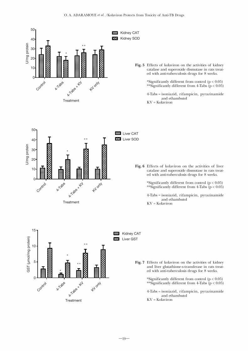
O. A. ADARAMOYE et al. /Kolaviron Protects from Toxicity of Anti-TB Drugs
―19―
Kidney CAT
Kidney SOD
*
**
0
10
20
30
50
40
U/m
g p
rote
in
Treatment
Contro
l
4-Ta
bs
4-Ta
bs +
KV
KV only
Liver CAT
Liver SOD
*
**
0
10
20
30
50
40
U/m
g p
rote
in
Treatment
Contro
l
4-Ta
bs
4-Ta
bs +
KV
KV only
Kidney CAT
Liver GST
*
*
**
**
0
5
10
15
GS
T (µ
mol
/mg
pro
tein
)
Treatment
Contro
l
4-Ta
bs
4-Ta
bs +
KV
KV only
Fig. 5 Effects of kolaviron on the activities of kidney catalase and superoxide dismutase in rats treat-ed with anti-tuberculosis drugs for 8 weeks.
*Significantly different from control (p < 0.05) **Significantly different from 4-Tabs (p < 0.05)
4-Tabs = isoniazid, rifampicin, pyrazinamide and ethambutol
KV = Kolaviron
Fig. 6 Effects of kolaviron on the activities of liver catalase and superoxide dismutase in rats treat-ed with anti-tuberculosis drugs for 8 weeks.
*Significantly different from control (p < 0.05) **Significantly different from 4-Tabs (p < 0.05)
4-Tabs = isoniazid, rifampicin, pyrazinamide and ethambutol
KV = Kolaviron
Fig. 7 Effects of kolaviron on the activities of kidney and liver glutathione-s-transferase in rats treat-ed with anti-tuberculosis drugs for 8 weeks.
*Significantly different from control (p < 0.05) **Significantly different from 4-Tabs (p < 0.05)
4-Tabs = isoniazid, rifampicin, pyrazinamide and ethambutol
KV = Kolaviron

O. A. ADARAMOYE et al. /Kolaviron Protects from Toxicity of Anti-TB Drugs
―20―
elevation of LPO and depletion of GSH in anti-TB drugs treated rats. The oxidative damage in these tissues might have resulted in the loss of structural integrity reflected by a marked increase in the leakage of hepatocellular enzymes. In addition, the enzymatic (superoxide dismutase, glutathione-s-transferase and glutathione peroxidase) defense mechanisms were also found to be compromised in the presence of anti-TB drugs. GSH, superoxide dismutase and glutathione peroxidase are known to protect the cells from the toxic effects of oxygen derived free radical and lipid peroxidation [44]. Although the liver is endowed with a unique capacity to regenerate thereby replenishing most of the protective systems, an overproduction of reactive species by anti-TB drugs may tilt the oxidant/ antioxidant balance of the cells. Based on this in vivo study, it is suggested that the underlying mechanism for hepato- and nephrotoxicity caused by these anti-TB drugs may be related to: 1) generation of free radicals and reactive metabolites, 2) imbalance in the oxidant/antioxidant defense, and 3) eventual peroxidation of membrane lipids that leads to the loss of tissues integrity. It is interesting to note that co-administration of KV with anti-TB drugs significantly attenuated the biochemical indices and oxidative stress markers in the rats. The protective role of KV in this study is consistent with previous studies on its anti-inflamma-tory, immuno-modulatory, free radical scavenging and hepatoprotective activities in vitro and in vivo [45, 46]. KV is known to suppress production of superoxide anion radical, exerts potent anti-inflammatory action that inhibits production of tumor necrosis factor alpha (TNF-a), and activation of NF-κB [19, 47]. These properties clearly explain the hepato- and nephropro-tective activities of KV in the present study. Also, KV has anti-microbial activity in addition to its anti-in-flammatory and antioxidant roles, making it a suitable candidate to alleviate the adverse effects of anti-TB drugs [48].
In conclusion, co-administration of KV with anti-TB drugs ameliorates lipid peroxidative damage, restores antioxidant status and normalizes markers of renal and hepatic injury in the rats. These findings show that KV may be a promising candidate for chemopre-vention of anti-TB drugs-induced tissues dysfunction. However, further detailed studies are required to estab-lish other mechanisms involve in the protection offers by this biflavonoid.
DECLARATION OF INTEREST
The authors have declared that there is no conflict of interest.
REFERENCES
1) World Health Organization. Global tuberculosis control: a short update to the 2009 report, Geneva. WHO 2009; 39: 1-15.
2) Soyinka A. Nigeria ranks fourth among TB high-burden coun-tries. Punch Newspaper 2007; 15: 3.
3) Okuonghae D, Omosigho S. Determinants of TB case detection in Nigeria: a survey. Glob J Health Sci 2010; 2: 123-8.
4) Wu S, Xia Y, Lv X, Tang S, Yang Z, Zhang Y, et al. Preventive use of hepatoprotectors yields limited efficacy on the liver toxicity of anti-tuberculosis agents in a large cohort of Chinese patients,” J Gastroenterol Hepatol 2015; 30: 540-5.
5) Chang CH, Chen YF, Wu VC, Shu CC, Lee CH, Wang JY, et al. Acute kidney injury due to anti-tuberculosis drugs: a five-year
experience in an aging population. BMC Infect Dis 2014; doi: 10.1186/1471-2334-14-23.
6) Enriquez-Cortina C, Almonte-Becerril M, Clavijo-Cornejo D, Palestino-Domínguez M, Bello-Monroy O, Nuño N, et al. Hepatocyte growth factor protects against isoniazid/rifampi-cin-induced oxidative liver damage. Toxicol Sci 2013; 135: 26-36.
7) Vuilleumier N, Rossier MF, Chiappe A, Degoumois F, Dayer P, Mermillod B, et al. CYP2E1 genotype and isoniazid-induced hepatotoxicity in patient treated for latent tuberculosis. Eur J Clin Pharmacol 2006; 62: 423-9.
8) Sheng YJ, Wu G, He HY, Chen W, Zou YS, Li Q Sheng, G, et al. The association between CYP2E1 polymorphisms and hepa-totoxicity due to anti-tuberculosis drugs: a meta-analysis. Infect Genet Evol 2014; 24: 34-40.
9) Feng D, Wang Y, Wang H, Weng H, Kong X, Martin-Murphy BV, et al. Acute and chronic effects of IL-22 on acetamino-phen-induced liver injury. J Immunol 2014; 193: 2512-18.
10) Heit C, Dong H, Chen Y, Shah YM, Thompson DC, Vasiliou V. Transgenic mouse models for alcohol metabolism, toxicity, and cancer. Adv Exp Med Biol 2015; 815: 375-87.
11) Dai Y, Cederbaum AI. Cytotoxicity of acetaminophen in human cytochrome P450 2E1-transfecred HepG2 cells. J Pharmacol Exp Ther 1995; 273: 1497-1505.
12) Bradford BU, Kono H, Isayama F, Kosyk O, Wheeler MD, Akiyama TE, et al. Cytohcrome P450 CYP2E1, but not adenine dinucleotide phosphate oxidase, is required for-ethanol-induced oxidative DNA damage in rodent liver. Hepatology 2005; 41: 336-44.
13) Iwu MM, Igboko OA. Flavonoids of Garcinia kola seeds. J Nat Prod 1982; 45: 650-51.
14) Cotterhill PJ, Scheinmann F, Stenhouse TA. Extractives from Guttiferae: kolaflavanone, a new biflavanone from the nuts of Garcinia kola Heckel. J Chem Soc Perkin Trans 1978; 1: 246.
15) Waterman PG, Hussain RA. Systematic significance of xantho-nes, benzophenones and biflavonoids in Garcinia. Biochem Sys Ecol 1983; 11: 21-30.
16) Nwankwo JO, Tahnteng JG, Emerole GO. Inhibition of afla-toxin B1 genotoxicity in human liver-derived HepG2 cells by kolaviron biflavonoids and molecular mechanisms of action. Eur J Cancer Prev 2000; 9: 351-61.
17) Adaramoye OA , Adeyemi EO. Hepatopro t e c t ion of D-galactosamine-induced toxicity in mice by purified fractions from Garcinia kola seeds. Basic Clin Pharmacol Toxicol 2006; 98: 135-41.
18) Adaramoye OA, Farombi EO, Nssien M, Idowu SO, Ademowo OG, Adeyemi EO. Hepatoprotective activity of purified fractions from Garcinia kola seeds in mice intoxicated with carbon tetra-chloride. J Med Food 2008; 11: 544-50.
19) Farombi EO, Shrotriya S, Surh YJ. Kolaviron inhibits dimethyl nitrosamine-induced liver injury by suppressing COX-2 and iNOS expression via NF-kappaB and AP-1. Life Sci 2009; 84: 149-55.
20) Farombi EO, Nwaokeafor IA. Anti-oxidant mechanisms of ko-laviron: studies on serum lipoprotein oxidation, metal chelation and oxidative membrane damage in rats. Clin Exp Pharmacol Physiol 2005; 32: 667-74.
21) Adaramoye OA, Farombi EO, Adeyemi EO, Emerole GO. Comparative study on the antioxidant properties of flavonoids of Garcinia kola seeds. Pak J Med Sci 2005; 21: 331-39.
22) Iwu MM, Igboko OA, Okunji CO, Tempesta MS. Antidiabetic and aldose reductase activities of biflavanones of Garcinia kola. J Pharm Pharmacol 1990; 42: 290-92.
23) Adaramoye OA, Lawal SO. Kolaviron, a biflavonoid fraction from Garcinia kola, protects against isoproterenol-induced injury by mitigating cardiac dysfunction and oxidative stress in rats. J Basic Clin Physiol Pharmacol 2015; 26: 65-72.
24) Lowry OH, Rosebrough NJ, Farr AL, Randall RJ. Protein mea-surement with the Folin phenol reagent,” J Biol Chem 1951; 193: 265-75.
25) Rutkowski RB, Debaare L. An ultra-micro colorimetric method for determination of total and direct serum bilirubin. Clin Chem 1966; 12: 432-38.
26) Mohun AF, Cook LJ. Simple method for measuring serum level of glutamate-oxaloacetate and glutamate-pyruvate transaminases
O. A. ADARAMOYE et al. /Kolaviron Protects from Toxicity of Anti-TB Drugs
―21―
in laboratories. J Clin Pathol 1957; 10: 394-99.27) Reitman S, Frankel S. A colorimetric method for the determi-
nation of serum level of glutamate-oxaloacetate and pyruvate transaminases. Am J Clin Pathol 1957; 28: 56-63.
28) Jaffe M. Ueber den Neiderschlag, welchen Pikrinsäure im normalen harn Erzeught und über eine neue Reaction des Kreatinins. Z Physiol Chem 1886; 10: 391-400.
29) Talke H, Schubert GE. Enzymatische Harnstoff bestimmung in Blut and serum in Optischen Test nach Warburg. Klin Wochschr 1965; 43: 174.
30) McCord JM, Fridovich I. Superoxide dismutase, an enzymatic function for erythrocuperin. J Biol Chem 1969; 244: 6049-55.
31) Aebi H. Catalase: Methods of enzymatic analysis, Bergmeyer HV(ed.). New York, Verlag Chemie, 1974; 673-684.
32) Habig WH, Pabst MJ, Jakoby WB. Glutathione-S-transferases. The first enzymatic step in mercapturic acid formation. J Biol Chem 1974; 249: 7130-9.
33) Rotruck JT, Pope AL, Ganther HE, Swanson AB, Hafeman DG, Hoekstra WG. Selenium: biochemical role as a component of glutathione peroxidise. Science 1973; 179: 588-90.
34) Moron MS, Depierre JW, Mannervick B. Levels of glutathione, glutathione reductase and glutathione-s-transferase activities in rat lung and liver. Biochim Biophys Acta 1979; 582: 67-78.
35) Buege JA, Aust SD. Microsomal lipid peroxidation. Methods Enzymol 1978; 30: 302-10.
36) Pal R, Rana SV, Vaiphei K, Singh K. Isoniazid-rifampicin in-duced lipid changes in rats. Clin Chim Acta 2008; 389: 55-60.
37) Tasduq SA, Kaizer P, Sharma SC, Johri RK. Potentiation of isoniazid-induced liver toxicity by rifampicin in a combinational therapy of anti-tubercular drugs (rifampicin, isoniazid and pyrazinamide) in wistar rats: A toxicity profile study. Hepatol Res 2007; 37: 845-53.
38) De Vriese AS, Robbrecht DL, Vanholder RC, Vogelaers DP, Lameire NH. Rifampicin-associated acute renal failure: patho-physiologic, immunologic, and clinical features. Am J Kidney Dis 1998; 31: 108-15.
39) Kwon SH, Kim JH, Yang JO, Lee EY, Hong SY. Ethambutol-induced acute renal failure. Nephrol Dial Transplant 2004; 19: 1335-6.
40) Chiba S, Tsuchiya K, Sakashita H, Ito E, Inase N. Rifampicin-
induced acute kidney injury during the initial treatment for pul-monary tuberculosis: a case report and literature review. Intern Med 2013; 52: 2457-60.
41) Zhang Y, Warren MS, Zhang X, Diamond S, Williams B, Punwani N, et al. Impact on creatinine renal clearance by the interplay of multiple renal transporters - A case study with INCB039110. Drug Metab Dispos 2015; 43: 485-9.
42) Jaramillo-Juárez F, Rodríguez-Vázquez ML, Rincón-Sánchez AR, Consolación Martínez M, Ortiz GG, Llamas J, et al. Acute renal failure induced by carbon tetrachloride in rats with hepatic cirrhosis. Ann Hepatol 2008; 7: 331-8.
43) Tasduq SA, Kaisar P, Gupta DK, Kapahi BK, Maheshwari HS, Jyotsna S, et al. Protective effect of a 50% hydroalcoholic fruit extract of Emblica officinalis against anti-tuberculosis drugs induced liver toxicity. Phytother Res 2005; 19: 193-7.
44) Halliwell B, Gutteridge JMC. Protection against oxidants in biological systems: The superoxide theory of oxygen toxicity. In: Free Radicals in Biology and Medicine, Halliwell B, Gutteridge JMC (eds). New York, Oxford University Press 1989; 86-187.
45) C . S. Nworu, P. A. Akah, C . O. Esimone, C . O. Okoli, and F. B. Okoye, “Immuno-modulatory activities of kolaviron, a mixture of three related biflavonoids of Garcinia kola Heckel,” Immunopharmacology and Immunotoxicology, vol. 30, no. 2, pp. 317-332, 2008.
46) E. O. Farombi, I. A. Adedara, B. O. Ajayi, O.R. Ayepola, and E. E. Egbeme, “Kolaviron, a natural antioxidant and anti-inflamma-tory phytochemical prevents dextran sulphate sodium-induced colitis in rats,” Basic and Clinical Pharmacology and Toxicology, vol. 113, no. 1, pp. 49-55, 2013.
47) S. B. Olaleye, S. A. Onasanwo, A. O. Ige, K. K. Wu, and C. H. Cho, “Anti-inflammatory activities of a kolaviron-inhibition of nitric oxide, prostaglandin E2 and tumor necrosis factor-alpha production in activated macrophage-like cell line,” African Journal of Medicine and Medical Sciences, vol. 39, pp. 41-46, 2010.
48) A. Oluwatosin, A. Tolulope, K. Ayokulehin et al., “Antimalarial potential of kolaviron, a biflavonoid from Garcinia kola seeds, against Plasmodium berghei infection in Swiss albino mice,” Asian Pacific Journal of Tropical Medicine, vol. 7, no. 2, pp. 97-104, 2014.