breast cancer in patients of rheumatoid arthritis with ...mj-med-u-tokai.com/pdf/420208.pdf ·...
TRANSCRIPT
―104―
Tokai J Exp Clin Med., Vol. 42, No. 2, pp. 104-108, 2017
Breast Cancer in Patients of Rheumatoid Arthritis with Methotrexate Therapy Mimicking Histopathological Changes after Neoadjuvant
Chemotherapy
Nobue KUMAKI*1, Chizuko OKAMATSU*1, Yutaka TOKUDA*2 and Naoya NAKAMURA*1
*1Department of Pathology, Tokai University School of Medicine *2Department of Breast and Endocrine Surgery, Tokai University, School of Medicine
(Received May 1, 2017; Accepted May 24, 2017)
Two breast cancer patients with a history of treatment for long-term rheumatoid arthritis (RA) had histolog-ical findings similar to histological changes seen in resected mammary gland specimens following neoadju-vant chemotherapy (NAC).The first patient was a 64-year-old woman who visited our hospital after feeling a lump in her left breast. The second patient was a 68-year-old woman who visited our hospital for an indentation in her left nipple. They were diagnosed with breast cancer following detailed examinations and underwent mastectomy. Both patients had a history of RA and were being treated with Methotrexate. The histological diagnoses of these patients were invasive ductal carcinoma, but frequent dispersal of cancer cell nests, stromal fibrosis, elasto-sis, edema and inflammatory cell infiltration were seen. Fibrosis was also found in the dissected lymph node. These histological findings were extremely similar to changes that occur in the mammary gland tissue after NAC; however, these patients had not undergone NAC. Methotrexate, which was being administered as an anti-rheumatic drug to the two patients, might have played a role similar to that of metronomic chemothera-py, which involves the continuous use of low-dose anti-cancer drugs, resulting in histological changes similar to those seen after NAC.
Key words: Breast cancer, Histopathological change, Neoadjuvant chemotherapy, Rheumatoid arthritis, Methotrexate
INTRODUCTION
Neoadjuvant chemotherapy (NAC) is an established treatment for breast cancer [1], with evaluations of surgical mammary following NAC frequently en-countered in routine pathological diagnoses. NAC-related histological changes are noted in carcinoma cells and non-neoplastic tissues [2], because the effects of anti-cancer drugs extend to cells beyond carcinoma cells. Carcinoma cells reveal degeneration and necro-sis, while non-neoplastic tissue shows inflammatory cell infiltration, stromal changes, and non-neoplastic glandular tissue changes. A wide area can be observed in surgical specimens of the mammary gland tissue, which allows NAC-related histological changes to be microscopically and easily seen. In this paper, we re-port our experience of two breast cancer patients who showed histological findings that mimics histological changes after NAC in surgical specimens though they were not treated with chemotherapy, together with a discussion of the literature.
CASE REPORTS
< Case 1 >The patient was a 64-year-old woman who visited
a hospital after feeling a lump in her left breast. Mammography revealed focal asymmetric densities in the lower area in the mediolateral oblique view and in
the outer area in the craniocaudal view. Ultrasound showed an 18-mm tumor in the CD area. A diagnosis of invasive ductal carcinoma (invasive carcinoma of no special type (NST) in the WHO classification) was made by performing needle biopsy, and the patient underwent mastectomy and sentinel lymph node bi-opsy. The patient had a 40-year history of rheumatoid arthritis (RA) prior to her breast cancer surgery and had undergone synovectomy approximately 25 years earlier. She had been prescribed methotrexate (MTX) at 6 mg/week starting 10 years ago.
< Case 2 >The patient was a 68-year-old woman who visited
a hospital after observing an indentation in her left nipple one year prior to her hospital visit. An elastic, hard induration of approximately 5 cm was felt in the left C area on palpation, with tightening of the skin. Ultrasound showed a tumor of approximately 50 mm in the AC–C areas. Needle biopsy revealed invasive ductal carcinoma, and the patient underwent left mastectomy and axillary lymph node dissection. The patient had an approximately 25-year history of RA prior to her breast cancer surgery and had been taking oral MTX (6 mg/week) at the time of surgery.
Nobue KUMAKI, Department of Pathology, Tokai University School of Medicine, 143 Shimokasuya, Isehara, Kanagawa 259-1193, JapanTel: +81-463-93-1121 Fax: +81-463-91-1370 E-mail: [email protected]
N. KUMAKI et al. /Breast Cancer in RA with MTX Therapy
―105―
PATHOLOGICAL FINDINGS
< Case 1 >A tumor with an indistinct border and tightening
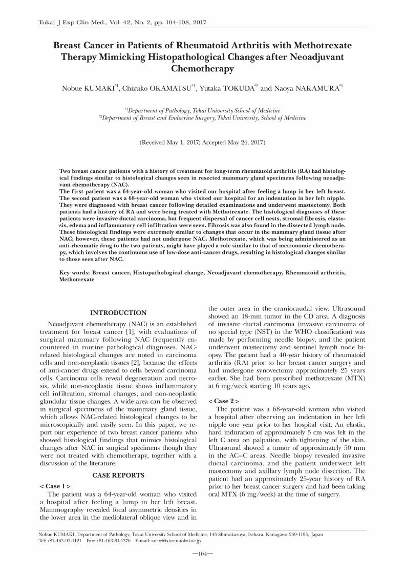
was macroscopically noted in the CD area of the left breast. Histologically, the center of the tumor showed coarse fibrosis (Fig. 1a), while invasive carcinoma cells arranged in trabecular or small cluster pattern were present in the periphery (Fig. 1b). Some cancer cells were enlarged with pyknosis and karyorrhexis sporad-ically seen. The ductal components of carcinoma cells were seen around the tumor. Dense or sparse fibrosis was noticeable surrounding the carcinoma cells, and chronic inflammatory cell infiltration and elastosis were observed. Background fibrosis was also noticeable in the area of the ductal components of the carcinoma cells, and ducts were surrounded by dense collagen fibers (Fig. 1c). Circular and concentric collagen fi-ber nests were sometimes seen (Fig. 1d), which were assumed to be scarred mammary ducts. Hemosiderin deposition and calcification were occasionally noted in the stroma. Non-neoplastic ducts were sparsely spread, with basement membrane hypertrophy of the small ducts. (Data including the histological type, tumor size and subtype were summarized in Table.)
< Case 2 >While no clear mass formation was macroscopically
visible in the C area, hardening of mammary gland tissue was noted. The hard area spread to the nipple, which appeared inverted. Histological findings showed diffuse infiltration of carcinoma cells arranged in cord-like and small cluster (Fig. 2a, b). Diffuse invasion of carcinoma cells was also seen. Fibrosis of the stroma was remarkable (Fig. 2a). Vacuolar change of carcino-ma cells was focally observed. Noticeable elastosis (Fig. 2c), together with edema, fibrosis, chronic inflamma-tory cell infiltration, and occasional hyalinization, was observed in the stroma. The same circular collagen fiber nests as in Case 1 were observed. Lymphatic invasion of the cancer was sporadically seen, together with metastasis of cancer to the axillary lymph nodes. Fibrosis was partially detected in the lymph nodes (Fig.2d). No noticeable histological changes were seen in the non-neoplastic mammary ducts.
DISCUSSION
After-NAC mammary gland tissue shows histolog-ical changes in stroma and non-neoplastic glandular tissues, in addition to carcinoma cells [2-4]. Carcinoma brings about cellular morphological changes, such as cell enlargement, vacuolation, pyknosis, and necrosis, and structural and distribution pattern changes, including structure deformation of the gland and cell dispersal. Stromal changes have been reported, including fibrosis, edema, elastosis, hyalinization,
Fig. 1 Histopathological findings in Case 1 a) Low-power view of the tumor, which showed geographic distribution of carcinoma cells
and central fibrosis. b)-d) High-power view of the tumor. b) Invasive carcinoma cells and stromal coarse fibrosis
with infiltration of inflammatory cells. c) Severe fibrosis of stroma and ductal component of carcinoma cells surrounded by dense collagen fibers. d) Circular and concentric colla-gen fiber nest, so-called “healing” of mammary duct (black arrowhead).
a b
c d
Figure 1. Histopathological findings in Case 1
N. KUMAKI et al. /Breast Cancer in RA with MTX Therapy
―106―
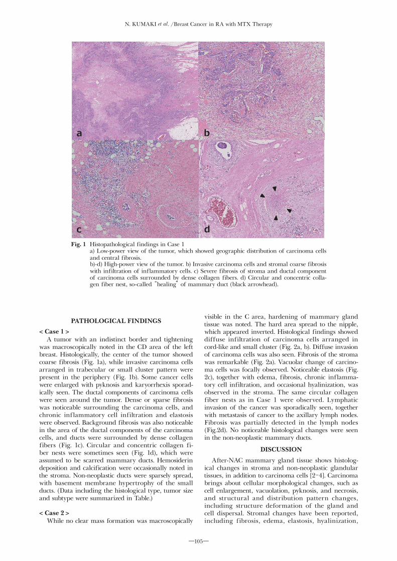
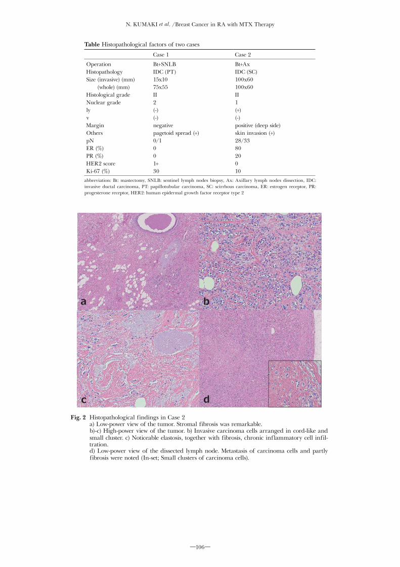
Table Histopathological factors of two cases
Case 1 Case 2
OperationHistopathologySize (invasive) (mm)
(whole) (mm)Histological gradeNuclear gradelyvMarginOtherspNER (%)PR (%)HER2 scoreKi-67 (%)
Bt+SNLBIDC (PT)15x1075x55II2(-)(-)negativepagetoid spread (+)0/1001+30
Bt+AxIDC (SC)100x60100x60II1(+)(-)positive (deep side)skin invasion (+)28/338020010
abbreviation: Bt: mastectomy, SNLB: sentinel lymph nodes biopsy, Ax: Axillary lymph nodes dissection, IDC: invasive ductal carcinoma, PT: papillotubular carcinoma, SC: scirrhous carcinoma, ER: estrogen receptor, PR: progesterone receptor, HER2: human epidermal growth factor receptor type 2
Fig. 2 Histopathological findings in Case 2 a) Low-power view of the tumor. Stromal fibrosis was remarkable. b)-c) High-power view of the tumor. b) Invasive carcinoma cells arranged in cord-like and
small cluster. c) Noticeable elastosis, together with fibrosis, chronic inflammatory cell infil-tration.
d) Low-power view of the dissected lymph node. Metastasis of carcinoma cells and partly fibrosis were noted (In-set; Small clusters of carcinoma cells).
c d
a b
Figure 2. Histopathological findings in Case 2
N. KUMAKI et al. /Breast Cancer in RA with MTX Therapy
―107―
infiltration of foamy cells and lymphocytes, hemosid-erin deposition, microcalcification, and angiogenesis. Hardening of the basement membrane of terminal duct-lobular units [5], and hyalinization of vascular walls can also be found. Honkoop et al. postulated that stromal changes are a physical defensive response to chemotherapy [6].
In our patients, the changes and degenerative findings of the carcinoma cells themselves were mild; however, dispersal of carcinoma cell nests was partly observed. Stomal fibrosis, edema, elastosis, and inflam-matory cell infiltration were noticeable, with tightened tissue structures. Circular or oval scarred nests formed of layers of collagen fibers were similar to the process of “healing” proposed by Muri et al. [7] and are pre-sumed to be traces of ductal components of carcinoma cells that have disappeared [8]. Fibrosis was also seen in the regions of lymph nodes that showed cancer me-tastasis in Case 2.
The above-mentioned histological findings appear extremely similar to those seen after NAC. Fibrosis and inflammatory cell infiltration can also occur as a biological reaction to procedures, including biopsy and surgery, but they are usually localized in such cases. The presented patients showed extensive and diffuse changes to the mammary gland and also showed his-tological changes in organs other than the mammary glands, such as lymph nodes. This suggests that the cause of histological changes had a systemic effect. Medications such as anti-cancer drugs are examples of systemic drugs used during breast cancer therapy; however, neither of the presented patients had under-gone NAC. Both patients had nonetheless suffered from RA for over 20 years and had been taking MTX. MTX is associated with the development of malignant tumors, including malignant lymphoma [9]; however, the risk of breast cancer in RA patients treated with MTX is not high compared with the risk in healthy individuals [10]. MTX has been used as an anti-cancer drug for breast cancer [1, 11], and it is used as an immunosuppressant in the treatment of RA, where it is continuously administered in doses at one-tenth or one-twentieth the dose given in the treatment of cancer [12]. This could explain the similar findings to those observed using metronomic chemotherapy (mCT).
Conventional chemotherapy involves the admin-istration of a drug at the maximum tolerated dose and repeated periodical washout periods to allow the body to recover from the side effects of the drug. On the other hand, therapy that involves the continuous or frequent administration of a low-dose anti-cancer drug without washout periods is referred to as mCT [13]. This therapy was proposed over 15 years ago based on experiments using xenotransplant models reported by Browder et al. [14] and Klement et al. [15] and was named “metronomic” by Hanahan et al. [16] The concentration of the drug in the blood is kept low by frequent daily doses, thus reducing the side effects [13]. Conventional chemotherapy targets proliferating tumor cells, whereas mCT targets endothelial cells and impairs angiogenesis to suppress tumor growth. Furthermore, mCT has other effects on cancer, includ-ing the enhancement of anti-cancer immune function [17] and reduction in blood estrogen levels. Clinical
trials have been reported using mCT for breast cancer under various clinical conditions [18], and clinical trials incorporating MTX have been performed in metastatic breast cancer patients [19]. The pathological complete response rate was 47.5 % under neoadjuvant conditions in triple-negative breast cancer patients [20], with some patients showing a therapeutic effect. Details of histological changes in breast cancer patients during mCT have not been presented in the literature; howev-er, extensive histological changes, including fibrosis in the mammary glands tissue of the presented patients, could be the same histological changes as those seen after NAC.
In conclusion, we presented two breast cancer patients with a history of long-term treatment for RA and who had histological findings similar to those seen after NAC. Oral MTX taken as an anti-rheumatic drug could have effects similar to those seen in che-motherapy, such as mCT. This suggests the importance of examining the clinical data, including patient back-ground, when observing and diagnosing pathological specimens.
REFERENCES1) Japanese breast cancer society, ed. Clinical Practice Guideline for
treatment of breast cancer (2015). Tokyo: Kanehara & Co., Ltd., 2015. [Published in Japanses]
2) Sahoo S, Lester SC. Pathology of breast carcinomas after neo-adjuvant chemotherapy. Arch Pathol Lab Med. 2009; 133: 633-642.
3) Fisher ER, Wang J, Bryant J, Fisher B, Mamounas E, Wolmark N. Pathology of preoperative chemotherapy. Findings from the national surgical adjuvant breast and bowel progect (NSABP) protocol B-18. Cancer. 2002; 95: 681-695.
4) Sethi D, Sen R, Parshad S, Khetarpal S, Garg M, Sen J. Histopathologic changes following neoadjuvant chemotherapy in various malignancies. Int J Appl Basic Med Res. 2012; 2: 111-6.
5) Brachtel E, Koerner FC. Pathologic Effects of Therapy. In: Hoda SA, Brogi E, Koerner FC, Rosen PP, eds. Rosen's Breast Pathology 4th ed.. Philadelphia: Lippincott Williams & Willkins, 2014: 1197-1215.
6) Honkoop AH, Pinedo HM, DeJong JS, Verheul HM, Linn SC, Hoekman K, et al. Effects of chemotherapy on pathologic and biologic characteristics of locally advanced breast cancer. Am J Clin Pathol. 1997; 107: 211-218.
7) Muri R, Aitkenhead AC. The healing of intra-ductal carcinoma of the mamma. J Pathol Bacteriol. 1934; 38: 117-127.
8) Horii R, Akiyama F, Kasumi F, Koile M, Sakamoto G. Spontaneous “healing” of breast cancer. Breast Cancer. 2005; 12: 140-144.
9) Smitten AL, Simon TA, Hochberg MC, Suissa S. A meta-analysis of the incidence of malignancy in adult patients with rheumatoid arthritis. Arthritis Res Ther. 2008; 10: R45.
10) Buchbinder R, Barder M, Heuzenoeder L, Wluka AE, Giles G, Hall S, et al. Incidence of melanoma and other malignancies among rheumatoid arthritis patients treated with methotrexate. Arthritis Rheum. 2008; 59: 794-799.
11) Moreno A, Escobedo A, Benito E, Serra JM, Guma A, Riuet F. Pathological changes related to CMF primary chemotherapy in breast cancer. Breast Cancer Res. Treat. 2002; 75: 119-125.
12) Smolen JS, Landewe R, Bijlsma J, Burmester G, Chatzidionysiou K, Dougados M, et al. EULAR recommendations for the man-agement of rheumatoid arthritis with synthetic and biological disease-modifying antirheumatic drugs: 2016 update. Ann Rheum Dis. 2017 [Epub ahead of print]
13) Maiti R. Metronomic chemotherapy. J. Pharmacol. Pharmacother. 2014; 5: 186-192.
14) Browder T, Butterfield CE, Kräling BM, Shi B, Marshall B, O'Reilly MS, et al. Antiangiogenic scheduling of chemotherapy improves efficacy against experimental drug-resistant cancer.

N. KUMAKI et al. /Breast Cancer in RA with MTX Therapy
―108―
Cancer res. 2000; 60: 1878-1886.15) Klement G, Baruchel S, Rak J, Man S, Clark K, Hicklin DJ, et
al. Continuous low-dose therapy with vinbrastine and VEGF receptor-2 antibody induce sustained tumor regression without over toxicity. J. Clin. Invest. 2000; 105: R15-R24.
16) Hanahan D, Bergers G, Bergsland E. Less is more, regularly: metronomic dosing of cytotoxic drugs can target tumor angio-genesis in mice. J. Clin. Invest. 2000; 105: 1045-1047.
17) Lake RA, Robinson BW. Immunotherapy and chemotherapy-a practical partnership. Nat Rev Cancer. 2005; 5: 397-405.
18) Munzone E, Colleoni M. Clinical overview of metronomic che-motherapy in breast cancer. Nat. Rev. Clin. Oncol. 2015; 12: 631-644.
19) Colleoni M, Rocca A, Sandri MT, Zorzino L, Masci G, Nole F, et al. Low-dose oral methotrexate and cyclophosphamide in metastatic breast cancer: antitumor activity and correlation with vascular endothelial growth factor levels. Ann Oncol. 2002; 13: 73-80.
20) Masuda N, Higaki K, Takano T, Matsunami N, Morimoto T, Ohtani S, et al. A phase II study of metronomic paclitaxel/cyclophosphamide/capecitabine followed by 5-fluorouracil/epirubicin/cyclophosphamide as preoperative chemotherapy for triple-negative or low hormone receptor expressing/HER2-negative primary breast cancer. Cancer Chemother Pharmacol. 2014; 74: 229-38.