search for appropriate experimental methods to create ...mj-med-u-tokai.com/pdf/310311.pdf · an...
TRANSCRIPT
― 118―
Tokai J Exp Clin Med., Vol. 31, No. 3, pp. 118-122, 2006
INTRODUCTION
Refractory peripheral arterial disease is becoming an important therapeutic target since its incidence has been markedly increasing due to the increase in aged population and patients with diabetes mellitus. Consequently various therapeutic approaches including angiogenic treatment with growth factors or cell trans-plantation have been attempted using mouse hind-limb ischemia models [1-4]. Scrutinizing these studies we found that experimental methods to create hind-limb ischemia are not standardized. Method of occlud-ing artery varies from ligation, or cutting, to excision of the artery. The targeted artery varies also from the iliac artery [1], the femoral artery [2-4], or the femoral with saphenous artery [5-9]. In some cases, both the femoral artery and vein were occluded. Strangling of the thigh itself was attempted in some studies [10, 11]. As the severity of ischemic damage cannot be uniform in different experimental methods, the comparison of effect among various therapies becomes very difficult.
Another important problem in previously used animal models is lack of data on blood flow when hind-limb is lost. In case of severe ischemic damage in mouse necrosis and loss of hind-limb often oc-cur within three days but in patients of peripheral artery disease ischemia is chronic and acute necrosis is seldom seen. In these patients the improvement of blood flow is one of the key indices in determining effective treatments. Therefore it is mandatory to have an animal model in which chronic hind-limb ischemia
is present but necrosis seldom occurs and sequential evaluation of blood flows is possible in order to evalu-ate various therapies.
In the present study, firstly we examined six meth-ods for inducing hind-limb ischemia in Balb/ca mice and evaluated the severity of ischemic change to search for a stable severe ischemia model. Secondly we select-ed three methods with mild ischemic changes among six methods which do not produce severe necrosis and examined degree of ischemia by measuring CPK release, muscle weight, and histological changes to find an appropriate mild ischemia model.
MATERIALS AND METHODS
AnimalsSeventy-five male Balb/ca mice (12 weeks old, 20 to
30 g, Japan Clea Inc, Ishibe) were used. All operations and measurements were performed under general an-esthesia (1.0 to 1.5% isoflurane, 60% dinitrous monox-ide, and 40% oxygen). The operation was performed, by only one investigator (T.G.), under a microscope (Konan Operation Microscope 707, Konan Keeler Co. LTD, Japan). To create ischemia, the vessels were cut or resected after ligation of the stumps with sterilized 6-0 silk suture (Azwel Inc, Osaka). The investigation conforms with The Guide for the Care and Use of Laboratory Animals published by the U.S. National Institutes of Health (NIH Publication No. 85-23, re-vised 1996).
Search for appropriate experimental methods to create stable hind-limb ischemia in mouse
Takako GOTO*1, Naoto FUKUYAMA* 2, Akira AKI*1, Kazuo KANABUCHI*1, Koji KIMURA*1, Hiroyuki TAIRA*1, Etsuro TANAKA* 3, Noriaki WAKANA* 3, Hidezo MORI* 4 and Hiroshi INOUE*1
Departments of *1 Surgery and *2 Physiology, Tokai University School of Medicine *3 Department of Nutritional Sciences, Tokyo University of Agriculture, Tokyo, Japan
* 4 Department of Cardiac Physiology, National Cardiovascular Center Research Institute, Suita, Japan
(Received June 26, 2006; Accepted July 19, 2006)
Objective: Stable animal models for refractory peripheral arterial disease are not established. A standardized animal model of hind-limb ischemia is required upon searching effective treatment for this condition. The aim of the study is to verify previously used hind-limb ischemia models to find a standard method. Methods: Using Balb/ca mice six various methods of inducing hind-limb ischemia were applied and two weeks after operation degree of ischemic damage were examined. Six methods include V group, A group, AV group, A-strip group, AV-strip group and Prox-A group (refer the text). Results: Degree of ischemia was evaluated macroscopically by judging toes, foot, knee, and total hind-limb necrosis. We found that severity of damage was markedly different among different methods. Furthermore the severity of necrosis was not uniform even in the same method group.Conclusions: The A-strip group in which the femoral artery from the bifurcation of the deep femoral artery to the saphenous artery was stripped appears to be suitable as a stable severe ischemia model. The A group in which the femoral artery were cut just below the bifurcation of the deep femoral artery appears to be suit-able as a chronic mild ischemia model.
Key words: angiogenesis, animal model, blood vessels, femoral artery, hind-limb ischemia
Takako GOTO, Department of Surgery, Tokai University School of Medicine, Isehara, Kanagawa 259-1193, Japan Tel: 0463-93-1121 ext:2280 Fax: 0463-95-7567 E-mail: [email protected]
T. GOTO et al. /Hind-limb ischemia in mouse
― 119―
EXPERIMENTAL PROTOCOLS
1. The six hind limb ischemia models (n= 60)The following six types of ischemia were created
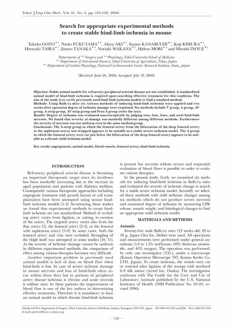
in the right lower limbs of 60 mice (schematic illus-trations are presented in Fig. 1). These included, (1) cutting the femoral vein at the distal site of the bifur-cation of the deep femoral vein (V-group, n=10), (2) cutting the femoral artery just below the bifurcation of the deep femoral artery (A-group, n=10), (3) cutting both the femoral artery and vein (AV-group, n=10), (4) resection of the femoral artery from the distal site of the bifurcation of the deep femoral artery to the sa-phenous artery (A-strip-group, n=10). By disecting the femoral artery as was shown in the Fig. 1B, branches including the popliteal artery were also obstracted and retrograde flows from these branches were completely avoidable. (5) resecting both the femoral artery and vein from the distal site of the bifurcation of the deep femoral artery to the saphenous artery (AV-strip-group, n=10), and (6) cutting the femoral artery at the proxi-mal site of the bifurcation of the deep femoral artery (Prox-A-group, n=10).
2. Macroscopic evaluation of ischemic severity Two weeks after the operation, the ischemic limb
was macroscopically evaluated by using graded mor-phological scales for necrotic area; grade 0: absence of necrosis, grade I: necrosis limiting to toes (toes loss), grade II: necrosis extending to a dorsum pedis (foot loss), grade III: necrosis extending to a crus (knee loss), grade IV: necrosis extending to a thigh (total hind-limb loss).
3. Blood flow measurementCalf blood flows on both sides were measured be-
low a patella with a noncontact laser Doppler flowme-ter (FLO-N1, Omegawave Corporation, Tokyo) before the operation, just after the operation, and two weeks post operatively, and were expressed as the ratio of the flow in the ischemic limb to that in the normal limb.
4. CPK release, muscle weights and histological evaluation in three mild ischemia groupsIn additional mice of V-, A-, and AV-groups (n
=5 each) blood samples were obtained from the
orbital plexus before the operation and 1, 2, and 7 days thereafter and concentrations of creatine phos-phokinase (CPK) were measured. At two weeks after the operation, the animals were sacrificed under an overdose of sodium pentobarbital and the anterotibial, gastrocnemius, and soleus muscles were dissected out and weighed. Histological analysis (HE staining) was performed in each muscle [12].
RESULTS
1) Severity of ischemic change in the six groupsTwo weeks after the operation necrotic changes were
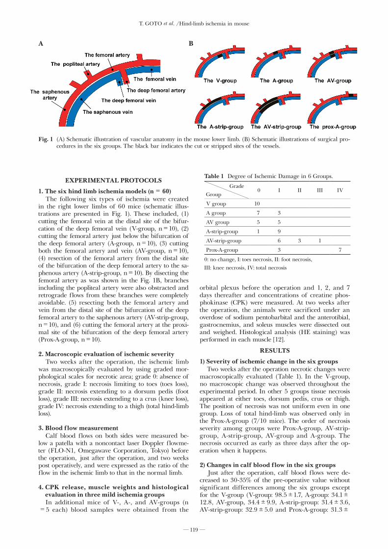
macroscopically evaluated (Table 1). In the V-group, no macroscopic change was observed throughout the experimental period. In other 5 groups tissue necrosis appeared at either toes, dorsum pedis, crus or thigh. The position of necrosis was not uniform even in one group. Loss of total hind-limb was observed only in the Prox-A-group (7/10 mice). The order of necrosis severity among groups were Prox-A-group, AV-strip-group, A-strip-group, AV-group and A-group. The necrosis occurred as early as three days after the op-eration when it happens.
2) Changes in calf blood flow in the six groupsJust after the operation, calf blood flows were de-
creased to 30-35% of the pre-operative value without significant differences among the six groups except for the V-group (V-group: 98.5±1.7, A-group: 34.1±12.8, AV-group, 34.4±9.9, A-strip-group: 31.4±3.6, AV-strip-group: 32.9±5.0 and Prox-A-group: 31.3±
Fig. 1 (A) Schematic illustration of vascular anatomy in the mouse lower limb. (B) Schematic illustrations of surgical pro-cedures in the six groups. The black bar indicates the cut or stripped sites of the vessels.
A B
Table 1 Degree of Ischemic Damage in 6 Groups.
GradeGroup
0 I II III IV
V group 10
A group 7 3
AV group 5 5
A-strip-group 1 9
AV-strip-group 6 3 1
Prox-A-group 3 7
0: no change, I: toes necrosis, II: foot necrosis, III: knee necrosis, IV: total necrosis
T. GOTO et al. /Hind-limb ischemia in mouse
― 120―
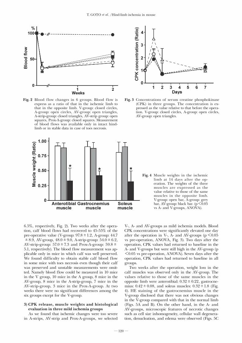
6.5%, respectively, Fig. 2). Two weeks after the opera-tion, calf blood flows had recovered to 45-55% of the pre-operative value (V-group: 97.8±1.2, A-group: 44.7±8.9, AV-group, 48.0±9.0, A-strip-group: 54.0±6.2, AV-strip-group: 57.0±7.3 and Prox-A-group: 59.8±5.1, respectively). The blood flow measurement was ap-plicable only in mice in which calf was well preserved. We found difficulty to obtain stable calf blood flow in some mice with toes necrosis even though their calf was preserved and unstable measurements were omit-ted. Namely blood flow could be measured in 10 mice in the V group, 10 mice in the A group, 8 mice in the AV-group, 8 mice in the A-strip-group, 7 mice in the AV-strip-group, 3 mice in the Prox-A-group. At two weeks there were no significant differences among the six groups except for the V-group.
3) CPK release, muscle weights and histological evaluation in three mild ischemia groupsAs we found that ischemic changes were too severe
in A-strips, AV-strip and Prox-A-groups, we selected
V-, A- and AV-groups as mild ischemia models. Blood CPK concentrations were significantly elevated one day after the operation in V-, A- and AV-groups (p<0.05 vs pre-operation, ANOVA, Fig. 3). Two days after the operation, CPK values had returned to baseline in the A- and V-groups but were still high in the AV-group (p<0.05 vs pre-operation, ANOVA). Seven days after the operation, CPK values had returned to baseline in all groups.
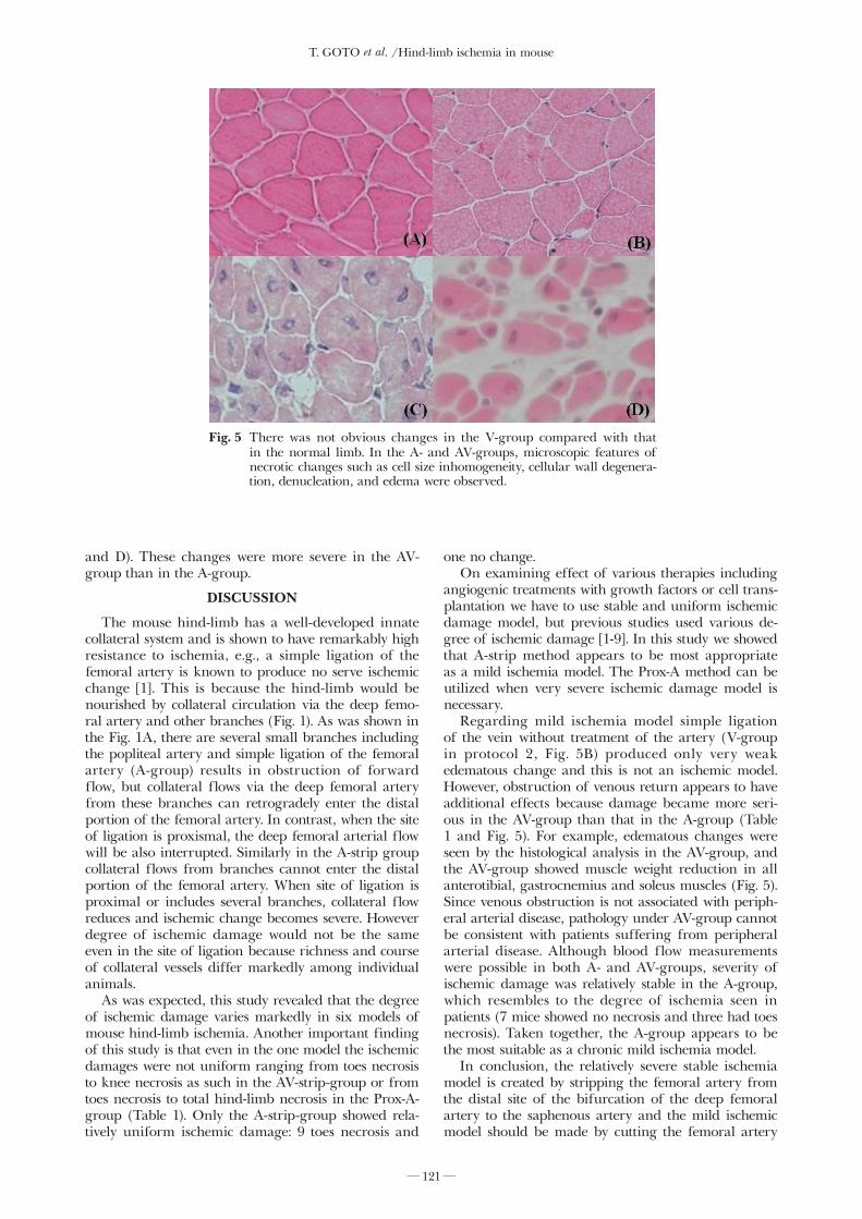
Two weeks after the operation, weight loss in the calf muscles was observed only in the AV-group. The values relative to those of the same muscles in the opposite limb were anterotibial: 0.32±0.22, gastrocne-mius: 0.42±0.08, and soleus muscles: 0.32±1.8 (Fig. 4). HE staining of the gastrocnemius muscle in the V-group disclosed that there was not obvious changes in the V-group compared with that in the normal limb (Figs. 5A and B). On the other hand, in the A- and AV-groups, microscopic features of necrotic changes such as cell size inhomogeneity, cellular wall degenera-tion, denucleation, and edema were observed (Figs. 5C
Fig. 2 Blood flow changes in 6 groups. Blood flow is express as a ratio of that in the ischemic limb to that in the opposite limb. V-group: closed circles, A-group: open circles, AV-group: open triangles, A-strip-group: closed triangles, AV-strip group: open squares, Prox-A-group: closed squares. Measurement of blood flows was available only in intact hind-limb or in stable data in case of toes necrosis.
Fig. 3 Concentrations of serum creatine phosphokinase (CPK) in three groups. The concentration is ex-pressed as the value relative to that before the opera-tion. V-group: closed circles, A-group: open circles, AV-group: open triangles.
Fig. 4 Muscle weights in the ischemic limb at 14 days after the op-eration. The weights of the three muscles are expressed as the value relative to those of the same muscles in the opposite limb. V-group: open bar, A-group: grey bar, AV-group: black bar. (p<0.05 vs A- and V-groups, ANOVA).
T. GOTO et al. /Hind-limb ischemia in mouse
― 121―
and D). These changes were more severe in the AV-group than in the A-group.
DISCUSSION
The mouse hind-limb has a well-developed innate collateral system and is shown to have remarkably high resistance to ischemia, e.g., a simple ligation of the femoral artery is known to produce no serve ischemic change [1]. This is because the hind-limb would be nourished by collateral circulation via the deep femo-ral artery and other branches (Fig. 1). As was shown in the Fig. 1A, there are several small branches including the popliteal artery and simple ligation of the femoral artery (A-group) results in obstruction of forward flow, but collateral flows via the deep femoral artery from these branches can retrogradely enter the distal portion of the femoral artery. In contrast, when the site of ligation is proxismal, the deep femoral arterial flow will be also interrupted. Similarly in the A-strip group collateral flows from branches cannot enter the distal portion of the femoral artery. When site of ligation is proximal or includes several branches, collateral flow reduces and ischemic change becomes severe. However degree of ischemic damage would not be the same even in the site of ligation because richness and course of collateral vessels differ markedly among individual animals.
As was expected, this study revealed that the degree of ischemic damage varies markedly in six models of mouse hind-limb ischemia. Another important finding of this study is that even in the one model the ischemic damages were not uniform ranging from toes necrosis to knee necrosis as such in the AV-strip-group or from toes necrosis to total hind-limb necrosis in the Prox-A-group (Table 1). Only the A-strip-group showed rela-tively uniform ischemic damage: 9 toes necrosis and
one no change. On examining effect of various therapies including
angiogenic treatments with growth factors or cell trans-plantation we have to use stable and uniform ischemic damage model, but previous studies used various de-gree of ischemic damage [1-9]. In this study we showed that A-strip method appears to be most appropriate as a mild ischemia model. The Prox-A method can be utilized when very severe ischemic damage model is necessary.
Regarding mild ischemia model simple ligation of the vein without treatment of the artery (V-group in protocol 2, Fig. 5B) produced only very weak edematous change and this is not an ischemic model. However, obstruction of venous return appears to have additional effects because damage became more seri-ous in the AV-group than that in the A-group (Table 1 and Fig. 5). For example, edematous changes were seen by the histological analysis in the AV-group, and the AV-group showed muscle weight reduction in all anterotibial, gastrocnemius and soleus muscles (Fig. 5). Since venous obstruction is not associated with periph-eral arterial disease, pathology under AV-group cannot be consistent with patients suffering from peripheral arterial disease. Although blood flow measurements were possible in both A- and AV-groups, severity of ischemic damage was relatively stable in the A-group, which resembles to the degree of ischemia seen in patients (7 mice showed no necrosis and three had toes necrosis). Taken together, the A-group appears to be the most suitable as a chronic mild ischemia model.
In conclusion, the relatively severe stable ischemia model is created by stripping the femoral artery from the distal site of the bifurcation of the deep femoral artery to the saphenous artery and the mild ischemic model should be made by cutting the femoral artery
Fig. 5 There was not obvious changes in the V-group compared with that in the normal limb. In the A- and AV-groups, microscopic features of necrotic changes such as cell size inhomogeneity, cellular wall degenera-tion, denucleation, and edema were observed.
T. GOTO et al. /Hind-limb ischemia in mouse
― 122―
just below the bifurcation of the deep femoral artery.
ACKNOWLEDGMENTS
The authors wish to thank Ms. Y Shinozaki for technical assistance. This work was supported by Grants-in-Aid for Scientific Research (15390066, 15659285, 16790761) from the MECSST; The Science Frontier Program of MECSST; Industrial Technology Research Grant Program in ’03 from NEDO of Japan; The Research Grants for Cardiovascular Disease (H16C-6), Health and Labour Sciences Research Grants (nano-001, genome-005 and Saisei-003) from the MHLW; the Promotion Fundamental Studies in Health Science of the OPSR, Japan; Tokyo University of Agriculture Soken-Project Research Aid.
REFERENCES1) Skjeldal S, Grogaard B, Reikeras O, Muller C, Torvik A, and
Svindland A. Model for skeletal muscle ischemia in rat hindlimb: evaluation of reperfusion and necrosis. Eur Surg Res 1991; 23: 355-365.
2) Kalka C, Masuda H, Takahashi T, Kalka-Moll WM, Silver M, Kearney M, et al: Transplantation of ex vivo expanded endothe-lial progenitor cells for therapeutic neovascularization. Proc Natl Acad Sci U S A 2000; 97: 3422-3427.
3) Iwaguro H, Yamaguchi J, Kalka C, Murasawa S, Masuda H, Hayashi S, et al: Endothelial progenitor cell vascular endo-thelial growth factor gene transfer for vascular regeneration. Circulation 2002; 105: 732-738.
4) Milia AF, Salis MB, Stacca T, Pinna A, Madeddu P, Trevisani M, et al: Protease-activated receptor-2 stimulates angiogenesis and accelerates hemodynamic recovery in a mouse model of
hindlimb ischemia. Circ Res 2002; 91: 346-352.5) Chatterjee BD and Chakraborti CK: Ischaemic mouse thigh
model for evaluation of pathogenicity of non- clostridial anaer-obes. Indian J Med Res 1989; 89: 36-39.
6) Murohara T, Asahara T, Silver M, Bauters C, Masuda H, Kalka C, et al: Nitric oxide synthase modulates angiogenesis in response to tissue ischemia. J Clin Invest 1998;101: 2567-2578.
7) Kanno S, Oda N, Abe M, Saito S, Hori K, Handa Y, et al: Establishment of a simple and practical procedure applicable to therapeutic angiogenesis. Circulation 1999; 99: 2682-2687.
8) Rivard A, Silver M, Chen D, Kearney M, Magner M, Annex B, et al: Rescue of diabetes-related impairment of angiogenesis by intramuscular gene therapy with adeno-VEGF. Am J Pathol 1999; 154: 355-363.
9) Byun J, Heard JM, Huh JE, Park SJ, Jung EA, Jeong JO, et al: Efficient expression of the vascular endothelial growth factor gene in vitro and in vivo, using an adeno-associated virus vector. J Mol Cell Cardiol 2001; 33: 295-305.
10) Wiersema AM, Oyen WJ, Dirksen R, Verhofstad AA, Corstens FH, and van der Vliet JA: Early assessment of skeletal muscle damage after ischaemia-reperfusion injury using Tc-99m-glucarate. Cardiovasc Surg 2000: 186-191.
11) Messina LM, Brevetti LS, Chang DS, Paek R, and Sarkar R: Therapeutic angiogenesis for critical limb ischemia: invited com-mentary. J Control Release 2002; 78: 285-294.
12) Kuwabara E, Furuyama F, Ito K, Tanaka E, Hattan N, Fujikura H, et al: Inhomogeneous vasodilatory responses of rat tail arter-ies to heat stress: evaluation by synchrotron radiation microan-giography. Jpn J Physiol 2002; 52: 403-408.
13) Kasahara H, Tanaka E, Fukuyama N, Sato E, Sakamoto H, Tabata Y, et al: Biodegradable gelatin hydrogel potentiates the angiogenic effect of fibroblast growth factor 4 plasmid in rabbit hindlimb ischemia. J Am Coll Cardiol 2003; 41: 1056-1062.