acute gastroenteritis associated with reovirus-like...
TRANSCRIPT

T. H. Flewett, A. S. Bryden, and Heather Davies
as the method does not reveal the serotype. The applicable as a diagnostic tool until antisera ofaccompanying paper discusses the value of the known specificity are available. Without these smallmethod for infection by reovirus-like viruses. isometric particles all look alike and cannot be
Electron microscopy of faeces will not be widely distinguished from each other.
IL Acute gastroenteritis associated with reovirus-like particles
T. H. FLEWETT, HEATHER DAVIES, A. S. BRYDEN, AND M. J. ROBERTSON
From the Regional Virus Laboratory, East Birmingham Hospital, Birmingham
SYNOPSIS Virus particles resembling reoviruses or orbiviruses were found in the faeces of 40 of 73patients under 6 years of age with acute gastroenteritis and in faeces of only two babies among31 patients under 6 years admitted to hospital with other diagnoses. In morphology the particlesresemble orbiviruses more closely than reoviruses, but differ in appearance from the orbiviruses inhaving a smooth, circular outline with a well marked continuous rim as seen in negatively stainedpreparations. They appear not to be serologically related to reovirus types 1, 2, or 3 and may bemembers of a new group.
Acute infectious gastroenteritis of young childrenis sometimes clearly associated with a bacterialpathogen, either a 'type-specific' strain of Escher-ichia coli or one of the non-lactose fermenters. Frommost patients, however, no pathogen can be isolated.It is generally presumed that a virus or viruses areresponsible, but although viruses of various kindshave occasionally been isolated evidence of a specificviral pathogen has usually been lacking.We have used the technique ofelectron microscopy
of faeces described in part I (Flewett, Bryden, andDavies, 1974a) to investigate patients with acutegastroenteritis occurring during the last 10 months.
Patients
municable diseases unit of the East BirminghamHospital under the care of Drs M. E. Barton, E. Carr-Saunders, R. Fothergill, A. M. Geddes, E. E. Hill, orProfessor H. V. Morgan. The gastroenteritis patientswere of various ages (fig 1). Their illnesses in generalconsisted of diarrhoea of acute onset, usually withvomiting and fever, sometimes up to 39 5°C (103°F)in the infants and younger children. The durationof the illness was usually short: most children weresent home after seven to 10 days in hospital. Aboutone quarter were admitted in a severely dehydratedstate (25% or greater dehydration) and requiredemergency fluid replacement by intravenous drip.
Methods
Seventy-three patients with acute gastroenteritis and31 with other conditions, all under 6 yr of age, havebeen studied. Fifty-nine gastroenteritis patients over6 years of age and 82 other patients were also ex-amined. The patients not suffering from gastroen-teritis were admitted with a wide variety of diagno-ses, mostly with febrile illnesses-respiratory tractinfections, meningitis, hepatitis, etc. All these hadbeen admitted to hospital, most of them to the com-
Virus suspensions from faeces were prepared forelectron microscopy as described by Flewett et al(1974a). For immunoelectron microscopy, the virussuspensions were resuspended in 1-5 ml phosphate-buffered saline (PBS) pH 7-2. One drop of serum atvarious dilutions was added to 0-5 ml of the re-suspended virus. After standing one to two hr atroom temperature and 40 overnight these suspen-sions were brought to 5 ml with PBS and were centri-
608
on 19 May 2018 by guest. P
rotected by copyright.http://jcp.bm
j.com/
J Clin P
athol: first published as 10.1136/jcp.27.8.608 on 1 August 1974. D
ownloaded from

Diagnostic electron microscopy offaeces
fuged at 50 000 rev/min for 30 minutes. The depositswere negatively stained for electron microscopy.Formvar membranes, though less stable, providedcleaner preparations than carbon membranes madehydrophilic by ionic discharge; every sort of fineparticle of macromolecular dimension appeared tobecome attached firmly to the carbon, whereas mostof this kind of fine detritus was washed off theformvar.
TISSUE CULTURETissue cultures from rhesus monkey kidney cell sus-
pensions were purchased from Flow LaboratoriesLtd, Irvine, Ayrshire, and cultures were also set upfrom trypsinized human embryo kidneys and lungs.Mouse L cells were kindly provided by ProfessorHenry Harris, FRS, and a thymidine-kinase-defi-cient line of L cells was kindly provided by ProfessorD. G. Harnden. These cells were all propagated inEagles MEM medium (Hanks' base) containing 10%foetal calf serum. Maintenance medium was EaglesMEM (Earle's base) without serum for monkeykidney cells, 2% foetal calf serum for human embry-onic lung and kidney, and 10% foetal calf serum forL cells.
Reovirus types 1, 2, and 3 and monkey antisera foreach type were kindly supplied by Dr MargueritePereira.
VIRUS ISOLATIONOne drop vol of virus suspensions, concentrated as
for electron microscopy, was inoculated into thetissue cultures listed above and also into sucklingmice; some cultures were inoculated with 0-2 ml volof supernatant fluids, diluted 1:10, after the first(clarifying) centrifugation.
Results
In faeces from 40 out of 73 patients under 6 yr withacute gastroenteritis, reovirus-like particles werefound. In four of these, adenoviruses were also seen.Reovirus-like particles were found in only onepatient over 6 yr of age (a man aged 20) with gastro-enteritis. Small particles, of parvovirus or entero-virus size, were found in 30% of all faeces examined(Flewett etal, l974a). Antiserawerenotusedto identifysuch particles. They may well have been bacterio-phages, and their prevalence did not differ signifi-cantly between the two groups of patients. Thesereovirus-like particles were found in faeces taken up
to nine days after the onset of diarrhoea.The distribution of the reovirus-like particles
among gastroenteritis and other patients is illus-trated in fig 1 and table I. The incidence during differ-ent months is illustrated in table lI. We do not believe
E 16
14
C 4
13-
0- I 1_-5 -3 3-4 4-6
Age in vears
Fig 1 Left-hand columns: gastroenteritis patients.Right-hand columns: patients with other diseases. Black:patients whose faeces contained reovirus-like particles.
Number Tested Number Positive
Gastroenteritis < 6 yr 73 40Gastroenteritis > 6 yr 59 1'Non-gastric < 6 yr 31 22Non-gastric > 6 yr 82 0
Table I Distribution o reovirus-like particles amongpatients with gastroenteritis and with other conditions1Man aged 20.'One-month-old baby with 'unsatisfactory development'.'One-month-old baby with Hirschsprung's disease. Green faeces werepresent, but gastroenteritis was not diagnosed clinically.
that the changes in incidence reflect an increasingexpertise in detecting them because they were oftenpresent in great numbers (fig 2) so that they couldhardly have been missed and were always looked forspecifically after being first discovered. Furthermore,observer bias was eliminated as far as possible;deposits were prepared by A.S.B. and marked bydaybook numbers only before being passed to themicroscopist (H.D.), who thus did not know whetherthe faeces came from gastroenteritis patients orothers.
609
on 19 May 2018 by guest. P
rotected by copyright.http://jcp.bm
j.com/
J Clin P
athol: first published as 10.1136/jcp.27.8.608 on 1 August 1974. D
ownloaded from
T. H. Flewett, Heather Davies, A. S. Bryden, and M. J. Robertson
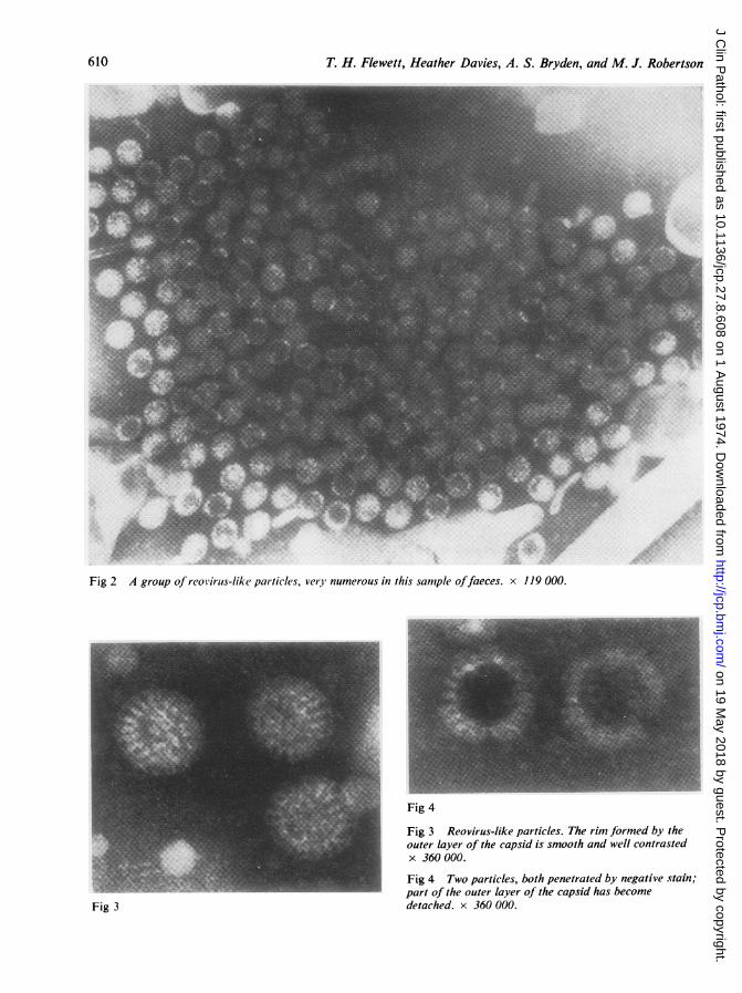
Fig 2 A group of reovirus-like particles, very numerous in this sample offaeces. x 119 000.
Fig 4
Fig 3 Reovirus-like particles. The rim formed by theouter layer of the capsid is smooth and well contrastedx 360 000.
Fig 4 Two particles, both penetrated by negative stain;part of the outer layer of the capsid has becomedetached. x 360 000.Fig 3
610
on 19 May 2018 by guest. P
rotected by copyright.http://jcp.bm
j.com/
J Clin P
athol: first published as 10.1136/jcp.27.8.608 on 1 August 1974. D
ownloaded from

Diagnostic electron microscopy offaeces
Fig 5 The outer capsid layer has been lost. The innerlayer of capsomeres can be seen attached to a thinmembrane surrounding a central space. One capsomerepresents the appearance ofa hollow cylinder (arrow).x 488 000.
Fig 6
MORPHOLOGY OF THE REOVIRUS-LIKE
PARTICLES
These were found in two forms, one in which thetwo layers of the capsid were complete, in diameter61-64 nm (fig 3) and the other 50-54 nm in diameter,from which the outer layer of the capsid appearedto be missing (fig 5). Both types of particles werefrequently seen together in the same preparation.Occasionally, particles were seen from which onlypart of the outer capsid layer had become detached(fig 4). Some particles were penetrated by the nega-tive stain; in these it appeared that the inner capsidsubunits were short, narrow, hollow, parallel-sided units attached at their inner end to a thin
Fig 7
Fig 6 A virion distorted by surface tension near theedge ofa droplet of negative stain. The subunits of theouter layer are placed directly above those of the innerlayer, like the cross-pieces upon capital Ts. x 360 000.
Fig 7 A group ofdetached capsomeric subunits. Thecircles are here resolved into subunits; the arrow indicatesa group ofsix. They could not be thus resolved in theintact virion. x 360 000.
membrane enclosing a central space about 38 nmin diameter. These subunits were about 5 nm long,4 nm wide, with a hole about 1 nm wide down thecentre (fig 5). The outer capsid subunits appeared tobe attached to the ends of these (fig 6). A detachedgroup of these is seen, end-on, in figure 7. The outersurface of the complete double-layered virions wascircular in outline, unlike reoviruses types 1, 2, and3 from tissue culture, whose outlines are distinctlyicosahedral. Also, unlike the reoviruses, the outercapsid subunits appeared to be continuous at theperiphery of the virions, giving the impression of acontinuous membrane surrounding the virus. Theparticles were easily deformed into an oval shape
611
on 19 May 2018 by guest. P
rotected by copyright.http://jcp.bm
j.com/
J Clin P
athol: first published as 10.1136/jcp.27.8.608 on 1 August 1974. D
ownloaded from

T. H. Flewett, Heather Davies, A. S. Bryden, and M. J. Robertson
Fig 8 A group of virus particles distorted by stresses Fig 10 Virus particles agglutinated by convalescentprobably due to uneven drying of negative stain. serum. The strands of antibody connecting the particlesx 300 000. are better visible here. x 300 000.
S _ F s ~~~~~Fig9 Virusparticlesagglutinated byconvalescent serum.A cylindricalstructure, probablycomposed ofcapsidprotein, is attached.Its diameter issimilar to that of thespherical particles.x 300 000.
w ~ ~~~~~~~~~~~~~~~~~~~~~~.... .w... ....
Fig 11 These particles show large-diameter capsomericgroupings, which in certain orientations of the particleappear as rings on the surface; in this these particlesresemble the orbiviruses, especially blue-tongue virus.x 360 000.
612
on 19 May 2018 by guest. P
rotected by copyright.http://jcp.bm
j.com/
J Clin P
athol: first published as 10.1136/jcp.27.8.608 on 1 August 1974. D
ownloaded from

Diagnostic electron microscopy offaeces
by surface forces during drying (fig 8). Here, the'surface membrane' effect is clearly visible. The cap-someres of the outer layer are placed directly uponthose of the inner layer.On the surface, both of the double-layered and
single-layered particles, the spaces between capso-meres (or holes in 'megameres') were somewhatlarger than they appear in reoviruses; the appearanceresembled more closely that of the published pic-tures of blue-tongue, Irituia, or haemorrhagic epi-zootic disease of deer (Murphy, Borden, Shope, andHarrison, 1971; Verweord, Els, de Villiers, andHuismans, 1972) than that of Colorado tick fevervirus. Radial division of surface or capsomeres onthe surface was never clearly apparent, as in Colo-rado tick virus on whole virions, though individualcapsomere subunits were visible when detached(fig 7).Sometimes the particles, when numerous, were
aggregated into clumps. No strands could be foundconnecting them to suggest that they might have beenlinked by globulin molecules, although in the thicklayer of negative stain surrounding 60-80 nm par-ticles fine strands might be difficult to discern,especially in the comparatively crudely purifiedmaterial under investigation. A tubular structurewas found in a clump of virions aggregated by con-valescent serum; this was presumably an aggregateof capsid protein, similar to those described in pre-parations of blue-tongue, Tribec, and other orbi-viruses (fig 9). Aggregates of what may have beencapsid protein were described by Fernelius, Ritchie,Classick, Norman, and Mebus (1972) in materialfrom tissue cultures infected by the newborn calfdiarrhoea agent, but these appeared as sheets ofprotein rather than tubules.
ATTEMPTS AT ISOLATIONAt the time of writing only one reo-like virus hasbeen isolated in tissue culture, though many speci-mens have been inoculated. This virus was detectedin the third blind subculture in human embryokidney (HEK) by electron microscopy of ultra-sonically disrupted cells. In further subcultures inboth HEK and MK cells a granular cytopathiceffect resembling that of reoviruses appeared. Itshaemagglutinin is neutralized to titre by antiserumto reovirus type 1. Other viruses were isolated froma few patients whose faeces contained the 'gastro-enteritis virus': adenoviruses, types 2 and 6, and 2untyped; Echo 11 virus and Escherichia coli sero-type 026 were both isolated from another.
Serological reactionsThe particles could be aggregated easily by conva-lescent sera, but only feebly by acute phase sera
from two patients (fig 10). However, strands re-sembling globulin fibres could be discerned on somevirus particles mixed with acute phase sera, takenthree to five days after the onset of disease. Thevirions were not agglutinated by antisera, derivedfrom hyperimmunized monkeys, to reoviruses types1, 2, and 3. These sera agglutinated their correspond-ing serotypes of reovirus particles; a heavy depositof globulin could be seen upon them by electronmicroscopy.
Discussion
Most young children with acute diarrhoea andvomiting in recent months in and around Birming-ham have had characteristic virus particles in theirfaeces, detectable by electron microscopy. As theperiod during which the virus is detectable is only afew days (in some patients at least) others may wellhave been missed. As virus particles must be verynumerous to be detectable by electron microscopy,they may have been missed in some patients be-cause they were too scanty. The occurrence of reo-virus-like particles in faeces could be used by thelaboratory, on the results so far obtained, for thediagnosis of viral gastroenteritis in young children.
Number Positive NegativeExamined
March-July 1973 16 5 (31 %) 11November 1973 15 9 (60%) 6December 1973 22 16 (73%) 6January 1974 19 9 (47%) 10
Table II Percentage offaeces from gastroenteritispatients with reovirus-like particles from March 1973to January 1974
It will be interesting to see whether they are presentor absent in outbreaks of acute gastroenteritisassociated with type-specific E. coli infection.We agree with Bishop et al (1973) that the reo-
virus-like particles are probably not true reoviruses;if we had been dealing with reovirus infection itseems unlikely that intensive attempts at isolationwould have been so unsuccessful. Furthermore,virus particles could not be agglutinated by antiserato reovirus types 1, 2, and 3. The single isolation of areovirus type 1 is probably merely an incidentalfinding. Simultaneous infection by two or even moreviruses in young children is not very rare. Morpho-logically, the particles are different and resembleblue-tongue virus more than the reoviruses types1, 2, and 3 (fig 11). But we hesitate to place themamong the orbiviruses (Borden, Shope, and Murphy,1971) without more evidence: for orbiviruses are all
613
on 19 May 2018 by guest. P
rotected by copyright.http://jcp.bm
j.com/
J Clin P
athol: first published as 10.1136/jcp.27.8.608 on 1 August 1974. D
ownloaded from

T. H. Flewett, Heather Davies, A. S. Bryden, and M. J. Robertson
acid-labile, and though acid-labile enteric pathogens,eg, Vibrio cholerae, are not unknown, most areacid-stable. Furthermore, in their distinct and char-acteristic smooth, circular outline, with well de-fined rim, the human viruses exactly resemble thegastroenteritis virus of calves (Femelius et al, 1972;Woode et al, 1974), which is known to be acid-stable (Welch and Thompson, 1973) and differsfrom the appearance of orbiviruses (Murphy et al,1971). Acid resistance may, of course, not benecessary for an enteric virus of young children,whose intestinal mobility is brisker and gastric acidweaker than that of adults.
These gastroenteritis viruses may well be membersof a new group of double-stranded RNA (diploma)viruses.
References
Bishop, R. F., Davidson, G. P., Holmes, I. H., and Ruck, B. J. (1973).Virus particles in epithelial cells of duodenal mucosa from chil-dren with acute non-bacterial gastroenteritis. Lancet, 2, 1281-1283.
Bishop, R. F., Davidson, G. P., Holmes, I. H., and Ruck, B. J. (1974).Detection of a new virus by electron microscopy of faecalextracts from children with acute gastroenteritis. Lancet, 1,149-151.
Borden, E. C., Shope, R. E., and Murphy, F. A. (1971). Physicochemi-cal and morphological relationships of some arthropod-borneviruses to bluetongue virus-a new taxonomic group: physico-chemical and serological studies. J. gen. Virol., 13, 261-271.
Feinstone, S. M., Kapikian, A. Z., and Purcell, R. H. (1973). HepatitisA: detection by immune electron microscopy of a virus-likeantigen associated with acute illness. Science, 182, 1026-1028.
Fernelius, A. L., Ritchie, A. E., Classick, L. G., Norman, J. O., andMebus, C. A. (1972). Cell culture, adaptation, and propagationof a reovirus-like agent of calf diarrhea from a field outbreakin Nebraska. Arch. ges. Virusforsch., 37, 114-130.
Flewett, T. H., Bryden, A. S., and Davies, H. A. (1973). Virus particlesin gastroenteritis. Lancet, 2, 1497.
Flewett, T. H., Bryden, A. S., and Davies, H. A. (1974a). Diagnosticelectron microscopy of faeces. 1. The viral flora of the faecesas seen by electron microscopy. J. clin. Path., 27, 603-608.
Flewett, T. H., Davies, H. A., Bryden, A. S., and Robertson, M. J.(1974b). Diagnostic electron microscopy of faeces. II. Acutegastroenteritis associated with reovirus-like particles. J.clin. Path., 27, 608-614.
Horne, R. W., and Nagington, J. (1959). Electron microscope studiesof the development and structure of poliomyelitis virus. J.molec. Biol., 1, 333-338.
Kapikian, A. Z., Gerin, J. L., Wyatt, R. G., Thornhill, T. S., andChanock, R. M. (1973). Density in cesium chloride of the 27nm '8FIIa' particle associated with acute infectious non-bacterial gastroenteritis; determination by ultracentrifugationand immune electron microscopy. Proc. Soc. e.p. Biol. (N. Y.),142, 874-877.
Kapikian, A. Z., Wyatt, R. G, Dolin, R., Thornhill, T. S., Kalica,A. R., and Chanock, R. M. (1972). Visualization by immuneelectron microscopy of a 27 nm particle associated with acuteinfectious nonbacterial gastroenteritis. J. Virol., 10, 1075-1081.
Murphy, F. A., Borden, E. C., Shope, R. E., and Harrison, A. (1971).Physicochemical and morphological relationships of somearthropod-borne viruses to bluetongue virus-a new taxo-nomic group. Electron microscopic studies. J. gen. Virol., 13,273-288.
Murphy, F. A., Coleman, P. H., Harrison, A. K., and Gary, G. W., Jr.,(1968). Colorado tick fever virus: and electron microscopicstudy. Virology, 35, 28-40.
Paver, W. K., Caul, E. O., Ashley, C. R., and Clarke, S. K. R. (1973)A small virus in human faeces. Lancet, 1, 237-240.
Welch, A. B., and Thompson, T. L. (1973). Physiochemical character-ization of a neonatal calf diarrhea virus. Canad. J. comp. Med.,37, 295-301.
Williams, R. E. 0. (1973). Benefit and mischief from commensal bac-teria. J. clin. Path., 26, 811-818.
Wood, H. A. (1973). Viruses with double-stranded RNA genomes. J.gen. Virol., 20, 61-85.
Woode, G. N., Bridger, J. C., Hall, G., and Dennis, M. J. (1974).The isolation of a reovirus-like agent associated with diarrhoeain colostrum-deprived calves in Great Britain. Res. vet. Sci.,16, 102-105.
Verwoerd, D. W., Els, H. J., de Villiers, E. M., and Huismans, H.(1972). Structure of the bluetongue virus capsid. J. Virol., 10,783-794.
Addendum
Since this paper was submitted for publication,Middleton, Szymanski, Abbott, Bortolussi, andHamilton (1974) have published similar evidencethat orbivirus-like particles were associated withacute gastroenteritis of infancy in Canada and werenot found in patients with non-enteric symptoms.Although their estimate of size is rather larger thanours, their picture leaves little doubt that the virusis the same.
Flewett, Bryden, Davies, Woode, Bridger, andDerrick (1974) have suggested that the closely relatedviruses of acute diarrhoea in children and calves aredistinct from the orbiviruses and have proposed thatthey should be called rotaviruses.
References
Middleton, P. J., Szymanski, M. T., Abbott, G. D., Bortolussi, R.,and Hamilton, J. R. (1974). Orbivirus acute gastroenteritis ofinfancy. Lancet, 1, 1241-1244.
Flewett, T. H., Bryden, A. S., Davies, H., Woode, G. N., Bridger, J.,and Derrick, J. (1974). Relation between viruses from acutegastroenteritis of children and newborn calves. Lancet, 2, 61-63.
614
on 19 May 2018 by guest. P
rotected by copyright.http://jcp.bm
j.com/
J Clin P
athol: first published as 10.1136/jcp.27.8.608 on 1 August 1974. D
ownloaded from