accurate splicing of hdac6 requires son
TRANSCRIPT

Wright State University Wright State University
CORE Scholar CORE Scholar
Browse all Theses and Dissertations Theses and Dissertations
2013
Accurate Splicing of HDAC6 Requires Son Accurate Splicing of HDAC6 Requires Son
Vishnu Priya Chowdary Battini Wright State University
Follow this and additional works at: https://corescholar.libraries.wright.edu/etd_all
Part of the Biology Commons
Repository Citation Repository Citation Battini, Vishnu Priya Chowdary, "Accurate Splicing of HDAC6 Requires Son" (2013). Browse all Theses and Dissertations. 1176. https://corescholar.libraries.wright.edu/etd_all/1176
This Thesis is brought to you for free and open access by the Theses and Dissertations at CORE Scholar. It has been accepted for inclusion in Browse all Theses and Dissertations by an authorized administrator of CORE Scholar. For more information, please contact [email protected].

Accurate splicing of HDAC6 requires Son
A thesis submitted in partial fulfillment
of the requirements for the degree of
Master of Science
By
Vishnu Priya Chowdary Battini
B.E., Vinayaka Mission University, 2010
2013
Wright State University

Wright State University
GRADUATE SCHOOL
November 22, 2013
I HEREBY RECOMMEND THAT THE THESIS PREPARED UNDER MY SUPERVISION BY
Vishnu Priya Chowdary Battini ENTITLED Accurate Splicing of HDAC6 requires Son BE
ACCEPTED IN PARTIAL FULFILLMENT OF THE REQUIREMENTS FOR THE DEGREE
OF Master of Science.
Paula A. Bubulya, Ph.D.
Thesis Director
David Goldstein, Ph.D., Chair
Department of Biological Sciences
College of Science and Mathematics
Committee on
Final Examination
Mill W. Miller, Ph.D.
Katherine J. D. A. Excoffon, Ph.D.
R. William Ayres, Ph.D.
Interim Dean, Graduate School
iii
ABSTRACT
Vishnu Battini, M.S., Department of Biological Sciences, Wright State University, 2013
Accurate Splicing of HDAC6 Requires Son.
Pre-mRNA splicing requires proper splice site selection mediated by many factors including
snRNPs and serine-arginine rich (SR) splicing factors. Son is the largest known SR splicing factor,
and it has several putative functional domains including an RS domain, a glycine rich patch (G-
patch) and double stranded RNA binding domain (DSRBD). One-third of Son’s amino acid
sequence consists of novel repetitive sequence motifs of unknown function (Sharma et al., 2010).
Son is essential for organization of pre-mRNA processing factors in nuclear speckles and for cell
cycle progression (Sharma et al., 2010; Sharma et al., 2011; Ahn et al 2011). Exon array analysis
of Son-depleted HeLa cells revealed changes in 1061 transcripts showing exon inclusion or
exclusion, and a total of 2067 splicing events that are potentially regulated by Son. We validated
that Son is required for appropriate splice site choice in transcripts for several chromatin-
modifying enzymes, including HDAC6, ADA and SETD8 (Sharma et al., 2011). However, the
mechanism by which Son maintains accurate splicing is unknown. We are systematically
generating model minigene cassettes for molecular and in situ analysis of Son-dependent splicing
regulation. We have constructed a HDAC6 minigene reporter that contains the genomic sequence
spanning exons 26 through 29. Following Son depletion in HeLa cells transiently transfected with
the HDAC6 minigene reporter construct, we observed skipping of exons 27 and 28 on both the
reporter and endogenous HDAC6 transcripts. Stable cell lines constructed using HDAC6 minigene
reporter construct showed exclusion of exons 27 and 28 upon Son depletion. In order to study Son-
dependent splicing on HDAC6 in situ, we aimed to localize splicing factor recruitment to the

iv
HDAC6 reporter minigene transcription site; however, RNA-FISH performed using probes
designed to bind to HDAC6 minigene transcripts did not show reproducible labeling of the
transcription locus. Finally, HEK293 cells stably expressing four different siRNA-refractory Son
deletion mutants were used to show rescue splicing of HDAC6 minigene transcripts.

v
TABLE OF CONTENTS
CHAPTER 1. INTRODUCTION…………………………………………………….. 1
1.1 Son regulates splicing and promotes cell cycle progression…………. 5
1.2 Histone Deacetylase………………………………………….……….. 11
1.3 Minigene assays…………………………………………….……...…. 15
CHAPTER 2. HYPOTHESES AND SPECIFIC AIMS………………....…………… 17
2.1 Hypotheses…………………………………………………..……...…..... 17
2.2 Specific aims……………………………………………….….…..……… 18
CHAPTER 3. MATERIALS AND METHODS………………………..….………… 23
3.1 HDAC6 subcloning………………………………….….…….……… 23
3.2 Cell culture............................................................................................ 24
3.3 Counting and plating cells..................................................................... 25
3.4 Freezing and thawing............................................................................ 25
3.5 Transfection.......................................................................................... 26
3.6 Son depletion........................................................................................ 26
3.7 RNA-FISH............................................................................................ 27
3.8 Stable cell line construction.................................................................. 28

vi
3.9 Immunofluorescence........................................................................... 29
3.10 RNA extraction................................................................................... 30
3.11 DNase treatment................................................................................. 31
3.12 RT-PCR and quantitative RT-PCR..................................................... 31
3.13 Primer design...................................................................................... 32
3.14 Microscopy......................................................................................... 37
3.15 Sequencing......................................................................................... 37
CHAPTER 4. RESULTS........................................................................................... 38
4.1 Construction of HDAC6 reporter transgene system.......................... 38
4.2 Son-depleted cells show exon skipping in endogenous
HDAC6 mRNA and transiently expressed HDAC6 reporter
Transcripts......................................................................................... 38
4.3 HeLa HDAC6 cell lines stably express HDAC6 minigene............... 44
4.4 Son-depleted cells show exon skipping in endogenous HDAC6
mRNA and stably expressed HDAC6 reporter transcripts............... 44
4.5 qRT-PCR analysis of HDAC6 exon skipping in
Son-depleted cells…………………………………………………. 58

vii
4.6 HeLa HDAC6 stable cell line with single reporter transcription locus... 61
4.6.1 Screening HeLa HDAC6 stable cell lines to identify
reporter transcription locus............................................................. 61
4.6.2 Screening HeLa HDAC6 stable populations to identify
reporter transcription locus............................................................. 62
4.6.3 Hybridization of transiently expressed HDAC6 reporter
minigene to identify transcription/splicing factories...................... 63
4.7 Stable cell lines constructed using four different deletion mutants
of Son...................................................................................................... 73
CHAPTER 5. DISCUSSION.......................................................................................... 76
CHAPTER 6. REFERENCES........................................................................................ 81

viii
LIST OF FIGURES
1. Motif structure of Son………………………………………………..….. 7
2. Motif structure of HDAC6………………………………………………. 13
3. SRSF1 and Son are recruited to the exogenous BTM minigene
transcription site…………………………………………………………. 20
4. Subcloning of the HDAC6 reporter minigene…………………………… 39
5. Skipping of exons 27 and 28 of the HDAC6 reporter minigene
in Son-depleted cells……………………………………………………... 41
6. Primary Screening of Stable cell lines…………………………………… 45
7. Secondary Screening of Stable cell lines………………………………… 47
8. Son depletion causes exon skipping in HDAC6 reporter cell line 69…… 49
9. Son depletion causes exon skipping in HDAC6 reporter cell line 65…… 52
10. Son depletion causes exon skipping in HDAC6 reporter cell line 57…… 55
11. qRT-PCR analysis of HDAC6 exon skipping in Son-depleted cells…….. 59
12. Screening HeLa HDAC6 stable cell lines to visualize the reporter
transcription locus by RNA-FISH………………………………………. 64
13. Screening HeLa HDAC6 stable populations by RNA-FISH to
identify reporter transcription locus…………………………………….. 67
14. RNA-FISH of transiently expressed HDAC6 reporter minigene
transcripts to visualize transcription/splicing factories…………………. 69

ix
15. RNA-FISH of transiently expressed HDAC6 reporter minigene
transcripts to visualize transcription/splicing factories…………………. 71
16. siRNA resistant Son Deletion Mutant constructs and their
expression in Stable HEK 293 Cell lines………………………….……. 74

x
LIST OF TABLES
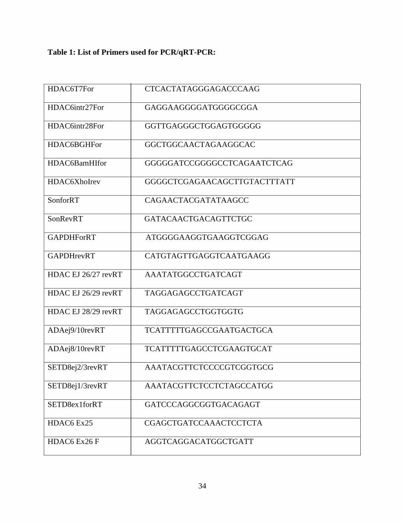
1. List of Primers used for PCR/qRT-PCR…………………………………. 34
2. List of RNA-FISH probes………………………………………………... 36

xi
ACKNOWLEDGEMENTS
I would like to thank Dr. Paula A. Bubulya for letting me work in her lab, for believing in me and
assigning me such a great project, for making sure I was funded during my Master’s program, for
being patient and understanding. I would also like to thank her for working with me to improve
my writing skills. I would like to thank Dr. Athanasios Bubulya for guiding me throughout my
Master’s, for his great suggestions, for teaching me molecular cloning techniques and for
explaining patiently how to choose controls for the experiments.
I would like to thank Dr. Mill W. Miller for helping me choose the right lab, for giving a lot of
moral support whenever I approached him, for assigning me GTA continuously during my
Master’s program, for being my thesis committee member, for giving great suggestions and for
writing recommendation letters for me. I would like to thank Dr. Katherine J. D. A. Excoffon for
being my committee member, for giving valuable suggestions on my project and for writing
recommendation letters whenever I asked.
I would like to thank Dr. Michael Markey for letting me use CGR facility and for helping me
troubleshoot qRT-PCR. I would like to thank Dr. Steven Berberich for providing us the plasmid
pcDNA3.1.
I would like to thank Dr. Sapna Varia firstly for her moral and emotional support throughout my
Master’s program. Secondly, for teaching me almost all techniques and sharing her success tricks
with me. I would like to thank Jennifer Pence for supporting my hypotheses every time, for being
there to troubleshoot various techniques and instruments, discuss and analyze my results. I would
also like to thank her for freezing and processing the stable cell lines when I was visiting my
parents in India.

xii
I would like to thank Dr. Alok Sharma for his valuable suggestions regarding my specific aims
during Rustbelt RNA meetings and for being patient whenever I contacted him asking for details
about siR-Son deletion mutant constructs. I would like to thank Andrew Snyder for helping me
find Son deletion mutant constructs and processing them. I would like to thank all other former
and current Bubulya lab members for their support. I would like to thank James Frisbie for his
suggestions and support.
I would like to thank Poornima Lakshminarayanan Kotha and Madhupriya Mahankali for helping
me take right decisions and giving lot of moral, emotional and technical support and for being
there for me 24X7. I would like to thank Ramya Ganesan for being a great roommate, friend and
for supporting me emotionally and financially. I would like to thank my former roommates Divya
Pemmaraju, Varsha Singireddy, Sushmitha Vegi, Rajitha Katipalli, Sucheta Gangulapattu, Usha
Vayuvegula for helping me adapt to the new environment when I came to the US for the first time.
I would like to thank my current roommates Pratyusha Talluri, Pavani Beesetty, Deepika
Beemanpally and Sravanthi Tatikonda for their suggestions and for making my life easier here. I
would like to thank all my friends, colleagues in India, whom I left behind when I moved to the
US.
Last but not least, I would like to thank my mom Vijaya Lakshmi Battini, my dad Sankara Rao
Battini, for giving me such a beautiful life, for their love, for inspiring me every single time and
for supporting all of my ‘big-stupid’ decisions. I would like to thank my sister Archana Battini
Dhulipalla and my brother-in-law Harish Dhulipalla for all kinds of support throughout.
1
CHAPTER 1: INTRODUCTION
Expression of protein coding genes in mammalian cells is a multistep process that begins with
transcription of genes into pre-messenger RNAs (pre-mRNAs). Pre-mRNA processing is
coordinated with transcription as well as within overall nuclear structure to improve the
efficiency of gene expression. Pre-mRNA processing factors modify nascent pre-mRNA to
produce a mature messenger RNA (mRNA) that is recognized by the nuclear export machinery
and transported to the cytoplasm for translation into protein. Three major steps of pre-mRNA
processing include 5’ capping, pre-mRNA splicing and polyadenylation at 3’ end of mRNA. My
project aims to understand how pre-mRNA splicing of a subset of transcripts is regulated by a
splicing factor called Son.
Pre-mRNA splicing is the removal of introns from nascent pre-mRNA by a multicomponent-
complex called the spliceosome (Reed, 2000; Görnemann et al., 2005). Spliceosomes are
comprised of proteins and small nuclear RNAs that are involved recognition of cis-acting
elements in pre-mRNAs for intron excision (Wahl et al., 2009). The three important sites within
introns that are recognized by components of the spliceosome include the 5’ splice site (also
called a donor site with consensus ‘GU’ residues), the 3’ splice site (also called an acceptor site
with consensus ‘AG’ residues) and the polypyrimidine tract containing a branch point adenosine
sequence. Pre-mRNA splicing begins with recognition of 5’ and 3’ splice sites in the nascent
mRNA by sRNPs, aided by serine-arginine-rich proteins (SR proteins) that help in recruitment of
snRNPs to nearby splice sites. The first spliceosome complex formed is the E complex (also
called the “commitment complex”) containing U1 snRNP that binds to the 5’ splice site.

2
Subsequently, BBP (branch point binding protein), Prp40 and U2AF recognize the branch point
sequence, polypyramidine tract and 3’ splice site, respectively. This in turn recruits U2 snRNP,
which binds to the branch point sequence forming complex A. Subsequently, recruitment of
U4/U6 and U5 snRNPs to complex A forms complex B. Eventually the 5’ and 3’ splice sites are
brought together, followed by the replacement of U1 snRNP by U6 snRNP and the release of U4
snRNP from the complex after which it is called as complex C. Complex C performs the two
catalytic reactions of splicing. In the first step, the 5’ splice site is cleaved through a trans-
esterification reaction and is ligated to an adenosine residue in the branch point sequence to form
an intron lariat. In the second step, the 3’ splice site is cleaved by another trans-esterification
reaction in which the 3’ end of the upstream exon is ligated to the 5’ end of the downstream
exon. The intron lariat is released and degraded via the nonsense mediated RNA decay (NMD)
pathway (Reed 2000; Jurica and Moore, 2003; Black 2003).
Alternative splicing is a natural phenomenon in eukaryotes that results in biodiversity of proteins
by selective inclusion or exclusion of particular exons or introns during pre-mRNA splicing.
Variation in 5’ and 3’ splice site selection in eukaryotic pre-mRNAs regulates gene expression
by altering the coding sequence of mRNA (as reviewed in Black, 2003). The resulting proteins
translated from alternatively spliced mRNAs have differential amino acid sequence and often
have diverse biological functions. While alternative splicing is influenced by cell type,
developmental stage and gender, the machinery used for alternative splicing is not different from
the basal splicing machinery. SR proteins and splicing repressors (heterogenous nuclear
ribonucleoproteins, or hnRNPs) determine which 5’ and 3’ splice sites will be targeted by the
splicing machinery during alternative splicing. The splice sites in nascent pre-mRNAs are
surrounded by several cis-acting elements called Exon/Intron Splicing Enhancers (ESEs/ISEs)

3
and Exon/Intron Splicing Silencers (ESSs/ISSs). These six-nucleotide sequences are binding
sites for splicing factors/repressors that regulate alternative splicing of pre-mRNAs. The
presence of these regulatory elements marks the boundaries of exons (Cartegni et al., 2002).
ESEs/ISEs act as binding sites for splicing factors such as SR proteins SRSF1 and SC35 while
ESSs/ISSs act as binding sites for splicing repressors such as DAPAZ1 and hnRNPA1.
Disruption of one or more cis-acting elements, or modulation of one or more proteins that bind to
them, results in either exon skipping or exon inclusion. Exon skipping is the phenomenon in
which alternative splicing of pre-mRNA results in exclusion of one or more exons from mature
mRNA. Alternative splicing is beneficial when multiple proteins are correctly produced from the
same pre-mRNA transcript. However, abnormal alternative splicing is implicated in diseases
such as cystic fibrosis and spinal muscular atrophy (reviewed in Faustino and Cooper, 2003;
Black, 2000).
Pre-mRNA processing factors localize in nuclear speckles, which are dynamic structures located
in the interchromatin regions of mammalian nuclei (reviewed in Spector and Lamond, 2010).
Proteomic analysis of nuclear speckles revealed the presence of at least 180 known proteins, of
which many are involved in pre-mRNA processing (Mintz et al., 1999; Saitoh et al., 2004).
Nuclear speckles do not contain DNA but are located in close proximity with active transcription
sites and form euchromatic neighborhoods (Haung and Spector, 1991; Thiry, 1995; Spector and
Lamond, 2010). The size and shape of nuclear speckles largely depends on various metabolic
and environmental factors including levels of gene expression. Live cell microscopy showed
formation of rounded and enlarged speckles when gene expression is inhibited suggesting that
nuclear speckles act as storage/assembly/modification sites for splicing factors (Spector and
Lamond 2010). Active genes are present at the periphery of nuclear speckles and polyA+ RNA

4
(but not DNA) localizes to nuclear speckles (Carter et al. 1991; Huang et al. 1994; Visa et al.
1993; reviewed in Thiry 1995). This suggests that some polyadenylated RNAs are enriched in
nuclear speckles after they are transcribed. Some of these RNAs are noncoding RNAs.
MALAT1 is one example of a long nuclear retained noncoding RNA that is enriched at nuclear
speckles and helps in recruitment of splicing factors to transcription sites and in alternative
splicing by regulating SF2/ASF phosphorylation (Hutchinson et al. 2007; Bernard et al. 2010;
Tripathi et al. 2010).
Splicing of pre-mRNA transcripts occurs co-transcriptionally. The largest subunit of RNA
polymerase II has a unique C terminal domain that acts as a docking site for splicing factors as
well as other RNA processing factors, and hence promotes co-transcriptional loading of pre-
mRNA processing factors onto nascent transcripts (Bentley, 2005; Reed, 2002; Misteli et al.,
1997; Misteli and Spector 1998). Live cell imaging of cells transfected with GFP-SF2/ASF in
BTK-IB cells show recruitment of GFP-SF2/ASF to the transcription site of BK virus early
genes. A trail of splicing factors was observed from adjacent nuclear speckles to the transcription
sites after 15-20 min of transcription activation (Misteli et al., 1997). Moreover, Misteli et al
(1997) showed that the transcriptionally active genes and their pre-mRNA are located at the
periphery of nuclear speckles (Misteli et al., 1997). The same was shown by another group that
BrUTP labeling (which marks transcription sites) is not associated with nuclear speckles during
short incubations. However, longer incubations with BrUTP marked the localization of
interchromatin granule-associated zones (Cmarko et al., 1999). Taken together, all of this data
supports the model that nuclear speckles supply splicing factors and other RNA processing
proteins to active transcription sites.

5
Son regulates splicing and promotes cell cycle progression
Son is among 33 novel proteins identified in the proteomic analysis of nuclear speckles (Saitoh
et al., 2004). Son is the largest known SR protein, and it has several putative functional domains
including an RS domain, a glycine rich patch (G-patch) and double stranded RNA binding
domain (DSRBD). RS domain contains alternative arginine and serine amino acid residues and
RS domains of different proteins interact with each other to provide protein-protein interactions
between different splicing factors. Phosphorylation of RS domains is essential for their function
and hence splicing activity is regulated through differential phosphorylation of RS domains
(Boucher et al., 2001; Blencowe, 2000; Graveley, 2000; Smith & Valcarcel, 2000). When
expressed in HeLa cells, YFP-Son co-localizes with SF2/ASF in nuclear speckles (Sharma et al.,
2010; Huen et al., 2010). Son also binds to the NHR4 domain of the leukemogenic protein
AML1-ETO and is important for cell growth (Ahn et al., 2008). One-third of Son’s amino acid
sequence consists of novel repetitive sequence motifs of unknown function that are unique to
Son as shown in Figure 1 (Saitoh et al., 2004). Depletion of Son in HeLa cells alters nuclear
speckle organization of splicing factors SF2/ASF, SC35 and U1-70K that reorganized to
doughnut-shape structures in the absence of Son (Sharma et al., 2010). The unique repeats in Son
were necessary to rescue the nuclear speckle organization (Sharma et al., 2010). Just as the
repeated heptad sequence in the C-terminal domain of the largest subunit of RNA polymerase II
serves as a platform for dynamic recruitment of proteins involved in co-transcriptional
processing of nascent transcripts, we speculate that the unique repeats in Son act as docking sites
for other proteins (Sharma et al, 2010; Sharma et al., 2011).

6
Since Son co-localizes to nuclear speckles with splicing factors such as SF2/ASF, SC35 and
small nuclear RNAs (snRNAs), Son is most likely a pre-mRNA splicing factor. Three separate
studies confirmed the role of Son in accurate splicing of human mRNAs and suggest that Son
plays an important role in regulating the expression of genes involved in maintaining genome
stability and cell cycle progression and pluripotency (Ahn et al., 2011; Sharma et al., 2011; Lu et
al 2013).

7
Figure 1: Motif structure of Son.
The amino terminus of Son contains six tandem repeat motifs between amino acids 334 and
1493. The carboxy-terminus of Son contains multiple domains such as a RS domain, a G-patch
and a DS RNA binding domain (Source: Sharma et al., 2011).

8

9
In the first study (Ahn et al., 2011), microarray analysis in Son depleted HeLa cells altered the
expression of 659 transcripts by more than 1.45-fold. Many of the affected transcripts fall into
pathways and functional categories such as apoptosis, cell cycle, cancer, DNA
replication/recombination/repair and amino acid metabolism. Most of the downregulated
transcripts are involved in cell cycle and DNA replication/recombination/repair and upregulated
transcripts belonged to the categories of cell signaling, cell death/survival and molecular
transport (Ahn et al., 2011). Validation of the microarray data performed by UV crosslinking and
immunoprecipitation followed by RT-PCR showed that Son is associated with mRNAs of
downregulated genes such as TUBG1, KATNB1, TUBGCP2, PCNT, AKT1 and AURKB (Ahn
et al., 2011).
In the second study (Sharma et al., 2011), exon array analysis of Son depleted HeLa cells
showed a 2-fold decrease in expression of 568 genes and a 2-fold increase in expression of 359
genes. The affected genes fall into several biochemical pathways such as apoptosis, cell cycle,
integrin mediated cell adhesion, smooth muscle contraction and G protein signaling (Sharma et
al., 2011). Validation of exon array data performed by quantitative RT-PCR using primer sets
within a single exon, for upregulated genes (due to Son depletion) namely GEMIN5, JL1A,
TNFRSF21, CCN61 and CYP1B1 showed 2-2.5 fold up regulation. Similarly, validation of
down regulated genes (due to Son depletion) such as CDK5 and TNCC1 showed nearly 50% and
60% reduction in transcript levels respectively (Sharma et al., 2011).
Exon array data performed by Sharma et al., (2011) additionally showed altered splicing (either
exon inclusion or exclusion) in 1061 genes and changes in 2067 splicing events. These results
suggest that Son plays a role in splice site selection during splicing of many transcripts (Sharma
et al., 2011). The down regulated genes due to Son depletion such as TUBG1, KATNB1,
10
TUBGCP2, AURKB, PCNT, AKT1, RAD23A and FANCG were tested for splicing defects with
RT-PCR using primers targeting two adjacent exons. Interestingly, intron retention was
observed, indicating Son is required for accurate intron removal in a subset of transcripts (Ahn et
al., 2011). Further studies on splice site selection accuracy in TUBG1 showed that Son depletion
results in improper removal of multiple introns indicating that Son acts as a coactivator in
constitutive splicing to avoid intron retention. Additionally, Son-dependent genes were predicted
(using ESEfinder) to contain weak 5’ and 3’ splice sites. Moreover, some splice sites were
predicted as ‘dual-specificity splice sites’ that can act as both 5’ and 3’ splice sites and thereby
baffle the splicing factors involved in splice site selection. When the weak splice sites were
mutated to become strengthened, they did not depend on Son for accurate splicing, suggesting
that Son is required only when transcripts have weak splice sites (Ahn et al., 2011). To determine
if Son is required in accurate splicing of all genes consisting of weak splice sites, Ahn et al.
(2011) have mutated the splice site of TUBA1B minigene, which is a Son-independent gene, to
weaken it. This mutated TUBA1B minigene did not show Son dependency for proper splicing
indicating that Son is essential for proper splicing of only a subset of genes (Ahn et al., 2011).
Furthermore, UV crosslinking and immunoprecipitation (CLIP) studies revealed that Son is
physically associated with the mRNAs that were down regulated following Son depletion,
suggesting a direct role for Son in splicing. Minigene assays (using the minigene reporter
constructs to study intron retention in vivo) for TUBG1 pre-mRNA containing exon7-intron7-
exon8 cassette or exon8-intron8-exon9 cassette showed that intron removal is directly dependent
on absence or presence of Son (Ahn et al., 2011). When functional domains of Son such as the
G-patch, the RS domain, or the DSRM were either deleted or cloned into expression vectors in
various combinations, it was observed that the RS domain and G-patch were important for

11
proper splicing of TUBG1 pre-mRNA, and the C-terminal region containing an RS domain, G-
patch and DSRM rescued splicing to a significant extent (Ahn et al., 2011).
Validation of the exon array data from Sharma et al. (2011) by RT-PCR showed altered splicing
in Son-depleted cells for mRNAs coding for chromatin modifying enzymes HDAC6, ADA and
SetD8. Exons 27 and 28 were skipped in HDAC6 mRNA, exon 2 was skipped in SetD8 mRNA
and exon 9 was skipped in ADA mRNA, indicating that Son maintains proper splice site
selection for all three transcripts (Sharma et al., 2011). Interestingly, Sharma et al., (2011) also
showed that Son depletion resulted in skipping of exon 6 in stably integrated rat beta-
tropomyosin (BTM) minigene reporter transcripts in HeLa cells. This suggested that Son is
involved in regulating alternative splicing of BTM, and that minigene constructs can be useful
tools for investigating splicing mechanisms in situ. Given that Son impacts all different types of
alternative splicing mechanisms, it is important to know if the mechanisms for such regulation
have any common RNA elements or if they involve common co-factors. Ahn et al., has already
investigated the importance of Son for proper intron removal in a subset of transcripts. Here I
propose to construct an HDAC6 reporter minigene to further investigate Son-dependent
regulation of exon skipping in situ.
In the third study (Lu et al., 2013), Son’s role in maintaining pluripotency was described. Son
depletion in human Embryonic Stem Cells (hESCs) results in loss of pluripotency and cell death.
Genome-wide RNA profiling in Son depleted hESCs identified a set of 1,994 introns from 1,127
genes that had splicing defects following Son depletion. OCT4, PRDM14, E4F1 and MED24 are
some of the genes associated with pluripotency that showed splicing defects due to Son depletion
(Lu et al., 2013).

12
Histone deacetylase 6:
Histone deacetylases (HDACs) deacetylate the nucleosomal histone tails at conserved lysine
residues. Transcription is regulated by the acetylation and deacetylation status of chromatin
making HDACs important in gene regulation. HDACs are also capable of deacetylating lysine
residues of transcription factors and thereby regulate their DNA binding ability. Eighteen
mammalian HDACs were identified and classified into three classes I, II (A and B) and III.
HDAC6 belongs to class IIB deacetylases and is a cytoplasmic protein (reviewed in Verdin et al.,
2003).
The gene encoding HDAC6 is 29,894 basepairs in length and is comprised of 29 exons. It lies on
X chromosome at position p11.23. The mature HDAC6 transcript is 4089 nucleotides and
encodes a protein of 1215 amino acids. A naturally occurring splice variant, hHDAC6p114, was
recently observed in A549 cells; this variant lacks the first 152 amino acids in its N-terminus
(Zhuang et al., 2010). Among all HDACs, HDAC6 is the only HDAC that contains two
functional histone deacetylase catalytic (CAT) domains and a C-terminal BUZ (binding-of-
ubiquitin zinc finger) domain (Figure 2; Hook et al., 2002). The two catalytic histone deacetylase
domains function in deacetylating α-tubulin present in microtubules, which play a key role in cell
motility (Hubbert et al., 2002). The HDAC6 BUZ-domain (amino acids 1134-1192) is encoded
by, 174 nucleotides present in exons 27 and 28 of HDAC6 mRNA. When Son is depleted in

13
Figure 2: Motif structure of HDAC6.
HDAC6 consists of a nuclear exclusion signal domain, two catalytic deacetylase domains, a
cytoplasmic anchoring signal domain and a C-terminal BUZ-domain. (Source: Boyault et al.,
2007).

14

15
HeLa cells, exons 27 and 28 of HDAC6 pre-mRNA are skipped (Sharma et al, 2011),
presumably deleting the BUZ domain under these conditions.
The BUZ-domain of HDAC6 associates with polyubiquitin and thus with ubiquitinated
misfolded proteins (Kawaguchi et al., 2003). Under normal conditions, misfolded proteins are
degraded by proteasomes. Under stress, formation of misfolded proteins increases and becomes
toxic to the cell (Ward et al., 1995). In several human disorders, the accumulation of misfolded
proteins leads to cell death. One mechanism to degrade the misfolded proteins is formation of
aggresomes (Kawaguchi et al., 2003). A model suggests that HDAC6 interacts with dynein
motor proteins as well as ubiquitinated misfolded proteins and directs them to the microtubule-
organizing center (MTOC) via microtubules, where aggresomes are formed (Kawaguchi et al.,
2003). Once aggresomes are formed, the misfolded proteins are degraded by the local proteases
and autophagy proteins. Mutant HDAC6 lacking either catalytic deacetylase domains or BUZ-
domain could not rescue aggresome formation, suggesting the importance of deacetylase
domains and BUZ-domains in aggresome formation (Kawaguchi et al., 2003).
Minigene assays
Several genes express more than one type of mRNAs through alternative splicing. Exon
recognition plays an important role in alternative splicing. Several factors influence the
recognition of exon such as strength of splice sites, sizes of alternatively spliced exons or the
introns on either side, and the presence of secondary structure (Cooper, 2005). Reporter
minigene assays involve construction of a reporter plasmid containing a small portion of
genomic DNA including the alternatively spliced exon(s), flanking introns and exons. These
reporter plasmids are then used in in vivo analysis of cis-regulatory elements (such as cryptic

16
splice sites, ESEs and ESSs) and trans-acting factors such as splicing factors and splicing
repressors (Cooper, 2005). Since endogenous transcripts cannot be visualized easily in situ and
the large size of the endogenous genes and transcripts makes it difficult to analyze the binding
partners and protein-RNA interactions, minigene assays are important tools to analyze the
proteins recruited to the transcription/splicing sites of the minigene. Moreover, inducing silent
mutations in the minigene can help us determine ESEs/ESSs that are essential for proper splicing
of the transcripts and can also determine the binding regions for several splicing factors and
splicing repressors (Cooper, 2005, Ahn et al., 2011).

17
CHAPTER 2: HYPOTHESES AND SPECIFIC AIMS
HYPOTHESES
Hypothesis 1: To construct an HDAC6 minigene reporter
Sharma et al (2011) showed that Son depletion causes alternative splicing of endogenous
HDAC6 and exogenous BTM minigene. Ahn et al. (2011) showed that Son is required for
constitutive splicing of both endogenous and exogenous TUBG1. Based on the above studies on
requirement of Son in splicing, I hypothesized that inclusion of exons 27 and 28 in HDAC6
minigene reporter transcripts requires Son.
Hypothesis 2: To analyze splicing of HDAC6 minigene reporter transcripts in Son-depleted
cells Based on the work performed by Sharma et al. (2011) and Ahn et al. (2011), as well as on
rescue experiments employing Son and its deletion mutants, I hypothesized that the full length
Son and its deletion mutant containing the C-terminal RS domain can rescue the exon skipping
in HDAC6 transcripts. Millevoi et al (2010) showed that hnRNPA1 and hnRNP H/F are involved
in the alternative splicing of BRCA1 transcripts. They found that single base-pair substitution
occurred in the gene and resulted in creation of binding sites for the two splicing regulators.
Sharma et al (2011) performed in situ studies to show that splicing factors such as SF2/ASF and
U1-70K are present at the BTM minigene transcription site both before and after Son depletion.
Recruitment of splicing factors to the co-transcriptional splicing sites results in accurate splice
site selection followed by spliceosome assembly, whereas recruitment of hnRNPs results in
formation of nonproductive spliceosomes or arrests the spliceosome assembly (Erkelenz et al.,

18
2013). Based on the experimental results from the above studies and previous work in our lab on
alternative splicing of HDAC6 in the absence of Son, I hypothesized that Son has a role in
blocking hnRNPs from binding to nascent pre-mRNAs and thereby regulates alternative splicing.
SPECIFIC AIMS
Aim 1: To construct an HDAC6 minigene reporter
I designed a minigene reporter system to investigate the mechanism by which Son regulates
inclusion of exons 27 and 28 in HDAC6 mRNA.
Aim 1a: I generated a HDAC6 minigene cassette as a model minigene reporter system to study
the mechanism of splicing regulation by Son. The HDAC6 minigene cassette, consisting of
genomic DNA encoding E26-I26-E27-I27-E28-I28-E29 (where E represents exon and I
represents intron), was cloned into pcDNA3.1. This reporter plasmid was transfected into HeLa
cells to test whether the exons 27 and 28 are skipped following Son depletion. RNA was
extracted from transfected cells, and RT-PCR was performed using primers specific to
exogenous minigene transcripts. Endogenous HDAC6 transcripts served as a positive control for
exon skipping after Son depletion.
Aim 1b: After confirming that the transiently transfected HDAC6 minigene reporter shows
skipping of exon 27 and 28, I stably transfected HeLa cells with the HDAC6 minigene reporter
plasmid and screened clones that are resistant to the drug G418 which is an analog to neomycin.
The selected clones were expanded for primary and secondary screening. Screening was
performed by extracting RNA from each separate clone for RT-PCR analysis to detect

19
exogenous HDAC6 transcripts using an upstream primer targeting T7 sequences encoded by the
plasmid and a downstream primer targeting exon 29. The stable cell lines expressing exogenous
HDAC6 minigene reporter transcripts were then characterized to verify that skipping of exons 27
and 28 occurs after Son depletion.
Aim 1c: RNA-FISH using the fluorescent tagged-oligos designed either at T7 promoter region of
exogenous HDAC6 minigene or in exon 26 was be performed in HeLa HDAC6 clones, HeLa
HDAC6 stable populations and HeLa cells transiently transfected with HDAC6 plasmid to
identify the HeLa HDAC6 clones containing a reporter locus (ideally it is expected to be
detected as a single dot in each nucleus as shown for BTM cell line in Fig. 3).

20
Figure 3: SRSF1 and Son are recruited to the exogenous BTM minigene transcription site.
A. RNA-FISH using a fluorescently conjugated probe targeting exon 5 of rat beta-tropomyosin
labeled the BTM reporter transcription site. B, C. Immunofluorescence localization of SRSF1
and Son in BTM HeLa cells. Arrows indicate the transcription site of the BTM reporter minigene
(Source: Sharma et al., 2011).

21

22
Aim 2: To analyze splicing of HDAC6 minigene reporter transcripts in Son-depleted cells
The first series of experiments determined which regions of Son are necessary for maintaining
correct splicing of HDAC6 transcripts. To achieve this, I transfected HEK 293 cells with siRNA-
refractory (siR) constructs including full length YFP-SiR-Son and three deletion mutants of Son
(YFP-SiR-Son-1-2008, YFP-SiR-Son-1-1493 and YFPSiR-Son-1-332) and selected stable clones
expressing the constructs using G418. The stable cell lines were transfected with two different
siRNA duplexes and a non-targeting luciferase gene siRNA (control) against Son followed by
RT-PCR using primers sets targeting exon 26-29 region of endogenous HDAC6. The
overexpression of siR-Son constructs and knockdown of endogenous Son were validated by
qRT-PCR using primer sets specific to exogenous and endogenous Son. These experiments help
us determine which deletion mutants can rescue exon skipping of HDAC6 and hence can directly
implicate Son in regulation of splicing.
The next series of experiments aims to observe alternative splicing of HDAC6 minigene in situ
and determine the role of Son in recruiting splicing enhancers/repressors to nascent HDAC6
transcripts. In the attempt to visualize the transcription loci of HDAC6 minigene (as in specific
aim 1C), the experiments were designed to perform immunofluorescence with antibodies against
Son and SF2/ASF along with RNA-FISH with T7 or E26 probes.

23
CHAPTER 3: MATERIALS AND METHODS
HDAC6 subcloning
Human HDAC6 genomic DNA containing exons 26, 27, 28, 29 and the intervening introns was
selected for minigene reporter system construction as Son depletion causes exclusion of exons 27
and 28 in HDAC6 transcripts. BAC clone RP11-416B14 containing HDAC6 gene was purchased
from Children's Hospital Oakland Research Institute (CHORI; Oakland, CA). The BAC clone
library was initially constructed by isolating genomic DNA from male human blood cells.
HDAC6 minigene was then amplified using 5’ CAAGAATCGGGCTTCTCTGA 3’present
in intron 25 region and 5’ TCTCCACCTGCTCAAAGTCA 3’present in intron 29 region
as forward and reverse primers respectively. The amplified PCR product of size 1,387 bp was
cloned into PCR2.1 vector (TA cloning kit, Invitrogen). The positive clone from TA cloning
was used as a template to amplify HDAC6 minigene. Primers were designed to amplify the
region containing exon 26 through 29 to avoid unnecessary confusion about splice site selection.
The forward primer 5’-GGGGGATCCGGGGCCTCAGAATCTCAG-3’contains a flanking
BamH1 restriction site (underlined) and the reverse primer 5’-
GGGGCTCGAGAACAGCTTGTACTTTATT-3’ contains a flanking Xho1 restriction site
(underlined). For PCR, input DNA (100 ng), forward and reverse primers (100 ng each),
magnesium chloride (50 mM, Finnzymes, PA), pfu Ultra Hotstart DNA Polymerase (Aligent
Technologies, CA), 10X pfu Ultra Hotstart DNA polymerase buffer, dNTPs (10mM, Aligent
Technologies, CA) and Molecular Biology Grade water (5 Prime, MD) were used. pcDNA3.1
was used as the vector to subclone HDAC6 minigene as it contains antibiotic resistant genes for

24
both ampicillin and neomycin which allows its selection in both bacterial and mammalian cells.
Empty pcDNA3.1-V5-His-A (Invitrogen, CA) and HDAC6 minigene amplified from TA-
HDAC6 clone were separately digested with BamH1 and Xho1. Gel extraction (Cat. No. 20021;
Qiagen, MD) was performed for both insert (HDAC6 minigene) and double digested pcDNA3.1.
Ligation was performed using T4 DNA ligase (Invitrogen, CA). The ligation reaction was
transformed to DH5α cells (Invitrogen, CA) following the manufacturer’s protocol. Bacterial
colonies were screened for the presence of the vector containing HDAC6 minigene (insert).
Plasmid DNA was isolated and digested with different enzymes to make sure the DNA bands of
expected size were obtained. Plasmid DNA isolated from the three positive bacterial clones was
sequenced to ensure no mutations were present.
Cell culture
HeLa cells obtained from Dr. David Spector (CSHL, NY) and HEK 293 cells (ATCC, VA) were
grown and maintained in 100 mm X 20 mm culture dishes in Dulbecco's Modified Eagle
Medium (DMEM; Hyclone, Thermoscientific, Utah) with 10% fetal bovine serum (FBS;
Hyclone, Thermoscientific, Utah) and 1% penicillin/streptomycin (Invitrogen, CA). All cells
were maintained in a humidified incubator (37oC) in the presence of 5% carbon dioxide. The
cells were passaged after reaching 80% confluency. To passage cells, the cells were briefly
washed 3 times with 1X phosphate buffered saline (PBS; 137 mM NaCl; 2.7 mM KCl; 4.3 mM
Na2HPO4; 1.47 mM KH2PO4, pH 7.4) and were treated with 2ml 0.25% trypsin/EDTA (Hyclone,
Thermoscientific, Utah) for 2 min at 37ºC. 5 ml DMEM containing 10% FBS was added to
inactivate trypsin and the cells were collected in to 15 ml conical tubes, spun for 2 min at 1500
rpm. Supernatant was aspirated and the pellet of cells was resuspended in fresh DMEM

25
supplemented with FBS and antibiotics, either plated in 100 X 20mm dishes for passage or
counted and plated in 6 well dishes.
Counting and plating cells
To plate cells for Son depletion and for RNA-FISH experiments, the cells were collected from
100 X 20mm plates, centrifuged and resuspended as described in the cell culture section above.
10µl of cell suspension was loaded onto each side of the Bright-Line Hemocytometer and the
cells were counted and averaged to find the number of cells per 1 ml. Cells were diluted with
DMEM + 10% FBS (for Son depletion) or with DMEM + 10% FBS and 1%
penicillin/streptomycin (for RNA-FISH) accordingly to get a final concentration of 1 X 10⁵ (for
Son depletion) or 1.5 X 10⁵ (for RNA-FISH).
Freezing and thawing cells
To freeze the cells, the cells were expanded to reach 80% confluency in 100 mm dishes and
washed with 1X PBS. After washing, the cells were trypsinized, collected in to 15 ml conical
tubes and centrifuge at 1500 rpm for 2 minutes. The supernatant was aspirated and 1 ml of ice-
cold freezing medium (95% FBS and 5% DMSO (Cat. No. D2650, Sigma, MO)) was added,
mixed and 0.5 ml was added to each cryovial. The cryovials were then placed in Styrofoam racks
(to slowdown the freezing process) and transferred to -80oC freezer.
To thaw, the vial was placed in 37oC water bath briefly, 5 ml DMEM +10% FBS +1% pen/strep
was added and transferred to a 15 ml conical tube. The tube was centrifuged for 2 min at 1500
rpm and the supernatant was aspirated. To the pellet, 10 ml fresh medium was added and plated
in 100 mm dish and placed in 37oC humidified incubator supplemented with 5% CO2. Thawing

26
process was carried out as fast as possible as DMSO present in the freezing medium is harmful
to cells at higher temperatures.
Transfections
HeLa cells were cultured (~80% confluency), trypsinized and collected as described previously.
The cells were resuspended in 1 ml of medium. Salmon sperm DNA was used along with
HDAC6 reporter plasmid to ensure equal distribution of the plasmid to cells. 2µl of salmon
sperm DNA (20µg/µl), 2µg of HDAC6 reporter plasmid and 250µl cell suspension were added
to a 4 mm gap cuvette. Electroporation was performed by using GenePulser X Cell (250V,
950μF and 4Ω; BioRad, CA). Transient transfections were optimized in HeLa cells to determine
the optimal amount of DNA as well as the post-transfection time required to detect expression of
reporter transcripts. For stable transfections, G418 (1mg/ml) was added to the medium for
selection.
Son depletion
Son depletion was achieved by RNAi using Son siRNA duplexes (si1 catalog # J-012983-05 or
si4 J-012983-08; Dharmacon RNA Technologies). A non-targeting siRNA duplex for luciferase
(catalog no. D-001210-02; Dharmacon RNA Technologies) was used as a control to ensure that
the changes observed are not simply due to activation of RNAi machinery. Son depletion was
validated by immunofluorescence with antibodies against Son and qRT-PCR using primer sets
targeting Son mRNA, standardized to GAPDH. The quantitative results (ct values) of GAPDH

27
qRT-PCR was subtracted from Son qRT-PCR results to determine Δct. ΔΔct was calculated by
subtracting the effect of control siRNA. Fold change was then determined by calculating 2-ΔΔct. A
graph is then plotted between the fold change and samples such as control, siRNA 1 and siRNA
4.
Son depletion in HEK293 cells stably expressing siRNA-refractory YFP-Son constructs was
achieved by using siRNA duplex 4, as the deletion mutants contain silent mutations in the region
of Son mRNA that is targeted by siRNA duplex 4. siRNA duplex 1 was also used to deplete both
exogenous and endogenous Son in control rescue experiments. A luciferase-targeting control
siRNA duplex was used as control to ensure that the changes observed are not simply due to the
activation of RNAi machinery. Overexpression of siRNA refractory YFP-Son constructs and
endogenous Son depletion was confirmed by qRT-PCR using primer sets targeting exogenous,
endogenous Son and GAPDH.
RNA-FISH
RNA-FISH was performed using the T7 probe that is specific to exogenous HDAC6 minigene
transcripts and/or E26 probe which binds to exon 26 region of both exogenous and endogenous
HDAC6 mRNA. All the positive HeLa HDAC6 clones were screened to identify those
containing a single reporter locus. The cells were fixed using 2% formaldehyde, washed three
times 5 min each with 1XPBS and permeabilized using Triton X-100 in the presence of 250mM
vanadyl–ribonucleoside complex (VRC) for 8 min to prevent RNase activity. Further, the cells
were washed with 1X PBS two times 5 min each and blocked using RNase free 0.5% BSA in 1X
PBS for 15 min. The cells were then washed in 1X SSC for 5 min. The hybridization mixture
was prepared by adding RNA-FISH probe(s), 2X SSC, 1mg/ml of tRNA, 10% dextran sulfate,

28
and 25% formamide. Hybridization was performed by adding 20μl hybridization mixture to
clean glass slides and inverting the coverslips (cells facing down) and then the coverslips were
sealed with rubber cement on all four sides. Glass slides were incubated at 37oC for three hours,
in a humidified chamber. The cells were washed in 2X SSC and 25% formamide for 30 min at
37oC followed by a 30 min wash in 2X SSC at room temperature. The cells were then briefly
washed in 2X SSC and then processed for immunofluorescence. Microscopy was performed on a
Deltavision RT microscope, and images were collected and processed using SoftWorx software.
Stable cell line construction
To construct HeLa HDAC6 stable cell line, HeLa cells were transfected with HDAC6 subclone
as described below. The next day, cells were split to 10 plates (100 X 100 mm) to make sure the
cells are separated enough to form individual colonies. Third day, Geneticin or G418 (1 mg/ml)
is added to the cells. G418 is an analog to neomycin sulfate and it blocks protein synthesis by
interfering with the function of 80S ribosomes. Neor gene present in HDAC6 plasmid produces
aminoglycoside 3'-phosphotransferase, APT 3' II which is resistant to G418. The cells were then
allowed to grow for 2-3 weeks within which the cells that integrate the plasmid into their genome
and make APT 3' II do survive and develop their own colonies while untransfected cells and
cells in which plasmid does not integrate into the genome die. The colonies were picked later
into 24 well dishes and allowed to grow until the wells were confluent. For primary screening of
clones stably expressing HDAC reporter transcripts, RNA was extracted from the 96 clones
followed by DNase treatment and RT-PCR using T7 forward primer and E29 reverse primer .
Positive clones from primary screening were expanded and screened a second time to confirm
the expression of minigene transcripts. The positives from secondary screening were expanded
and frozen down.

29
Immunofluorescence
Coverslips were washed thoroughly in one part nitric acid and two parts hydrochloric acid for
two hours, rinsed in distilled water continuously until the pH returns to 7.0. The coverslips were
then stored in 70% ethanol and flamed before using to remove ethanol. Cells grown on
coverslips were washed once with 1X PBS and fixed with 2% paraformaldehyde (in 1X PBS) for
15 min followed by three washes with 1X PBS 5 min each. The cells were then permeablized for
5 min with 0.2% TritonX-100 and washed three times with 0.5% normal goat serum (NGS,
Gibco) in 1X PBS. Washes with normal goat serum help in blocking the proteins. The coverslips
were then placed with the cell side up in a humidified chamber prepared by putting a wet filter
paper in a 15 cm plate. 40µl of primary antibodies diluted in 0.5% NGS-1X PBS were added to
the cells and incubated for one hour at room temperature. Rabbit polyclonal antibodies were
developed (Covance; Denver, PA) against the C-terminal peptide sequence
SPNKKHAKATAATV of Son (Wu13; 1:100), and against the N- terminal peptide sequence
CEESESKTKSH of Son (Wu14; 1:1000) and monoclonal anti-SF2/ASF AK103 (1:2500;
provided by A. Krainer, Cold Spring Harbor Laboratory, Cold Spring Harbor, NY). Coverslips
were placed back into the appropriate dishes and washed three times with 0.5% NGS in 1X PBS.
40µl of secondary antibodies diluted in 0.5% NGS -1X PBS were added to cells placed in
humidified chamber cell side up. Fluorescently- conjugated secondary antibodies donkey anti-
mouse (Cy5) and donkey anti-rabbit (FITC or TxRed) proteins (1:500; Jackson ImmunoResearch
Laboratories, West Grove, PA) were used. After one hour incubation with secondary antibodies,
the coverslips were placed back into the appropriate dishes and washed two times with 0.5%
NGS in 1X PBS for 5 min each. The cells were then washed for 5 min in 4', 6-diamidino-2-
phenylindole (DAPI; 10 μg/ml diluted 1:1000 in 1X PBS) to stain DNA and washed once with

30
1X PBS. To mount the coverslips, either anti-fade mounting medium (p-phenylenediamine,
glycerol, pH. 8.0 - 9.0) or N-propyl-gallate mounting medium (for CY5-conjugated antibodies)
was used and the coverslips were placed cell side down on clean microscope glass slides with 8-
10 µl mounting medium. Excess mounting medium was removed by using a filter paper and the
edges of coverslips were painted with clear nail polish. The slides were then stored in -80oC or
imaged using DeltaVision RT microscope (Applied Precision).
RNA Extraction
RNA extraction was performed by using Qiagen RNAeasy mini prep kit according to
manufacturer instructions (Qiagen, CA). Briefly, cells cultured in 6 well dishes were trypsinized
and collected by centrifuging for 5 min at 1500 rpm. The supernatant was completely aspirated
and the cell pellet was resuspended in 350 µl buffer RLT. The cell suspension was homogenized
by vortexing for 30 sec and 350 µl 70% ethanol was added. 700 µl of the above solution was
loaded into RNAeasy spin column placed in a 2 ml collection tube, centrifuged for 15 sec at full
speed (13,200 rpm) and the flow through was discarded. 700 µl of buffer RW1 was added to the
spin column, centrifuged for 15 sec at full speed and the flow through was discarded. The spin
column was then washed 2 times with 500 µl of buffer RPE and centrifuged (first time for 15 sec
and second time for 2 min) at full speed. To elute RNA, the spin column was placed in 1.7 ml
collection tube, 30 µl RNase-free water was added and the tube was centrifuged for 1 min at
14,000 rpm in an Eppendorf microcentrifuge. An optional elution step was performed by adding
30 µl of RNase-free water and collecting RNA in a fresh tube. Eluted RNA was stored at -80oC.

31
DNase treatment
The concentration of RNA extracted as described above, was measured using a Nanodrop
spectrophotometer (Thermo Scientific). Buffer, Turbo DNase activation buffer (10%), 5 µg of
RNA, Turbo DNase (1µl) were mixed and brought to 50 µl final volume with RNase free water.
The samples were incubated at 37oC for 30 min followed by the addition of DNase inactivation
reagent (10%) and incubated for 2 min at room temperature with occasional mixing. The samples
were centrifuged for 1.5 min at 10,000 X g and the supernatant containing DNA-free RNA was
transferred to new 1.5 ml tubes and stored at -80oC.
RT-PCR and quantitative RT-PCR
RT-PCR was performed by using qScript-One step RT-PCR system and qScript-two step RT-
PCR system (Quanta Biosciences, MD Cat. No. 95047, 95073). For one-step RT-PCR, 100 ng of
DNase treated RNA was used as template, 3 µM forward and reverse primers, 2X Sybrgreen
buffer, 1 µl reverse transcriptase and RNase-free water were used to make 10 µl reactions.
Master mix was prepared by adding appropriate amounts of SybrGreen buffer, reverse
transcriptase, forward and reverse primers and water, mixed and centrifuged briefly and added to
each of the Cepheid tubes. 100 ng of RNA was added to each of the tubes and centrifuged
briefly. The tubes were then loaded in Cephed one StepRT-PCR machine (Chepheid, CA).
Amplification was performed for 40 cycles As exon skipping occurs in HDAC6 transcripts up on
Son depletion, reverse transcription-PCR results in amplification results show more than one
PCR product when certain primer pairs were used. In these cases, the RT-PCR products were
applied to either 2% agarose gels or 10% native polyacrylamide gels (30% Polyacrylamide, 10μl
TEMED, 10% APS, 5X TBE) at 80V for 90 min in 1X TBE. The gels were stained on shaking

32
platform by adding 2 μl of 10 mg/ml ethidium bromide to 300 ml of buffer 1X TBE for 5 min
before exposing to ultraviolet light.
For two-step RT-PCR, cDNA was prepared by mixing 4 µl cDNA fast mix, 100 ng DNase-
treated RNA, 1 µl reverse transcriptase and RNase-free water to make up the reactions to 20 µl.
The samples were incubated in DNA engine DYAD thermo cycler (MJ Researchers, MA) with
cycle conditions: 5 min at 22oC; 30 min at 42oC and 5 min at 85oC. cDNA was either stored at -
20oC or diluted 3 fold and used as template for qPCR. qPCR was performed in 96 well plates in a
qPCR machine(App lied Biosciences). For qPCR, master mixes were prepared separately for
each primer set. 5 µl of PerfecTa fast mix, 3 µM forward primer, 3 µM reverse primer and
RNase-free water were mixed per reaction to make mastermix and aliquoted to the reaction wells
followed by addition of 3µl of diluted cDNA template. The quantitative results (ct values) of
GAPDH qRT-PCR were subtracted from Son qRT-PCR results to determine Δct. ΔΔct was
calculated by subtracting the effect of control siRNA. Fold change was then determined by
calculating 2-ΔΔct.
Primer and probe design
All primers designed were checked for at least 50% GC content and melting temperature (above
450C), The primers were checked for possibility of forming self-dimers, heterodimers and
hairpin structures by using http://www.idtdna.com/analyzer/Applications/OligoAnalyzer/. Hairpin
structures form when the melting temperature of hairpin structures suggested is close to that of
the primer. Self-dimers or hetero-dimers form when the ΔG value is lower than -10 k.
Furthermore, the specificity of the primer sequences were tested using
http://blast.ncbi.nlm.nih.gov/Blast.cgi website. Nucleotide blast, Refseq mRNA, Homo sapiens

33
were selected to blast the primer sequence. For non-specific binding to occur both forward and
reverse primer should bind to the same gene/mRNA or genes that are adjacent.
34
Table 1: List of Primers used for PCR/qRT-PCR:
HDAC6T7For CTCACTATAGGGAGACCCAAG
HDAC6intr27For GAGGAAGGGGATGGGGCGGA
HDAC6intr28For GGTTGAGGGCTGGAGTGGGGG
HDAC6BGHFor GGCTGGCAACTAGAAGGCAC
HDAC6BamHIfor GGGGGATCCGGGGCCTCAGAATCTCAG
HDAC6XhoIrev GGGGCTCGAGAACAGCTTGTACTTTATT
SonforRT CAGAACTACGATATAAGCC
SonRevRT GATACAACTGACAGTTCTGC
GAPDHForRT ATGGGGAAGGTGAAGGTCGGAG
GAPDHrevRT CATGTAGTTGAGGTCAATGAAGG
HDAC EJ 26/27 revRT AAATATGGCCTGATCAGT
HDAC EJ 26/29 revRT TAGGAGAGCCTGATCAGT
HDAC EJ 28/29 revRT TAGGAGAGCCTGGTGGTG
ADAej9/10revRT TCATTTTTGAGCCGAATGACTGCA
ADAej8/10revRT TCATTTTTGAGCCTCGAAGTGCAT
SETD8ej2/3revRT AAATACGTTCTCCCCGTCGGTGCG
SETD8ej1/3revRT AAATACGTTCTCCTCTAGCCATGG
SETD8ex1forRT GATCCCAGGCGGTGACAGAGT
HDAC6 Ex25 CGAGCTGATCCAAACTCCTCTA
HDAC6 Ex26 F AGGTCAGGACATGGCTGATT

35
HDAC6 Ex29 Rev CCGTATTCTGGGGCTTAGTG

36
Table 2: List of RNA-FISH probes:
T7 TxRed, (TxRed)AGCTTGGGTCTCCCTATAGTGAGTCGTATT
HDAC E26 TxRed, (TxRed)CTAGTAGGTTCTCCTCTCCTGGGGCCTGAG

37
Microscopy
Images were collected in z-stacks using a 60X objective (1.4 numerical aperture; Olympus,
Tokyo, Japan) on a DeltaVision RT microscope (Applied Precision). The raw data from z-stacks
were displayed as volume projections.
Sequencing
The HDAC6 reporter minigene plasmid construct was sequenced to confirm that no mutations
are present. Midipreps were prepared for three of the positive clones (2, 21 and 36). Since the
size of the HDAC6 minigene is 1.3Kb, a forward primer was designed at the T7 promoter region,
two forward primers were designed at a distance of 400bp each and a reverse primer was
designed at the BGH signal. 1.2μg of plasmid DNA and 100 pmole primer was added and sent
for sequencing (Retrogen,inc, CA). Results were analyzed using multiple sequence alignment
tool (http://www.ebi.ac.uk/Tools/msa/clustalw2/).

38
CHAPTER 4: RESULTS
4.1 Construction of a HDAC6 reporter transgene system
Son was previously reported to be required for proper splicing of HDAC6 transcripts (Sharma et
al., 2011). To further understand the mechanism of aberrant splicing caused when Son is
depleted, I designed reporter minigene assays. HDAC6 subclone containing genomic segment
encompassing exons 26-29 was constructed in pcDNA 3.1 and the positives from subcloning are
shown in Figure 4. Genomic sequence for HDAC6 was amplified by PCR and subcloned into
pcDNA3.1. Plasmid DNA was digested with BamH1 and Xho1, and colonies containing a 1.3
Kb insert along with a 5.5Kb band were chosen as positive clones (Fig. 4A, 4B and 4C). The
positive clones 2, 21 and 36 were digested with various restriction enzyme combinations to
confirm the presence of the HDAC6 minigene. Furthermore, three clones were sequenced to
confirm that no mutations were introduced during PCR amplification.
4.2 Son-depleted cells show exon skipping in endogenous HDAC6 mRNA and transiently
expressed HDAC6 reporter transcripts
To examine splicing changes in exogenous HDAC6 transcripts upon Son depletion, HeLa cells
were transiently transfected with HDAC6 reporter minigene plasmid, followed by siRNA
transfection to deplete Son. Reduction of Son transcripts was confirmed by qRT-PCR with
primers targeting Son mRNA (Fig. 5A) and immunofluorescence where Son depleted individual
cells show loss of speckle pattern when compared to mock and control oligo transfected cells
(Fig. 5B). Son depleted samples showed a clear difference in splicing of reporter transcripts as
seen by polyacrylamide gel electrophoresis of RT-PCR products. While cells treated with mock

39
Figure 4: Subcloning of the HDAC6 reporter minigene.
HDAC6 minigene PCR products were subcloned in to pcDNA3.1. A, B, C: Miniprep DNA
isolated from amp-resistant bacterial colonies was double digested with Xho1 and BamH1. Gel
images show positive clones containing two fragments; one corresponding to plasmid backbone
(5.5 Kb) and the other to the minigene (1.3 Kb). Some of the clones contain an extra band at 6.8
Kb which is the linearized plasmid. Contols included undigested empty vector (lane 1 of Panels
A, B and C), linearized empty vector (lane 2 of Panels A, B and C) and empty vector digested
with each of the enzymes (lanes 13 and 14 of panel C).

40

41
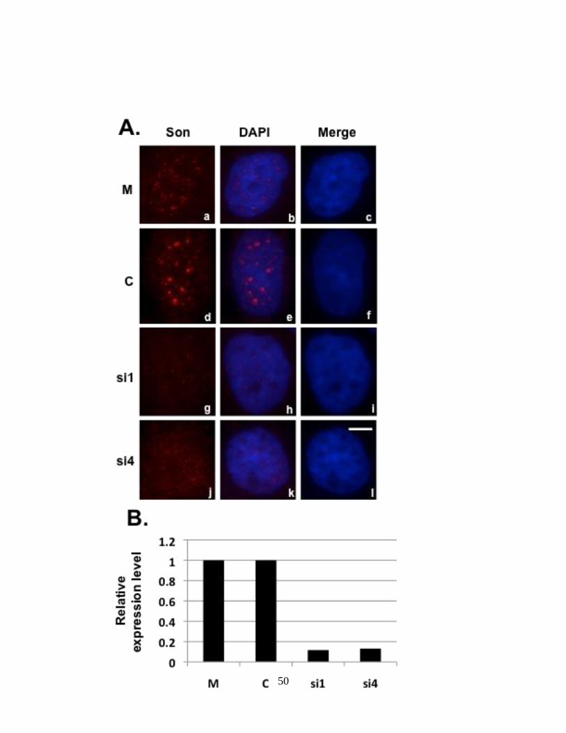
Figure 5: Skipping of exons 27 and 28 of the HDAC6 reporter minigene in Son-depleted
cells.
HeLa cells transiently transfected with HDAC6 reporter plasmid and Son siRNA duplexes were
either processed for immunofluorescence (A) or harvested to perform qRT-PCR (B) to show Son
depletion. Cells treated with Son siRNAs and immunostained with anti-Son antibodies in (A) do
not show Son labeling in nuclear speckles (g and j) when compared to mock and control siRNA-
treated cells (a and d). DNA was stained with DAPI (c, f, I and l). Bar = 5µm. qRT-PCR in (B)
showed reduction of Son mRNA levels in siRNA-treated samples when compared to that of
mock and control siRNA treated samples. (C) qRT-PCR performed in triplicate using primers
that amplify only reporter minigene transcripts show exon skipping in Son siRNA-treated
samples. Lanes labeled as “–” (2, 4, 6, 8 and 10) represent reactions in which reverse
transcriptase was omitted. (D) RT-PCR performed with primers that amplify both endogenous
and reporter minigene transcripts show exon skipping in siRNA-treated samples. Lanes labeled
“U” represent untreated samples that were transfected with HDAC6 reporter plasmid and
harvested. Lanes labeled as “–” (2, 4, 6, 8 and 10) represent reactions in which reverse
transcriptase was omitted.

42

43

44
transfection or control oligo showed inclusion of exons 27-28, these exons were skipped in Son
depleted cells (Fig. 5C). Splicing changes observed in exogenous HDAC6 minigene transcripts
are consistent with that of endogenous HDAC6 transcripts (Fig. 5D and Sharma et al., 2011).
4.3 HeLa HDAC6 cell lines stably express HDAC6 minigene
To further investigate the mechanism of Son-mediated splicing in situ, it is important to
construct cell lines stably expressing HDAC6 minigene. HeLa cells were hence transfected with
HDAC6 subclone and G418 (1 mg/ml) was added to medium to select clones stably expressing
HDAC6 minigene. Primary screening by RT-PCR for RNA extracted from each of the clones
show unspliced, fully spliced and exon skipped products in clones 6, 19, 57, 65, 69, 79, 80 and
93 (Fig. 6A, 6B and 6C). Secondary screening was performed in which the positives from
primary screening were expanded and RNA was extracted. RT-PCR for the clones mentioned
above showed stable expression of HDAC6 minigene in clones 19, 57, 65, 69, 79 and 80 (Fig.
7A, 7B and 7C).
4.4 Son-depleted cells show exon skipping in endogenous HDAC6 mRNA and stably
expressed HDAC6 reporter transcripts
Three of the HeLa HDAC6 clones that stably express HDAC6 minigene were transfected with
Son siRNA to test splicing patterns of HDAC6 minigene. Son depletion in three of the HeLa
HDAC6 stable cell lines (Clones 69, 57 and 65) resulted in skipping of exons 27 and 28 in both
exogenous (Fig. 8C, 9C and 10C) and endogenous HDAC6 transcripts (Fig. 8D, 9D and 10D).

45
Figure 6: Primary Screening of Stable cell lines.
RNA extracted from G-418-resistant colonies was used to perform RT-PCR to amplify HDAC6
minigene transcripts. The bands migrating at 528 bp correspond to properly spliced reporter
transcripts and the bands at 259 bp correspond to products in which exons 27 and 28 are skipped
(A, B and C). RT-PCR was performed on RNA isolated from cells that were transiently
transfected with the HDAC6 reporter plasmid (panel A: lanes 1 and 2; panel B: lane 1; panel C:
lanes 1 and 2) as well to indicate the sizes of expected bands for comparison with bands from
stable clones. Additional bands in some of the clones may be due to the presence of genomic
DNA in the samples.

46

47
Figure 7: Secondary Screening of Stable cell lines.
Positive clones from the primary screening of stable reporter cell lines were expanded. RNA was
extracted to perform RT-PCR to amplify HDAC6 minigene transcripts. The bands migrating at
528 bp correspond to properly spliced reporter transcripts and the bands at 259 bp correspond to
products in which exons 27 and 28 are skipped (A, B and C). RT-PCR was performed on RNA
isolated from cells that were transiently transfected with the HDAC6 reporter plasmid (panel A,
B, C: lane 1) as well to indicate the sizes of expected bands for comparison with bands from
stable clones. Additional bands in some of the clones may be due to the presence of genomic
DNA in the samples.

48

49
Figure 8: Son depletion causes exon skipping in HDAC6 reporter cell line 69.
HeLa HDAC6 clone 69 stably expressing HDAC6 reporter plasmid was transfected with Son
siRNA duplexes and processed for either immunofluorescence (A) or harvested to perform qRT-
PCR (B) to show Son depletion. Cells treated with Son siRNA and immunostained with anti-Son
antibodies in (A) do not show Son labeling in nuclear speckles (g and j) when compared to mock
and control siRNA treated cells (a and d). DNA was stained with DAPI (c, f, I and l). Bar = 5µm.
qRT-PCR in (B) showed reduction of Son mRNA levels in siRNA-treated samples when
compared to mock and control siRNA treated samples. (C) qRT-PCR performed in triplicate
using primers that amplify only reporter minigene transcripts show exon skipping in Son siRNA-
treated samples. (D) RT-PCR performed with primers that amplify both the endogenous as well
as reporter minigene transcripts show exon skipping in siRNA-treated samples. Lanes labeled
“U” represent the samples for which RNA was extracted without treating with siRNA reagents.
50

51

52
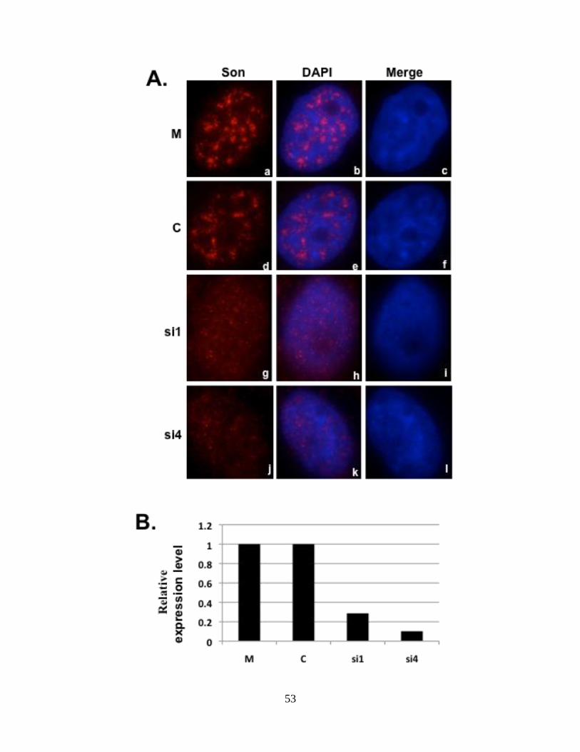
Figure 9: Son depletion causes exon skipping in HDAC6 reporter cell line 65.
HeLa HDAC6 clone 65 stably expressing HDAC6 reporter plasmid was transfected with Son
siRNA duplexes and processed for either immunofluorescence (A) or harvested to perform qRT-
PCR (B) to show Son depletion. Cells treated with Son siRNA and immunostained with anti-Son
antibodies in (A) do not show Son labeling in nuclear speckles (g and j) when compared to mock
and control siRNA treated cells (a and d). DNA was stained with DAPI (c, f, I and l). Bar = 5µm.
qRT-PCR in (B) showed reduction of Son mRNA levels in siRNA-treated samples when
compared to mock and control siRNA treated samples. (C) qRT-PCR performed in triplicate
using primers that amplify only reporter minigene transcripts show exon skipping in Son siRNA-
treated samples. (D) RT-PCR performed with primers that amplify both the endogenous as well
as reporter transcripts show exon skipping in siRNA-treated samples. Lanes labeled “U”
represent the samples for which RNA was extracted without treating with siRNA reagents.
53

54

55
Figure 10: Son depletion causes exon skipping in HDAC6 reporter cell line 57
HeLa HDAC6 clone 57 stably expressing HDAC6 reporter plasmid was transfected with Son
siRNA duplexes and processed for either immunofluorescence (A) or harvested to perform qRT-
PCR (B) to show Son depletion. Cells treated with Son siRNA and immunostained with anti-Son
antibodies in (A) do not show Son labeling in nuclear speckles (g and j) when compared to mock
and control siRNA treated cells (a and d). DNA was stained with DAPI (c, f, I and l). Bar = 5µm.
qRT-PCR in (B) showed reduction of Son mRNA levels in siRNA-treated samples when
compared to mock and control siRNA treated samples. (C) qRT-PCR performed in triplicate
using primers that amplify only reporter minigene transcripts show exon skipping in Son siRNA-
treated samples. (D) RT-PCR performed with primers that amplify both the endogenous as well
as reporter transcripts show exon skipping in siRNA-treated samples. Lanes labeled “U”
represent the samples for which RNA was extracted without treating with siRNA reagents.

56

57

58
Exon skipping of HDAC6 reporter transcripts in Son depleted cells when expressed transiently
or stably indicates that the HDAC6 minigene reporter construct contains the necessary elements
required to study Son-dependent splicing of exons 27 and 28. Since multiple stable cell lines
expressing the HDAC6 reporter minigene show the same result, there is probably no connection
between chromatin context and splicing regulation by Son for splicing of exons 27 and 28 of
HDAC6 transcripts.
4.5 qRT-PCR analysis of HDAC6 exon skipping in Son-depleted cells
To quantify the relative expression level of properly spliced and exon skipped products in Son
depleted cells that stably express HDAC6 reporter minigene, exon-junction primers were
designed that allow the amplification of single products as shown in fig. 11A. Properly spliced
HDAC6 minigene contains all exons i.e., exons 26, 27, 28 and 29 allowing us to design an exon
junction primer that includes the last 12 nt of exon 26 and first 12 nt of exon 27. Since exon 27 is
absent in the exon skipped product, the EJ26/27 primer specifically complements the properly
spliced HDAC 6 transcripts. Likewise, exons 26 and 29 are joined when exon skipping occurs.
An exon junction primer (EJ26/29) was designed containing the last 12 nt of exon 26 and the
first 12 nt of exon 29, it will amplify only the reporter transcripts in which exons 27 and 28 are
skipped. Furthermore, to differentiate between endogenous and exogenous HDAC6 transcripts,
a primer was designed to complement sequences within exon 25 to amplify only endogenous
HDAC6 transcripts (since HDAC6 minigene does not contain exon 25), and another primer was
designed to complement sequences just following the T7 promoter region of the HDAC6
minigene to amplify only exogenous HDAC6 trnascripts (since the T7 sequences are absent in
59
Figure 11: qRT-PCR analysis of HDAC6 exon skipping in Son-depleted cells.
qRT-PCR performed on HeLa HDAC6 stable cells post-Son depletion. A) Four sets of exon
junction primers were used to amplify splice variants for exogenous reporter transcripts as well
as for endogenous HDAC6 transcripts. B) Relative transcript level for properly spliced verses
exon skipped transcripts is measured in both endogenous and exogenous HDAC6.

60

61
endogenous HDAC6 transcripts). qRT-PCR was performed in the Son-depleted HeLa
HDAC6 stable cell line (clone 69) using combination of primers for HDAC6. Son depletion
was validated by qRT-PCR. For both the endogenous and the exogenous HDAC6 transcripts
in Son-depleted cells, the level of properly spliced transcripts were low while levels of
improperly spliced transcripts were high in comparison to respective transcript levels in cells
treated with luciferase siRNA (control) (Fig. 11B). This indicates the efficacy of the
minigene reporter system we generated in mimicking the endogenous HDAC6 splicing
patterns.
4.6 HeLa HDAC6 stable cell line with single reporter transcription locus
The stable cell lines constructed using HDAC6 reporter minigene construct were used to
observe reporter transcription loci inside the cells. To achieve this, RNA fluorescence in situ
hybridization (RNA-FISH) was initially performed for all six positive clones.
4.6.1 Screening HeLa HDAC6 stable cell lines to identify reporter transcription locus
Two RNA-FISH probes were designed one at the T7 promoter region which binds only to
reporter minigene transcripts and the other probe in exon 26 region of HDAC6 which can
hybridize to both exogenous and endogenous HDAC6 transcripts. The stable clones were
grown on coverslips and RNA-FISH combined with immunofluorescence was performed as
described in the methods section. For immunofluorescence, antibodies against Son and
SF2/ASF were used to observe co-localization patterns to HDAC6 transcripts. Since BTM
cell line constructed by Dr. Paula Bubulya consists of the same plasmid backbone and

62
includes the T7 promoter region, RNA-FISH was performed on this cell line as a control.
The BTM cell line is considered a positive control for HDAC6 reporter since it can be used
to validate the working conditions of FISH probes and other reagents used. RNA-FISH in
HeLa cells was performed as a negative control that will not contain T7 sequences and can be
used to establish background fluorescence levels of RNA-FISH probes.
Unfortunately, RNA-FISH performed on stable cell HDAC6 reporter lines showed no
consistent labeling of transcription loci. BTM cell line hybridized with T7 probe showed a
bright transcription locus (Fig. 12: panel 1) and co-localization with Son (Fig.12 panel 2) and
SF2/ASF (Fig. 12 panel 3) indicating that the probes and other reagents used were working
appropriately. HeLa cells in the absence of probe did not show any transcription loci (Fig. 12
panels 9, 10 and 11) and HeLa cells hybridized with E26 probe show random labeling of
bright dots, some of which co-localized with Son and SF2/ASF (Fig. 12 panels 5, 6 and 7).
HDAC6 stable cell lines hybridized with E26 probe showed either 2-3 bright dots that
resembled a nuclear speckle pattern. The speckle pattern observed colocalizes with Son and
SF2/ASF while some of the bright dots colocalize with Son and SF2/ASF (Fig.12 panels a-
x).
4.6.2 Screening HeLa HDAC6 stable populations to identify reporter transcription locus
Since we did not successfully generate a stable clone with single transcription locus, we
proceeded to make stable populations with HDAC6 reporter minigene construct and visualize
the transcription loci of HDAC6 minigene in situ. HeLa cells were transfected with HDAC6
reporter construct and selected using G418 (1 mg/ml). The cells were then trypsinized,
collected and frozen stocks were made. RNA-FISH with different probe concentrations (10

63
ng, 20 ng and 40 ng), combined with immunofluorescence with antibodies against Son and
SF2/ASF was performed. RNA-FISH with HDAC6 probe showed speckle like structures
(Fig.13 panels a, e and i) that co-localized with Son and SF2.ASF (Fig.13 panels b, c, d, f, g,
h, j, k and l).
4.6.3 Hybridization of transiently expressed HDAC6 reporter minigene to identify
transcription/splicing factories
RNA-FISH for both stable populations and stable cell lines being unsuccessful, we moved on
to cells transiently expressing HDAC6 reporter minigene. Transient transfection of
minigenes such as BTM have previously been used to create large transcription factories
within nuclei that can be detected by RNA-FISH (Huang and Spector, 1996). HeLa cells
transfected with 2 μg of HDAC6 reporter plasmid DNA were fixed after 48 hours to perform
RNA-FISH with different probe concentrations (10 ng, 20 ng and 40 ng) and
Immunofluorescence with antibodies against Son and SF2/ASF. RNA-FISH with E26 probe
showed 2-3 bright dots (Fig.14 panels a, e and i) some of them colocalizing with Son and
SF2/ASF (Fig.14 panels b, c, d, f, g, h, j, k and l). One more condition was tested by varying
the amount of DNA used for transfection. HeLa cells were transfected with different
concentrations of HDAC6 reporter plasmid DNA (1, 2, 5, 7 and 10 μg), and RNA-FISH
combined with immunofluorescence was performed after two days. Again, transcription
factories were not observed, but we observed a nuclear speckle pattern for HDAC6 reporter
transcripts (Fig.15 panels a, e, i, m and q).

64
Figure 12: Screening HeLa HDAC6 stable cell lines to visualize the reporter transcription
locus by RNA-FISH.
HeLa HDAC6 stable cell lines were processed for RNA-FISH to label HDAC6 minigene
transcripts combined with dual label immunofluorescence localization of Son and SF2/ASF.
Panels 1, 5, a, e, i, m, q and u show RNA-FISH signal in different cell lines as labeled. Panel 9
shows HeLa cells not labeled with any probe (to determine background fluorescence). Panels 2,
6, 10, b, f, j, n, r and v show localization of Son in different cell lines. Panels 3, 7, 11, c, g, k, o, s
and w show localization of SF2/ASF in different cell lines. Bar = 5μm.

65
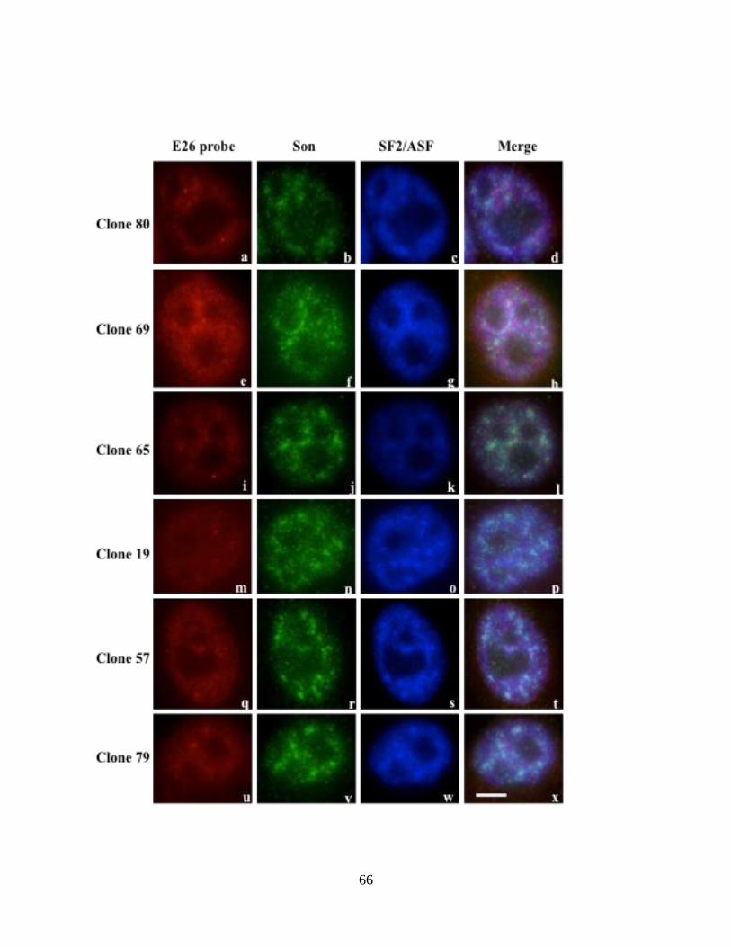
66

67
Figure 13: Screening HeLa HDAC6 stable populations by RNA-FISH to identify reporter
transcription locus.
HeLa HDAC6 stable populations were processed for RNA-FISH to label HDAC6 minigene
transcripts combined with dual label immunofluorescence localization of Son and SF2/ASF.
Panels a, e and i show HDAC6 minigene transcripts of stable populations when 10, 20 or 40 ng
of E26 probe was used for RNA-FISH, respectively. Panels b, f and j show localization of Son in
stable populations. Panels c, g and k show localization of SF2/ASF in stable populations. Bar =
5μm.

68

69
Figure 14: RNA-FISH of transiently expressed HDAC6 reporter minigene transcripts to
visualize transcription/splicing factories.
HeLa cells were transiently transfected with 2 μg of HDAC6 minigene reporter plasmid. After 48
hours, the cells were fixed and processed for RNA-FISH to label HDAC6 minigene transcripts
combined with dual-label immunofluorescence to visualize localization of Son and SF2/ASF.
Panels a, e and i show transiently expressed HDAC6 minigene transcripts when 10, 20 and 40 ng
of E26 probe was used for RNA-FISH, respectively. Panels b, f and j show localization of Son.
Panels c, g and k show localization of SF2/ASF. Bar = 5μm.

70

71
Figure 15: RNA-FISH of transiently expressed HDAC6 reporter minigene transcripts to
visualize transcription/splicing factories.
HeLa cells were transiently transfected with various amounts (1, 2, 5, 7 and 10 μg) of HDAC6
minigene reporter plasmid. After 48 hours, the cells were fixed and processed for RNA-FISH to
label HDAC6 minigene transcripts combined with dual label immunofluorescence to visualize
localization of Son and SF2/ASF. Panels a, e and i show transiently expressed HDAC6 minigene
RNA when either 1, 2, 5, 7 or 10 μg of HDAC6 minigene reporter plasmid was transfected,
respectively. Panels b, f and j show localization of Son. Panels c, g and k show localization of
SF2/ASF. Bar = 5μm.

72

73
4.7 Stable cell lines constructed in HEK 293 with YFP tagged Son deletion mutant
constructs express YFP-Son constantly
HEK 293 cells were transfected with four YFP tagged siR resistant Son deletion mutant
constructs namely YFP-siR-Son Fl, YFP-siR-Son 1-2008, YFP-siR-Son 1-1493 and YFP-
siR-Son 1-332 (see Figure 16A). Stable clones were selected using G418. Primary and
secondary screening through fluorescent microscopy showed consistent expression of the
constructs. Three positive clones were picked for each construct after secondary screening.
Clones 5, 27 and 64 (YFP-siR-Son Fl), 4, 32 and 69 (YFP-siR-Son 1-2008), 15, 48 and 58
(YFP-siR-Son 1-1493), 8, 16 and 46 (YFP-siR-Son 1-332) were selected (Figure 16B).

74
Figure 16: siRNA resistant Son Deletion Mutant constructs and their expression in
Stable HEK 293 Cell lines.
A. SiRNA resistant Son full-length mutant construct consists of all the domains of Son and
in tagged to YFP. siRNA resistant Son 1-2008 deletion mutant construct consists of the
region from 1 to 2008 amino acids and lacks C-terminal G-patch and double stranded
DNA binding domain. siRNA resistant Son 1-1493 deletion mutant construct consists of
the region from 1 to 1493 amino acids and lacks C-terminal RS domain, G-patch and
double stranded DNA binding domain. siRNA resistant Son 1-332 deletion mutant
construct consists of the region from 1 to 332 amino acids and lacks the repeat motifs and
C-terminal RS domain, G-patch and double stranded DNA binding domain.
B. Stable cell lines constructed using the four deletion mutant constructs were screened for
consistent overexpression. The stable clones 5, 27 and 64 of full length-siR Son show
speckled pattern confirming the overexpression of Son protein. The stable clones 4, 32
and 69 of 1-2008-siR Son show speckled pattern confirming the overexpression of Son
protein. The stable clones 15, 48 and 58 of 1-1493-siR Son show speckled pattern
confirming the overexpression of Son protein. The stable clones 8, 16 and 46 of 1-332-
siR Son show expression of protein in both nucleus and cytoplasm confirming the loss of
speckled pattern of overexpressed Son deletion mutant.

75
A
B

76
CHAPTER 5: DISCUSSION
Son is the largest SR protein identified in proteomic analysis of nuclear speckles (Saitoh et al.,
2004). Our lab has previously showed that depletion of Son causes reorganization of nuclear
speckles in to a doughnut shaped phenotype (Sharma et al., 2010). In the process of
understanding the cellular functions of Son, microarray and exon array analysis performed in
Son depleted cells showed decrease or increase in transcript levels as well as altered splicing of
transcripts that encode proteins important for cellular processes such as cell cycle. Our lab has
previously validated exon skipping in transcripts for three endogenous chromatin-regulating
genes including HDAC6, ADA and SETD8 that is caused when Son is depleted (Sharma et al.,
2011). Ahn et al (2011) discussed the effect of Son depletion on splicing of cell cycle regulatory
proteins such as TUBG1, KATNB1, TUBGCP2, PCNT, AKT1 and AURKB.
Understanding the mechanism by which Son regulates splicing events of several selected genes
requires a set of experiments to help define cis-acting elements (ESEs/ISEs) in transcripts that
interact with Son for accurate splicing, as well as the domains of Son that mediate splicing
activities, I aimed to determine the domains/repeats important for Son dependent splicing in
HDAC6. Since Son is known to alter splicing of transcripts with weak splice sites (Sharma et al.,
2011 and Ahn et al., 2011), we analyzed the splice sites of HDAC6 between exons 26 and 29
through ESEfinder. ESEfinder predicted weak splice sites at 5’splice site of exon 27 and 3’
splice site of exon 28 that possibly explains why HDAC6 splicing is dependent on presence of
Son. Further experiments need to be done to validate the presence of weak splice sites in
HDAC6 and Son dependency for accurate splicing. This can be performed by introducing silent
mutations in the predicted weak splice sites of HDAC6 and determining Son dependency for

77
accurate splicing. Moreover, Son depletion causes exclusion of exons 27 and 28 in HDAC6
transcripts that code for the BUZ domain of HDAC6 protein. Since the transcripts without exons
27 and 28 are still in frame at the junction of exons 26 and 29, we predict that the mRNA is
translated and the protein produced lacks BUZ domain. There is no evidence of natural
occurrence of HDAC transcripts lacking exons 27-28 in the literature. However, HDAC6 protein
lacking the BUZ domain is predicted to be non-functional in binding to ubiquitin, thus
interfering with the process of recruiting mis-folded proteins to aggresomes. Aggresomes are
formed as a response to formation of misfolded proteins. Failure to degrade misfolded proteins
results in neuronal cell death in many neurodegenerative diseases. Polyubiquitinated misfolded
proteins are transported to aggresomes by dynein motor proteins. HDAC6 binds to dynein motor
proteins via a dynein-motor binding (DMB) domain present in between the two deacetylase
domains, and it binds to polyubiquitinated misfolded proteins through its BUZ domain. Thus
HDAC6 helps recruit misfolded proteins to dynein motor proteins followed by their transport to
aggresomes (Kawaguchi et al., 2003; Rodriguez-Gonzalez et al., 2008).
To gain insight into splicing functions for Son, we relied on reporter minigene assays. Our goal
was to construct a reporter minigene system inside cells that could be used for RNA-FISH
combined with immunofluorescence with antibodies against Son, several other splicing factors
and splicing repressors such as hnRNPs in order to determine the effects of Son depletion on co-
recruitment of splicing regulators to transcription sites. To achieve this goal, we constructed a
minigene reporter system containing the genomic DNA spanning exons 26 through 29 of
HDAC6. After transiently transfecting HDAC6 minigene reporter plasmid and depleting Son in
HeLa cells, exons 27 and 28 were excluded in both endogenous and exogenous HDAC6
transcripts indicating the presence all the elements required to study Son-regulated splicing in the

78
minigene. We further tested exon exclusion in HeLa cells stably expressing our reporter
construct and found that exons 27 and 28 are excluded in endogenous and exogenous HDAC6
transcripts. One important conclusion that can be drawn from these observations is that since
multiple stable cell lines expressing the HDAC6 reporter minigene show the same result, there is
probably no connection between chromatin context and splicing regulation by Son for splicing of
exons 27 and 28 of HDAC6 transcripts.
To achieve the goal of visualizing Son-mediated splicing in situ, we stably transfected the
HDAC6 minigene reporter in HeLa cells. However, we were unable to show the transcription
locus of the minigene. Careful analysis of RNA-FISH experiments performed in parental HeLa
cells show bright dots in individual nuclei that possibly could be considered as the transcription
locus for endogenous HDAC6. However, the dots do not co-localize with Son and SF2/ASF
consistently every time we perform RNA-FISH experiment, or in majority of cells on the same
coverslip. We therefore do not consider the dots we see as transcription loci of HDAC6, but
rather as an unexplained RNA-FISH signal. The next interesting observation is from RNA-FISH
results of certain clones and cells transiently expressing HDAC6 reporter minigene. We observed
a nuclear speckle pattern after performing RNA-FISH with HDAC6 probes. Moreover, speckle
pattern is observed in parental HeLa cells hybridized with HDAC6 probe (e26-TxRed probe)
which suggests a functional role of endogenous HDAC6 mRNA in nuclear speckles or post-
transcriptional splicing of HDAC6 mRNA in nuclear speckles. The speckle pattern was also
observed in cells that were treated for RNA-FISH but not for simultaneous immunofluorescence
localization of splicing factors (data not shown) which rules out the probability of speckle pattern
being a cross-talk from other channels. However, since nuclear speckles contain a large number

79
of different proteins and RNAs that might bind non-specifically to FISH probes, the possibility
non-specific binding of HDAC6 probe to nuclear speckles could not be ruled out.
I had planned to observe alternative splicing of HDAC6 minigene in situ and determine the role
of Son in recruiting splicing enhancers/repressors to nascent HDAC6 transcripts. Since we could
not reliably visualize the transcription locus of HDAC6 reporter minigene inside the cells, these
experiments could not be performed. Once the transcription locus is developed and visualized,
the following experimental design can be followed to understand the effect of Son depletion in
the recruitment of other splicing factors. Cy5-conjugated exon junction probes, namely EJ26/27
and EJ26/29, need to be used to visualize constitutively and alternatively spliced regions of
HDAC6 minigene in situ. Individual probes EJ27/26, EJ29/26, E26, E27 and E29 work as
controls to optimize the RNA-FISH conditions, to rule out the possibility that the experimental
probes EJ26/27 and EJ26/29 bind only partially to nascent transcripts. Once hybridization
conditions are set, exon skipping in control versus Son-depleted cells has to be evaluated. In a
second set of experiments, performing RNA-FISH combined with immunofluorescence labeling
antibodies specific to Son, SF2/ASF, SC35 and hnRNP A1 will determine how Son depletion
alters their localization patterns. SF2/ASF and SC35 are nuclear speckle proteins known to
promote splicing while hnRNP A1 is a splicing repressor (Sharma et al., 2010 and Millevoi et al.,
2010). Performing this experiment in parallel for multiple factors, and comparing one splicing
factor/repressor and Son per coverslip in both control and Son depleted cells, will allow
examination of relative levels of these proteins at the HDAC6 minigene locus in control versus
Son depleted cells. If SF2/ASF or SC35 decrease at the locus when Son is depleted, we can
conclude that Son is required for the accumulation of these factors and that this may be important
for inclusion of HDAC6 exons 27 and 28. We can also conclude that Son is a scaffolding protein

80
that recruits splicing factors to the transcription/splicing sites and hence when Son is absent,
SF2/ASF or SC35 will not be recruited to the splice sites that influence inclusion of exons 27 and
28, resulting in exon skipping. Since hnRNPA1 is a splicing repressor, increase in its levels at
the locus would suggest that presence of Son at nascent transcripts provides a signal that
prevents hnRNP recruitment to promote correct splicing.
The goal of this project was to begin building reporter systems to observe Son-dependent
splicing events, both at the molecular level as well as in situ. Since I successfully constructed and
characterized the HDAC6 minigene for molecular analysis, more features can be introduced that
will allow more in-depth analysis. For example, site-directed mutagenesis in ESE or ESS
sequences can be introduced to learn exactly which nucleotides in the transcripts are needed for
proper splicing regulation. In addition, MS2 stem loops could be introduced for purifying
reporter mRNAs from control and Son-depleted cells, followed by mass spectrometry analysis to
determine what splicing complexes are lost/gained from reporter transcripts in Son-depleted
cells. In addition, MS2-binding protein can be introduced by transfection to directly visualize
reporter mRNAs by microscopy in live cells instead of using RNA-FISH approaches. Finally,
constructing a cell line similar to U2OS 2-6-3 (Janicki et al., 2004) will be needed to examine the
dynamics of transcription activation and alternative splicing in situ on the HDAC6 reporter
minigene locus.

81
CHAPTER 6: REFERENCES
1. Ahn EY, DeKelver RC, Lo MC, Nguyen TA, Matsuura S, Boyapati A, Pandit S, Fu XD
and Zhang DE (2011). SON Controls Cell-Cycle Progression by Coordinated Regulation of
RNA Splicing. Molecular Cell, 42, 185–198.
2. Ahn EY, Yana M, Malakhova OA, Loa MC, Boyapati A, Ommen HB, Hines R, Hokland
P, and Zhang DE (2008). Disruption of the NHR4 domain structure in AML1-ETO abrogates
SON binding and promotes leukemogenesis. Proceedings of the National Academy of Sciences,
105, 17103–17108.
3. Bentley DL (2005). Rules of engagement: co-transcriptional recruitment of pre-mRNA
processing factors. Elsevier Ltd, 17, 251–256.
4. Berdichevskiĭ FB, Chumakov IM, Kiselev LL (1988). Decoding of the primary structure
of the son3 region in human genome: identification of a new protein with unusual structure and
homology with DNA-binding proteins. Molecular Biology, 22, 794-801.
5. Bernard D, Prasanth KV, Tripathi V, Colasse S, Nakamura T, Xuan Z, Zhang MQ, Sedal
F, Jourdren L, Coulpier F, Triller A, Spector DL, Bessis A (2010). A long nuclear retained no-
coding RNA regulates synaptogenesis by modulating gene expression. EMBO Journal, 18, 3082-
3093.
6. Black DL (2003). Mechanisms of Alternative Pre-messenger RNA Splicing. Annual
Reviews of Biochemistry, 72, 291-336.
7. Blencowe BJ (2000). Exonic splicing enhancers: Mechanism of action, diversity and role in
human genetic diseases. Trends in Biochemical Sciences, 25, 106-110.

82
8. Boucher L, Ouzounis CA, Enright AJ and Blencowe BJ (2001). A genome-wide survey of
RS domain proteins. RNA, 7, 1693-1701.
9. Boyault C, Sadoul K, Pabion M and Khochbin S (2007). HDAC6, at the crossroads
between cytoskeleton and cell signaling by acetylation and ubiquitination. Oncogene, 26, 5468–
5476.
10. Cartegni LS, Chew L and Krainer AR (2002). Listening to silence and understanding
nonsense: exonic mutations that affect splicing. Nature Review Genetics, 3, 285–298.
11. Carter KC, Taneja KL, Lawrence JB (1991). Discrete nuclear domains of poly (A) RNA
and their relationship to the functional organization of the nucleus. Journal Cell Biology, 115,
1191–1202.
12. Cmarko D, Verschure PJ, Martin TE, Dahmus ME, Krause S, Fu XD, Driel RV, and
Fakan S (1999). Ultrastructural Analysis of Transcription and Splicing in the Cell Nucleus after
Bromo-UTP Microinjection. Molecular Biology of the Cell, 10, 211–223.
13. Cooper TA (2005). Use of minigene systems to dissect alternative splicing elements
Science Methods, 37, 331–340.
14. Erkelenz S, Mueller WF, Evans MS, Busch A, Schoneweis K, Hertel KJ and Schaal H
(2013). Position dependent splicing activation and repression by SR and hnRNP proteins rely on
common mechanisms. RNA, 19, 96-102.
15. Faustino NA and Cooper TA (2003). Pre-mRNA splicing and human disease. Genes and
Development, 17, 419-437.
16. Görnemann J, Kotovic KM, Hujer K and Neugebaue KM (2005). Cotranscriptional
spliceosome assembly occurs in a stepwise fashion and requires the cap binding complex.
Molecular Cell, 19, 53–63.

83
17. Graveley BR (2000). Sorting out the complexity of SR protein functions. RNA, 6, 1197–
1211.
18. Herrmann CH, Mancini MA (2001). The Cdk9 and cyclin T subunits of TAK/P-TEFb
localize to splicing factor-rich nuclear speckle regions. Journal Cell Science, 114, 1491–1503.
19. Hock R, Wilde F, Scheer U, Bustin M (1998). Dynamic relocation of chromosomal
protein HMG-17 in the nucleus is dependent on transcriptional activity. EMBO Journal, 17,
6992–7001.
20. Hook SS, Orian A, Cowley SM, and Eisenman RN (2002). Histone deacetylase 6 binds
polyubiquitin through its zinc finger (PAZ domain) and copurifies with deubiquitinating
enzymes. Proceedings of the National Academy of Sciences, 99, 13425–13430.
21. Huang S and Spector DL (1991). Nascent pre-mRNA transcripts are associated with
nuclear regions enriched in splicing factors. Genes and Development, 5, 2288-2302.
22. Huang S and Spector DL (1996). Intron-dependenRt ecruitmento f Pre-mRNAS plicingF
actors to Sites of Transcription. Journal of Cell Biology, 133, 719-732.
23. Huang S, Deerinck MH, Ellisman MH, Spector DL (1994). In vivo analysis of the
stability and transport of nuclear poly (A) RNA. Journal Cell Biology, 126, 877–899.
24. Hubbert C, Guardiola A, Shao R, Kawaguchi Y, Ito A, Nixon A, Yoshida M, Wang XF
and Yao TP (2002). HDAC6 is a microtubule-associated deacetylase. Nature, 417, 455-458.
25. Huen MSY, Sy SMH, Leung KM, Ching YP, Tipoe GL, Man C, Shuo Dong S and Junjie
Chen(2010). SON is a spliceosome-associated factor required for mitotic progression. Cell
Cycle, 9, 2679-2685.

84
26. Hutchinson JN, Ensminger AW, Clemson CM, Lynch CR, Lawrence JB, Chess A.
(2007). A screen for nuclear transcripts identifies two linked noncoding RNAs associated with
SC35 splicing domains. BMC Genomics, 8, 39.
27. Janicki SM, Tsukamoto T, Salghetti SE, Prasanth KV, Ried T, Shav-Tal Y, Bertrand E,
Singer RH and Spector DL (2004). From silencing to gene expression: real-time analysis in
single cells. Cell, 116, 683-698.
28. Jurica MS and Moore MJ (2003). Pre-mRNA Splicing: Awash in a Sea of Proteins.
Molecular Cell, 12, 5–14.
29. Kawaguchi Y, Kovacs JJ, McLaurin A, Vance JM, Ito A and Yao TP (2003). The
deacetylase HDAC6 regulates aggresome formation and cell viability in response to misfolded
protein stress. Cell, 12, 727-38.
30. Lamond AI and Spector DL (2003). Nuclear speckles: a model for nuclear organelles.
Nature reviews: Molecular Cell Biology, 4, 605-612.
31. Maniatis T, Reed R (2002). An extensive network of coupling among gene expression
machines. Nature, 416, 499-506.
32. Millevoi S, Bernat S, Telly D, Fouque F, Gladieff L, Favre G, Vagner S and Toulas C
(2010). The c.5242C>A BRCA1 missense variant induces exon skipping by increasing splicing
repressors binding. Breast Cancer Research and Treatment, 120, 391–399.
33. Mintz PJ, Patterson SD, Neuwald AF, Spahr CS, and Spector DL (1999). Purification and
biochemical characterization of interchromatin granule clusters. EMBO Journal, 18, 4308–4320.
34. Misteli T (2000). Cell biology of transcription and pre-mRNA splicing: nuclear
architecture meets nuclear function. Journal of Cell Science 113, 1841-1849.

85
35. Misteli T and Spector DL (1998). The cellular organization of gene expression. Current
opinion in cell biolgy, 10, 323-331.
36. Misteli T, Caceres JF and Spector DL (1997). The dynamics of a pre-mRNA splicing
factor in living cells. Nature, 387, 523-527.
37. Pendergrast PS, Wang C, Hernandez N, Huang S (2002). FBI-1 can stimulate HIV-1 Tat
activity and is targeted to a novel subnuclear domain that includes the Tat-P-TEFb-containing
nuclear speckles. Molecular Biology of the Cell, 13, 915–929.
38. Reed R (2000). Mechanisms of fidelity in pre-mRNA splicing. Elsevier Science Ltd., 12,
340-345.
39. Rodriguez-Gonzalez A, Lin T, Ikeda AK, Simms-Waldrip T, Fu C, and Sakamoto KM
(2008). Role of the Aggresome Pathway in Cancer: Targeting Histone Deacetylase 6–Dependent
Protein Degradation. Cancer Research, 68, 2557-2560.
40. Saitoh N, Spahr CS, Patterson SD, Bubulya P, Neuwald AF and Spector DL (2004).
Proteomic Analysis of Interchromatin Granule Clusters. Molecular Biology of the Cell, 15, 3876-
3890.
41. Sanchez-Alvarez M, Goldstrohm AC, Garcia-Blanco MA, Sune C (2006). Human
transcription elongation factor CA150 localizes to splicing factor-rich nuclear speckles and
assembles transcription and splicing components into complexes through its amino and carboxyl
regions. Molecular and Cellular Biology, 26, 4998–5014.
42. Sharma A, Markey M, Torres-Muñoz K, Varia S, Kadakia M, Bubulya A and Bubulya
PA (2011). Son maintains accurate splicing for a subset of human pre-mRNAs. Journal of Cell
Science, 124, 4286–4298.

86
43. Sharma A, Takata H, Shibahara K, Bubulya A and Bubulya PA (2010). Son is essential
for nuclear speckle organization and cell cycle progression. Molecular Biology of the Cell, 21,
650-663.
44. Smith CW, Valcarcel J (2000). Alternative pre-mRNA splicing: The logic of combinatorial
control. Trends in Biochemical Sciences, 25, 381–388.
45. Spector DL and Lamond AI (2010). Nuclear speckles. Cold Spring Harbor Perspectives
in Biology, 10.
46. Thiry M (1995). Behavior of interchromatin granules during the cell cycle. European
Journal of Cell Biology, 68, 14-24.
47. Tripathi V, Ellis J, Shen Z, Song D, Freier SM, Bennett CF, Sharma A, Bubulya PA,
Blencowe BJ, Prasanth SG, Prasanth KV (2010). Nuclear-retained non-coding RNA regulates
alternative splicing by modulating SR splicing factor phosphorylation. Molecular Cell, 6, 925-
938.
48. Verdin E, Dequiedt F and Kasler HG (2003). Class II histone deacetylases: versatile
regulators. Trends in genetics, 19, 286-293.
49. Verdin E. (2006). Histone Deacetylases: Transcriptional Regulation and Other Cellular
Functions, Humana Press Inc., Totowa, NJ.
50. Visa N, Puvion-Dutilleul F, Harper F, Bachellerie J-P, Puvion E (1993). Intranuclear
distribution of poly A RNA determined by electron microscope in situ hybridization.
Experimental Cell Research, 208, 19–34.
51. Wahl MC, Will CL and Luhrmann R (2009). The Spliceosome: Design Principles of a
Dynamic RNP Machine. Cell, 136, 701–718.