abnormalities of vascular system
TRANSCRIPT
The Venous Diseases of Lower Limbs. A Concise Guide
Raffaele Grande1,2,*; Alessandro Carra2; Luigi Marcuccio2; Silvio Vitale2; Lucia Butrico2
1Department of Surgery “Pietro Valdoni”, “Sapienza” University of Rome.
2Unit of Vascular and Endovascular Surgery, Casilino Hospital, Rome.
*Correspondence to: Raffaele Grande, Department of Surgery “PietroValdoni” “Sapienza” University of Rome Viale
del Policlinico 15100161 Rome, Italy.
Tel: +393275543467; Fax: +390649970794; Email: [email protected]
Chapter 5
Abnormalities of VascularSystem
Abstract
Venous thromboembolism and chronic venous disease of lower limbs are the pathological conditions with the highest epidemiological impact in industrialized countries. Although they are very frequent in daily clinical practice, the diagnosis and treatment of the various diseases is not always easy to manage. The purpose of this work is to propose a simple guide that illustrates the cardinal and updated principles of the management of venous diseases of the lower limbs to the various medical personalities who approach the venous problems of the lower limbs.
Keywords: Deep Venous Thrombosis; Pulmonary Embolism; Chronic Venous Disease; Algorithm
1. Introduction
The venous diseases of lower limbs always represent the most frequent pathology which many doctors approach regardless of their specific specialization [1].
Chronic venous disease (CVD) is a common problem affecting adult population, es-pecially in Western countries [2] the prevalence of CVD among individuals younger than 30 years was <10% in men and women; prevalence in men and women aged ≥ 70 years is 57% and 77%, respectively [3]. CVD symptoms range from varicose veins to leg oedema, and se-rious dermal manifestations consisting of hyperpigmentation, eczema, lipodermatosclerosis
2
ww
w.openaccessebooks.comAbnormalities of Vascular System
Gra
nde
R
and venous skin ulceration that remains a significant worldwide health problem resulting in significant morbidity [4]. The most frequently encountered symptoms associated with varicose veins include leg swelling, pain, itching, nocturnal cramping, and leg heaviness.
Venous Thromboembolism (VTE) includes several pathological conditions such as Deep venous thrombosis (DVT) of the lower extremities, Pulmonary embolism (PE) and Post-thrombotic Syndrome (PTS): they are another highly prevalent venous conditions encountered by health care providers of all disciplines [5-6].
Although the different gnosological entities are well known and discussed, the phisi-cians often encounters hard difficulties in differential diagnosis with other pathologies with very similar symptom spectrum, especially at the beginning of the career.
The purpose of this work is to create a concise and updated guide to the main patholo-gies affecting the venous system of the lower limbs with clinical, instrumental and therapeutic interpretation.
2. Methods
Scopus (www.scopus.com), Embase (www.embase.com), and Medline (www.ncbi.nlm.nih.gov/ pubmed) databases were used for the International literature review. No language restrictions were applied. Manual searching of reference lists for relevant studies and previous reviews was also performed. The primary search term was conducted for any combination of the words ‘‘venous thromboembolism” and ‘‘deep venous thrombosis” and “chronic venous disease” and ‘‘management’’. Studies were included if they contained adequate information regarding symptoms, diagnostic algorithms, and the type of therapeutic approach (pharmaco-logical or surgical).
3. Venous Thrombo-Embolism (VTE)
VTE refers to the pathological condition characterized by the acute formation of throm-bus in the venous district; this condition includes two entities: Deep Vein Thrombosis (DVT) and Pulmonary Embolism (PE).
The venous thrombotic event is the result of a mix of endothelial damage, hypercoagu-lability and venous stasis, as hypothesized by Virchow in his triad [7] and the physiological antithrombotic activity of the venous endothelium can rapidly become prothrombotic with the production of tissue factors such as von Willebrand, fibronectin and P selectin.
3.1. Deep Venous Thrombosis (DVT)
DVT remains a frequent and serious disease, with a high annual incidence (300,000-600,000 per year), but it is difficult to estimate due to the numerous episodes that have gone
3
Abnormalities of Vascular System
unnoticed and often little attention to clinical diagnosis. The initial phase of DVT can occur, in complicated cases, as PE, phlegmasia cerulea dolens or paradoxical embolism; long-term complications may include recurrent thromboembolic disease, chronic PE, and Post Throm-botic Syndrome (PTS) [8].
3.2. Risk factors
3.2.1. Main
- Thrombophilia (congenital or acquired)
- Surgery (neurosurgery, major orthopedic surgery of the leg, thoracic, abdominal or pel-vic surgery for cancer, cardiovascular and kidney transplantation)
- Trauma
- Prolonged bed rest
- Presence of active cancer
- Presence of important co-morbidities (Heart failure, Myocardial infarction, Stroke, acute or chronic lung diseases, Inflammatory bowel diseases, other rare diseases)
- Obesity
- Neurological diseases with paresis of the lower limbs
- Age
- Pregnancy and the puerperium
- Contraceptive or hormone replacement therapy
3.2.2. Secondary
- Personal or family (1st degree) history of VTE
- Central venous catheter
- Previous superficial venous thrombosis
- Presence of varicose veins
4
Abnormalities of Vascular System
3.3. Diagnosis
3.3.1. Edema or pain
Location depends from the side of venous thrombosis, the edema is hard and does not create "fovea". The pain often has no specific characteristics and it varies from a feeling of heaviness to functional impotence. Compression of the gastrocnemius on the tibial bone plane evokes a lively pain in the patient with DVT (Bauer's sign), the Homans sign (dorsiflexion of the foot) helps in the diagnosis (the patient in a dorsal decubitus with legs bent at 90 degrees). Another important clinical sign is usually hyperthermia (38 ° C), feeling of anguish and tachy-cardia.
Often patients with cancer, elderly, obese, have symptoms of DVT.
3.3.2. Distension of the superficial veins
Superficial venous hypertension resulting from acute obstruction causes distension. The differential diagnosis in this case will be more difficult in patients who already have varicosi-ties of the lower limbs. Peripheral cyanosis following veno-lymphatic stasis is frequent. The clinical examination, in which the signs are not always present, must be conducted compara-tively in the two limbs.
3.3.3. Advanced stages of DVT
It is possible, albeit less frequently than in the "benign" forms, that the picture of DVT is complicated by limb ischemia. The pain is persistent, extensive, acute and most often localized in the groin. It is described as "excruciating". Edema sets in quickly, not presenting a fovea; it is woody or rubbery, with a shiny skin. Muscle lodges are strained. Sometimes there are der-matological alterations with petechiae or even bleeding bubbles. Cyanosis develops rapidly, distal at first then to the entire lower limb with decreased local temperature and hypoesthesia. Peripheral pulses are reduced, even abolished. The differential diagnosis is represented by ar-terial spasm or arterial embolism, situations in which there is no edema, the superficial veins are flattened and the wrists are abolished early. The evolution towards the SPT is frequent.
3.4. Laboratory and Instrumental Diagnosis
3.4. 1. Ultrasound examination
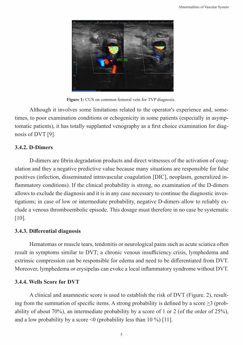
Ecography with Doppler spectrum can facilitate the examination in anatomically dif-ficult sides and it provides morphological informations. The diagnostic criteria are the lack of venous compressibility at the support of the probe (compression ultrasound (CUS)) or the visualization of an intravenous echogenic zone that represents a thrombus (Figure 1).
5
Abnormalities of Vascular System
Figure 1: CUS on common femoral vein for TVP diagnosis.
Although it involves some limitations related to the operator's experience and, some-times, to poor examination conditions or echogenicity in some patients (especially in asymp-tomatic patients), it has totally supplanted venography as a first choice examination for diag-nosis of DVT [9].
3.4.2. D-Dimers
D-dimers are fibrin degradation products and direct witnesses of the activation of coag-ulation and they a negative predictive value because many situations are responsible for false positives (infection, disseminated intravascular coagulation [DIC], neoplasm, generalized in-flammatory conditions). If the clinical probability is strong, no examination of the D-dimers allows to exclude the diagnosis and it is in any case necessary to continue the diagnostic inves-tigations; in case of low or intermediate probability, negative D-dimers allow to reliably ex-clude a venous thromboembolic episode. This dosage must therefore in no case be systematic [10].
3.4.3. Differential diagnosis
Hematomas or muscle tears, tendonitis or neurological pains such as acute sciatica often result in symptoms similar to DVT; a chronic venous insufficiency crisis, lymphedema and extrinsic compression can be responsible for edema and need to be differentiated from DVT. Moreover, lymphedema or erysipelas can evoke a local inflammatory syndrome without DVT.
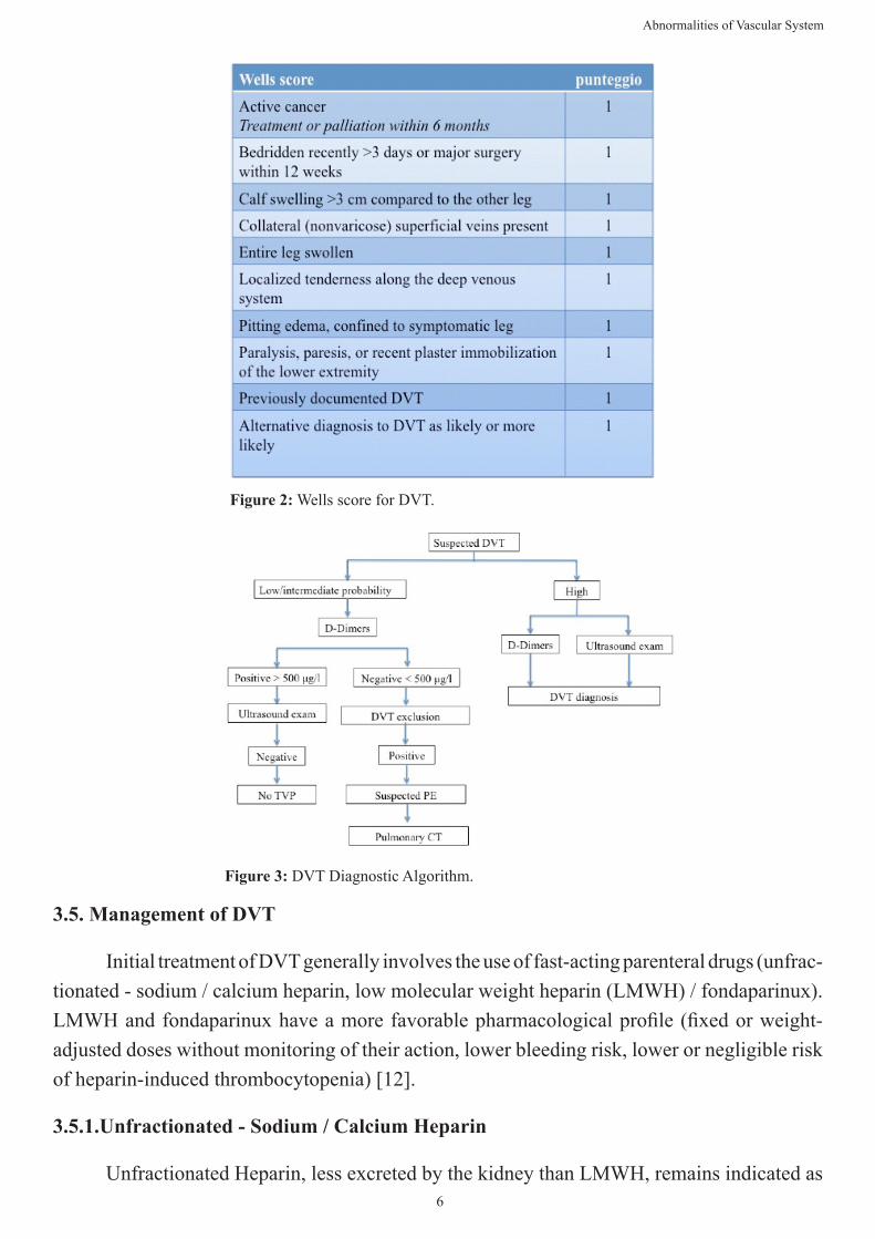
3.4.4. Wells Score for DVT
A clinical and anamnestic score is used to establish the risk of DVT (Figure. 2), result-ing from the summation of specific items. A strong probability is defined by a score ≥3 (prob-ability of about 70%), an intermediate probability by a score of 1 or 2 (of the order of 25%), and a low probability by a score <0 (probability less than 10 %) [11].
6
Abnormalities of Vascular System
Figure 2: Wells score for DVT.
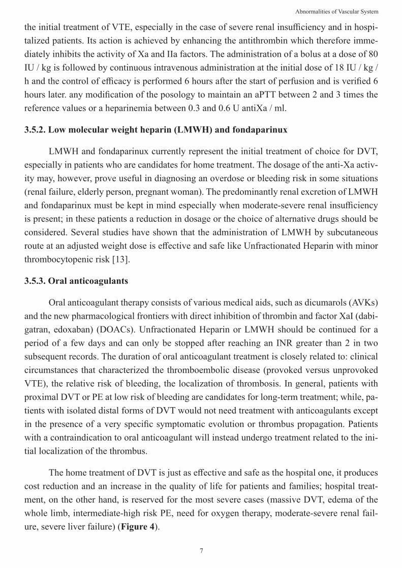
Figure 3: DVT Diagnostic Algorithm.
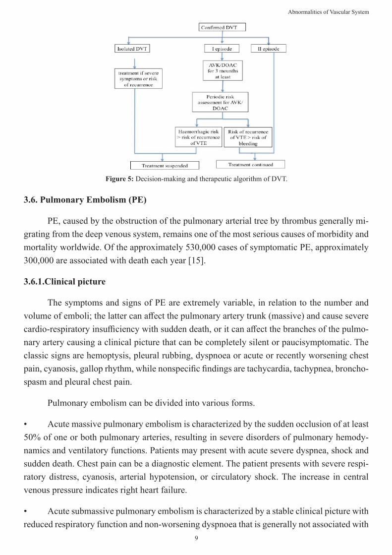
3.5. Management of DVT
Initial treatment of DVT generally involves the use of fast-acting parenteral drugs (unfrac-tionated - sodium / calcium heparin, low molecular weight heparin (LMWH) / fondaparinux). LMWH and fondaparinux have a more favorable pharmacological profile (fixed or weight-adjusted doses without monitoring of their action, lower bleeding risk, lower or negligible risk of heparin-induced thrombocytopenia) [12].
3.5.1.Unfractionated - Sodium / Calcium Heparin
Unfractionated Heparin, less excreted by the kidney than LMWH, remains indicated as
7
Abnormalities of Vascular System
the initial treatment of VTE, especially in the case of severe renal insufficiency and in hospi-talized patients. Its action is achieved by enhancing the antithrombin which therefore imme-diately inhibits the activity of Xa and IIa factors. The administration of a bolus at a dose of 80 IU / kg is followed by continuous intravenous administration at the initial dose of 18 IU / kg / h and the control of efficacy is performed 6 hours after the start of perfusion and is verified 6 hours later. any modification of the posology to maintain an aPTT between 2 and 3 times the reference values or a heparinemia between 0.3 and 0.6 U antiXa / ml.
3.5.2. Low molecular weight heparin (LMWH) and fondaparinux
LMWH and fondaparinux currently represent the initial treatment of choice for DVT, especially in patients who are candidates for home treatment. The dosage of the anti-Xa activ-ity may, however, prove useful in diagnosing an overdose or bleeding risk in some situations (renal failure, elderly person, pregnant woman). The predominantly renal excretion of LMWH and fondaparinux must be kept in mind especially when moderate-severe renal insufficiency is present; in these patients a reduction in dosage or the choice of alternative drugs should be considered. Several studies have shown that the administration of LMWH by subcutaneous route at an adjusted weight dose is effective and safe like Unfractionated Heparin with minor thrombocytopenic risk [13].
3.5.3. Oral anticoagulants
Oral anticoagulant therapy consists of various medical aids, such as dicumarols (AVKs) and the new pharmacological frontiers with direct inhibition of thrombin and factor XaI (dabi-gatran, edoxaban) (DOACs). Unfractionated Heparin or LMWH should be continued for a period of a few days and can only be stopped after reaching an INR greater than 2 in two subsequent records. The duration of oral anticoagulant treatment is closely related to: clinical circumstances that characterized the thromboembolic disease (provoked versus unprovoked VTE), the relative risk of bleeding, the localization of thrombosis. In general, patients with proximal DVT or PE at low risk of bleeding are candidates for long-term treatment; while, pa-tients with isolated distal forms of DVT would not need treatment with anticoagulants except in the presence of a very specific symptomatic evolution or thrombus propagation. Patients with a contraindication to oral anticoagulant will instead undergo treatment related to the ini-tial localization of the thrombus.
The home treatment of DVT is just as effective and safe as the hospital one, it produces cost reduction and an increase in the quality of life for patients and families; hospital treat-ment, on the other hand, is reserved for the most severe cases (massive DVT, edema of the whole limb, intermediate-high risk PE, need for oxygen therapy, moderate-severe renal fail-ure, severe liver failure) (Figure 4).
8
Abnormalities of Vascular System
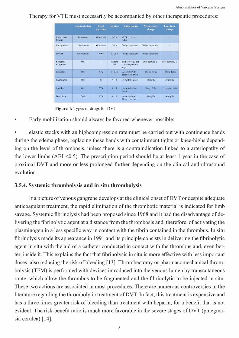
Therapy for VTE must necessarily be accompanied by other therapeutic procedures:
Figure 4: Types of drugs for DVT
• Early mobilization should always be favored whenever possible;
• elastic stocks with an highcompression rate must be carried out with continence bands during the edema phase, replacing these bands with containment tights or knee-highs depend-ing on the level of thrombosis, unless there is a contraindication linked to a arteriopathy of the lower limbs (ABI <0.5). The prescription period should be at least 1 year in the case of proximal DVT and more or less prolonged further depending on the clinical and ultrasound evolution.
3.5.4. Systemic thrombolysis and in situ thrombolysis
If a picture of venous gangrene develops at the clinical onset of DVT or despite adequate anticoagulant treatment, the rapid elimination of the thrombotic material is indicated for limb savage. Systemic fibrinolysis had been proposed since 1968 and it had the disadvantage of de-livering the fibrinolytic agent at a distance from the thrombosis and, therefore, of activating the plasminogen in a less specific way in contact with the fibrin contained in the thrombus. In situ fibrinolysis made its appearance in 1991 and its principle consists in delivering the fibrinolytic agent in situ with the aid of a catheter conducted in contact with the thrombus and, even bet-ter, inside it. This explains the fact that fibrinolysis in situ is more effective with less important doses, also reducing the risk of bleeding [13]. Thrombectomy or pharmacomechanical throm-bolysis (TFM) is performed with devices introduced into the venous lumen by transcutaneous route, which allow the thrombus to be fragmented and the fibrinolytic to be injected in situ. These two actions are associated in most procedures. There are numerous controversies in the literature regarding the thrombolytic treatment of DVT. In fact, this treatment is expensive and has a three times greater risk of bleeding than treatment with heparin, for a benefit that is not evident. The risk-benefit ratio is much more favorable in the severe stages of DVT (phlegma-sia cerulea) [14].
9
Abnormalities of Vascular System
Figure 5: Decision-making and therapeutic algorithm of DVT.
3.6. Pulmonary Embolism (PE)
PE, caused by the obstruction of the pulmonary arterial tree by thrombus generally mi-grating from the deep venous system, remains one of the most serious causes of morbidity and mortality worldwide. Of the approximately 530,000 cases of symptomatic PE, approximately 300,000 are associated with death each year [15].
3.6.1.Clinical picture
The symptoms and signs of PE are extremely variable, in relation to the number and volume of emboli; the latter can affect the pulmonary artery trunk (massive) and cause severe cardio-respiratory insufficiency with sudden death, or it can affect the branches of the pulmo-nary artery causing a clinical picture that can be completely silent or paucisymptomatic. The classic signs are hemoptysis, pleural rubbing, dyspnoea or acute or recently worsening chest pain, cyanosis, gallop rhythm, while nonspecific findings are tachycardia, tachypnea, broncho-spasm and pleural chest pain.
Pulmonary embolism can be divided into various forms.
• Acute massive pulmonary embolism is characterized by the sudden occlusion of at least 50% of one or both pulmonary arteries, resulting in severe disorders of pulmonary hemody-namics and ventilatory functions. Patients may present with acute severe dyspnea, shock and sudden death. Chest pain can be a diagnostic element. The patient presents with severe respi-ratory distress, cyanosis, arterial hypotension, or circulatory shock. The increase in central venous pressure indicates right heart failure.
• Acute submassive pulmonary embolism is characterized by a stable clinical picture with reduced respiratory function and non-worsening dyspnoea that is generally not associated with
10
Abnormalities of Vascular System
an acute pulmonary heart.
• Non-massive acute pulmonary embolism is characterized by symptoms due to ischemia or infarction of a small lung segment. In a healthy person, small pulmonary embolisms affect-ing less than 50% of the circulation are often asymptomatic.
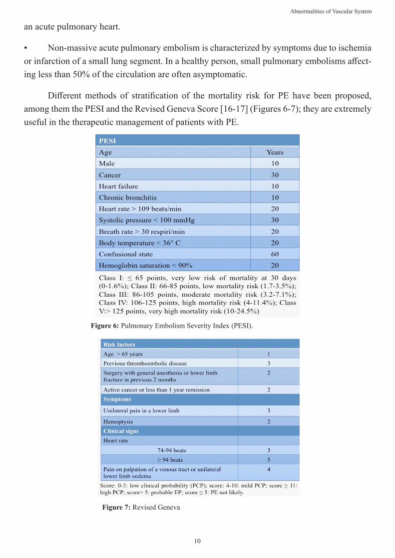
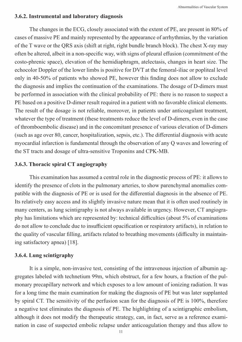
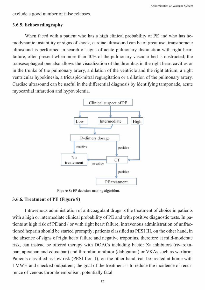
Different methods of stratification of the mortality risk for PE have been proposed, among them the PESI and the Revised Geneva Score [16-17] (Figures 6-7); they are extremely useful in the therapeutic management of patients with PE.
Figure 7: Revised Geneva
Figure 6: Pulmonary Embolism Severity Index (PESI).
11
Abnormalities of Vascular System
3.6.2. Instrumental and laboratory diagnosis
The changes in the ECG, closely associated with the extent of PE, are present in 80% of cases of massive PE and mainly represented by the appearance of arrhythmias, by the variation of the T wave or the QRS axis (shift at right, right bundle branch block). The chest X-ray may often be altered, albeit in a non-specific way, with signs of pleural effusion (commitment of the costo-phrenic space), elevation of the hemidiaphragm, atelectasis, changes in heart size. The echocolor Doppler of the lower limbs is positive for DVT at the femoral-iliac or popliteal level only in 40-50% of patients who showed PE, however this finding does not allow to exclude the diagnosis and implies the continuation of the examinations. The dosage of D-dimers must be performed in association with the clinical probability of PE: there is no reason to suspect a PE based on a positive D-dimer result required in a patient with no favorable clinical elements. The result of the dosage is not reliable, moreover, in patients under anticoagulant treatment, whatever the type of treatment (these treatments reduce the level of D-dimers, even in the case of thromboembolic disease) and in the concomitant presence of various elevation of D-dimers (such as age over 80, cancer, hospitalization, sepsis, etc.). The differential diagnosis with acute myocardial infarction is fundamental through the observation of any Q waves and lowering of the ST tracts and dosage of ultra-sensitive Troponins and CPK-MB.
3.6.3. Thoracic spiral CT angiography
This examination has assumed a central role in the diagnostic process of PE: it allows to identify the presence of clots in the pulmonary arteries, to show parenchymal anomalies com-patible with the diagnosis of PE or is used for the differential diagnosis in the absence of PE. Its relatively easy access and its slightly invasive nature mean that it is often used routinely in many centers, as lung scintigraphy is not always available in urgency. However, CT angiogra-phy has limitations which are represented by: technical difficulties (about 5% of examinations do not allow to conclude due to insufficient opacification or respiratory artifacts), in relation to the quality of vascular filling, artifacts related to breathing movements (difficulty in maintain-ing satisfactory apnea) [18].
3.6.4. Lung scintigraphy
It is a simple, non-invasive test, consisting of the intravenous injection of albumin ag-gregates labeled with technetium 99m, which obstruct, for a few hours, a fraction of the pul-monary precapillary network and which exposes to a low amount of ionizing radiation. It was for a long time the main examination for making the diagnosis of PE but was later supplanted by spiral CT. The sensitivity of the perfusion scan for the diagnosis of PE is 100%, therefore a negative test eliminates the diagnosis of PE. The highlighting of a scintigraphic embolism, although it does not modify the therapeutic strategy, can, in fact, serve as a reference exami-nation in case of suspected embolic relapse under anticoagulation therapy and thus allow to
12
Abnormalities of Vascular System
exclude a good number of false relapses.
3.6.5. Echocardiography
When faced with a patient who has a high clinical probability of PE and who has he-modynamic instability or signs of shock, cardiac ultrasound can be of great use: transthoracic ultrasound is performed in search of signs of acute pulmonary disfunction with right heart failure, often present when more than 40% of the pulmonary vascular bed is obstructed; the transesophageal one also allows the visualization of the thrombus in the right heart cavities or in the trunks of the pulmonary artery, a dilation of the ventricle and the right atrium, a right ventricular hypokinesia, a tricuspid-mitral regurgitation or a dilation of the pulmonary artery. Cardiac ultrasound can be useful in the differential diagnosis by identifying tamponade, acute myocardial infarction and hypovolemia.
Figure 8: EP decision-making algorithm.
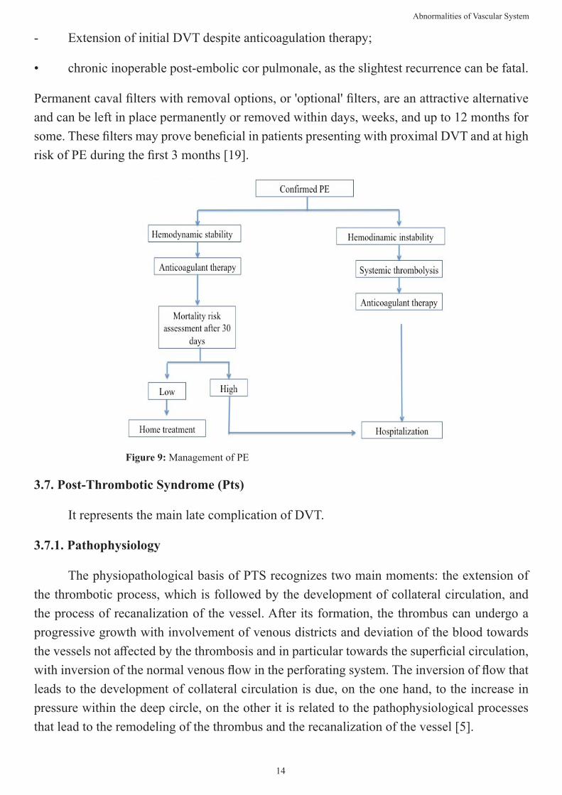
3.6.6. Treatment of PE (Figure 9)
Intravenous administration of anticoagulant drugs is the treatment of choice in patients with a high or intermediate clinical probability of PE and with positive diagnostic tests. In pa-tients at high risk of PE and / or with right heart failure, intravenous administration of unfrac-tioned heparin should be started promptly; patients classified as PESI III, on the other hand, in the absence of signs of right heart failure and negative troponins, therefore at mild-moderate risk, can instead be offered therapy with DOACs including Factor Xa inhibitors (rivaroxa-ban, apixaban and edoxaban) and thrombin inhibitor (dabigatran) or VKAs such as warfarin. Patients classified as low risk (PESI I or II), on the other hand, can be treated at home with LMWH and checked outpatient; the goal of the treatment is to reduce the incidence of recur-rence of venous thromboembolism, potentially fatal.
13
Abnormalities of Vascular System
For patients classified as high-risk PE, revascularization of the pulmonary trunks via systemic fibrinolysis with Tissue Recombinant Plasminogen Activator (r-tPA) is the treatment of choice. The procedure is able to quickly reduce the extent of the thrombus, the extent of right heart dysfunction and pulmonary vascular resistance ensuring, in comparison with treat-ment with the systemic anticoagulant alone, an important reduction in mortality; despite this, the risk of severe and intracranial bleeding remains respectively 20% and 3-5% (especially at doses> 100 m in 2 hours) while the risk of minor and major bleeding events for treatment with anticoagulant remains respectively 15 % and 1-5%. In patients with sub-massive PE and severe myocardial necrosis and / or right cardiac dysfunction, treatment with systemic fibrino-lysis will vary from case to case.
Pulmonary embolectomy has been used in cohorts of patients with "central" pulmonary embolus: this procedure obviously requires a median sternotomy and cardiopulmonary bypass. Embolectomy can be proposed in patients with submassive PE and with contraindications to systemic fibrinolysis or when fibrinolysis has not brought great clinical benefit, in patients with thrombi located in the right atrium and ventricle or when patients are not candidates for catheterization procedure.
3.6.6. Catheter Directed Therapy (CDT).
CDT is an emerging treatment, generally reserved for patients with massive or sub-mas-sive PE and contraindication to systemic fibrinolysis or where the latter has not been effective in ensuring an improvement in cardiac performance. Recent evidence confirms the clinical success of CDT in patients with massive PE, with a success rate of 86.5% and 2.4% of major complications, when compared to systemic fibrinolysis (20% risk of major complications such as severe bleeding and 3-5% of hemorrhagic stroke). In patients with submassive PE with right ventricular overload, CDT may be proposed to reduce the rate of recurrent PE, DVT and PE-related death that is commonly associated with persisting right ventricular dysfunction.
3.6.7. Caval Filters
The implantation of caval filters requires very specific indications:
- Absolute contraindication to anticoagulant treatment in the course of a proximal venous thrombosis;
- An ineffectiveness of the anticoagulant treatment;
- The appearance of PE under anticoagulant treatment is a legitimate indication, although very rare. The systematic performance of a lung scan in the case of proximal venous thrombosis is a reference examination. In case of doubt, the use of pulmonary angiography is essential;
14
Abnormalities of Vascular System
- Extension of initial DVT despite anticoagulation therapy;
• chronic inoperable post-embolic cor pulmonale, as the slightest recurrence can be fatal.
Permanent caval filters with removal options, or 'optional' filters, are an attractive alternative and can be left in place permanently or removed within days, weeks, and up to 12 months for some. These filters may prove beneficial in patients presenting with proximal DVT and at high risk of PE during the first 3 months [19].
Figure 9: Management of PE
3.7. Post-Thrombotic Syndrome (Pts)
It represents the main late complication of DVT.
3.7.1. Pathophysiology
The physiopathological basis of PTS recognizes two main moments: the extension of the thrombotic process, which is followed by the development of collateral circulation, and the process of recanalization of the vessel. After its formation, the thrombus can undergo a progressive growth with involvement of venous districts and deviation of the blood towards the vessels not affected by the thrombosis and in particular towards the superficial circulation, with inversion of the normal venous flow in the perforating system. The inversion of flow that leads to the development of collateral circulation is due, on the one hand, to the increase in pressure within the deep circle, on the other it is related to the pathophysiological processes that lead to the remodeling of the thrombus and the recanalization of the vessel [5].
15
Abnormalities of Vascular System
The onset of insufficiency of the deep venous circulation is closely related to the speed of the recanalization process and relapses.
3.7.2. Clinical diagnosis
The main predisposing factors for the development of an SPT are:
• Recurrent ipsilateral DVT, especially in patients> 65 years
• Iliac-femoral DVT
• Non-optimal anticoagulant therapy
• High BMI
The clinical picture of PTS can be characterized by symptoms and signs that differ from patient to patient and can also take on serious forms that are a source of important disability. The patient may report a sense of heaviness and fatigue that increase with prolonged standing, the seasons and hot environments and the menstrual cycle. There may be deep burning pain during walking (which develops due to venous hypertension), which reaches its peak during the contraction phase of the calf muscles (venous claudication). Following the development of ulcers, bacterial superinfection phenomena may occur.
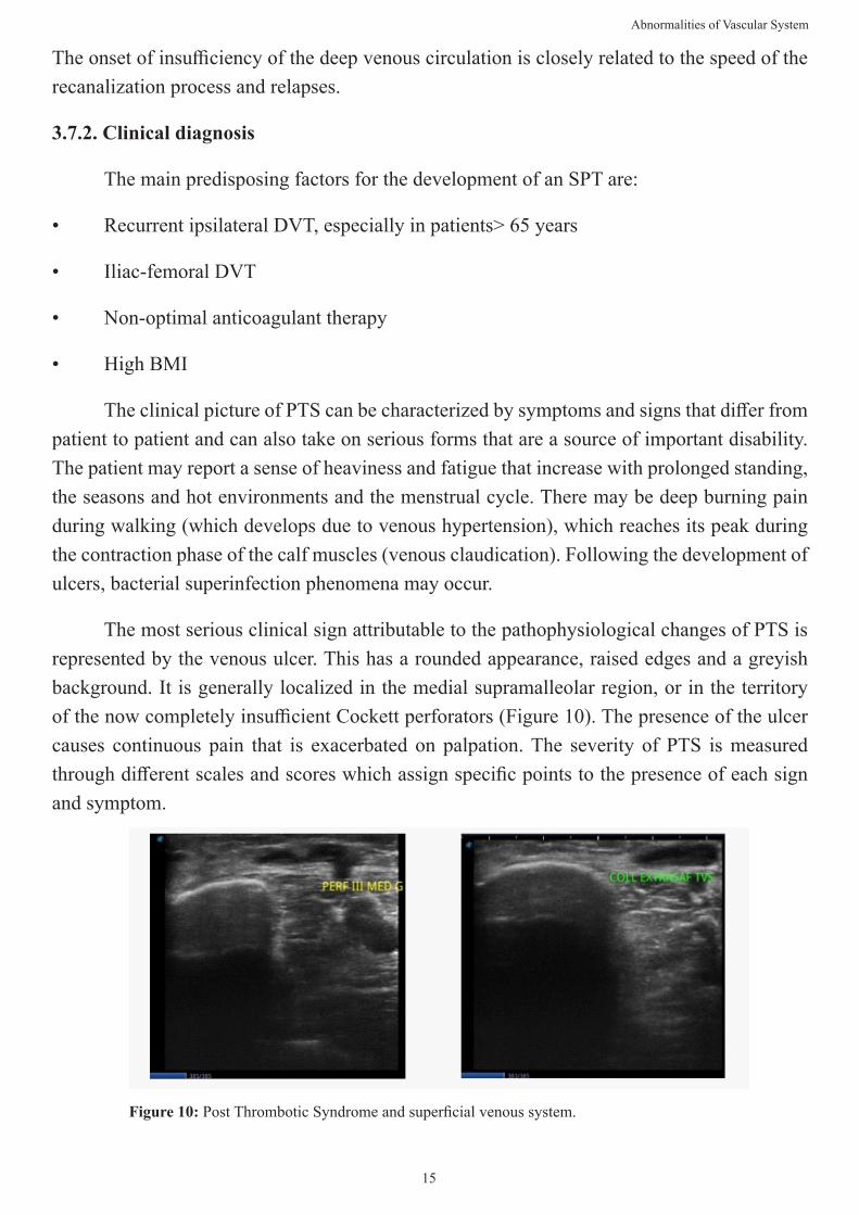
The most serious clinical sign attributable to the pathophysiological changes of PTS is represented by the venous ulcer. This has a rounded appearance, raised edges and a greyish background. It is generally localized in the medial supramalleolar region, or in the territory of the now completely insufficient Cockett perforators (Figure 10). The presence of the ulcer causes continuous pain that is exacerbated on palpation. The severity of PTS is measured through different scales and scores which assign specific points to the presence of each sign and symptom.
Figure 10: Post Thrombotic Syndrome and superficial venous system.
16
Abnormalities of Vascular System
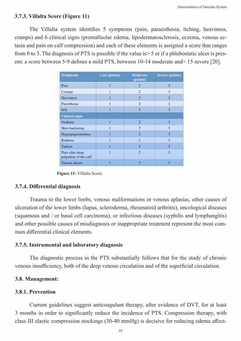
3.7.3. Villalta Score (Figure 11)
The Villalta system identifies 5 symptoms (pain, paraesthesia, itching, heaviness, cramps) and 6 clinical signs (premalleolar edema, lipodermatosclerosis, eczema, venous ec-tasia and pain on calf compression) and each of these elements is assigned a score that ranges from 0 to 3. The diagnosis of PTS is possible if the value is> 5 or if a phlebostatic ulcer is pres-ent; a score between 5-9 defines a mild PTS, between 10-14 moderate and> 15 severe [20].
Figure 11: Villalta Score.
3.7.4. Differential diagnosis
Trauma to the lower limbs, venous malformations or venous aplasias, other causes of ulceration of the lower limbs (lupus, scleroderma, rheumatoid arthritis), oncological diseases (squamous and / or basal cell carcinoma), or infectious diseases (syphilis and lymphangitis) and other possible causes of misdiagnosis or inappropriate treatment represent the most com-mon differential clinical elements.
3.7.5. Instrumental and laboratory diagnosis
The diagnostic process in the PTS substantially follows that for the study of chronic venous insufficiency, both of the deep venous circulation and of the superficial circulation.
3.8. Management:
3.8.1. Prevention
Current guidelines suggest anticoagulant therapy, after evidence of DVT, for at least 3 months in order to significantly reduce the incidence of PTS. Compression therapy, with class III elastic compression stockings (30-40 mmHg) is decisive for reducing edema affect-
17
Abnormalities of Vascular System
ing the distal portions of the lower limb, promoting venous return by improving the function of muscle pump.
Thrombolysis is able to improve venous circulatory hemodynamics while preserving chronic valve damage and, in fact, guarantees the reduction of the risk of PTS. Since patients with large DVT, especially those at the iliac-femoral level, are by definition a higher risk of developing PTS, rapid thrombolysis reduces PTS in itself. To date, modern endovascular tech-niques have totally supplanted the use of the tissue plasminogen activator thanks to the greater effectiveness confirmed by many RCTs.
Percutaneous Mechanical Thrombectomy (PMT) is an additional minimally invasive treatment but with a lower grade of recommendation.
3.8.2. Treatment
Patients with a clear diagnosis of PTS currently benefit from limited therapeutic possi-bilities in addition to those previously described. The use of neuro-muscular electrostimulators can reduce the chronic symptoms of PTS; stenting of one or more venous segments for chronic occlusive syndrome improves the deep venous circulation and consequently the symptoms; surgery, with the use of biological valves or neovalves, have still had little success despite the better clinical outcomes compared to more traditional surgical procedures.
4. Chronic Venous Disease (CVD)
Chronic Venous Disease (CVD) is a pathological condition characterized by peripheral venous hemodynamic dysfunction of the lower limbs with a spectrum of clinical signs ranging from varicose veins to premalleolar edema up to the most dramatic phases such as hyperpig-mentation, eczema, lipodermatosclerosis and ulcers.
4.1. Pathophysiology
In the superficial venous system there is a lower pressure regimen than in the deep venous one, so exposure to higher pressures can cause dilatation and tortuosity: valve insuffi-ciency causes flow reversal with venous hypertension and altered load on the venous walls. At the beginning the vein opposes hypertrophying the parietal muscle component (phase of com-pensation), but over time the vein dilates, lengthens, vents and degenerates (decompensation phase = varices). The evolution of varices does not always foresee reflux from the saphenous-femoral junction because, in addition to the long reflux from above, it is possible to hyperflux from the bottom upwards, typical in venous alterations that occur in pathologies of the foot from altered posture. Varicose veins and venous dilations can be the result of impaired outflow at the crossing points or in perforating points, frequent in case of hyperextension of the knees in people who use very high heels or have a varus valgus foot.
18
Abnormalities of Vascular System
4.2. Classification
Varicose syndromes are classified according to three main aspects:
A) Etiology
- Essential or primary varices: these are the most frequent and are caused by a structural and / or histological alteration of the venous walls and valves which makes them unable to resist pressure increases. These alterations derive from multiple factors that act mainly on the varicose diathesis of the individual which is a congenital hypoplasia of the connective tissue. The main cofactors are pregnancy, constipation, sedentary occupation, obesity, flat foot and erect status.
- Secondary varices: these are the consequence of a thrombotic event localized in the deep venous circulation that hinders the normal flow of blood, which, through the perforating veins, finds its outlet in the superficial circulation, overloading it and causing varicosis.
B) Anatomo-physiology
- Varicose veins (subcutaneous or subfascial): characterized by long refluxes due to insuf-ficiency of the femoral and / or popliteal crosse.
- Reticular varicose disease(subcutaneous and intradermal varicose veins): characterized by reticular varicosities without compromising the saphenous-femoral or saphenous-popliteal junctions, therefore devoid of long refluxes (intact saphenous).
- Mixed varicose disease (subcutaneous, intradermal and subfascial varices): varicose disease with associated long refluxes due to insufficiency of the saphenous-femoral crosse.
- Telangiectasias: characterized by fine intradermal varices, red or blue, located mainly on the inside of the knee, outside the thigh and on the lateral side of the leg. Their diameter does not exceed a millimeter; they are usually tributary to reticular varices (blue vessels) or dilated capillaries due to tissue hypoxia or lymphovenous stasis (increase in pressure).
C) Morphology
- Truncular varices: typical of saphenous veins (diameter> 3mm)
- Reticular varices: dilated and tortuous (diameter <5mm)
- Telangiectasias: permanently dilated intradermal venules less than 1 mm in diameter. diameter between 0.5-1mm).
19
Abnormalities of Vascular System
4.3. Clinical and Instrumental Diagnosis
The inspection allows to evaluate broadly the varicose geography, to quantify its stasis and chronic venous insufficiency; allows to define the foot support defects and the state of the subcutaneous tissue. Varicose gavoccioli, venous trunks, ulcers, pigmentations, foot condi-tion and posture, symmetry of the subgluteal sulcus, flexion-extension of the knees, possible nail diseases, coloration of the fingers, state of adipose tissue, presence of edema, phototype, presence of any livedo reticularis or histamine dermal reactivity, tendency to pigmentation or keloid.
• Trendelenburg test
This maneuver is used to distinguish a superficial venous reflux from an incompetence of the valves of the deep venous system. With the patient in the supine position, the lower limb must be raised until the superficial varicose veins are emptied, then a lace is placed on the upper third of the thigh. In the upright position, a filling of the varicose superficial venous circulation will indicate an incompetence of the valves of the deep venous system or of the perforating veins. If, on the other hand, the superficial circle fills up only after removing the lace, this will indicate an incontinence of the circle itself.
• Manouvre of Pèrthes
It serves to distinguish the antegrade or retrograde direction in a superficial varice. An ante-grade flow indicates that the system is actually a natural by-pass for overcoming a deep vein thrombosis. This indication appears of enormous importance if we consider that, in the case of thrombosis of a tract of deep vein, a superficial vein, albeit varicose, represents an important venous drainage route and therefore should not be sclerosed or surgically removed. A tourni-quet is placed in the proximal part of the varicose vein. After walking the patient, the emptying of the superficial varicose venous system will indicate the patency of the deep venous system, on the contrary, the engorgement and congestion of the varicose vein will demonstrate the presence of an obstruction of the deep venous system.
• Linton test
If the Pèrthes maneuver is positive, the patient is placed in the supine position with the tour-niquet in place and the examined lower limb is raised. If the varices distal to the lace do not empty within a few seconds, an obstruction of the deep venous system must be considered.
• Pratt test
It is used for the research of venous refluxes of perforators. When the patient is lying down, a lace is applied to the root of the thigh and then the whole limb is bandaged with an
20
Abnormalities of Vascular System
elastic compression bandage. The patient is placed on his feet and the elastic band slowly un-winds from the top. A reflux denotes the presence of an insufficient perforator. By applying laces to the various levels, all incontinent perforators can be located.
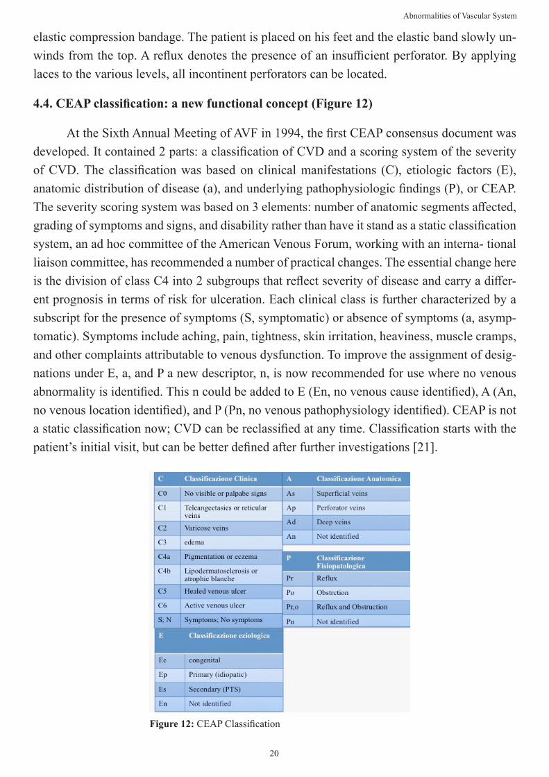
4.4. CEAP classification: a new functional concept (Figure 12)
At the Sixth Annual Meeting of AVF in 1994, the first CEAP consensus document was developed. It contained 2 parts: a classification of CVD and a scoring system of the severity of CVD. The classification was based on clinical manifestations (C), etiologic factors (E), anatomic distribution of disease (a), and underlying pathophysiologic findings (P), or CEAP. The severity scoring system was based on 3 elements: number of anatomic segments affected, grading of symptoms and signs, and disability rather than have it stand as a static classification system, an ad hoc committee of the American Venous Forum, working with an interna- tional liaison committee, has recommended a number of practical changes. The essential change here is the division of class C4 into 2 subgroups that reflect severity of disease and carry a differ-ent prognosis in terms of risk for ulceration. Each clinical class is further characterized by a subscript for the presence of symptoms (S, symptomatic) or absence of symptoms (a, asymp-tomatic). Symptoms include aching, pain, tightness, skin irritation, heaviness, muscle cramps, and other complaints attributable to venous dysfunction. To improve the assignment of desig-nations under E, a, and P a new descriptor, n, is now recommended for use where no venous abnormality is identified. This n could be added to E (En, no venous cause identified), A (An, no venous location identified), and P (Pn, no venous pathophysiology identified). CEAP is not a static classification now; CVD can be reclassified at any time. Classification starts with the patient’s initial visit, but can be better defined after further investigations [21].
Figure 12: CEAP Classification
21
Abnormalities of Vascular System
4.4.1. Ultrasound examination
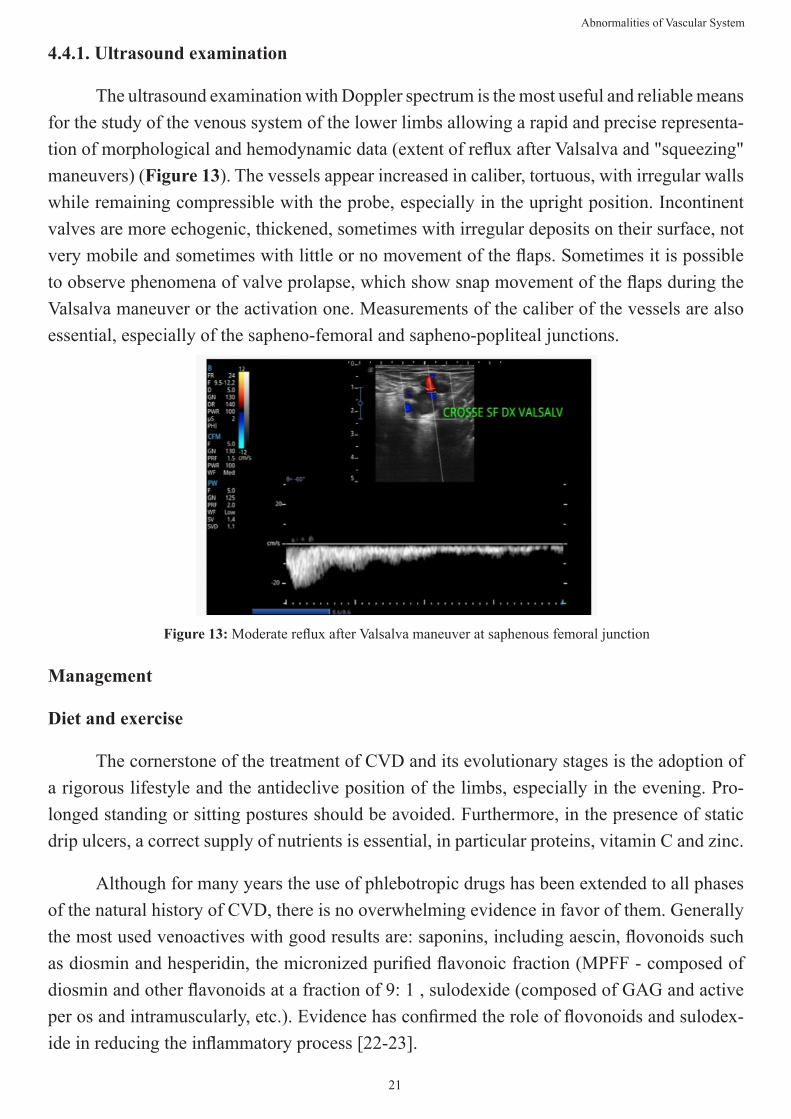
The ultrasound examination with Doppler spectrum is the most useful and reliable means for the study of the venous system of the lower limbs allowing a rapid and precise representa-tion of morphological and hemodynamic data (extent of reflux after Valsalva and "squeezing" maneuvers) (Figure 13). The vessels appear increased in caliber, tortuous, with irregular walls while remaining compressible with the probe, especially in the upright position. Incontinent valves are more echogenic, thickened, sometimes with irregular deposits on their surface, not very mobile and sometimes with little or no movement of the flaps. Sometimes it is possible to observe phenomena of valve prolapse, which show snap movement of the flaps during the Valsalva maneuver or the activation one. Measurements of the caliber of the vessels are also essential, especially of the sapheno-femoral and sapheno-popliteal junctions.
Figure 13: Moderate reflux after Valsalva maneuver at saphenous femoral junction
Management
Diet and exercise
The cornerstone of the treatment of CVD and its evolutionary stages is the adoption of a rigorous lifestyle and the antideclive position of the limbs, especially in the evening. Pro-longed standing or sitting postures should be avoided. Furthermore, in the presence of static drip ulcers, a correct supply of nutrients is essential, in particular proteins, vitamin C and zinc.
Although for many years the use of phlebotropic drugs has been extended to all phases of the natural history of CVD, there is no overwhelming evidence in favor of them. Generally the most used venoactives with good results are: saponins, including aescin, flovonoids such as diosmin and hesperidin, the micronized purified flavonoic fraction (MPFF - composed of diosmin and other flavonoids at a fraction of 9: 1 , sulodexide (composed of GAG and active per os and intramuscularly, etc.). Evidence has confirmed the role of flovonoids and sulodex-ide in reducing the inflammatory process [22-23].
22
Abnormalities of Vascular System
4.4.2. Compression Therapy
Compression therapy is the basis of any treatment for patients suffering from CVD and in the various pathological entities that characterize it (from varicose veins, to persistent edema to ulcer). Recent evidence suggests the use of moderate elastic compression of 20-30 mmHg in the case of symptomatic varicose disease and as a first treatment strategy, adjuvant with ablative surgery, in the case of ulcers. Attention should be paid to the concomitant pres-ence of arteriopathy.
4.5.Treatment of Cvd
In the presence of moderate CVD (CEAP C1-C2) the intervention, in the different op-tions available (interruption of the Great Saphenous Vein at the level of the saphenous-femoral crosse with or without stripping, endovascular obliteration by laser method (EVLA) or radio-frequency (RF) , sclerotherapy) is reserved for symptomatic patients, persistence of symptoms despite adequate pharmacological and elastic compression therapy or signs of rapid evolution towards advanced stages.
Generally the surgical indication is instead indicated in patients diagnosed with severe CVD (CEAP> 3).
4.6. Surgical Approach to CVD
The first phase consists in the crossectomy of the saphenous vein and in the ligation of the tributaries to the sapheno-femoral junction; it is a surgical act that requires extreme care, in order not to leave varicose trunks which, in a second time, could give a recurrence by recanalizing the underlying vessels. Subsequently, the insufficient saphenous vein can be stripped or its phlebectomy by means of mini-incisions in the varicose tract. The operation can be conducted under local, spinal or general anesthesia depending on the clinical picture and the opinion of the anesthetist. Stripping can be of the long type when the vein is completely removed, or of the short type with partial removal of the vessel.
An incision is made in the groin to isolate the internal saphenous vein and at the level of the popliteal fossa (posterior part of the knee) for the isolation of the external saphenous vein; some small incisions are useful for binding and removing any collateral varicosities. Once the upper and lower extremities of the vein have been identified, a probe (stripper) is inserted inside it, from bottom to top. At the upper end of the probe, which protrudes at the level of the inguinal incision, the severed head of the vein is tied and a metal head is fixed; this head, when the probe is withdrawn, drags all the saphenous vein downwards, tearing the connections with the collateral veins, until it is completely extracted. At the end of the surgery, the small skin incisions are sutured and a compression bandage is made, which is usually removed after
23
Abnormalities of Vascular System
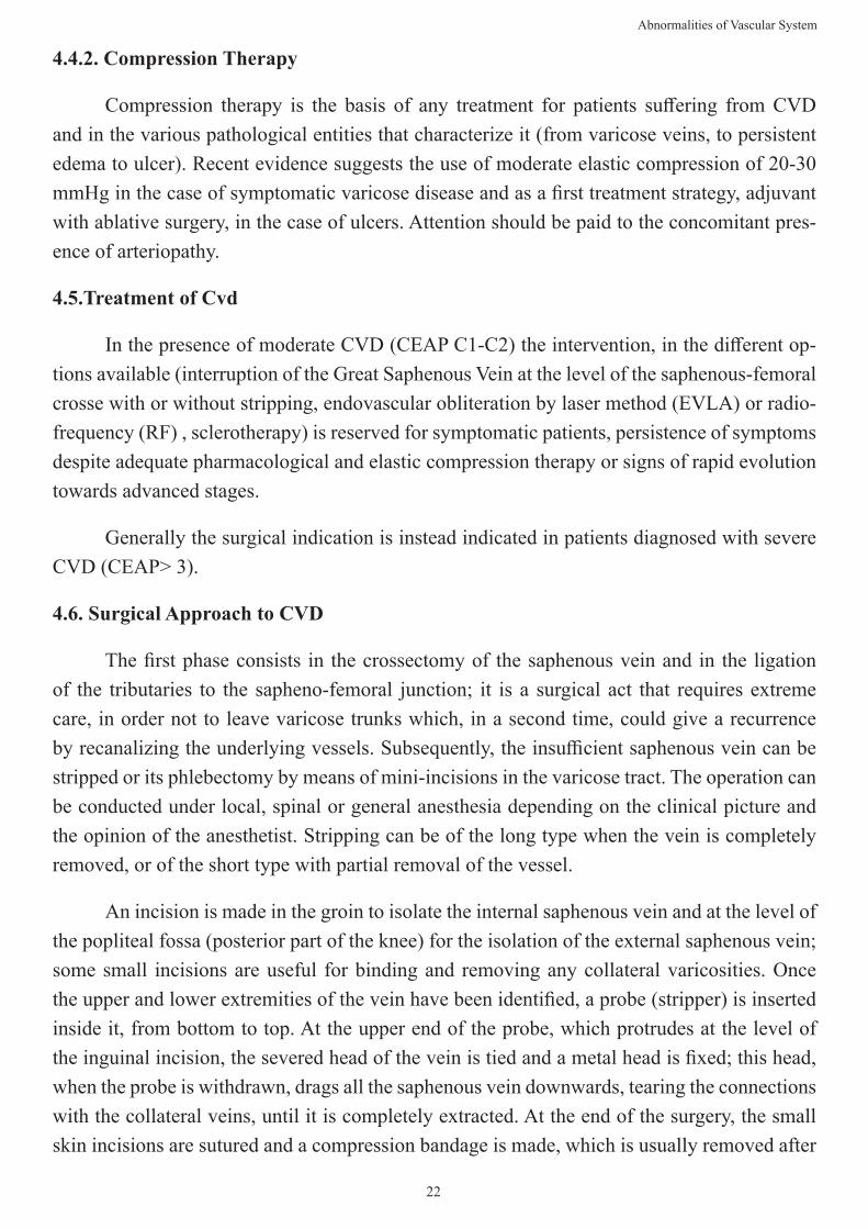
about a week, together with the sutures [Figure 14].
Figure 14: Surgical sapheno – femoral junction exposure
4.6.1.Phlebectomy of saphenous and incontinent perforating collaterals
The preoperative study using echocolor Doppler allows to precisely highlight the exact site of refluxes and to save healthy vein sections if phlebectomy is not associated with strip-ping of the great saphenous vein. All the varicosities to be treated are "mapped" with a marker. Local anesthesia is first practiced along the most evident varicose trunks with a very diluted anesthetic solution in order to detach the varices from the subcutaneous tissue.
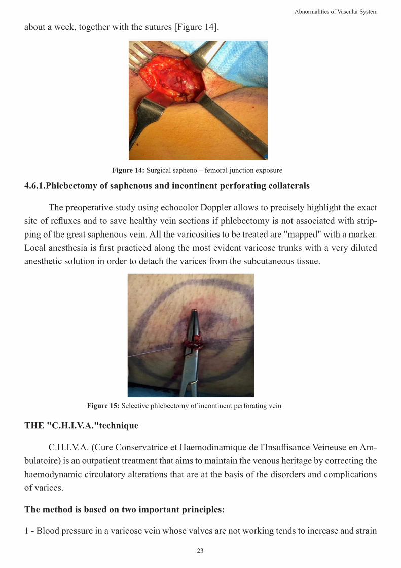
Figure 15: Selective phlebectomy of incontinent perforating vein
THE "C.H.I.V.A."technique
C.H.I.V.A. (Cure Conservatrice et Haemodinamique de l'Insuffisance Veineuse en Am-bulatoire) is an outpatient treatment that aims to maintain the venous heritage by correcting the haemodynamic circulatory alterations that are at the basis of the disorders and complications of varices.
The method is based on two important principles:
1 - Blood pressure in a varicose vein whose valves are not working tends to increase and strain
24
Abnormalities of Vascular System
the veins.
The first goal of the therapy is, therefore, to decrease the hydrostatic pressure by split-ting the blood column (and therefore the vein) into several segments.
2 - Between the healthy veins and the varicose veins of the saphenous there are connections or Shunts through the perforating veins (communication branches between the deep and super-ficial venous circulation) and the saphenous-femoral crosse (where the saphenous vein opens into the femoral vein) ; for which the blood carried upwards by the deep veins of the leg, tends to flow back into the varices. The other goal of C.H.I.V.A. is represented by the interruption of such shunts. The application of these principles is possible through the ecocolor Doppler tending to the identification of the main "vanishing points". The surgical act consists of simple ligations of the veins in the points previously identified and mapped [24].
4.7. Endovascular Procedures
To date, endovascular obliteration can benefit from two main procedures: Laser (EVLA) (20% of cases) and Radiofrequency (RF) (30% of cases). The physical mechanism with which the thermal damage is produced is able to trigger the parietal inflammation and therefore scle-rosis of the treated vein.
To date, RF, particularly in the United States, if anatomically feasible (saphenous di-ameter> 5 mm and <12 mm, absence of tortuosity, distance from the skin> 0.5 cm), is the treatment of choice for the incompetence of the superficial venous system and has, in fact, exceeded, in terms of short and medium-term results for what concerns the relapses of the saphenous-femoral and saphenous-popliteal crosse, the classic open surgical interventions. RF not only allows the procedure to be carried out on an outpatient basis and under local an-esthesia, but guarantees reduction of pain, less incidence of bruising and a quick possibility of returning to work [25].
4.8. Sclerotherapy
Sclerosing therapy is essentially based on the intravenous injection of irritating chemicals ca-pable of causing reactive endothelitis with exfoliation of the endothelium itself and formation of the so-called "sclerus". The indications for variceal sclerosing therapy are as follows:
- telangiectasias, even if relevant from an aesthetic point of view, when they are associated with the symptoms of CVD;
- reticular varices (1-3 mm);
- saphenous varices;
25
Abnormalities of Vascular System
- extra-saphenous varices;
- emergency treatment in case of variceal rupture hemorrhage.
4.8.1.Complications after surgery for CVD
Thromboembolic complications. The risk of DVT and PE is very low, ranging from 0.4% to 5.3%, if a postoperative ultrasound is performed. Risk factors significantly associated with the onset of postoperative DVT are family history of DVT and an operative indication for CEAP 5 or 6. Outside of these criteria, systematic anticoagulant treatment after surgery is not warranted, especially if the procedure is performed under local or locoregional anesthesia, with early gait and wearing elastic restraint.
Hematomas. They are always present, albeit without sequelae, after a classical exeresis surgery and practically absent after conservative surgery of the saphenous trunks. The elastic compression partially prevents them and reduces the pain they are responsible for.
Local infectious complications. Rare, they occur, most of the time, after repeated sur-gery in the groin fold and in the obese and / or diabetic patient and are favored by lymphatic collections or losses.
Lymphatic complications. Early, these result in a collection (lymphocele) or / and a lo-calized loss of lymphatic fluid (lymphorrhea), most often in the inguinal incision, due to opera-tive trauma of the vessels or lymph nodes or, also, during phlebectomies in certain locations (dorsum of the foot, tibial crest).
Neurological complications. These are superficial sensory nerve lesions, responsible for sensitivity disorders in 36.5% of cases, paresthesia in 8% of cases and dysesthesia in 1.6% of cases. These disorders subside, most of the time, within a few weeks, sometimes after several months.
Aesthetic complications. These are late. The worsening of pre-existing telangiectasias, the occurrence of a network of neothelangiectasias (matting) or pigmentation, especially on the medial aspect of the thigh, are not exceptional on the venous excision path. Skin complica-tions have also been reported along the stripping process: localized hypertrichosis, scleroder-miform dermatitis, vitiligo.
4.8.2.Results in the territory of the great saphenous vein
A recent evidence conducted by Mendes confirmed the benefit of open surgery over medical treatment [26], moreover, comparing short stripping vs long stripping, other authors showed that the risk of internal saphenous nerve injury at one month was not significantly dif-ferent between short or long excision (4.5% vs 19%, p > 0.05), with, in addition, a regression
26
Abnormalities of Vascular System
of symptoms in almost half of the cases at five years [27]. Several randomized studies have compared surgical venous removal with one or more endovascular techniques with results in favor of the latter with regard to pain. No statistically significant differences were observed in terms of quality of life. In patients undergoing sclerotherapy alone, relapses are twice as high as in patients undergoing classic surgical or endovascular procedures [25, 27].
5. Follow-Up
Varicose recurrence after open or endovascular surgery is frequent, estimated at 35% at two years, 65% at 11 years and even 77% at 34 years. The causes are a progression of varicose disease in 15% of cases, the appearance of a neovascularization related to the development of incompetent tortuous veins from the groin or popliteal fossa in 13% of cases and a technical or tactical error respectively in 5 , 3% and in 4% of cases. Therefore, clinical and ultrasound monitoring is required in the short, medium and long term.
6. Acknowledgements
Thanks to Medi Italia srl for the constant technical support in daily clinical and scientific activity
7. Conflict of Interest
The authors declare that they have no competing interests.
8. Funding
This work received no funding.
9. References
1. Homans J. Diseases of the veins. N Engl J Med. 1946;235:163-7.
2. Ruggiero M, Grande R, Naso A, Butrico L, Rubino P, Placida GD, Cannistrà M, Serra R. Symptoms in patients with skin changes due to chronic venous insufficiency often lead to emergency care service: an Italian observational study. Int Wound J 2016;13:967-71.
3. Serra R, Grande R, Butrico L, Fugetto F, de Franciscis S. Epidemiology, diagnosis and treatment of chronic venous disease: a systematic review. Chirurgia, 2016;29:34-45.
4. Amato B, Coretti G, Compagna R, Amato M, Buffone G, Gigliotti D, Grande R, Serra R, de Franciscis S. Role of matrix metalloproteinases in non-healing venous ulcers. Int Wound J 2015;12:641-5.
5. de Franciscis S, Gallelli L, Amato B, Butrico L, Rossi A, Buffone G, Caliò FG, De Caridi G, Grande R, Serra R. Plasma MMPs and TIMPs evaluation in patients with deep venous thrombosis. Could they have a predictive role in the development of Post Thrombotic Syndrome? Int Wound J. 2016;13:1237-45.
6. Busceti MT, Grande R, Amato B, Gasbarro V , Buffone G, Amato M, Gallelli L, Serra R, de Franciscis S. Pulmonary Embolism, Metalloproteinases And Neutrophil Gelatinase Associated Lipocalin. Acta Phlebol. 2013;14:115-21.
27
Abnormalities of Vascular System
7. Lurie JM, Png CYM, Subramaniam S, Chen S, Chapman E, Aboubakr A, Marin M, Faries P, Ting W. Virchow’s triad in “silent” deep vein thrombosis. J Vasc Surg Venous Lymphat Disord. 2019;7:640-5.
8. Di Nisio M, van Es N, Büller HR. Deep vein thrombosis and pulmonary embolism. Lancet. 2016;388(10063):3060-73.
9. Needleman L, Cronan JJ, Lilly MP, Merli GJ, Adhikari S, Hertzberg BS, DeJong MR, Streiff MB, Meissner MH. Ultrasound for Lower Extremity Deep Venous Thrombosis: Multidisciplinary Recommendations From the Society of Radiologists in Ultrasound Consensus Conference. Circulation. 2018;137:1505-15.
10. Linkins LA, Takach Lapner S. Review of D-dimer testing: Good, Bad, and Ugly. Int J Lab Hematol. 2017;39 Suppl 1:98-103.
11. Chopard R, Albertsen IE, Piazza G. Diagnosis and Treatment of Lower Extremity Venous Thromboembolism: A Review. JAMA. 2020;324:1765-76.
12. Serhal M, Barnes GD. Venous thromboembolism: A clinician update. Vasc Med. 2019;24:122-31.
13. Pernès JM, Auguste M, Kovarski S, Borie H, Renaudin JM, Coppe G. Acute deep vein thrombosis and endovascular techniques: It is time for a new aggiornamento! Diagn Interv Imaging. 2012;93:725-33.
14. Lopez R, DeMartino R, Fleming M, Bjarnason H, Neisen M. Aspiration thrombectomy for acute iliofemoral or cen-tral deep venous thrombosis. J Vasc Surg Venous Lymphat Disord. 2019;7:162-8.
15. Essien EO, Rali P, Mathai SC. Pulmonary Embolism. Med Clin North Am. 2019;103:549-64.
16. Jiménez D, Aujesky D, Moores L, Gómez V, Lobo JL, Uresandi F, Otero R, Monreal M, Muriel A, Yusen RD; RIE-TE Investigators. Simplification of the pulmonary embolism severity index for prognostication in patients with acute symptomatic pulmonary embolism. Arch Intern Med. 2010;170:1383-9.
17. Abolfotouh MA, Almadani K, Al Rowaily MA. Diagnostic Accuracy of D-Dimer Testing and the Revised Geneva Score in the Prediction of Pulmonary Embolism. Int J Gen Med. 2020;13:1537-43.
18. Kumamaru KK, Saboo SS, Aghayev A, Cai P, Quesada CG, George E, Hussain Z, Cai T, Rybicki FJ. CT pulmonary angiography-based scoring system to predict the prognosis of acute pulmonary embolism. J Cardiovasc Comput To-mogr. 2016;10:473-9.
19. Young T, Sriram KB. Vena caval filters for the prevention of pulmonary embolism. Cochrane Database Syst Rev. 2020;10:CD006212.
20. Galanaud JP, Holcroft CA, Rodger MA, Kovacs MJ, Betancourt MT, Wells PS, Anderson DR, Chagnon I, Le Gal G, Solymoss S, Crowther MA, Perrier A, White RH, Vickars LM, Ramsay T, Kahn SR. Comparison of the Villalta post-thrombotic syndrome score in the ipsilateral vs. contralateral leg after a first unprovoked deep vein thrombosis. J Thromb Haemost. 2012;10:1036-42.
21. Lurie F, Passman M, Meisner M, Dalsing M, Masuda E, Welch H, Bush RL, Blebea J, Carpentier PH, De Maeseneer M, Gasparis A, Labropoulos N, Marston WA, Rafetto J, Santiago F, Shortell C, Uhl JF, Urbanek T, van Rij A, Eklof B, Gloviczki P, Kistner R, Lawrence P, Moneta G, Padberg F, Perrin M, Wakefield T. The 2020 update of the CEAP clas-sification system and reporting standards. J Vasc Surg Venous Lymphat Disord. 2020;8:342-52.
22. Serra R, Gallelli L, Conti A, De Caridi G, Massara M, Spinelli F, Buffone G, Caliò FG, Amato B, Ceglia S, Spaziano G, Scaramuzzino L, Ferrarese AG, Grande R, de Franciscis S. The effects of Sulodexide on both clinical and molecular parameters in patients with mixed arterial and ulcers of lower limbs. Drug Des Devel Ther. 2014;8:519-27.
23. Serra R, Grande R, Butrico L, Buffone G, Caliò FG, Squillace A, Rizzo BA, Massara M, Spinelli F, Ferrarese AG, De Caridi G, Gallelli L, de Franciscis S. Effects of a new nutraceutical substance on clinical and molecular parameters in patients with chronic venous ulceration. Int Wound J 2016; 13:88-96.

24. Faccini FP, Ermini S, Franceschi C. CHIVA to treat saphenous vein insufficiency in chronic venous disease: charac-teristics and results. J Vasc Bras. 2019;18:e20180099.
25. Vemulapalli S, Parikh K, Coeytaux R, Hasselblad V, McBroom A, Johnston A, Raitz G, Crowley MJ, Lallinger KR, Jones WS, Sanders GD. Systematic review and meta-analysis of endovascular and surgical revascularization for patients with chronic lower extremity venous insufficiency and varicose veins. Am Heart J. 2018;196:131-43.
26. Mendes CA, Martins AA, Fukuda JM, Parente JB, Munia MA, Fioranelli A, Teivelis MP, Varella AY, Caffaro RA, Kuzniec S, Wolosker N. Randomized trial of radiofrequency ablation versus conventional surgery for superficial venous insufficiency: if you don’t tell, they won’t know. Clinics (Sao Paulo). 2016;71:650-6.
27. Ay Y, Gunes E, Turkkolu ST, Selcuk E, Calim M, Akal R, Aydin C, Inan B, Koksal C, Kahraman Ay N. Comparative efficacy and life quality effects of surgical stripping, radiofrequency ablation, and cyanoacrylate embolization in patients undergoing treatment for great saphenous vein insufficiency. Phlebology. 2021;36:54-62.
28
Abnormalities of Vascular System