frequent and widespread vascular abnormalities in human...
TRANSCRIPT

1
Frequent and Widespread Vascular Abnormalities in Human
STAT3 Deficiency
Running title: Chandesris et al.; Vascular abnormalities in human STAT3 deficiency
Marie-Olivia Chandesris, MD1,2,3,*; Arshid Azarine, MD, MSc4,*; Kim-Thanh Ong, MD,
PhD5,*; Soraya Taleb, PhD6,*; Pierre Boutouyrie, MD, PhD5; Elie Mousseaux, MD, PhD4,7;
Mélissa Romain, MSc6; Erwan Bozec, PhD5; Stéphane Laurent, MD, PhD 5; Nathalie Boddaert,
MD, PhD8,9; Caroline Thumerelle, MD, PhD10; Isabelle Tillie-Leblond, MD, PhD11; Cyrille
Hoarau, MD, PhD12; Yvon Lebranchu, MD, PhD12; Nathalie Aladjidi, MD13; François Tron,
MD, PhD14; Vincent Barlogis, MD15; Gérard Body, MD16; Marine Munzer, MD17; Roland
Jaussaud, MD, PhD18; Felipe Suarez, MD, PhD1,2,3; Olivier Clement, MD, PhD4; Olivier
Hermine, MD, PhD1,2,3,19; Alain Tedgui, PhD6; Olivier Lortholary, MD, PhD2,3,20; Capucine
Picard, MD, PhD2,3,21,22,#; Ziad Mallat, MD, PhD2,6,23,#; Alain Fischer, MD, PhD2,3,24,25,#
1Hematology Dept, Necker Children's Hospital, Assistance Publique Hôpitaux de Paris (APHP);2Paris Descartes University, Paris Cité Sorbonne, Necker Faculty of Med; 3Ctr de
référence des déficits immunitaires héréditaires (CEREDIH), Necker Children's Hospital, APHP; 4Radiology Dept, Georges Pompidou European Hospital, APHP; 5Pharmacology Dept, Georges
Pompidou European Hospital, Paris Descartes University, APHP;6INSERM U970, Paris-Descartes University; 7INSERM U678, Georges Pompidou European Hospital, APHP; 8Pediatric
Radiology Unit, Necker Children's Hospital, APHP; 9INSERM U1000, Paris-Descartes University, Paris;10Pediatric Pneumology Dept, Jeanne de Flandres Hospital; 11Pneumology Unit, Calmette Hospital, Lille, France;12Immunology Unit, Tours Hospital, Tours;13Pediatric Hemato-
oncology Dept, Pellegrin Hospital, Bordeaux; 14Immunology Unit, Rouen Hospital, Rouen;15Pediatric Hemato-oncology Dept, Timone Hospital, Marseille;16Pediatric Pneumology Dept, Châlons-en-Champagne Hospital, Châlons-en-Champagne;17Pediatric Onco-Hematology
Dept, Pediatric Hospital; 18Internal Med Dept, Robert Debré Hospital, Reims;19CNRS UMR 8147, Paris Descartes University, Necker Med Faculty, Necker; 20Dept of Infectious Diseases & Tropical Med, Necker - Enfants Malades Hospital & Inst Pasteur, CNRMA, CNRS URA3012,
APHP;21Study Ctr of Primary Immunodeficiencies, Necker - Enfants Malades Hospital, APHP;22Lab of Human Genetics of Infectious Diseases, Necker Branch, INSERM U980, Paris,
France; 23Dept of Med, University of Cambridge, Cambridge, United Kingdom; 24Immuno-
, ; NN
-Leblblblblblblblblblblblblblblblblblblbblblbllononononononononononononononnononononnnoonond,d,d,dd,d,d,d,d,d,d,d,d,d,d,d,ddddd,dddd MMMMMMMM MMMMMMMMMMMMMMMD,D,DD,D,D,D,D,DD,D,D,D,DDDD,DDDD,D PPPPPPPPP PPPPPP13131313131313313131331331313133D ; Yvon Lebranchu MD PhD ; Nathalie Aladjidi MD ; F
c M
P P
D 2
D ; , , ; , ,
D ; YvYvYvYvYvononononon LLLLLebeeee ranchu, MD, PhDDDD ; Nathalie AAAAAladjidi, MD ; F
ceneneneent Barlllllogogogoggisiii , MDMDMDMDMD155555; GéGéGéGéGéraraardrdrdrdd B odody,y,y,y,y, MMM MMDDDDD161616161 ;; MaMaMaMaMariiiiinennnn M M MMMununununnzezezezezer,r,r,rr, M
PhhhhhDDDDD18; ; ; ;; FeFeFeFeFelililililipepepeppe S S SSSuuuauu rererererez,z,z,z,z, MMMMMD,D,D,D,D, P P PPPhDhDhDhDhD1,21,21,2,,,3; ; ;;; OlOlOlOlOlivivivivivieieieieier rrr r ClClClCClemememememenenenenent,t,t,tt M M MMMD,D,D,DD, P P P
D1,2,3,19; Alllllaiaiaiaiain nnnn TeTeTeTeTedgdgdgdgdguiuiuiuiui,, PhPhPhPhPhDDDDD6666;;; ;; OlOlOlOlOliviviviivieeeeerrr rr LoLoLoLoLortrtrtrthohohohoholalalallaryryryryry,,, ,, MD, PhD2
DDDD2,3,21,2222222222,#,#,#,#,#;;;; ZiZiZiZiZiadadadadad M MMMMalalalllalalalaat,tt,t MM MMMD,D,D,DD P PPPPhDhDhDhDhD2,62,62,666,23,23,232323,#,#,#,#,#;;;; AlAlAlAlAlaiaiaiaiainnnn FiFiFiFiFiscscscheheheheher,rrr, MM MD,D,D,
by guest on June 26, 2018http://circgenetics.ahajournals.org/
Dow
nloaded from
by guest on June 26, 2018http://circgenetics.ahajournals.org/
Dow
nloaded from
by guest on June 26, 2018http://circgenetics.ahajournals.org/
Dow
nloaded from
by guest on June 26, 2018http://circgenetics.ahajournals.org/
Dow
nloaded from
by guest on June 26, 2018http://circgenetics.ahajournals.org/
Dow
nloaded from
by guest on June 26, 2018http://circgenetics.ahajournals.org/
Dow
nloaded from
by guest on June 26, 2018http://circgenetics.ahajournals.org/
Dow
nloaded from
by guest on June 26, 2018http://circgenetics.ahajournals.org/
Dow
nloaded from
by guest on June 26, 2018http://circgenetics.ahajournals.org/
Dow
nloaded from
by guest on June 26, 2018http://circgenetics.ahajournals.org/
Dow
nloaded from
by guest on June 26, 2018http://circgenetics.ahajournals.org/
Dow
nloaded from
by guest on June 26, 2018http://circgenetics.ahajournals.org/
Dow
nloaded from
by guest on June 26, 2018http://circgenetics.ahajournals.org/
Dow
nloaded from

2
Hematology Pediatric Dept, Necker - Enfants Malades Hospital, APHP; 25INSERM U768, Necker - Enfants Malades Hospital, Paris, France
* These authors contributed equally; # These authors contributed equally
Corresponding author:
Alain Fischer
Immuno-Hematology Pediatric Department / INSERM U768
Hôpital Necker - Enfants Malades
149 rue de Sèvres
F-75015 Paris
France, EU
Phone: +33 144 494 822
Fax: +33 144 495 070
E-mail: [email protected]
Journal Subject Code: [109] Clinical genetics; [30] CT and MRI; [50] Cerebral Aneurysm, AVM, & Subarachnoid hemorrhage; [51] Cerebral Lacunes; [58] Computerized tomography and Magnetic Resonance Imaging; [79] Aneurysm, AVM, hematoma
Abbreviations: AA: artery aneurysm; AAA: abdominal aortic aneurysm; AD-HIES: autosomal dominant - hyper-IgE syndrome; AE: artery ectasia; Ang: angiotensin; CAD: coronary artery disease; CCA: common carotid artery; CD: cluster of differentiation; CEMRA: contrast-enhanced MRA; CMC: chronic mucocutaneous candidiasis; CMR: cardiac magnetic resonance; CNS: central nervous system; cPP: central pulse pressure; CT: computed tomography; Di: internal diameter; DVT: deep venous thrombosis; ENT: ear, nose and throat; FLAIR: flow-attenuated inversion recovery; Gd: gadolinium; hsCRP: high-sensitivity C-reactive protein; IgE: immunoglobulin E; IFN: interferon; IL: interleukin; IMT: intima media thickness; i.p.: intraperitoneal; IU: international unit; iv: intravenous; LGE: late gadolinium enhancement; MMP: matrix metalloproteinase; MSCT: multislice computed tomography; MRA: magnetic resonance angiography; MRI: magnetic resonance imaging; NIH: national institutes of health; P/Pt: patient; PAI-1: plasminogen activator inhibitor-1; PBMC: peripheral blood mononuclear cell; PE: pulmonary embolism; PID: primary immunodeficiency; STAT3: signal transducer and activator of transcription 3; SOCS3: suppressor of cytokine signaling-3; sVCAM-1: soluble vascular cell adhesion molecule-1; Tg: transgenic; TGF: transforming growth factor; TIMP-1: tissue inhibitor of MMP-1; TNF: tumor necrosis factor; WB-MRA: whole-body magnetic resonance angiography; WM: white matter; WMH: white matter hyperintensity.
@
o ro de Imaging; [79] Aneurysm AVM hematoma
@@@@@ncncncncnckk.kkk apapapapaphppppp.fr r
ode: [109] CCCCClililililinininininicacacaccal ll ll gegegegegenenenenenetititititicscscscsc ;;;; [ [[ [3030303030] ]]]] CTCTCTCTCT aaaa andndndndnd M M M M MRIRIRIRIRI; [50] Ceroiooidd hehemomomorrrrrrhahagegegeg ; ; ; [5[5[5[ 1]1]1] CC Cerererebebrararall LL Lacacacunununeseses; ; ; [5[5[5[[ 8]8]8]] CC Comomompupupupp ttterererizizededde IImamagigingng;; [7[7[7[79]9]9]9] AAAAneneururysysmm AAAAVMVMVMVM hehehh mamatotomama
by guest on June 26, 2018http://circgenetics.ahajournals.org/
Dow
nloaded from

3
Abstract:
Background - STAT3 deficiency is responsible for autosomal dominant hyper-IgE syndrome
characterized by recurrent bacterial and fungal infections, connective tissue abnormalities, hyper-
IgE and Th17 lymphopenia. Although vascular abnormalities have been reported in some
patients, the prevalence, characteristics and etiology of these features have yet to be described.
Methods and Results - We prospectively screened 21 adult STAT3-deficient patients (median
age: 26 years; range 17 - 44) for vascular abnormalities. They were explored with whole-body
magnetic resonance imaging angiography, coronary multislice computed tomography and echo-
tracking-based imaging of the carotid arteries. We also assayed for serum biomarkers of
inflammation and endothelial dysfunction. Finally, murine models of aortic aneurysm were
studied in the presence and absence of inhibitors of STAT3-dependent signaling. Brain
abnormalities (white matter hyperintensities, lacunar lesions suggestive of ischemic infarcts,
atrophy) were found in 95% of patients. Peripheral and brain artery abnormalities were reported
in 84% of patients, whereas coronary artery abnormalities were detected in 50%. The most
frequent vascular abnormalities were ectasia and aneurysm. The carotid intima-media thickness
was markedly decreased, with a substantial increase in circumferential wall stress indicating the
occurrence of hypotrophic arterial remodeling in this STAT3-deficient population. Systemic
inflammatory biomarker levels correlated poorly with the vascular phenotype. In vivo inhibition
of STAT3 signaling or blockade of IL-17A resulted in a marked increase in aneurysm severity
and fatal rupture in mouse models.
Conclusions - Vascular abnormalities are highly prevalent in STAT3-deficient patients. This
feature is consistent with the greater susceptibility to vascular aneurysm observed after inhibition
of STAT3-dependent signaling in mouse models.
Key words: STAT3; autosomal dominant hyper-IgE syndrome; primary immunodeficiency; vascular abnormalities; white matter hyperintensities
ndnndndndndndndndndndndnndndndnddndn enenennenenenenenenennenenenenenenennnne t t t tt tttt t t t t t siisisisisisisiiisisiisissssisss gngngngngngngngngngngngngngnngngngngnngnggg alalalalaalalalalalalaaaa inninininininininininnninnninninninnggggggggggggggggggg
gestiiiiiiiiiiiiiiivevevevevevveveveveveveveveeevevveveevee o o oo o oooo oo o oooo oof f ff ff ff fff f f ff f ff isisisisisiisisisiisissiisisisisschhchchchchchhchhhchchchchchhchchchchccchem
e
w eas co er re te .
n m
a , s
innnnn 9 999 95%5%5%5%5% o of ffff pppapp tients. Peripheralll aand brainnnn a rtttttererereery abnormalitie
whhhhheeere eas cororronaarrry aaarrrtery yyyy ababbnonnormmaalitttttieieieieies sss weweere dddeteteteeecee teddd dd inn 50%0%0%.
norm llalities wewwww re eeeeectcccc asia and d annneuryyyysmsmsmsmsm. The cacacaaarotiiid intima-m
asaasaseded, wiwiiththttt aa a ss sububstststananantitialal i iiincncncrerereasasaseee ininn c c cirircucucumfmfmfferererenenentitialal ww walalll stststrereressss
by guest on June 26, 2018http://circgenetics.ahajournals.org/
Dow
nloaded from
4
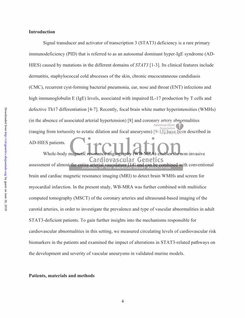
Introduction
Signal transducer and activator of transcription 3 (STAT3) deficiency is a rare primary
immunodeficiency (PID) that is referred to as an autosomal dominant hyper-IgE syndrome (AD-
HIES) caused by mutations in the different domains of STAT3 [1-3]. Its clinical features include
dermatitis, staphylococcal cold abscesses of the skin, chronic mucocutaneous candidiasis
(CMC), recurrent cyst-forming bacterial pneumonia, ear, nose and throat (ENT) infections and
high immunoglobulin E (IgE) levels, associated with impaired IL-17 production by T cells and
defective Th17 differentiation [4-7]. Recently, focal brain white matter hyperintensities (WMHs)
(in the absence of associated arterial hypertension) [8] and coronary artery abnormalities
(ranging from tortuosity to ectatic dilation and focal aneurysms) [9-13] have been described in
AD-HIES patients.
Whole-body magnetic resonance angiography (WB-MRA) enables the non-invasive
assessment of almost the entire arterial vasculature [14] and can be combined with conventional
brain and cardiac magnetic resonance imaging (MRI) to detect brain WMHs and screen for
myocardial infarction. In the present study, WB-MRA was further combined with multislice
computed tomography (MSCT) of the coronary arteries and ultrasound-based imaging of the
carotid arteries, in order to investigate the prevalence and type of vascular abnormalities in adult
STAT3-deficient patients. To gain further insights into the mechanisms responsible for
cardiovascular abnormalities in this setting, we measured circulating levels of cardiovascular risk
biomarkers in the patients and examined the impact of alterations in STAT3-related pathways on
the development and severity of vascular aneurysms in validated murine models.
Patients, materials and methods
ary artery abnonooooooonoooonooonn r
[9-13]3]3]3]3]3]3]33]3]3]3]3]3]]33]3]3]3] hh h hh hhhhh hhhhh hh havavavavavavavavavavavavavavavavavaavve eeeee e e e eee eeee bebbebebebebebebebebebebebebbebebebebbbbb e
m o
t i
maaaaagngngngngneteteteteticicicicic rrrresesesesonononono anaaaa cecececee aaa aanngnn ioioioioiogrgrgrgrgrapapapapaphyhyhyhyhy (W(W(W(W(WB-B-B-B-B-MRMRMRMRMRA)A)A)A)A) eeeeenananananablblblblblesesesss tt ttthehehehee nn nnno
tttt tt thehe ee entntntirireee arararteteteririalal vv vasasascucuculalatttururureee [1[1[1[ 4]4]4] a a andnd cc cananan b beee cococombmbinineded ww wiiittt
by guest on June 26, 2018http://circgenetics.ahajournals.org/
Dow
nloaded from

5
Patients
Twenty-one adult patients (aged over 16) registered in the national French primary
immunodeficiency (CEREDIH) database as having a heterozygous STAT3 mutation were
enrolled in a French, nationwide, prospective, multicenter study performed between August 2009
and December 2010. The study was approved by the investigational review board at Necker
Children's Hospital (Paris, France). All subjects gave their written informed consent. The use of
a computer database to store personal information was authorized by the French National Data
Protection Commission (CNIL authorization #908256).
The main features of each patient were recorded and scored using the National Institutes
of Health (NIH) scoring system: all the scores were then added together to yield an overall score.
Scores over 40 indicate that the subject is likely to carry an AD-HIES phenotype, scores of 20 to
40 points are inconclusive and scores below 20 points indicate that the subject is unlikely to have
an AD-HIES phenotype [15].
For the echo-tracking procedure, healthy control subjects (drawn from a reference
database) were matched for gender, age and mean blood pressure. When two or more control
subjects were available for a given patient, we chose the person whose arterial parameters were
closest to those of the patient.
Imaging
The imaging procedures included brain MRI, gadolinium-enhanced whole-body MRA and late
gadolinium enhancement (LGE) cardiac MRI (CMRI) in a single session, followed by iodine-
enhanced prospective cardiac MSCT using a weight-based, low-dose, sequential, prospective
protocol. Details of the protocols are given in the Supplementary Appendix [16-23]. Nineteen
ed using the NaNaNaaNaaaNaNaNaNaNNaNNNN t
ogethhhhhhhhhhhhhhhhhherereerererererererererererererererer t t tt t t t t t t t ttt tto oo oo ooo o oooo ooooo yiyiyiyiyiyiyiyiyiyiyiyiyiyyiyiyiyiyiy eleeleleleleleleleleleleleleelleleleleee dddd dd
a e
l s
y
atatatatateee ee that ttttthehehehehe suuuuubjbjbjbjbjececececect t t t isisisisis l l lllikikikikkelee y y y y y tott ccaarryryryryry a aa aan nnnn ADADADADA -HIHHHH ESESESESES p p ppphehehehehenononononotytytytytypepppp
lusisisisisiveveveveve aaaaandndndnnd ssscococococorereerees sss bebebebebelololololoww ww 2020202020 pp pppoioioioiointntntntnts ss ininininindidididdicacacacacatetetetete tttthahahahahat ttt thththththe eeee sususssubjbjbjbbjececececctttt ii iiisttt
yyyypepepe [[ [151515].].].]]
by guest on June 26, 2018http://circgenetics.ahajournals.org/
Dow
nloaded from

6
patients underwent all imaging procedures. MRI was not performed in P18 because of
claustrophobia and MSCT was not performed in P16 because of iodinated contrast agent-related
anaphylaxis. Overall vascular lesion scores were calculated by adding up the number of arteries
with major lesions. Symmetrical bed lesions were counted as two defects and multiple lesions
affecting first-, second- or third-order arteries were counted as a single defect. The overall brain
lesion severity was scored by grading the WMHs in both periventricular and subcortical areas
and the number of lacunar lesions (see the Supplementary Appendix).
Carotid artery ultrasound and hemodynamic measurements
Ultrasound scans were performed in order to determine the geometric and stiffness
parameters of the common carotid arteries (CCAs) in the 21 patients and 21 age-, gender- and
arterial blood pressure-matched healthy controls drawn from a reference database. Detailed
protocols are given in the Supplementary Appendix [24].
Blood sample assays
In addition to standard blood analysis, serum from STAT3-deficient patients and gender-
and age-matched healthy blood donors was assayed for circulating levels of a panel of
biomarkers related to inflammation, extracellular matrix remodeling and endothelial dysfunction.
Detailed procedures are given in the Supplementary Appendix.
Experimental models of aortic aneurysm and rupture
We used validated murine models of angiotensin (Ang) II–induced aortic aneurysm and
rupture [25,26]. Mice were treated with either a cell-permeable STAT3 inhibitor that targets the
he geoeeoeoeoeoeoeoeoeoeoeoeoeoeoeooooomemmemememememememememmmememememememeemetrtrtrtrtrtrtrtrtrtrtrtrtrtrtrtrtriciciciciciciciciccicicciccicicicc a a a a a a aa a a a a a aaa aaandnn
m -
r s
n
mmmmmmmmmmon carararara otototototiddddd a aa aartrtrtrtrtererererrieieieieies ss s s (C(CCCCCACACAAAs) inn ththhththe eeee 2121212121 pp ppatatatattieieieieients s s ss ananananand dddd 2121212121 a a a a agegggg -
re-mamamamamatctctctctchehehehh d ddd heheheheealalalallththththy yyyy cococococontntntnttrorororor lslslslsls d ddddrararararawnwnwnwnwn ff fffrororororom mmmm aa aaa rererereefefefefferererereencncncncncee eee dadadadadatatatataabababababas
nnn tt thehe SuSuSuppppppppp lelelememementntntararary y y y ApApAppppepepep ndndndixixix [2[2[24]4]4]].
by guest on June 26, 2018http://circgenetics.ahajournals.org/
Dow
nloaded from

7
STAT3-SH2 domain and prevents its association with upstream kinases (three intraperitoneal
(i.p.) injections per week of 25 mg/kg Stattic (Calbiochem)) or a mouse monoclonal anti-mouse
IL-17A antibody (two i.p. injections per week of 200 μg per mouse), as previously described
[27]. We also studied mice with severe reductions in STAT3-dependent signaling caused by
transgenic over-expression of SOCS3 (Tg-SOCS3) [28,29].
Statistic analysis
Values were expressed as a percentage or the mean ± SEM, as appropriate. Statistical
tests included Mann-Whitney U, chi-squared and Fisher’s exact tests. Multiple-group
comparisons were performed with a general linear model analysis of variance. Kaplan-Meier
survival curves were built and analyzed using a log-rank (Mantel-Cox) test. The threshold for
statistical significance was set to p<0.05.
Results
Patients
The patients’ characteristics are summarized in Table 1. Twelve men and 9 women with
a median age of 26 (range: 17-44) and an NIH score over 40 (mean: 70.8; range: 52-86) were
enrolled in the study. No acute, invasive infections were noted at the time of the imaging
procedures but we observed skin manifestations (n=4), chronic lower respiratory tract symptoms
(n=4) and CMC (n=4). Cardiovascular risk factors included active smoking (n=5), hypertension
controlled with dihydropyridine therapy (n=1) and overweight (n=4). Incidental deep venous
thrombosis (DVT) was reported in three patients (P7, P16 and P17) and consisted of leg phlebitis
tests. Multipleeeeee g
s of fffffff vavaaavavvavavavavavavavavaavavavavavvaaririrrrirrrrirrririririririanananananananananananananananananaanancececececceccececececececeeeceeeecce. . . ... . ...... ... K
c
bbbbbuuuiuu lt andndndndn a a a aananananaalylylylylyzezezezezed d d d d usususususinininining ggg a aaa lolog-rranknknknknk ( ( ( ((MaMaMaMaMantntntnttelelelelel-C-- oxoxoxoxox) ) ) ) ) tetetet ststststst. . . ThThThThThe
ce wwwwwasasasasas sssssetetetee ttttooo oo p<p<p<p<p<0.000 0505050505.
by guest on June 26, 2018http://circgenetics.ahajournals.org/
Dow
nloaded from

8
(n=3; 6 DVT events) and pulmonary embolism (PE) (n=2; 3 PE events). None of the study
patients had experienced or showed cardiac or neurologic vascular events, migraine or cognitive
defects.
Central nervous system abnormalities
Brain abnormalities (Figure 1, Table 2, Supplementary Table 1) were detected on MRI
in 19 of the 20 patients (95%), with WMHs in 18 patients, lacunar lesions suggestive of ischemic
infarcts in 3 patients and atrophy in 11 patients (ventricle dilation/subcortical atrophy in 9 and/or
cortical atrophy in 6). Brain vascular abnormalities were observed in 7 patients including 4 with
a cerebral artery aneurysm, 4 with arterial dilations and/or tortuosities and 2 with a vascular
agenesia. The WMHs were variously located in the frontal lobes (100% of affected cases),
parietal lobes (37.5%) and/or temporal lobes (19%). Seven patients had mild WMH lesions, 7
had moderate lesions and 4 had severe lesions. In a given patient, the WMH lesions tended to be
equally disseminated between the periventricular and subcortical areas (Figures 1A to 1D). In
two patients, asymptomatic aneurysms of the right mid-cerebral artery (in P2) and the basilar
trunk (in P16) prompted us to recommend intravascular coil embolization (Figures 1E to 1F).
Unfortunately, P16 died from aneurysm rupture one month after diagnosis and before the
endovascular procedure could be performed.
Cardiac and arterial lesions
Cardiac and coronary artery abnormalities (Table 2, Supplementary Table 2) were
detected in 10 of the 20 patients. Thirty coronary artery dilations were detected by MSCT in 8
patients, including 7 coronary artery ectasias in 4 patients, 16 coronary artery aneurysms in 5
d in 7 patients ssssss ssss in
sitieeeeeeees sss ssssssssssss ananaanannanananananaaanananananananana d dd dddddd d dddddddddd 2 222 2222222222 22 22 wiwwwiwiwiwiwiwiwiwiwiwiwwiwwiwiwwwww th
H t
% M
s o
Hsssss ww w wwere vavavavav riririririououououuslslslslsly y y y y lololoolocacacacacateteteteted d ddd innnnn ththe ffrononononontatatatatal llll lololololobebebebes s s ss (1(( 0000000000% % % %% ofofofff aaa aaffffffffffeceeee t
%) aaaaandndndndnd/o/o/o/o/orr rrr teteteempmpmpmpmpororororalllll ll lllobobobobobes (( (((1919191919%)%)%)%)%). SeSeSeeevevevevevennn nn papapapapatititititienenenenentstttt hhhhhadadadadad m mmmmililililild ddd WMWWWW
ssss aa andnd 44 4 h hhhadad ss sevevevererereee lelesisionononsss. II Innn aa a g g gggivivvenenen p p ppatatatieieeeentntnt, ,, ththeee WMWMWMHHH lelesisiio
by guest on June 26, 2018http://circgenetics.ahajournals.org/
Dow
nloaded from

9
patients and moderate coronary artery dilations in 4 patients (Figures 1G to 1L). Only one
instance of mild stenosis was detected. It is noteworthy that four focal areas of LGE were
observed at the apex of the left ventricle in 4 patients by CMRI; this is highly suggestive of small
myocardial infarcts (Figure 1M). In 3 of these cases, left main or left anterior descending
aneurysms were detected by MSCT (Figure 1L). Peripheral vascular abnormalities (Table 2,
Supplementary Table 3) were detected with a 4-step WB-MRA protocol (Figure 1N) in 17 of
the 20 patients (85%), with artery ectasia in 6 patients (one per patient), 9 artery aneurysms in 7
patients including 4 aneurysms located in the brain, 5 artery stenoses >50% in 4 patients, 9 artery
stenoses <50% in 7 patients, one dissection and one arteriovenous fistula (Figures 1O to 1R).
Minor artery abnormalities were also detected (10 tortuous arteries in 7 patients and 15 artery
lumen irregularities in 8 patients). Atherosclerosis in these patients was mild or absent.
Echo-tracking findings
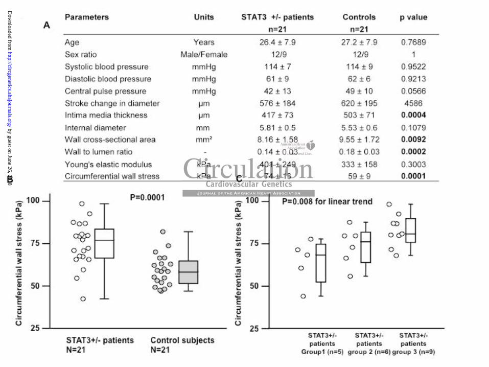
No stenoses, plaques, ectasia or aneurysms were observed in the CCA. However, the
study patients had a markedly thinner intima media thickness (IMT) and a marginally greater
internal carotid diameter, relative to healthy controls (Table 2, Figure 2A). The extent of the
reduction in IMT varied from one patient to another but averaged -86 micrometers ( m) (a 17%
reduction, p=0.004). As a consequence, the wall cross-sectional area was 15% lower in STAT3-
deficient patients than in controls (8.1 ± 1.6 versus 9.6 ± 1.7 mm², respectively; p=0.0092) and
the wall-to-lumen ratio was 21% lower (p=0.002). Since arterial blood pressure did not differ
significantly when comparing patients and controls, circumferential wall stress was significantly
(p=0.001) higher (by 26%) in patients than in control subjects (Figure 2B). Patients and controls
did not differ significantly in terms of central blood pressure parameters (pulse pressure and
us fistula (Figuuuuuuuuuuuuurerrrrrrrrrrr
es innnnnnnnnnnnnnnn 7 777777777777777777 p p ppp p p p pp p p p p p p p ppataatatatatatatatatatatataaataata ieieieieieiieieieeiieieeiieieieieentntntntntntntntntntntntntnnnnntnntnts a
i a
i
innn nn 888 88 patiienenenenentststststs)... AA A AAthththththerererererosososososclclclclcleeerorororosisiss s inin thehehehehesesesesese pppppatatata ieieieieentntntnnts wawawawawas s s ss mimimimimildldldldd ooo o or a
innngsgsgs
by guest on June 26, 2018http://circgenetics.ahajournals.org/
Dow
nloaded from

10
augmentation index), carotid stiffness indexes and aortic pulse wave velocity (Figure 2A). No
significant association between carotid IMT and markers of disease severity (i.e. the NIH score)
was found. Circumferential wall stress level was significantly and positively associated with (i)
the number of affected arterial territories identified on imaging (increase for each additional
affected artery territory: 2.5 kPa, 95% CI [0.2 to 4.8], p=0.03) and (ii) the severity of cerebral
lesions (p=0.04) (Supplementary Figure 1). The total R2 was 0.42. Circumferential wall stress
rose in a stepwise manner with both vascular and cerebral lesion severity (Figure 2C, p=0.008).
In summary, it was found that virtually all patients had vascular abnormalities associated
with WMH. These lesions were mostly arterial dilations that often led to ectasia and aneurysms
in both the coronary arteries and the periphery. The presence of abnormalities correlated with the
observation of higher circumferential wall stress and a thinner IMT, as determined by echo-
tracking.
Laboratory test data
The STAT3-deficient patients had normal standard blood parameters, with the exception
of hypereosinophilia (n=13), hyper-alpha-2 globulinemia (n=12) and elevated fibrinogen (n=4).
Serum lipid, glucose and uric acid levels were in the normal range. Patients and controls did not
significantly differ in terms of circulating levels of inflammatory cytokines. Levels of soluble
vascular cell adhesion molecule-1 and of matrix remodeling markers (matrix metalloproteinase
(MMP) -1, tissue inhibitor of MMP-1 and plasminogen activator inhibitor-1 (PAI-1)) were
higher in patients than in controls, with the exception of lower levels of MMP-3
(Supplementary Table 4). Circulating levels of PAI-1 and IL-1 were inversely correlated with
carotid IMT in female STAT3-deficient patients (r= 0.73, p=0.045).
en led to ectasiaiaiaaaaaaiaiaaaiaaaii
abnooooooooooooormrmrmrmrmrrmrmrmrmrmrmrmrmmmmrmrmmrmalalalalalalalalalalalalalalallalala itititiititititititititiitiititieieieieieiieieieeiieieeieieieieeesssssssssssssssssss c cco
r er ciciciciircummmmmfefefefeererererer ntntntntntiaiaiaiaialll wawawawawallllllllll s s s trtrtrtt eeeesssssssss a ndnd a ttttthihihihihinnnnnnnnnnerererere I II IIMTMMMM , , ,, , asasasasas d d d d deteteteetererererermimimimimine
by guest on June 26, 2018http://circgenetics.ahajournals.org/
Dow
nloaded from

11
STAT3 signaling and susceptibility to vascular aneurysm in mouse models
In vivo inhibition of STAT3 with a small molecule (Stattic) that targets the SH2 domain
led to a significant (p=0.01) increase in the severity of abdominal aortic aneurysms (AAAs) in a
validated Ang II - and TGF- -dependent C57Bl/6 mouse model [26], when compared with
treatment with vehicle (Figure 3A). This finding suggests that STAT3 signaling has a critical
role in the maintenance of vessel integrity. Accordingly, transgenic SOCS3-overexpressing mice
that exhibit reduced STAT3 phosphorylation had more severe aneurysms and more fatal vessel
ruptures than C57Bl/6 wild-type mice (p<0.0001) in the same Ang II-infusion model [26]
(Supplementary Figure 2). Since impaired Th17 differentiation is a hallmark of STAT3
deficiency [4,6,7], we also addressed the role of IL-17A in aneurysm formation. Administration
of a neutralizing anti-mouse IL-17A antibody significantly increased aneurysm severity in
response to Ang II infusion, compared with mice treated with control IgG (p=0.001). Following
IL-17A neutralization, we observed more intense vascular inflammation and greater infiltration
of CD3+ T lymphocytes; this observation further supports a vascular protective role for IL-17
signaling (Figure 3B).
Discussion
The present report describes the systematic, whole-body screening of STAT3-deficient
patients for vascular abnormalities. We also sought to identify a mechanistic relationship
between this type of abnormality and alterations in STAT3-dependent signaling. Importantly, we
found that adult STAT3-deficient patients frequently exhibit brain hyperintensities (suggestive of
nnnnnnnnnnnnnnnnnnnnggggg ggggggggggggggg IIIIIIIIIIIIIIIIIIIIIIIIIIIIIIIIIIIIIIIIII -i-i-i-i-i-i-i-i-iii-i-i-i-i-i-i-i-i-i--i-i-- nfnfnfnfnfnfnfnfnfnfnfnfnfnfnfnfnnnfnnnnfususususususususususususuususususuusssususuusioioioioioioioioioioioioioioioioiioioiioonn nnnnnnn mmmmmmmmmmmmmmmmm
gure 2) Since impaired Th17 differentiation is a hallmark of
w
i s
nfusion, compared with mice treated with control IgG (p 0.0
gureeee 22 2).).).).). SSSSininninince impaired Th17 dddddifiiii ferentiation n nnn is a hallmark of
weeeee aaaaalso addrrreesseseed thththththe roleee offf IIL--117A A A AA ininininin aanenn urrrysysysm mmmm fooooormrmmrmmaaatiooonnn. f
i-mouse IL-1-1-1-1-17A7A7A7A7A a a a aantntntntntibibibibibododododody y y yy sisisisisigngngngng ifififfficicicicicanananana tltltltltlyy yyy ininininincrcrcrcrcreaeaeaee seseseseed ddd d anaaaa eurysm s
nfnnfususioion,n, cc comompaparerredd ddd wiiwithththhth m miiiiicecece tttttrereatattededddd w witititiith hhh cocontttnttroroll lll IgIIgGGG GG (p(p=00 00.0
by guest on June 26, 2018http://circgenetics.ahajournals.org/
Dow
nloaded from
12
small vessel alterations) and have an abnormally high prevalence of medium- and large-artery
dilations throughout the vascular tree. Analysis of the mechanical properties of the CCA revealed
hypotrophic remodeling of the carotid artery wall which is consistent with defective
mechanotransduction and susceptibility to aneurysm formation.
Brain abnormalities have already been observed in 36 AD-HIES patients for whom
STAT3 mutation status data were not reported [1,8]. The WMHs varied greatly in severity and
ranged from isolated, punctuate spots to multifocal, confluent lesions with a periventricular
distribution in many cases, as seen in other WM diseases and notably leukoaraiosis [30].
Leukoaraiosis is observed in elderly subjects and is considered to be a small-vessel disease
resulting from a widespread, chronic brain hypoperfusion, with disruption of the blood-brain
barrier. The fact that small lacunae were also observed further evokes a primary vascular origin.
Thus, the hypothesis of an inflammatory or immune disease of the CNS appears to be less
probable. Moreover, it is striking to note the absence of vascular risk factors in the present cohort
of STAT3-deficient patients (with the exception of smoking in 25% of the group) [21,30].
We also identified vascular ectasia, aneurysms, irregularities and stenoses in brain,
peripheral and coronary arteries. Coronary artery abnormalities (notably aneurysms) have been
previously reported in AD-HIES patients, including a recently studied group of 26 STAT3-
deficient subjects [9,10,12,13]. Cerebral vascular events (stroke, retinal occlusion and internal
carotid aneurysm) were first reported in 3 AD-HIES patients [1] and 5 other cases with lacunar
infarction were subsequently identified [8]. In the present cohort (in which all the patients had
genetically confirmed STAT3 deficiency), it is noteworthy that all anatomical areas appear to be
potentially affected, with a trend towards more damage in small- and medium-diameter vessels.
These findings prompt us to conclude that STAT3-deficient patients are prone to developing a
o be a small veseeseseseeeeeeeeee
disruuuuuuuuuuuuptptptptptptptptptptptptptptptttptptptptpp ioioioiooioiioioioiioioioioioioioioion n n nnnnnn n nnn nnnn nn ofofofofofofofofoffoofoffofofofoffofoff tttttttttttttttttttthhhhhhhehhhhhhhhhhhh
h
sssssmmmamm ll lacacacaca unununununaeaeaeaeae www wwererererere e ee e alalalalalsososooo obbbbbsess rvvededdd f f fffurururururthththhherererere e ee eevovovovovokeeeees ssss a a aa a prprprprprimimimimimararararary
offf ff ananananan iiiiinfnnfnfnflalalaammmmmmmmmmatatatata orrrrryyy yy oorooo iiiiimmmmmmumumumumunenenenene d ddisisisisiseaeaeaeaeasesesesese ooooof ffff ththththheee ee CNCNCNCNCNS SSSS apapappppepepeppeararararars ss
itit i isss stststririiikikingngnggg tt tooo nn notototeee ththeee ababsesesencncnceee ofofoff v v vasasascucuculalall rrr ririsksk f facacactototorsrsrs i innn thtt
by guest on June 26, 2018http://circgenetics.ahajournals.org/
Dow
nloaded from

13
systemic, vascular disease - regardless of the presence or absence of vascular risk factors and,
apparently, infectious complications. However, a potential infectious etiology should always be
screened for, since three cases of mycotic aneurysm have been reported [8,31,32].
The results of CCA hemodynamic measurements revealed an abnormally low IMT and a
marked greater circumferential wall stress in STAT3-deficient patients; these findings suggest a
defect in tensile stress mechanotransduction and a subsequent propensity for arterial dilation and
tortuosity in many arterial sites. These data are reminiscent of the inherited Ehlers-Danlos
vascular-type syndrome [23,33] in which hyper-extensibility with scoliosis and other
musculoskeletal disorders are major features - a strikingly similar situation to STAT3-deficiency.
The vascular abnormalities observed here in STAT3-deficient patients may therefore result from
a systemic connective tissue disorder that includes arterial fragility. The correlation between
greater circumferential wall stress and (i) the extent of peripheral arterial lesions and (ii) the
severity of brain lesions suggests a causal link in the occurrence of vascular lesions such as
ectasia and aneurysm. Lastly, this situation contrasts with the greater IMT observed in vascular
diseases associated with infections or atheroma [34-37].
The animal model studies reported here strongly suggest that alterations in STAT3-
dependent signaling are directly involved in the vascular phenotype of STAT3-deficient patients.
Although impaired IL-17 production by T cells is the most important consequence of defective
STAT3-dependent transcription in patients, IL-17's specific role remains to be clearly defined.
The murine data evidenced the aggravation of aneurysm formation following neutralization of
IL-17A and thus suggest that Th17 deficiency has an important role in mediating the vascular
phenotype. However, other IL-17-unrelated STAT3-dependent pathways may also be involved.
Further studies based on a mouse model with dominant-negative mutations of STAT3 should be
r situation to STSSTSTSTSSSSSSSSSS
atientsttstststststststststststststssss mmmmmm mmmm mm m m mm mmmayayayayayayayayayayayayayayayayayayayyyyyyy t tt t tt t tt t ttttttthehheheheheheeheehehehehehehehehehehehhh re
v i
i
o o
ve ee ee tititititissue ddddisisisisisororororrdededededer r r rr thththhthatatatatat i i iiinnnnclclclcc udududddesee aarrterererereriaiaiaiaiallll l frfrfrfrfragaagaggililillilititititity. TTTTThehehehehe c c cc corororororrererererelallll ti
ial llll wawawawawallllllllll sssstrtrtrresesesesss ssss anananannd (i(i(i(i(i) )))) thththththe eeee exexexxextetetetetentntntntnt o oooof ffff pepepepeperiririririphphphphpherererereralalalaa aa aartrtrtrtrterererereriaiaiaiaialll ll lelelellesisisisiononononons
onoononsss sususugggggggggesesestttsss aaa cacacausususalal l lininnnkk inin tt thheee ocococcucucurrrrrrenenencecece oo off vavavascscscululararar l lesesesioioo
by guest on June 26, 2018http://circgenetics.ahajournals.org/
Dow
nloaded from

14
able to confirm or negate this hypothesis and help identify the specific cell-type(s) responsible
for the vascular phenotype.
The frequency and severity of vascular abnormalities associated with STAT3 deficiency
observed here have clinical implications and likely justify the development of screening,
surveillance and therapeutic strategies. Two cohort members were found to require endovascular
treatment for a brain aneurysm, unfortunately one died before the procedure. Moreover, STAT3
deficiency appears as an emerging, hereditary cause of brain small-vessel disease whose long-
term repercussions remain to be determined. In the literature, WM lesions have been associated
with subtle neurological deficits (memory decline, cognitive disability, depressive symptoms)
that may initially go unnoticed by both patients and physicians [21,38,39]. Systematic screening
for memory and cognitive dysfunctions could thus be offered with a view to define their
prognostic significance. Despite the absence of any evidence for effective, preventive strategies,
there may be a case for primary prophylaxis of WMH progression, ischemic strokes, vascular
leukoencephalopathy and aneurysm rupture by evicting vascular risk factors and initiating
antithrombotic and anti-hypertensive treatments, if needed. On the basis of the murine data, the
use of an angiotensin II receptor antagonist could legitimately be investigated. This type of
preventive measure will have to be carefully evaluated, given the risk of internal hemorrhage
associated with the use of antithrombotic agents [40].
In conclusion, adult patients with a STAT3 deficiency were found to have a high
prevalence of vascular abnormalities throughout the arterial tree. These abnormalities were
characterized by hypotrophic remodeling of the artery wall, increased circumferential stress and
enhanced susceptibility to dilation and aneurysm formation. Some of these aneurysms affect
heart and brain vessels and might incur a high vascular risk. Animal model studies strongly
ability, depressisisiisiiiisisisisisss v
21,3888888888888888888,3,333,3,3,3,3,3,3,33,3,33,3,3,3,3,33,, 9]9]9]9]9]9]9]9]9]9]9]9]9]9]9]9]9]9]9]]]. .. . ... .. ..... . SySySySySySySySySySySySySySySySySySyyySyySyystststsssssstsssssssss e
n i
n e
f o
nininiiititiiiive dysysysysy fufufufufuncncnccctititititiononononons ss s s cococococoulululululd dd thththhhusuu bbe ofofofofoffefefefeferererereredd ddd wiwiwiwiwithththtt aaaaa v v vv vieieieieiew ww w tototototo dd d d defeeee i
nce.eeee D DDDDesesesesespipipipp tetetet t tttheheheheh aaaabsbsbsbsbsenennenenccecc ooooof ffff ananananany yyyy evevevevevidididididenenenenencecececece ff fffororororor ee eeefffffffffececececectitititiiveveveveve, prprprppreveveveveve
fofforrr prprprimimmmararary y y y prprprpp opopophyhyyyylalaxixisss ofofo WMWMWMHHH prprprogogoggrereressssssioion,n,n,, i iscscschehemimiccc ss strtrtrooo
by guest on June 26, 2018http://circgenetics.ahajournals.org/
Dow
nloaded from
15
suggest a direct role for STAT3-dependent signaling in the vascular phenotype and should
facilitate the identification of specific molecular targets for disease modulation.
Acknowledgments: We thank the patients and their families for their trust and cooperation. We also thank members of INSERM U768, GHMI-INSERM U980 and the Immunohematology Unit at Necker Hospital for helpful discussions. We thank Dr Fanny Lanternier, MD, Dr Caroline Charlier, MD, PhD, and Dr Blandine Denis, MD, from the department of infectious diseases and Tropical Medicine at Necker Hospital. We thank Dr Isabelle Melki, MD, for clinical help with describing the patients. We thank Corinne Jacques, AS, Chantal Harre, AS, Stéphanie N’daga, AS, and Aminata Diabate, AS, for excellent technical assistance. We thank Dr Nizar Mahlaoui, MD, MSc, for maintenance of the CEREDIH database.
Funding Sources: INSERM unit U768 received INSERM core funding and an advanced ERC grant. The Laboratory of Human Genetics of Infectious Diseases received funding from the March of Dimes, the Dana Foundation and INSERM. Ziad Mallat received a fellowship from the French National Research Agency (Agence Nationale de la Recherche).
Conflict of Interest Disclosures: None
References:
1. Grimbacher B, Holland SM, Gallin JI, Greenberg F, Hill SC, Malech HL, et al. Hyper-IgE syndrome with recurrent infections--an autosomal dominant multisystem disorder. N Engl J Med. 1999;340:692-702. 2. Minegishi Y, Saito M, Tsuchiya S, Tsuge I, Takada H, Hara T, et al. Dominant-negative mutations in the DNA-binding domain of STAT3 cause hyper-IgE syndrome. Nature. 2007;448:1058-1062. 3. Holland SM, DeLeo FR, Elloumi HZ, Hsu AP, Uzel G, Brodsky N, et al. STAT3 mutations in the hyper-IgE syndrome. N Engl J Med. 2007;357:1608-1619. 4. Ma CS, Chew GY, Simpson N, Priyadarshi A, Wong M, Grimbacher B, et al. Deficiency of Th17 cells in hyper IgE syndrome due to mutations in STAT3. J Exp Med. 2008;205:1551-1557. 5. Minegishi Y, Saito M, Nagasawa M, Takada H, Hara T, Tsuchiya S, et al. Molecular explanation for the contradiction between systemic Th17 defect and localized bacterial infection in hyper-IgE syndrome. J Exp Med. 2009;206:1291-1301.
ases receivedddd f aaaaaaaaaaaaaaaaattttt ttttttttttttttt rerererrrererererererererrrrreeerererececececececeecececeecececceecccececececceeivvivivivviviviviiiviiviviviviviiivvvivvedededededededededededededededededdedededdedde aaaa aaa aaaa a aaaa fffffffffffffffffffffelelelelelelelelelellelelelelelelelelllerchehheheheheheheheheheheheheheheeee).).))).).).).).).).).).).).).).).).))
DDDDDisisisii closururururureseseese : :: NoNoNoNoNonenenenee
by guest on June 26, 2018http://circgenetics.ahajournals.org/
Dow
nloaded from

16
6. Milner JD, Brenchley JM, Laurence A, Freeman AF, Hill BJ, Elias KM, et al. Impaired T(H)17 cell differentiation in subjects with autosomal dominant hyper-IgE syndrome. Nature. 2008;452:773-776. 7. de Beaucoudrey L, Puel A, Filipe-Santos O, Cobat A, Ghandil P, Chrabieh M, et al. Mutations in STAT3 and IL12RB1 impair the development of human IL-17-producing T cells. J Exp Med. 2008;205:1543-1550. 8. Freeman AF, Collura-Burke CJ, Patronas NJ, Ilcus LS, Darnell D, Davis J, et al. Brain abnormalities in patients with hyperimmunoglobulin E syndrome. Pediatrics. 2007;119: e1121-1125. 9. Ling JC, Freeman AF, Gharib AM, Arai AE, Lederman RJ, Rosing DR, et al. Coronary artery aneurysms in patients with hyper IgE recurrent infection syndrome. Clin Immunol. 2007;122:255-258.
10. Young TY, Jerome D, Gupta S. Hyperimmunoglobulinemia E syndrome associated with coronary artery aneurysms: deficiency of central memory CD4+ T cells and expansion of effector memory CD4+ T cells. Ann Allergy Asthma Immunol. 2007;98:389-392.
11. Alomar-Melero E, Martin TD, Janelle GM, Peng YG. An unusual giant right coronary artery aneurysm resembles an intracardiac mass. Anesth Analg. 2008;107:1161-1162.
12. Gharib AM, Pettigrew RI, Elagha A, Hsu A, Welch P, Holland SM, et al. Coronary abnormalities in hyper-IgE recurrent infection syndrome: depiction at coronary MDCT angiography. Am J Roentgenol. 2009;193:W478-481. 13. Freeman AF, Avila EM, Shaw PA, Davis J, Hsu AP, Welch P, et al. Coronary Artery Abnormalities in Hyper-IgE Syndrome. J Clin Immunol. 2011;31:338-345.
14. Lehrke S, Egenlauf B, Steen H, Lossnitzer D, Korosoglou G, Merten C, et al. Prediction of coronary artery disease by a systemic atherosclerosis score index derived from whole-body MR angiography. J Cardiovasc Magn Reson. 2009;11:36. 15. Grimbacher B, Schaffer A, Holland SM, Davis J, Gallin JI, Malech HL, et al. Genetic linkage of hyper-IgE syndrome to chromosome 4. Am J Hum Genet. 1999;65:735-744. 16. Fazekas F, Chawluk JB, Alavi A, Hurtig HI, Zimmerman RA. MR signal abnormalities at 1.5 T in Alzheimer's dementia and normal aging. Am J Roentgenol. 1987;149:351-356. 17. Austen WG, Edwards JE, Frye RL, Gensini GG, Gott VL, Griffith LS, et al. A reporting system on patients evaluated for coronary artery disease. Report of the Ad Hoc Committee for Grading of Coronary Artery Disease, Council on Cardiovascular Surgery, American Heart Association. Circulation. 1975;51:5-40. 18. Díaz-Zamudio M, Bacilio-Pérez U, Herrera-Zarza MC, Meave-González A, Alexanderson-
E syndrome assssssssssssssosssssssssTTTTTTTTTTTTTTTTTTTTTTT cc ccccccccccccccccccccelelelelelelelelelelelelelelelellelelelllle lslslslsllslslslslslslslslslslslslslslslslsl aa a aaaaa a aaaaaaaaaaandndndndndndndndndndndndndndndndndndndnddndndn e e e ee ee eeeexpxxpxpxpxpxxxpxxxpxxxpxxpxxpxx aaaaaaaaaaaaaaaaa007;9999999999;9;99999999998:8:8:8:88:8:88:8:88:8:8:8:88::88:38383838388383838383838383833838338888389-99-9-9-9-9-9-99-9-9-9-9-99-9999 39399399993939939399393999393939393933992
E ar t
i oe M
Roentgenol 2009;193:W478 481
E,E,E,E,E, M MM M Martin nnnn TDTDTDTDT , , , , , JaJaJaJaJanenenenenelllllllllle e ee e GMGMGMGMGM, PePPP ngng YYYYYG.G.G.G.G. AAAA An nn n n ununununnusuuuu uauaauaual llll gigigigigiannannant t t t t riririririghgggg taaaaannn n intracarrrddiacacc mmmaasa s. AnAnAneeesthtth AAnnalglglglglg. 2022 08088;10007:1:1:1:11161-1-1-11-111116222...
igrew RI, EEEEElalalalalaghghghghgha a a a a A,A,A,A,A, H H H H Hsususususu A,A,A,A,A, W WWWelelelelelchchchchch PPPPP,,, HoHoHoHoHollllllllllananananand dd d SMSMSMSMSM, et al. Coererer II-IgEgEgE r r recececurururrererentntnt i infnfececectitiononon sysysyyyndndrororomememe::: dedepipipictctctioionnn atatat cc corororonononarararyyy MMMM
Rooenentgtgenenolloll 202020200909090 ;1;11939393 WW:WW:W474747478-888 484848481111
by guest on June 26, 2018http://circgenetics.ahajournals.org/
Dow
nloaded from

17
Rosas E, Zambrana-Balta GF, et al. Coronary artery aneurysms and ectasia: role of coronary CT angiography. Radiographics. 2009;29:1939-1954. 19. Satran A, Bart BA, Henry CR, Murad MB, Talukdar S, Satran D, et al. Increased prevalence of coronary artery aneurysms among cocaine users. Circulation. 2005;111:2424-2429. 20. Pahlavan PS, Niroomand F. Coronary artery aneurysm: a review. Clin Cardiol. 2006;29:439-443.
21. de Groot JC, de Leeuw FE, Oudkerk M, van Gijn J, Hofman A, Jolles J, et al. Cerebral white matter lesions and cognitive function: the Rotterdam Scan Study. Ann Neurol. 2000;47:145-151. 22. Kim KW, MacFall JR, Payne ME. Classification of white matter lesions on magnetic resonance imaging in elderly persons. Biol Psychiatry. 2008;64:273-280.
23. Yan AT, Shayne AJ, Brown KA, Gupta SN, Chan CW, Luu TM, et al. Characterization of the peri-infarct zone by contrast-enhanced cardiac magnetic resonance imaging is a powerful predictor of post-myocardial infarction mortality. Circulation. 2006;114:32-39. 24. Ong KT, Perdu J, De Backer J, Bozec E, Collignon P, Emmerich J, et al. Effect of celiprolol on prevention of cardiovascular events in vascular Ehlers-Danlos syndrome: a prospective randomised, open, blinded-endpoints trial. Lancet. 2010;376:1476-1484. 25. Daugherty A, Manning MW, Cassis LA. Angiotensin II promotes atherosclerotic lesions and aneurysms in apolipoprotein E-deficient mice. J Clin Invest. 2000;105:1605-1612. 26. Wang Y, Ait-Oufella H, Herbin O, Bonnin P, Ramkhelawon B, Taleb S, et al. TGF-beta activity protects against inflammatory aortic aneurysm progression and complications in angiotensin II-infused mice. J Clin Invest. 2010;120:422-432. 27. Taleb S, Romain M, Ramkhelawon B, Uyttenhove C, Pasterkamp G, Herbin O, et al. Loss of SOCS3 expression in T cells reveals a regulatory role for interleukin-17 in atherosclerosis. J Exp Med. 2009;206:2067-2077. 28. Seki Y, Inoue H, Nagata N, Hayashi K, Fukuyama S, Matsumoto K, et al. SOCS-3 regulates onset and maintenance of T(H)2-mediated allergic responses. Nat Med. 2003;9:1047-1054. 29. Tanaka K, Ichiyama K, Hashimoto M, Yoshida H, Takimoto T, Takaesu G, et al. Loss of suppressor of cytokine signaling 1 in helper T cells leads to defective Th17 differentiation by enhancing antagonistic effects of IFN-gamma on STAT3 and Smads. J Immunol. 2008;180:3746-3756. 30. Van der Knaap MS, Valk J. Magnetic Resonance of Myelination and Myelin Disorders. Third Edition. Chapter 98. White Matter Lesions of the Elderly page 759-766. ISBN-10 3-540-22286-3 Springer Berlin Heidelberg New York.
nance imaging g g g g g gg g g gg gg gg gg i000000000000000000060606060606060600606060606060606060606006600 ;1;1;1;1;1;1;1;1;1;1;1;1;1;1;1;1;111;111414141414141414141414141411414141414141411 :3:3:3:3:33:3:3:3:333:33:3:3333333:32-2-2-2-2-222-22-2-222-2222-22222 393939399393933939393939393939393939399339.
J, De Backer J, Bozec E, Collignon P, Emmerich J, et al. Effd rl
a ro
J, DDDDe e e e e BaBaBaBaBackckcckc ererererer J, Bozec E, Colligngngngngnon P, Emmeeeeerirrrr ch J, et al. Effdiiiiiooovoo ascuuuuulalalaaarr rrr evvvvvenenenenentstttt iiiiin nnnn vavavavavascsccsculululuularaa EEhlererererers-s-s-s-s-DaDaDaDaDanlnlnlnlososososos s ynynynynyndrdrdrdrdromomomomome:e:e:e:e: a a a a a p rlinininininddeddd d-endpppointntts trtrtrial. Laanccceet. 202 1010000;3;3;3;3;376776:147776-6-6-14844444.
anning MW,W,W,W,W, C C CCCasasasasassisisisisis ss LALALALALA... . . AnAnAnAnAngigigigigiotottottenenenenensissss n nnnn IIIIIIIIII p p ppprororororomomomommotetetetetes s s ss atherosclerooooprprprotototeiein n n EEE-dedefificicienenenttt mimicecece. JJJ ClClininin II Invnvnvesesesttt. 202020000000;1;1;1; 050505:1:1:1606060555-1616161111
by guest on June 26, 2018http://circgenetics.ahajournals.org/
Dow
nloaded from

18
31. Connolly B, Manson D, Khattak S, Burrows P. Bronchial artery aneurysm in hyperimmunoglobulinemia E syndrome. Pediatr Radiol. 1994;24:592-593. 32. El Noor IB, Venugopalan P, Johnston WJ, Froude JR. Ventricular aneurysm and myocarditis in a child with the hyperimmunoglobulin E syndrome. Eur Heart J. 1995;16:714-715. 33. Boutouyrie P, Germain DP, Fiessinger JN, Laloux B, Perdu J, Laurent S. Increased carotid wall stress in vascular Ehlers-Danlos syndrome. Circulation. 2004;109:1530-1535. 34. Cao JJ, Arnold AM, Manolio TA, Polak JF, Psaty BM, Hirsch CH, et al. Association of carotid artery intima-media thickness, plaques, and C-reactive protein with future cardiovascular disease and all-cause mortality: the Cardiovascular Health Study. Circulation. 2007;116:32-38. 35. Kumeda Y, Inaba M, Goto H, Nagata M, Henmi Y, Furumitsu Y, et al. Increased thickness of the arterial intima-media detected by ultrasonography in patients with rheumatoid arthritis. Arthritis Rheum. 2002;46:1489-1497. 36. Seth S, Goyal NK, Jagia P, Gulati G, Karthikeyan G, Sharma S, et al. Carotid intima-medial thickness as a marker of disease activity in Takayasu's arteritis. Int J Cardiol. 2006;108:385-390. 37. Cheung YF, Wong SJ, Ho MH. Relationship between carotid intima-media thickness and arterial stiffness in children after Kawasaki disease. Arch Dis Child. 2007;92:43-47. 38. Vermeer SE, Longstreth WT Jr, Koudstaal PJ. Silent brain infarcts: a systematic review. Lancet Neurol. 2007;6:611-619. 39. Debette S, Markus HS. The clinical importance of white matter hyperintensities on brain magnetic resonance imaging: systematic review and meta-analysis. BMJ. 2010;341:c3666. 40. Freeman AF, Kleiner DE, Nadiminti H, Davis J, Quezado M, Anderson V, et al. Causes of death in hyper-IgE syndrome. J Allergy Clin Immunol. 2007;119:1234-1240.
a aaaaaaaaaaaaa S,SSSSSSSSSSSSSSSSSSSSS e e ee e e eee eeeeeeeeeeet t ttttt tttttttttt tttttttt alalalalalalalaalalalalaalalalala . ... CaCaCaCaCaCaCaCaCaCaCaCaCaCaCaCaCaCaCCCaCCaCaroroororororoorororoorororororooottititttttt ddddddddddddddddddddnt J CaCaCaCaCaCCaCaCaCaCaCaCaCaCaaCaCCaCaaCaardrdrdrdrdrdrdrdrddrdrdrdrdrddrdrdddiooioioioioioioioioiooioioioioiiolllllllllllllllll. . . .. . .. .... ... 222220222222222222222
n th -
n m
nggg gg SSSJSS , HoHoHoHoHo M M MMMH.H.H.H.H. R RR RRelelelelelatatataatioioioioionsnsnsnn hihihip p p pp beeetwweeeeeeeeeen n nn n caaaaarorororor titititiddd dd intititititimamamamama-mmmmmededededediaiaiaiaia thhhhhilililililddrd en aftereer KKawawawasaa akki dididiseasaa e.. ArArrrrchchchchch D DDiss Chihihilddddd. 200707070707;9;992:4443-
ngstreth WT TTT T JrJrJrJrJr, ,, KoKoKoKoKoudududududstststststaaaaaaaaaal l l l l PJPJPJPJP .. SiSiSiSiilelelelelentntntntnt b b b b brararararaininininn iiiiinfnfnfnfnfararararrctctctctctsss:ss a system;6;6;6:6:6:6111111-6-6-6191919.
by guest on June 26, 2018http://circgenetics.ahajournals.org/
Dow
nloaded from

19
Table 1: Clinical characteristics and genotype data for the 21 STAT3-deficient patients.
Abbreviations: Age: age at the time of the study; DNA: DNA-binding domain; ENT: ear, nose and throat infections; F: female; I teeth: primary teeth; M: male; NHL: non-Hodgkin’s lymphoma; P: patient; SH2: Src Homology 2; TA: transactivator; yrs: years; +: presence. Normal serum IgE level is less than 150 kUI/l.
Patient Gender Country of origin Age (yrs) STAT3 mutation
domain NIH score
IgE level (KUI/l)
Previous infections
Skin signs Developmental signs
NHL
skin
lung
deep
abs
cess
es
ENT
neon
atal
rash
derm
atiti
s
dysm
orph
ia
reta
ined
I te
eth
bone
frac
ture
oste
open
ia
hype
rext
ensi
bilit
P1 M France 37 V637M/wt SH2 74 17,046 + + + + + + + + + P2 M France 38 K642E/wt SH2 66 4,599 + + + + + + + + + + P3 F France 26 V637M/wt SH2 73 521 + + + + + + + + + + P4 F French West Indies 20 S668Y/wt SH2 74 16,112 + + + + + + + + + P5 M French West Indies 25 R382W/wt DNA 84 22,000 + + + + + + + + + + P6 M France 23 R382W/wt DNA 68 7,892 + + + + + + + + P7 F Pakistan 38 Vdel463/wt DNA 79 27,920 + + + + + + + + + P8 M France 30 Vdel463/wt DNA 64 4,703 + + + + + + + + P9 F France 45 I665N/wt SH2 65 19,972 + + + + + + +
P10 F France 22 R382W/wt DNA 86 8,463 + + + + + + + + + P11 M France 19 N472D/wt DNA 55 3,990 + + + + + + P12 F Morocco 19 T714I/wt TA 78 18,762 + + + + + + + + P13 F France 24 R382W/wt DNA 73 4,525 + + + + + + + + P14 M Algeria 18 R382W/wt DNA 74 14,055 + + + + + + + + + P15 M France 29 R382Q/wt DNA 67 10,174 + + + + + P16 M France 39 F384L/wt DNA 72 803 + + + + + + + + + + P17 F France 28 R382W/wt DNA 73 15,494 + + + + + + P18 F France 17 T620A/wt SH2 52 4,515 + + + + + + P19 M France 19 R382W/wt DNA 37 26,677 + + + + + + + P20 M France 19 K709E/wt TA 77 1,209 + + + + + + + P21 M France 30 I568F/wt Linker 41 11,228 + + + + + + +
444444444444444,5,5,5,5,5,5,5,5,5555,5,5,55,555, 99999999999999999999999999999999 ++ + ++++ +++++++++++5212121212121212121211111212121212121222 +++++++++++++++++++
S668Y/wt SH2 74 16,112 +SSSS66666 8Y8Y8YY8Y/w/// t SH2 74 16,112 +R 883882W2W2W2W2W/w/w/wwttt tt DNDNDD A A AA 8484848484 2222,,,,,00000000000 0000 +++++R382WWW/w/w/ t DNDND A AA 6868666 7 7777 88,88 2292 + + VVVVVdededededel4l4l4l4l 6363636363/w/w/w/w/ t tttt DNDNDNDNDNAA AAA 7979797979 222227,7,92929292920 0000 +++++Vdel4633333/w/w/w/w/wttt tt DNDNDNDNDNA AAAA 6464646464 4 4444,7,7,7,7,7030000 +I6666665N5N5N5N5N/w/w/w/w/wt tttt SHSHSHSHSH2 22 22 6565656565 111119,9,9,9,9 97979797972 2222 +
by guest on June 26, 2018http://circgenetics.ahajournals.org/
Dow
nloaded from

20
Table 2: Imaging findings in a population of 21 STAT3-deficient patients investigated with cardiac MSCT, WB-MRA, brain MRI and late gadolinium-enhanced (LGE) CMRI.
PtsBrain findings Extracoronary vascular findings Cardiac and coronary artery findings
WMH score
lacunar lesion
cerebral atrophy
aneurysm ectasia stenosis other findings
CA aneurysm
CA ectasia
LGE other findings
P1 Moderate - + - + - - - + - + P2 Moderate - - + - + + + + + - P3 Mild - - - - + + - - - - P4 - - - - - - - - - - - P5 Moderate - + + + + + - + - - P6 Mild - + - - - - + - - + P7 Mild - + + - + + - - - - P8 Mild - - + + - + + - + - P9 Severe - - - - + + - - - -
P10 Mild - - - + + + - - - + P11 Severe + - - - - + - - - - P12 - - - - - - - - - + - P13 Severe - + - + - + + - - + P14 Moderate + + - - + + - - - - P15 Moderate - - + - + + + - + - P16 Mild - + + + + + No MSCT data - - P17 Severe - - + - + + - - - - P18 No MRI data - - - P19 Moderate + - - - - - - - - - P20 Mild - + - - + + - - - - P21 Moderate - - - - + - - + - + Abbreviations: CA: coronary artery; CMR: cardiac magnetic resonance imaging; LGE: late gadolinium enhancement; MRI: magnetic resonance imaging; MSCT: multislice computed tomography; P: patient; WB-MRA: whole body magnetic resonance angiography; -: absence; +: presence. Notes: - Other findings refer to vascular lumen irregularities, tortuosities, dissections or arteriovenous fistulae. - P16 did not undergo MSCT because of iodinated contrast agent-related anaphylaxis and P18 did not undergo MRI because of claustrophobia.
+ -- +++++++++++++++
+ +--
- -- - - - -
+ + +
+ + + ++ + - + ++++ +- -- - -- + + + ++ ------ ++ ++ + + + + ++ + + +++ - - ---- - - - + + + ++ - - - - - ---
++ ++ +++
by guest on June 26, 2018http://circgenetics.ahajournals.org/
Dow
nloaded from

21
Figure Legends:
Figure 1: Brain, cardiac and vascular imaging findings. Panels A to D are axial FLAIR brain
MR images. Panels A and B demonstrate the widespread presence of severe WM lesions in
P11, including parietal confluent WMHs (arrow). Note also the left frontal periventricular
lacunar lesion (arrowhead). Panel C shows a hyperintense periventricular lining (arrow),
associated with mild cortical atrophy in P6. Panel D demonstrates a moderate form of WM
lesion, including numerous subcortical, frontal, punctuate lesions in P2 (arrow). Panel E is a
(3D-TOF) MRA image of the cerebral arteries in P2,
showing a 7 mm right mid-cerebral aneurysm (arrow). Panel F is a 3D reconstruction
from WB-MRA data showing the head and neck vessels of P16 and demonstrating a 17x19
mm saccular aneurysm (asterisk) of the basilar trunk (more clearly visible in enlarged oblique
sagittal view in the red outlined box). Panels G to L are cardiac MSCT images. Panel G
shows a 3D reconstruction of the coronary arteries of P21, demonstrating distal right coronary
artery (RCA) ectasia. Moderate dilation of the first marginal artery is seen in the same patient
(panel H, a curved multiplanar image). Multiple coronary artery aneurysms and ectasia were
found in P2 (Panel I, 3D reconstruction; Panels J-K, curved multiplanar reconstructions),
including ectasia of the RCA (arrow), an aneurysm of the acute marginal branch of the RCA
(arrowhead) and ectasia of the mid left anterior descending (LAD) artery (black arrow). A
view of the whole LAD lumen (panel L) shows 5 different dilated lesions located in the
proximal, mid and distal LAD artery, whereas apical LGE is suggestive of focal apical
necrosis in P2 (Panel M, LGE-CMR, three-chamber long-axis view). Panel N shows a 3D
reconstruction (oblique sagittal view) from four-step WB-MRA and depicting the entire
arterial tree in P21. Panel O is an enlarged frontal view of the neck vessels of P21, showing
common carotid artery stenosis (arrowhead). Panel P is an enlarged sagittal view of the
sssssssssssssssss aa a aa a a a a a aa a a aa aa aa 333 333 333 3 3 33 33 3333D DDDD D DDD D DDDDDDDDDDDD reerererererereereeerereererrrrrrr cococococococococoococococococoocooconssnsnsnsnsnsnsnsnnsnsnsnsnnsnn trtrtrtrtrtrrrtrtrrtrtrtrrtrrtrtrrucucucucuucucucucucuuucuucuuucuucc
and dededededdededededededededeededededededdemomommomomomomomommomomomomomomomomomoom nsnsnsnsnsnsnsnsnsnsnnnsnsnnsnsnsnsnnsnnnn trtrtrtrtrrtrtrtrtrtrtrtrtrttrttrrrtrratatataaaatatatataataatataatatata ing
u a
h P
n
g
uryryryryrysm (asteteteeeririiskkkkk)))) ) ofofofofof ttttthehehehehe b b b b basasasaa ilii ararararr t truunnk (((((momomomomorerererere c ccleleleeaaaaarllly vivivivivisisisisiiblblblblble eeee ininininn ee eeenlnnnn a
he rededededed oo ooouutu liiinenened dddd bobobobox)x))x)). PaPaPaPaPanelslslslsl GGGGG tt ttto oo LL LLL ararararreeee e cacacacacardrdrdrdrdiaiaiaiaiac ccc MMMSMM CTCTCTCTCT iiiiimamamagegegegesss. PP P
nnnnstststruruructctctioioiooon nn n ofofof tt thehehe cococorororonananaryryryyy a a artrtrterererieieies ss ofofof P P PP212121, ,, dededemmmonononstststrararatititingngnggg d d disisistatatalll rrrr
by guest on June 26, 2018http://circgenetics.ahajournals.org/
Dow
nloaded from

22
abdominal region, showing lumen irregularities in the superior mesenteric artery in the same
patient. Panel Q shows an early venous return in the right leg in P15 (arrows) that is highly
suggestive of arteriovenous fistula (AVF). The green outlined box shows an enlarged axial
image of the right calf of P15, demonstrating a connection (via an AVF) between the posterior
tibial artery and the great saphenous vein. Panel R shows dilation of the sinus of Valsalva
(measuring 45 mm) in P5 (MSCT reconstruction).
Figure 2: Echo-tracking findings. A: common carotid artery parameters in 21 STAT3-
deficient patients and age-, gender- and blood-pressure-matched healthy controls. Panel B:
circumferential wall stress values (calculated with the Lamé equation (mean blood pressure
(MBP) * internal diameter/2 * intima media thickness)) in STAT3-deficient patients and
healthy controls. C: values of circumferential wall stress according to the severity of arterial
lesions detected in MRA and MSCT scans and the severity of central nervous system lesions
(white matter hyperintensities and silent brain infarctions). The scoring system was as
follows: group 1 STAT3-deficient patients are those with moderate brain and vascular
abnormalities; group 2 STAT3-deficient patients are those with moderate brain abnormalities
and severe vascular abnormalities; group 3 STAT3-deficient patients are those with severe
brain and vascular abnormalities. Median scores were used as cut-off values. It is noteworthy
that none of the patients was in the upper category for central nervous system lesions and the
lower category for vascular lesions.
Figure 3: STAT3 inhibition and IL-17 neutralization each increase the occurrence and
severity of aneurysms in mice treated with angiotensin II. A: the left-hand panel depicts
increasing levels of aortic aneurysm severity (from 0, no aneurysms, to IV, aneurysm rupture;
see the Methods section) in C57Bl/6 mice treated with either a specific STAT3 inhibitor
aaaaaaaaaaaaaaaaaaaaatittitititititititititiititititit onononononononononononnonnonononnonnnn ( ( ( (( ( (( ( ( ( ( ( ( ((mememememememememmememememememeemmmmmmmmeanananananananananannnnananannanana bbbbb bbbbbbbbbbbbloololololololoololololololoolololollol oooooooooooooo
T3-ddddddddddddddefefefefefefefeffefefefefefeffefefefefeeficiciiciciciciciciciciciicicicicicicicccicieieieieeieieieieieieieieieieieieieeeentntntntntntntntntntntntntntnntntnntnnnnnn pppppppppppppppppppppatataaaatataaaaataataatatatata ie
y
n s
p w
CCCCC: values ooooof f fff cicicicicircrcrcrcrcumumumumumfefefefeferererererentntntntntiaiaal ll wwawww lll strrrrreseseesessssss aaaaccccccc orororrdididididingngnggg tt tt tooo oo thththththe e e ee seseseeseveveveveverirrrr ty
n MRMRMRMRMRA AAAA annnd dddd MSMSMSMMSCTCTCTCC ssssscaacacacans aaaaandndndndnd t ttthhhhheee ssseveveveveveririririritytytytyty ooooofff ff cececec ntntntntraaaaall lll nenenenenervvvououous ssss sysysys
peppeperiririntntntenenensisisiss tititieseses aa andndnd ss silililenenenttt brbrbraiaiainnn inininfafafarcrcrctititiononono s)s)s). ThThTheee scscscorororinining g g gg sysysyyystststememem w ww w
by guest on June 26, 2018http://circgenetics.ahajournals.org/
Dow
nloaded from

23
(Stattic) or vehicle (dimethyl sulfoxide, DMSO) in the Ang II + anti-TGF- model of
aneurysm formation. B: the right-hand panel shows a reduction in STAT3 phosphorylation
(P-STAT3) in splenocytes from mice treated with Stattic (vs. DMSO). C, Levels of aortic
aneurysm severity in male APOE-/- mice treated with either a neutralizing anti-IL17A or a
control antibody in the AngII model of aneurysm formation. The right-hand panels evidence
the increased vascular infiltration of CD3+ T cells following the inhibition of IL17.
by guest on June 26, 2018http://circgenetics.ahajournals.org/
Dow
nloaded from

by guest on June 26, 2018http://circgenetics.ahajournals.org/
Dow
nloaded from
by guest on June 26, 2018http://circgenetics.ahajournals.org/
Dow
nloaded from

by guest on June 26, 2018http://circgenetics.ahajournals.org/
Dow
nloaded from

FischerClément, Olivier Hermine, Alain Tedgui, Olivier Lortholary, Capucine Picard, Ziad Mallat and Alain
Tron, Vincent Barlogis, Gérard Body, Marine Munzer, Roland Jaussaud, Felipe Suarez, Olivier Thumerelle, Isabelle Tillie-Leblond, Cyrille Hoarau, Yvon Lebranchu, Nathalie Aladjidi, François
Mousseaux, Mélissa Romain, Erwan Bozec, Stéphane Laurent, Nathalie Boddaert, Caroline Marie-Olivia Chandesris, Arshid Azarine, Kim-Thanh Ong, Soraya Taleb, Pierre Boutouyrie, Elie
Frequent and Widespread Vascular Abnormalities in Human STAT3 Deficiency
Print ISSN: 1942-325X. Online ISSN: 1942-3268 Copyright © 2011 American Heart Association, Inc. All rights reserved.
TX 75231is published by the American Heart Association, 7272 Greenville Avenue, Dallas,Circulation: Cardiovascular Genetics
published online November 14, 2011;Circ Cardiovasc Genet.
http://circgenetics.ahajournals.org/content/early/2011/11/14/CIRCGENETICS.111.961235World Wide Web at:
The online version of this article, along with updated information and services, is located on the
http://circgenetics.ahajournals.org/content/suppl/2011/11/14/CIRCGENETICS.111.961235.DC1Data Supplement (unedited) at:
http://circgenetics.ahajournals.org//subscriptions/
is online at: Circulation: Cardiovascular Genetics Information about subscribing to Subscriptions:
http://www.lww.com/reprints Information about reprints can be found online at: Reprints:
document. Permissions and Rights Question and Answer this process is available in the
located, click Request Permissions in the middle column of the Web page under Services. Further information aboutnot the Editorial Office. Once the online version of the published article for which permission is being requested is
can be obtained via RightsLink, a service of the Copyright Clearance Center,Circulation: Cardiovascular Genetics Requests for permissions to reproduce figures, tables, or portions of articles originally published inPermissions:
by guest on June 26, 2018http://circgenetics.ahajournals.org/
Dow
nloaded from

1
SUPPLEMENTAL MATERIAL
Frequent and widespread vascular abnormalities in human STAT3 deficiency.
Supplementary imaging methods:
Magnetic resonance imaging:
P18 did not undergo MRI because of claustrophobia. Magnetic resonance imaging was
performed on a 1.5 T scanner system (Excite HDx, General Electric Medical Systems,
Waukesha, WI). Brain imaging was first achieved using a neurovascular coil, by acquiring a
single series of axial fluid-attenuated inversion recovery (FLAIR) images, followed by an
axial multi-slab three-dimensional time-of-flight (3D-TOF) non-enhanced intracranial MR
angiography. WB-MRA was then performed in four steps by using a neurovascular coil for
station 1 (head and neck) and a body coil for stations 2 (chest and abdomen), 3 (pelvis and
thighs) and 4 (calves) (Figure 1N) during administration of a total dose of 0.2 mmol/kg of
gadoterate meglumine (Dotarem® from Guerbet, Villepinte, France). The neurovascular coil
was then replaced with a cardiac-phased array coil to screen for small, asymptomatic
myocardial infarcts (as previously described [16]) by using conventional 2D late gadolinium
enhanced imaging (LGE) 10 minutes after the initial gadolinium bolus injection.
Cardiac multislice computed tomography (MSCT) imaging:
P16 did not undergo MSCT because of iodinated contrast agent-related anaphylaxis. All
cardiac MSCT scans were performed on a 64-row system (LightSpeed VCT,
GE Healthcare, Milwaukee, Wisconsin, USA) during intravenous (IV) injection of 1.5 mL/kg
of 350 mg iodine per mL contrast medium (iobitridol from Guerbet, France) at a flow rate of

2
5 mL/s. A low X-ray dose sequential protocol was applied by using prospectively
electrocardiography-gated acquisition of 0.625 mm-thick slices. This led to a mean radiation
exposure of 159 mGy.cm dose-length product (range: 97-272), corresponding to an effective
dose of 2.7 mSv (range: 1.6-4.6). To decrease the heart rate, all patients but P12 (an asthma
sufferer) received an IV injection of 5 mg of atenolol 10 minutes before initiation of the
MSCT scan.
All original and post-processed MSCT and MRA images were analyzed on a dedicated
workstation. In cases of vessel dilation, we estimated the maximum cross-sectional diameter
of the lesion and that of the adjacent normal segment. All potential lesions were considered in
a consensus analysis by at least two radiologists with expertise in cardiovascular and brain
imaging (AA, NB, EM). Peripheral and coronary arteries were divided into 58 and 17
segments, respectively [17]. An arterial dilation exceeding 1.5 times the diameter of a normal
arterial segment was classified as an aneurysm if it involved less than 50% of the total length
of the segment and as an artery ectasia if it involved more than 50% of the total length of the
segment. When the entire length of a peripheral artery was dilated, it was classified as an
artery ectasia when the maximum diameter of the artery exceeded the diameter of the contra-
lateral artery by a factor of 1.5 [18-20]. Furthermore, moderate focal artery dilation, abnormal
arterial tortuosity and lumen irregularities were considered in cases of dilation of between 1.0
and 1.5 times the adjacent artery segment. Arterial stenosis was qualified as a reduction of
more than 50% or less than 50% in the lumen diameter. Brain MR images were classified in a
consensus analysis by two experts (AA, NB). As previously reported [16,21,22], WMHs were
rated visually on separately axial FLAIR images for periventricular and subcortical areas and
graded as mild, moderate and severe as a function of their size (i.e. punctuate versus
confluent) and number. Cortical atrophy, ventricular dilation and lacunar lesions were also

3
identified. In a consensus analysis by two experts (AA, EM), late gadolinium enhancement
(LGE) on CMRI was defined as the presence of myocardial hyper-intensity in two different
cardiac views [23].
Supplementary methods for carotid artery ultrasound and hemodynamic
measurements:
Measurements were performed in a dedicated, air-conditioned room. Blood pressure
was measured using a Colin oscillometric device (Press-Mate 8800 from Omron, Rosny,
France). Central pulse pressure (cPP) was measured using a Sphygmocor device (Atcor
Medical, Sydney, Australia). The right common carotid artery was measured with a high-
precision echotracking device (the Artlab system from Esaote PIE Medical, Maastricht,
Netherlands), as previously described [24]. The following parameters were measured or
calculated: intima media thickness (IMT), diameter (internal (Di) and external (De)), wall to
lumen ratio (2IMT/Di), distensibility (Dist=dD/Di*cPP), elastic modulus
(Einc=[3(1+Di²/(De²-Di²))]/Dist) and circumferential wall stress (CWS=Di*MBP/2*IMT).
Supplementary methods for biomarker analysis:
Total IgE, glucose, lipid and uric acid levels, blood cell counts, hemostatic and
inflammatory (serum protein electrophoresis, fibrinogen, C-reactive protein) parameters were
measured in all patients. Furthermore, serum from STAT3-deficient patients and from gender-
and age-matched healthy blood donors was sampled in order to assess the circulating levels of
a panel of biomarkers related to inflammation, extracellular matrix remodeling and
endothelial dysfunction (high-sensitivity C-reactive protein (hsCRP), IL-1α, IL-1β, IL-4, IL-
5, IL-6, IL-9, IL-10, IL-12p70, IL-13, IL-18, IL-22, IL-23, RANTES (regulated upon
activation, normal T-cell expressed, and secreted), tumor necrosis factor (TNF) -α, TNF-β,

4
matrix metalloproteinase (MMP) -1, MMP-2, MMP-3, MMP-8, MMP-9, tissue inhibitor of
MMP-1 (TIMP-1), plasminogen activator inhibitor-1 (PAI-1), soluble vascular cell adhesion
molecule-1 (sVCAM-1), using FlowCytomix technology (Bender MedSystems), Fluorokine®
MultiAnalyte Profiling kits (R&D Systems for MMP-1, MMP-2, MMP-3, MMP-8), an
immunonephelometric assay for hsCRP (Siemens) and/or high-sensitivity ELISA assays (R &
D Systems for sVCAM-1).

5
Supplementary Table 1: Brain abnormality screening results in a cohort of 20 STAT3-deficient patients explored with brain MRI.
White matter hyperintensities Patient Periventricular Subcortical
Lacunar lesion Cortical atrophy
Ventricle dilation
Brain lesion score
P1 ++ / Linear / Frontal ++ / Punctate / Frontal - Present Present Moderate P2 ++ / Punctuate/ Frontal & Parietal ++ / Punctate & Confluent / Frontal & Parietal - - Present Moderate P3 - + / Punctate & Confluent / Frontal - - Present Mild P4 - - - - Present Mild P5 ++ / Punctate / Frontal & Parietal ++ / Punctate / Frontal & Parietal - Present Present Moderate P6 + / Linear & Confluent / Frontal &
Parietal ++ / Punctate / Frontal - Present - Mild
P7 + / Punctiform / Frontal & Parietal ++ / Punctate / Frontal - Present - Moderate P8 + / Punctiform / Frontal - - - - Mild P9 +++ / Confluent / Frontal & Parietal +++ / Confluent / Frontal & Parietal - - - Severe P10 - + / Punctate / Frontal & Temporal - - - Mild
P11
+++ / Confluent / Frontal +++ / Confluent / Frontal & Parietal 1 lesion / Periventricular / Frontal
- - Severe
P12 - - - - - Absence P13
+ / Punctiform / Frontal +++ / Punctate & Confluent / Frontal &
Parietal - Present Present Severe
P14 ++ / Linear / Frontal ++ / Punctate / Frontal 1 lesion / Cortical / Frontal Present Present Severe P15 ++ / Linear, Frontal ++ / Punctate & Confluent / Frontal - - Present Moderate P16 + / Punctate / Frontal + / Punctate / Frontal - - Present Mild P17 ++ / Punctiform / Frontal +++ / Punctate / Frontal & Temporal - - - Severe P19 ++ / Linear / Frontal ++ / Punctate / Frontal 1 lesion / Cortical / Frontal - - Severe P20 - + / Punctate / Frontal - - Present Mild P21 ++ / Punctiform & Linear / Frontal ++ / Punctiform / Frontal, Temporal &
Parietal - - - Moderate
Abbreviations: MRI: magnetic resonance imaging; P: patient; WMHs: white matter hyperintensities; 0: absence. Notes: - The intensity of WMHs was expressed by using 1 to 3 cross (+), depending on the size (ranging from punctuate to confluent) and the number of the lesions: - for no WMH lesions; + for mild, with one to 3 lesions; ++ for moderate, with more than 3 lesions and/or a few confluent foci; +++ for severe, with large confluent WMHs. - The overall brain lesion severity score was rated as follows: absence, mild, moderate or severe. - P18 did not undergo MRI because of claustrophobia.

6
Supplementary Table 2: Cardiac and coronary artery abnormality screening results in 10 of the 20 STAT3-deficient patients investigated with cardiac MSCT and late gadolinium enhanced CMRI.
Abbreviations: CA: coronary artery; CMR: cardiac magnetic resonance; Cx: circumflex; LAD: left anterior descending coronary artery; LGE: late gadolinium enhancement; LM CA: left main coronary artery; mm: millimeters; MSCT: multislice computed tomography; P/Pt: patient; R: right; RCA: right coronary artery; VSD: ventricular septal defect. Notes: - P16 did not undergo cardiac MSCT because of iodinated contrast agent-related anaphylaxis. - No cardiac or coronary artery abnormalities were detected in P3, P4, P7, P9, P11, P14, P17, P18, P19 and P20. - The coronary imaging results in P12 were influenced by a contra-indication to beta-blockers (due to asthma) and thus an insufficient decrease in heart rate. - In P18, only 5 coronary segments could be analyzed, due to artifacts.
Pt CA aneurysm (maximum diameter, in mm)
CA ectasia (maximum diameter in mm)
Late gadolinium enhancement (LGE)
Other findings
P1 Distal RCA Moderate dilation of distal first marginal branch < 50% ostial proximal LAD stenosis
P2
Acute marginal segment of RCA (5) Distal Cx (4) Proximal LAD (7) 3 lesions of distal LAD (6.5; 6; 5.5) 2 lesions of first marginal (5.5; 5.5)
Mid RCA (7) Distal RCA (4) Mid LAD (8)
Apical (Segment 17)
P5 LM CA (7) P6 Distal Cx (3.5)
Distal LAD (5) Moderate dilation of mid and distal LAD
P8 LM CA (6) Mid Cx (3.5) First marginal branch (2.5)
Apical (Segment 17)
P10 Bicuspid aortic valve with raphe Membranous VSD
P12 Apical (Segment 17)
P13 Proximal LAD (5) Moderate dilation of distal RCA and mid LAD P15 Mid LAD (4.5) Apical
(Segment 17)
P21 LM CA (5.5) Distal RCA (5.5)
Moderate dilation of mid Cx and mid RCA
Mis en forme : Portugais(Brésil)

7
Supplementary Table 3: Extracoronary vascular abnormalities in 17 of the 20 STAT3-deficient patients investigated with WB-MRA. Pt Artery aneurysm
(maximal diameter in mm) Artery ectasia
(maximal diameter in mm)
Artery stenosis Lumen irregularities Other findings
P1 R int carotid ( 7.5) P2 R mid cerebral (7) > 50% R common carotid R vertebral P3 < 50% ostial celiac trunk short R and L vertebral Tortuous and early bifurcation of L ant
cerebral , Tortuous R and L vertebral P5 Valsalva sinus (45)
Pulmonary (31)
Celiac trunk (9) < 50% R Ext iliac L subclavian L vertebral Int carotid, SMA
Local dissection of L subclavian
P7 Proximal SMA (10) < 50% R superficial femoral R vertebral A Absence of post tibial P8 Proximal SMA (12) Superficial femoral (8) R vertebral (V4)
L vertebral (V3) Tortuous R and L int carotid
P9 < 50% ostial L and R vertebral Tortuous R int carotid P10 R common iliac (11) < 50% L vertebral 2 L mid cerebral P11 Tortuous R int carotid P12 Jugular asymmetry with enlarged R jugular
vein and many collaterals P13 Celiac trunk (9) Agenesia of L post communicating
Tortuous R int carotid P14 > 50% R subclavian R and L vertebral P15 Ostial enlargement of L ant
cerebral (3) < 50% ostial R vertebral R vertebral (V4) Arteriovenous fistula between R post tibial
artery and greater saphenous vein P16 Pre-stenotic R hepatic (6.5)
Basilar trunk (19 x 17) Upper branch of the R fibular (5.5)
> 50% R hepatic Tortuous R int carotid Azygos continuation of the inf vena cava
P17 At the junction of L ant communicating and ant cerebral (2)
< 50% long sub-bulbar R int carotid
Early venous return of the L ovarian vein Agenesia of the L proximal ant cerebral
P20 > 50% L vertebral (V2) > 50% celiac trunk
L vertebral (V1) R vertebral (V3)
Highly tortuous R int carotid (loop)
P21 < 50% L common carotid < 50% celiac trunk
Abbreviations: Ant: anterior; ext: external; inf: inferior; int: internal; L: left; mm: millimeters; P/Pt: patient; post: posterior; R: right; SMA: superior mesenteric artery; V: vertebral artery segment; WB-MRA: whole-body magnetic resonance angiography; > greater than; < less than.

8
Notes: - P18 did not undergo MRA because of claustrophobia. - No extracoronary artery abnormalities were detected in P4, P6 and P19. - P15: The fistula between the right posterior tibial artery and the internal saphenous vein was confirmed using Doppler ultrasound.

9
Supplementary Table 4: Biomarkers related to inflammation, extracellular
matrix remodeling and endothelial dysfunction
Data are given as the mean ± SEM; *p<0.05; † in mg/l.

10
Supplementary Figure 1:
Scatter plots showing that the circumferential wall stress level was significantly and
positively associated with the severity of cerebral lesions (p=0.04) and the number of affected
arterial territories identified on imaging (increase for each additional affected artery territory:
2.5 kPa, 95% CI [0.2 to 4.8], p=0.03). A Spearman rank test, yielding R values of 0.641
(p=0.0017) for the severity of the cerebral lesions and 0.442 (p=0.045) for the number of
arterial territories was used followed by a bivariate analysis using a robust regression
procedure. The total R square is derived from the bivariate analysis.
40.0
60.0
80.0
100.0
0.0 6.7 13.3 20.0
sigma vs C_L
C_L
sigm
a
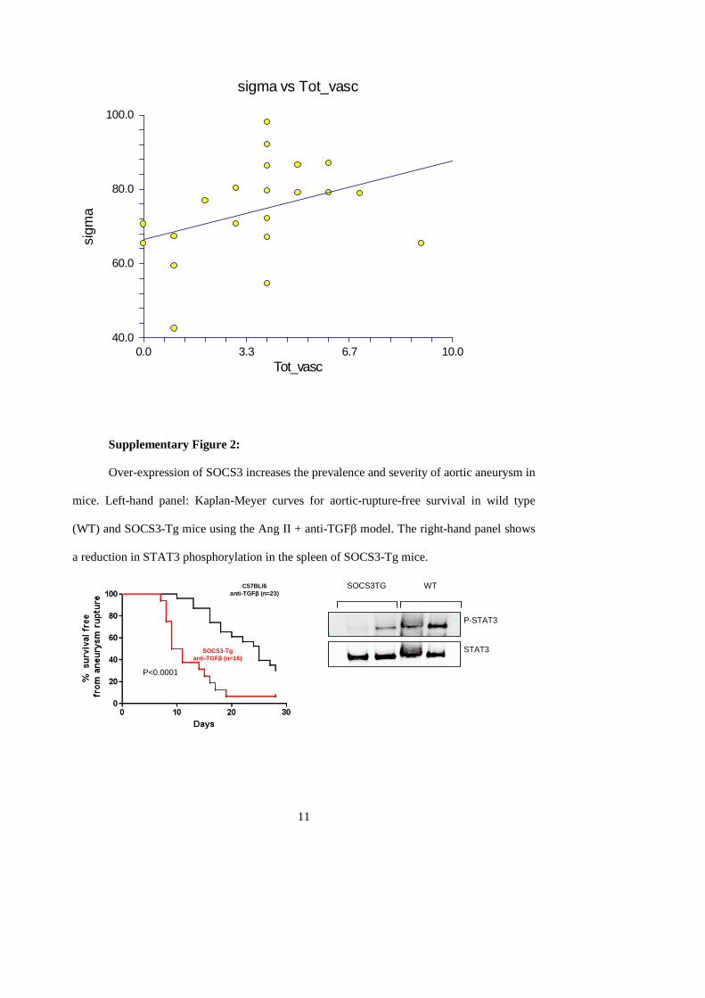
11
40.0
60.0
80.0
100.0
0.0 3.3 6.7 10.0
sigma vs Tot_vasc
Tot_vasc
sigm
a
Supplementary Figure 2:
Over-expression of SOCS3 increases the prevalence and severity of aortic aneurysm in
mice. Left-hand panel: Kaplan-Meyer curves for aortic-rupture-free survival in wild type
(WT) and SOCS3-Tg mice using the Ang II + anti-TGFβ model. The right-hand panel shows
a reduction in STAT3 phosphorylation in the spleen of SOCS3-Tg mice.
C57BL/6 anti-TGFβ (n=23)
SOCS3-Tg anti-TGFβ (n=16)
STAT3
P-STAT3
SOCS3TG WT
P<0.0001