a study on rotavirus gastroenteritis in children...
TRANSCRIPT

A STUDY ON ROTAVIRUS GASTROENTERITIS IN
CHILDREN UNDER FIVE YEARS IN COIMBATORE.
Dissertation submitted in
Partial fulfillment of the Regulations required for the award of
M.D. DEGREE
In
MICROBIOLOGY– BRANCH IV
The Tamil Nadu
DR. M.G.R. MEDICAL UNIVERSITY
Chennai
APRIL 2017.

CERTIFICATE
This is to certify that the enclosed work “A STUDY ON
ROTAVIRUS GASTROENTERITIS IN CHILDREN UNDER FIVE
YEARS IN COIMBATORE” submitted by Dr.R. Senthilkumar to The
Tamilnadu Dr.M.G.R. Medical University is based on bonafide cases
studied and analyzed by the candidate in the Department of Microbiology,
Coimbatore Medical College Hospital, during the period from July 2015 to
June 2016 under the guidance and supervision Dr.N. Mythily M.D.,
Professor, Department of Microbiology.
Guide
Dr.N.MYTHILY., M.D., Professor, Department of Microbiology Coimbatore-14
Dr.A,EDWIN JOE, M.D(FM).,BL. Dr.A.DHANASEKARAN, M.D.,DCH DEAN, Professor and HOD, Coimbatore Medical College and Hospital Department of Microbiology Coimbatore – 14. Coimbatore Medical College, Coimbatore – 14.

DECLARATION
I, Dr R. SENTHILKUMAR solemnly declare that the dissertation
entitled “A STUDY ON ROTAVIRUS GASTROENTERITIS IN
CHILDREN UNDER FIVE YEARS IN COIMBATORE” was done by
me at Coimbatore Medical College Hospital, during the period from July 2015
to June 2016 under the guidance and supervision of Dr.N.MYTHILY, M.D.,
Professor, Department of Microbiology, Coimbatore Medical College,
Coimbatore.
This dissertation is submitted to The Tamilnadu Dr.M.G.R. Medical
University towards the partial fulfillment of the requirement for the award
of M.D Degree (Branch - IV ) in Microbiology.
I have not submitted this dissertation on any occasion to any
University for the award of any degree.
Place:
Date: Dr.R. Senthilkumar


ACKNOWLEDGEMENT

ACKNOWLEDGEMENT
I express my deep debt of gratitude to our respectful
Dean,Dr.A.Edwin Joe,M.D(FM).,BL., for permitting me to do this study.
I wish to place my deep sense of gratitude and sincere thanks to
Dr.A.Dhanasekaran,M.D.,DCH, Professor and Head of the Department of
Microbiology, for the constant encouragement and timely advice given to
me during the course of my post-graduation and for selection and
preparation of this dissertation.
I express my deep sense of gratitude and indebtedness to Professor
Dr.N. Mythily M.D., for her constant guidance, valuable advice and inspiration
throughout my study. Also I sincerely thanks to Dr.V.Sadhiqua, DGO.,MD.,
Associate Professor for her support, encouragement and expert guidance which
helped me to complete this study.
I sincerely place my thanks to Associate Professor
Dr.Sankar.P, M.D., for timely advice and valuable suggestion in completion
of this dissertation.
I express my sincere thanks to my Assistant Professors,
Dr.S.Deepa, M.D., Dr.N.Bharathisanthose, M.D., Dr.B.PadminiM.D.,
Dr.C.Ashok Kumar,M.D and Dr.R.Radhika,MD., for their valuable
suggestion and help.
I would like to thank HOD, faculties and Post Graduates of Paediatrics
Department for their cooperation and support in completing my study.

My special thanks to my Post Graduate Colleagues
Dr.M.Banumathy, Dr.V.M.Theeba, and to my Junior Post Graduates in
the Department of Microbiology for their co-operation in completing my
study.
I take this opportunity to thank all the technical staffs in the
Department of Microbiology who gave me their kind co-operation
throughout my study.
I affectionately thank my family members who are giving me their
constant support to my entire post-graduation course, love and
encouragement without which this work would not have been successful.
I am thankful to God, who have been with me all throughout my
way to reach the destination.

Submission author:Assignment t it le:Submission tit le:
File name:File size:
Page count:Word count:
Character count:Submission date:
Submission ID:
Digital ReceiptThis receipt acknowledges that Turnit in received your paper. Below you will f ind the receiptinf ormation regarding your submission.
The f irst page of your submissions is displayed below.
201414253 Md Microbiology R.Sen…2015-2015 plagiarismA STUDY ON ROTAVIRUS GASTR…rota_26_sept.docx219.17K9114,47580,20326-Sep-2016 04:08PM709755207
Copyright 2016 Turnitin. All rights reserved.


CONTENTS

S.NO CONTENTS PAGE NO
1. INTRODUCTION 01
2. AIMS AND OBJECTIVES 07
3. REVIEW OF LITERATURE 08
4. MATERIALS AND METHODS 43
5. RESULTS 60
6. DISCUSSION 67
7. SUMMARY 83
8. CONCLUSION 85
9. BIBLIOGRAPHY
10. ANNEXURE
11. MASTERCHART

LIST OF TABLES
S.NO TABLES
1. Distribution of Acute Gastroenteritis In Accordance With Age and
Gender
2. Age Distribution in RVGE Cases
3. Seasonal Distribution of Rotavirus
4. Geographical Distribution of Rotavirus.
5. RVGE and water consumption.
6. Rotavirus Gastroenteritis cases related with feeds.
7. Clinical findings in Rotavirus Positive and Negative cases
8. Comparison of ELISA and ICT in Detection of Rotavirus Antigen
9. Genotypic distribution of Rotavirus

LIST OF CHARTS
S.NO CHARTS
1. Age and Sex Distribution in RVGE Cases
2. Sex Distribution of study population and Rotavirus positive cases
3. Seasonal Trends of Rotavirus
4. Geographical Distribution of Rotavirus
5. Educational Status of the Parents
6. Socioeconomic Status of parents
7. Rotavirus Gastroenteritis and feeds
8. Comparison of ELISA and ICT in Detection of Rotavirus
Antigen
9. Genotypic distribution of Rotavirus.

LIST OF FIGURES
S.NO FIGURES
1. Immunochromatography Kit With Its Contents
2. Immunochromatography Cassettes
3. ELISA Kit and its contents
4. Stool Samples and Microtitre Plate
5. ELISA Reader
6. ELISA Microtitre Plate with Controls and Samples
7.
a. RT - PCR Rotavirus: Plate Setup and Thermal Profile
b. RT - PCR Group a Rotavirus Amplification Plots
c. Group A Rotavirus Genotyping
8.
a. RT - PCR Rotavirus: Plate Setup & Thermal Profile
b. RT PCR Group A Rotavirus Amplification Plots
c. Group A Rotavirus Genotyping
9. EM Picture of Rotavirus
10. a. Pathogenesis of Rotavirus
b. Villous Atrophy in Rotavirus Gastroenteritis
11. PCR Machine

LIST OF ABBREVIATIONS
AGE Acute gastroenteritis
DLP Double Layered Particles
EIA Enzyme Immunoassay
ELISA Enzyme Linked Immunosorbent Assay
EM Electron Microscope
E-type Electropherotype
IAP Indian Academy of Paediatrics
ICG Immunochromatography
LA Latex Agglutination
NIH National Institute of Health
NSP Non Structural Proteins
ORT Oral Rehydration Therapy
RVGE Rotavirus Gastroenteritis
RT-PCR Reverse Transcryptase Polymerase Chain Reaction
SG Sub Group
SLP Single Layered Particles
ssRNA Single Stranded RNA
TLP Triple Layered Particulars
VP Viral Proteins
WHO World Health Organisation

INTRODUCTION

1
INTRODUCTION
Rotavirus remains the world’s commonest cause of gastroenteritis
among children predominantly in developing countries. Globally rotavirus alone
is responsible for around 139 million cases or almost 40% of gastroenteritis related
hospitalization, in the paediatric age group every year. The impact of Rotavirus
infection in children is far greater in India accounting for about 26 % of all
gastroenteritis related hospitalization among children.
In addition, diarrhoea plays a major role in malnutrition among children
which predisposes them to other infectious diseases and increases childhood
morbidity and mortality. Rotavirus infects almost all children at least once by the
age of five years. It is prevalent in all geographical regions of the world and almost
all socioeconomic groups.
Children attending daycare are at a higher risk of hospitalization due to
Rotavirus infection. Asymptomatic infection in newborn nurseries occurs round
the year and infection can spread to families causing gastroenteritis in adult
caregivers of children which might be the reason for endemic cases.
Though the Rotavirus infection is self-limited, it can cause severe
dehydration and electrolyte imbalance leading to complications such as shock,
cardiac failure, seizures and aspiration of vomitus resulting in aspiration
pneumonitis. Serologic evidences indicate that frequent rotavirus infection leads to

2
development of coeliac disease in genetically predisposed children. It can also
cause chronic diarrhoea in immuno compromised children and relatively severe
gastroenteritis in adults with immuno suppression. Studies indicate that Rotavirus
accounts for 4-7% of traveler’s diarrhoea.
However, the significance of the rotavirus gastroenteritis is said to be under
estimated in our country since most of the children with symptoms of
gastroenteritis are not brought to the health care set-up for treatment. Routine
screening for Rotavirus infection is also not being done in symptomatic children.
The word ‘Rota’ means wheel due to its distinct wheel like shape. It was
discovered in Nebraska in United States among ‘Calves’ in 1969 and first human
cases were described in 1973 in children with acute gastroenteritis.
Rotavirus belongs to the family Reoviridae and the virion is a non-
enveloped icosahedral particle. It consists of three concentric protein layers, which
encloses 11 segments of ds-RNA together with polymerase [VP-1] and capping
enzyme [VP-3] complexes. These segments encode six structural proteins VP-1 to
VP-4, VP-6 and VP-7 and six non-structural proteins NSP-1 to NSP-6. The
innermost layer is VP2 which encloses two proteins VP-1 and VP-3. The middle
layer is VP-6 and though not exposed on the viral surface, VP-6 is the target of the
most abundant antibodies produced by Rotavirus infection. The outermost layer
[glycoprotein] is made up of VP-7 and VP-4 spikes which protrude from the virion.
VP-4 is the major cell attachment protein and virulence determinant.

3
Based on the VP-6 capsid gene, the virus has been classified into seven
major genogroups A to G; among them Group A, B and C infect human beings.
Group A is most commonly detected among children with endemic gastroenteritis.
Group B and C are associated with epidemics of gastroenteritis affecting all age
groups. The strain ADRV - Adult Diarrhoea Rota Virus of Group B has been
linked to large outbreaks of severe diarrhoea in China as well as smaller outbreaks
and endemic disease in Bangladesh and India. Group C causes less severe
gastroenteritis in both adults and children. Serotype-G2 predominates in outbreaks
of Group A rotavirus gastroenteritis among adults and is found to be particularly
virulent.
On the basis of outer shell proteins VP-7 and VP-4, the Group A viruses are
further classified into 27 G and 35 P genotypes respectively. Worldwide the most
prevalent genotypes causing majority of infections are G1P[8], G2P[4], G3P[8],
G4P[8], and G9P[8].
For efficient rotavirus infection, priming of the virus by intestinal trypsin is
necessary. It cleaves the spike protein,VP-4 into an aminoterminal fragment VP-8
and carboxy terminal fragment vp-5 .The VP-5 fragment involves in the binding
of virus with the intestinal epithelial receptor possibly an ‘intergrin’. The
pathogenesis of rotavirus is complex and multifactorial. The possible potential role
of the virus being enterotoxin, malabsorption [sodium and glucose] related to
mucosal damage of intestinal epithelial cells, depletion of disaccharidases ,etc.

4
Immunity in Rotavirus infection is complex; Innate, cellular and humoral
mechanisms participate in elimination of infection. The humoral immunity plays a
major role in protection from severe disease. Protection of Rotavirus infection after
natural infection or immunization is more reliable against viruses of the same
serotype as the immunizing strain. The duration of immunity to Rotavirus is
limited; repeated symptomatic infections commonly occur in both children and
adults, but generally with less severity. On the other hand, cross protection between
serotypes occurs after multiple infections.
In Rotavirus reinfection is common. Asymptomatic infections are common
in infants below 6 months of age; the time during which the maternal antibody
confers protection. Such infections protect against severe infection but do not offer
protection against reinfection. Young children can suffer up to five re-infections
within 2 years of age and 90% of children by the age of three years show antibodies
to rotavirus. Local immune factors like secretory IgA and interferon also play an
important role in protection against rotavirus infection.
From some clinical studies of Rotavirus infection, breast feeding was found
to be associated with lesser frequency of vomiting and less severe dehydration with
a lower risk of hospitalization.
Rotavirus shows an unusual genomic diversity worldwide. During the
Rotavirus outbreaks, multiple strains which show combination of several G and P
types are seen.G1,G2,G3 and G4 strains account for 97.5% of all rotavirus

5
infections in Asia, North America, and Europe.G5,G8,G9 and G12 strains are
prevalent in several areas and G9 strains occasionally show predominance.
Epidemiologically G1 strain is most commonly prevalent worldwide followed by
G9 and G2. Children infected with G1 strains have a greater risk of developing
gastroenteritis with severe dehydration than those infected with other strains of
Rotavirus.
Rotavirus gastroenteritis shows marked seasonal variation. In temperate
countries epidemic peak occurs only in winter whereas in India it occurs
throughout the year but more during November to February.
Rotaviruses are highly infectious (even <100 viruses can cause infection);
the best documented mode of viral transmission is faeco-oral-route. Rotaviruses
are stable in environment and they can survive on toys, books, clothes and surfaces
for a longer period. Perhaps they are relatively resistant to most soaps and
disinfectants; so there is a possible spread of infection through Health care staff
and caregivers of the children especially when proper hand hygiene is not being
followed.
Rotavirus shows variation in prevalent strains from time to time and region
to region. Genetic re-assortment is more common due to antigenic drift;
occasionally genogroup re-assortment between human and animal strains has
been observed. This contributes to a continuous introduction of genetic diversity

6
which necessitates the ongoing surveillance to assess the efficacy of rotavirus
vaccine
Rotavirus gastroenteritis cannot be eliminated through improvement in
sanitation alone since there is a possible spread of virus through, aerosols, vomitus
and contact with fomites and environmental contamination. Moreover there is no
specific treatment for Rotavirus infection except supportive measures such as
rehydration with ORS/IVF; so it is necessary to develop a safe and effective
vaccine to decrease the occurrence and severity of rotavirus gastroenteritis in the
paediatric population.
The Prophylactic Rotavirus vaccination within the perspective of
National Immunization Program in developed countries has been associated with
significant reduction in mortality and morbidity as well as hospitalization among
children under five years.
Considering the severity of Rotavirus infection and its associated mortality
and morbidity in children under five years, as well as the importance of genetic re-
assortment, this study is undertaken to assess the prevalent genotypes of Rotavirus
responsible for acute gastroenteritis within the time period in Coimbatore.

AIM & OBJECTIVES
7
AIM AND OBJECTIVES
AIM
To analyse in detail about the prevalence, demographic profile, seasonal
trends and detection methods of Rotavirus Gastroenteritis in children under
five years in Coimbatore.
OBJECTIVES
1. To detect the Rotaviral antigen from stool specimen in children
under 5 years with acute gastroenteritis by ENZYME IMMUNO
ASSAY.
2. To compare Enzyme Linked Immunosorbent Assay (ELISA) and
Immunochromotography (ICG) in detection of rotaviral antigen from
stool specimen.
3. To analyse in detail about the demographic profile and seasonal
trends of Rotavirus diarrhea in our place.
4. To study the proportion of G/P types among positive cases and
their association with the clinical severity of Rotavirus diarrhea.

REVIEW OF LITERATURE

8
REVIEW OF LITERATURE
HISTORY
In 1963, the virus with distinct morphologic features was first observed
by Adam and Kraft by electron microscopy in small intestine and rectal
swab specimens from mice and monkeys. These agents were called as infant
mice virus and simian agent 11 accordingly which were later identified as
Rotaviruses. They were described as 70nm particles with a unique
morphology (Latin word-Rota meaning wheel) - “wheel like appearance”[1].
In 1969, these virus particles were demonstrated in the stool sample
of calves’ with diarrhea, thus relating these virus particles with diarrhoeal
disease in cattle [2].
In 1973, the association between these calf viruses and human
diarrhoeal disease were documented (Bishop et al., 1973). They demonstrated
the Rotavirus particles from duodenal mucosa biopsy specimens and later in
stool specimens of children with acute gastroenteritis in Melbourne, Australia
( Flewett et al, bishop et al).
Later it was found that the human Rotaviruses and the animal
Rotaviruses shared a group antigen and they were classified as members of
Rotavirus genus within the family Reoviridae [1].

9
In 1980, the virus particles were morphologically indistinguishable
from the established Rotavirus strains except lacking the common group
antigen which was discovered in pigs. This led to the identification of six
additional Rotavirus groups (B to G) based on a common group antigen,
with previous Rotavirus strains classified as group A[2]. Group A, B, and
C have been associated with human diseases while other groups have been
linked with diseases of animals [3].
EPIDEMIOLOGY
Acute gastroenteritis is one of the most common illnesses of humans
and its morbidity and mortality are higher among children and the elderly
[Wilhelmi et al 2003]. Rotavirus is the important causative agent of diarrhoea
with severe dehydration in children in developed as well as developing world
[Alrifai et al 2009]. About four billion episodes of diarrhoea occur every
year worldwide, of which more than 90% are from developing countries [7].
According to the Global Burden of Disease Study 2010, Diarrhoea is in the
fourth common place in terms of maximum Disability Life Years (DALY)
lost.
Rotavirus is the commonest etiological agent of severe diarrhoea in
infants and young children worldwide [4] and accounts for 40% of
gastroenteritis related hospitalization [19]. In India, 30% of hospitalized
diarrhoeal cases are attributable to Rotaviruses [5].

10
In developing countries, Rotavirus alone is responsible for around half
a million deaths annually. In India, among more than 2.3 million annual
deaths in children [4], diarrhoea is the third leading cause of childhood
mortality and accounts for about 3,34,000 deaths[5].
In addition, diarrhoea plays a major role in malnutrition among
children and predisposes them to other infectious diseases which leads to
increase in childhood morbidity and mortality [7].
ETIOLOGY
Diarrhoea may be caused by Bacteria, Viruses and Parasites and rarely
Fungi [8]. Among them viruses are the major etiological agents contributing
to 70% of all diarrhoeal cases worldwide (Webb and Star, 2005).
Viruses causing diarrhoea in children :
Rotavirus, Adenovirus, Astrovirus , Calcivirus (Norovirus & Sapovirus),
Coronavirus and Torovirus[8]
ADENOVIRUS
They are non-enveloped ds-DNA viruses approximately 70-90 nm in
diameter; spherical in the form of icosahedran
Adenovirus can cause epidemic and sporadic diarrhoea in infants and
outbreaks are associated with type 3&7 infections

11
Enteric adenovirus types 40&41 account for about 5-15% of all
diarrhoeal cases in children[9&10]
ASTROVIRUS
They belong to the family Astroviridae and the genome is positive sense
ss-RNA. Under electron microscope, the virus has a star like morphology.
Human viruses are classified in the Mamastrivirus genus of family
Astroviridae; eight serotypes have been identified so far; among which,
serotype-1 is the most common.
Astroviruses are linked with epidemics in paediatric wards, day care
centers and nursing homes and contributes 7-15% of diarrhoeal cases
especially in children less than 1 year[10].
CALCIVIRUS
The virion particles are small (27-40nm in diameter), non-enveloped of
icosahedral symmetry with 32 cup shaped depressions on the surface
In human infection 2 genera of calcivirus have been linked:
1. Norovirus
2. Sapovirus

12
NOROVIRUS
They are small round structured virus which are the major cause
of non-bacterial epidemic gastroenteritis[10]
It includes Norwalk and Snow mountain viruses
NORWALK VIRUS
They are highly infectious and commonly associated with
nosocomial infections[11]
It affects older children and adults
SNOW MOUNTAIN VIRUS
Epidemic gastroenteritis in children and adults were recorded and
shown to be due to snow mountain virus[12]
SAPPOVIRUS
It was first identified in Sapporo, Japan and hence the name (27-
40 nm in diameter)[13]
Causes sporadic cases and occasional outbreaks of diarrhoea
predominantly among closed population in infants, young children
and elderly [13].

13
CORONAVIRUS
The name corona is used because of crown like projections on the
surface under electron microscope with helical nucleocapsid of 120-
160 nm in diameter[14]
They are linked with diarrhoeal cases in paediatric age group[29]
TOROVIRUS
They are enveloped positive strand RNA viruses, approximately 100-
140nm and associated with hospital acquired gastroenteritis in
paediatric patients[15]
ROTAVIRUS
It is well documented that Rotaviruses are the major cause of diarrhea
in human infants, young calves and piglets [16]. Rotaviruse is the agent of
infantile diarrhoea of human, epizootic diarrhoea of infant mice, Nebraska
calf diarrhoea and SA 11 virus of monkeys [17].
Despite gains in controlling diarrhoeal deaths, the disease burden still
remains high. Studies indicate there is an approximately 139 million cases
of gastroenteritis in children worldwide (Institute of Medicine, 1986).
Although the prevalence of Rotavirus diarrhea is similar in the developing
as well as developed countries (Alrifai et al.,), mortality is comparatively

14
higher in developing countries which accounts for around 6,11,000 deaths in
children less than five years[17].
There is widespread distribution of Rotavirus in the community. Almost
all children have at least one episode of Rotavirus infection within 5 years
of age [16]. The high prevalence of Rotavirus is due to persistence of
subclinical infection throughout the lifetime.
A study done in Washington over a period of 8 years shows 34.5%
of 1537 children found to be Rotavirus positive in their stool
specimens.
A study done in Japan over a period of six years showed 45% of
1910 children to be positive for Rotavirus from stool specimens.
An Australian study showed 39.6% of positivity for Rotavirus in stool
samples, among 3785 children with acute diarrhoea.
Hospital based studies in Africa, Asia and Latin America report 25%-
55% of hospitalization in children due to Rotavirus infection.
In countries of the eastern Mediterranean region, studies conducted
among children with acute diarrhoea shows Rotavirus in 40% of
inpatients and 23% of outpatients by the age of 3 years.
In a population based study among infants and young children in
Saudi Arabia, Rotavirus was detected in 95% of stool specimens. In

15
Jordan, in hospitalized children below five years, Rotavirus was
detected in 39.8% of stool specimens.
In India, a health care facility-based study of children has exhibited
Rotavirus infection to account for around 26% of all diarrhoea-related
hospitalization [18].
Primarily Rotavirus infection in younger children between 6 months
to 2 years of age is about 55% whereas it is 18.7% in children less than
6 months and 16% in the age group of 5-13 years. During the first five
years of life, almost every child acquires rotavirus infection.
The prevalence of Rotavirus infection is similar both in developing as
well as developed countries before vaccination [15]. Although the rotavirus
vaccines have been implemented in many middle income countries in Central
and South America, vaccines are not yet available in countries of Asia and
Africa. Widespread vaccination would markedly lower the global mortality
and financial burden due to Rotavirus infection.
Estimates indicate, there is an approximately 1,22,000, 27,000 and
2,000 deaths related to Rotavirus infection in India, China and Pakistan
respectively.
Adult contacts may be infected, as evidenced by seroconversion [28],
but they rarely exhibit symptoms, and virus is infrequently detected in their

16
stools. However epidemics of severe disease have been recorded in adults,
particularly in closed populations, such as in geriatric ward, healthcare
workers, travelers, military people and parents of infected children [1].
SEROTYPES OF ROTAVIRUS
The serotype classification is based on the surface protein vp7 (G-
type) and VP4 (P-type) which are the target of neutralizing antibodies [20].
There are 25 different G- serotypes and 35 P serotypes found at present
among group A Rotavirus of humans and animals. The prevalent G and P
type combination in population is somewhat lower than the expected number
of G and P type combination. Approximately, 12 G type (1 to 6, 8 to 12
and 20) and 15 P-types (1 to 6, 8 to 11,13,14,19,25 and 28) have been
linked with human infection[10].
The global distribution of human Rotavirus serotype appears to be
consistent. Since, 1989 till now, serotypes G1,G2,G3 and G4 constantly make
up the predominant strains detected especially in developed countries.
In 124 studies [1989-2004] on the global Rotavirus Serotypes and
Genotypes distribution, G1 strains were the most commonly detected among
45,571 typed strains [30]. Despite this, in the last decade G9 strains have
emerged as a predominant serotype in some countries such as Australia,

17
United States, Ghana, India and Brazil, in some parts of Africa, G8 strains
have emerged as predominant strains.
Initially, serotype analyses of clinical isolates for their P types (VP4)
were not widely performed because of the lack of appropriate mono specific
serologic reagents. However, RT PCR for P (VP4) genotype had made it
possible. Based upon the segment analysis of VP4, P1A[8] and P1B[4]strains
are the most frequently detected strains in humans. The P1A[8] genotype is
almost always found in association with G1,G2,G3,G4 and G9 strains
whereas P1B[4] genotype characteristically has specificity with serotype G2.
In the review of 16,474 strains evaluated so far, both VP4 and VP7
specificities indicate that the predominant P-G combinations, G1P[8], G2P[4],
G3P[8] and G4P[8] contribute 88.5% of all the serotypes and recent evidence
suggests that G9 P[8] strains are also detected in humans frequently[20].
It is assumed that the studies on serotypic classification gained much
attention because such classification will be relevant and correlate well in
relation to protective immunity in Rotavirus infection. The recent
documentation provides that G and P monotype vaccine is associated with
efficient protection against all Rotavirus infections [5].
In humans, the heterotypic immunity plays a major role in the
epidemiology of Rotavirus infection [5]. In spite of the potential serotypic

18
diversity of Rotavirus, immunity appears rapidly after limited exposure only
to a restricted number of serotypes.
SUB GROUPS
Group A has 4 sub-groups based on the vp5 diversity.
SG1, SG2, SG1 &2 and NON1 & NON2
SG1, SG2 possibly infect humans [21].
GENOGROUPS
Three genogroups (two major & one minor) have been established in
human strains by the RNA-RNA hybridization technique using the reference
strain WA DS-1 and AV-1. Human Rotavirus belongs to the WA genogroup,
have origin with porcine Rotavirus and those belonging to DS1 genogroup,
have origin with bovine Rotavirus [22].
ELECTROPHEROTYPE (E-TYPE)
Two major electropherotypes, ‘long’ and ‘short’ have been
demonstrated by separation of Rotavirus genome using polyacrylamide gel
electrophoresis [23]. The ‘long’ pattern corresponding to the sub group I and
the ‘short’ pattern corresponding to the sub group II of the Group A human
Rotavirus [24].

19
ROTAVIRUS INFECTION IN ADULT
Adults get frequent reinfection with Rotavirus but are asymptomatic
or affected less severely; subclinical infections are most common among
them. Outbreaks in adults have also been described; associated with 4-7%
of traveler’s diarrhoea [25].
Symptomatic Rotavirus gastroenteritis was found in some adults
undergoing bone marrow transplantation and patients with malignant disease
undergoing chemotherapy due to immunosuppression.
Group B-Rotavirus (Adult Diarrhoea Rotavirus-ADRV) have been
implicated in several large outbreaks involving up to 20,000 individuals of
severe gastroenteritis in adults in China[29]; also in smaller outbreaks and
endemic cases in Bangladesh and India[25]. Group B- Rotavirus (Group B-
RV) infection occurs primarily as epidemics or sporadic cases, possibly due
to contamination of water sources.
NOSOCOMIAL INFECTION
Rotavirus often causes Hospital acquired infection. Nosocomial
outbreaks have been recorded in newborn nurseries in Italy and neonatal
ICUs in United States. Such infections mainly involve young infants (0-5
months of age) [25].

20
TRANSMISSION OF ROTAVIRUS
The best documented mode of Rotavirus transmission is faeco-oral
route [26]. Rotaviruses are highly contagious; stable in ambient temperature
and needs low dose for human infection (<100 virus particles is enough to
cause infection) [27]. The source of infection for the young infant is either
from older sibling or patient with subclinical infection. There has been
speculation about Rotavirus transmission by respiratory route but it is not the
usual mode of transmission [25].
Community wide water borne epidemics of group A Rotavirus
(G-A RV) have been documented in Turkey in 2011. Effective disinfection
of contaminated material and proper hand washing are the most important
measures to control Rotavirus infection especially in hospitals and
institutions [1].
VIRAL SHEDDING
Virus is shed in very large amount in faeces (1012 particles per gram
faeces)[29]. The duration of viral shedding may persist upto days and is
prolonged in immuno-compromised children [29].

21
RESISTANCE OF ROTAVIRUS
Rotaviruses are stable at 50 deg C, pH range 3-9 and to some lipid
solvents like ether, chloroform but inactivated by 95% ethanol, phenol and
chlorine [29].
INCUBATION PERIOD
The average incubation period is 1-3 days [29].
DISTRIBUTION
Rotavirus is distributed throughout the world and the major causative
agent of severe diarrhoea with dehydration in infants and young children.
According to WHO reports, there are 2 billion cases of diarrhoeal disease
every year globally in children less than five years [31]; it also estimated
that 85% of global Rotaviral deaths occur in Africa, Asia and Latin America [32].
SEASONAL VARIATION
Before the introduction of vaccination, Rotavirus showed marked
seasonal variation in developed countries with the epidemic peaks occur in
the cooler month of the year [29]. The cause for this seasonal pattern is not
known. Following the introduction of Rotavirus vaccination the seasonal
pattern has decreased markedly.

22
On the other hand in the tropics, no or minimal seasonal variations
has been noted [33-35]. This may be due to high birth rates as well as high
transmission rates found in developing countries.
AGE, SEX AND SOCIOECONOMIC STATUS
In developed countries, Rotavirus gastroenteritis most frequently affects
infants and young children between 6 months to 2 years of age[29] followed
by the infants younger than 6 month of age. Male children were found to
be twice as susceptible and more likely to be hospitalized than female
children [1]. Living in crowded condition may predispose them to earlier
Rotavirus infection.
ANIMAL SUCEPTIBILITY
Rotaviruses have a wide range of hosts such as mice, calves, swine
and piglets
Most isolates have been recovered from newborn animals with
diarrhoea
Cross-species infections can occur in experimental inoculations [19]
Newborn animals often exhibit subclinical infection, reflecting the
presence of maternal antibody; overt disease is more common during
the weaning period [19]
23
PROPAGATION IN CELL CULTURE
Rotaviruses are fastidious in nature and difficult to cultivate
Most of the group A human Rotaviruses can be cultivated if it is
pretreated with a proteolytic enzyme like trypsin[19]
MALNUTRITION
Malnutrition plays an important role by increasing the severity of
Rotavirus gastroenteritis [36]. Repeated diarrhoeal infection may be a
precipitating factor leading to the development of malnutrition as the infection
damages the intestinal mucosa and absorptive cells are being lost over a
period.
MOLECULAR EPIDEMIOLOGIC STUDIES
The gel electrophoresis is the method followed very earlier to study
the molecular epidemiology of human Rotavirus [29]. Molecular
epidemiological studies have analyzed isolates, based on differences in the
migration of the 11 segments of ds-RNA genome in polyacrylamide gel
electrophoresis. The differences in electropherotypes can be used to
differentiate group A viruses from other groups [3]. But they cannot be used
to predict serotypes. Later, RNA/RNA hybridization techniques were
developed which employs labeled ss-RNA viral transcripts as probe for
genetic ds-RNA. These studies identified two major genogroups (family) of

24
human Rotavirus and showed that human strains lack homology with animal
strains [3]. Recent advances replaced the electrophoretic and RNA/RNA
hybridization techniques.
MORPHOLOGY OF ROTAVIRUS
The Rotavirus has a distinct morphologic feature and three types of
particles can be observed under electron microscopy.
The complete particle appears like a ‘wheel with spokes’ and a well-
defined smooth outer rim [1].
The complete virion is also called as triple layered particle (TLP)
The virion lacking the outer shell are called as double layered particle
(DLP); they are also called as ‘rough particles’ because their peripheral
trimeric subunits of the inner capsid project outside
Single layered particles (SLP/core) are seen less often. They lack
genomic RNA and are aggregated [25].
The rotavirus virion is a non-enveloped icasohedral particle, size
approximately 770 Å in diameter excluding the vp4 spikes belong to
Reoviridae family [21]. It consists of 3 concentric protein layers which
envelop segmented ds-RNA together with polymerase and capping
enzyme complex .This segments encode six structural proteins (VP-1
to VP-4, VP-6 and VP-7) and 6 non-structural proteins (NSP1 to
NSP6) [22].

25
The innermost layer consists of 120 copies of VP-2 arranged in one
icasohedral lattice. The VP-1 polymerase and VP3 capping enzyme
are anchored inside VP-2 shell adjacent to the pores at the icasohedral
vertices [37].
The middle layer consists of 780 copies of VP-6 which forms the
thick trimeric pillars in the icasohedral lattice [3].
The VP-6 although not exposed on the viral surface, is the target of
the most abundant antibodies produced by Rotavirus infection [3].
The genome VP-1 and VP-3 together with inner protein layer make
up the transcriptionally active double layered particle (DLP).
The outermost layer is thin, and it consists of 780 copies of a coat
glycoprotein VP-7, and sixty vp4 spikes which protrude from virion
[21].
The VP-4 and VP7 translocate the DLP across a host cell membrane
and into the cytoplasm [21].
VP4 is the major cell attachment protein and virulence determinant
and growth restriction factor in cell culture [17].
During the entry into a host intestinal cell, the viral proteins vp4 and
vp7 are shed from the virion which in turn leads to activation of the
viral polymerase [21].

26
ROTAVIRUS PROTEINS AND FUNCTIONS [2, 38]
VP 1 is a core protein, which act as RNA dependent RNA polymerase;
helps in SS-RNA binding and couples with VP 3
VP 2 is present in core, with the major function includes RNA
binding and required for replicase activity of VP 1
VP 3 is located in core, activates Guanyl transferase and methyl
transferase
VP 4 is situated in the outer capsid. It helps in host cell attachment;
It causes Haemagglutination and produce neutralizing antibodies
VP 6 is present in inner capsid, required for transcription
VP 7 located in the outer capsid, produces neutralizing antibodies
NSP 1 (NS 53) is a Non-structural protein, it promotes RNA binding
NSP 3 (NS 34) ,a Non-structural protein, helps in RNA binding;
stimulates NTPase and Helicase activity
NSP 4 is a Non-structural protein, involved in accumulation of
intracellular Calcium, RNA replication, and enterotoxin production.
NSP 5 - Non-structural protein, helps in RNA binding; activation of
protein kinase and interacts with VP 2 and NSP 6
NSP 6 - Non-structural protein, interacts with NSP 5

27
PATHOGENESIS
The pathogenesis of Rotavirus is complex and incompletely understood.
The virion infects villous enterocytes and causes cell destruction. The release
of progeny takes place by cell lysis [29].
For efficient Rotavirus infection to occur, priming of virus by intestinal
trypsin is necessary which cleaves the spike protein VP-4 into two fragments
namely VP-8-aminoterminal and vp5-carboxyterminal [38]. The vp8 contains
haem-agglutination domain which forms the spike heads and vp5 contains a
membrane interaction domain which forms the spike body. Cleavage and
rearrangement of vp4 increases the rigidity of VP4 spikes [29].
VIRAL ENTRY AND REPLICATION
The cell entry pathway of Rotavirus involves virus interaction with
host cell surface. Rotavirus strains related with non-human animals, attaches
to host cell through vp8 fragment which bind neuraminidase-sensitive sialic
acids on cell membrane glycoprotein or glycolipids [38].
Rotavirus strains linked with human infection, bind an alternative
receptor possibly an Integrin using the VP5 fragment. During cell entry the
VP5 undergoes a significant fold back re-arrangement, translocating its
hydrophobic apex from one end to the other end of the molecule [39].

28
After attachment and penetration, un-coating of virus particles takes
place in lysosomes of the cell cytoplasm. The outer cell of virus is removed
and core associated RNA transcriptase is activated. The transcriptase
transcribes mRNA molecules from negative strand of ds-RNA segment [29].
The core contains all necessary enzymes for transcribing, capping and
extruding the mRNA from the core and leaving the ds-RNA genome segments
inside [29]. After being extruded from the core, the mRNA is translated into
primary gene products. A viral replicase is responsible for synthesizing
negative sense strands to form the double strand genome segments and this
process take place in partially completed core structures [29].
The mechanism of assembly of the correct compliment of genome
segments into a developing new viral core is probably through the viral
polypeptides which may self - assemble to form the inner and outer capsid
shells [29].
Rotavirus produces inclusion bodies in the cytoplasm. Rotavirus
morphogenesis involves budding of single shelled particles into the rough
endoplasmic reticulum. The pseudo envelopes are then removed and outer
capsids are added. This distinct pathway is used because the major outer
capsid proteins of Rotavirus are glycosylated [29].
29
The possible role of Rotaviruse in pathogenesis of gastroenteritis:
1. Viral enterotoxin [41]
2. Malabsorption due to intestinal mucosal damage [40]
3. Depletion of the enzyme ‘disaccharridases’ [38]
In Rotavirus infected pigs, study of duodenal biopsy specimen by light
microscopy revealed, shortened and blunted villi with cuboid epithelium,
crypt hypertrophy and mononuclear cell infiltration in lamina propria [38].
NSP4 – A ‘viroporin’ is a novel protein which mediates cell death
by increasing intracellular calcium and affecting plasma membrane
permeability as well as tight junctions of villous cells. It is a viral enterotoxin
which increases the enteric secretion of fluids through a ‘signal transduction
pathway [29].
Rotavirus reduces the host protein synthesis through NSP3 which is
shown by marked decrease in the level of host cell protein and increase in
the level of viral protein[38]. Rotavirus NSP1 interacts with IRF3 (Interferon
Regulatory Factor) and leads to degradation of IRF3 which interferes with
the innate host immune mechanisms [3].
Rotavirus infection causes alterations in the tight junctions of the small
intestinal epithelial cells. The intestinal epithelial cells are sloughed off due

30
to loss of tight junctions during the period of viral replication which leads
to decreased glucose and sodium absorption [38}.
The effect of Rotavirus infection in human intestinal epithelial cell lines shows that the following factors are involved: [38]
1.Interleukin-8
2.Growth related peptide-2
3.Osteopontin
4.Chemokine
Of which, the chemokine secreted by infected enterocytes plays an
active role in pathogenesis of Rotavirus gastroenteritis
DIVERSITY OF ROTAVIRUS [41]
The Mechanism Involved In Diversity Of Rotavirus Includes The
Following: [20]
1. Point mutation accumulation leads to formation of new genetic lineages
and emergence of mutants which helps virus to escape from humoral
antibody response
2. Genetic re-assortment between two different human Rotavirus strains
infection at a time
3. Possible genetic re-assortment of human Rotavirus strains among
animal strains contributing to diversity.

31
IMMUNITY
The immune response to Rotavirus infection is complex and multi
factorial: Innate, cellular and humoral immunity contributes to eliminate the
infection. Humoral immunity has a dominant role among them to prevent
severe disease on successive infection or on primary infection after
immunization [29].
Cross protection between different serotypes occurs after successive
infection. The duration of immunity against Rotavirus infection is limited;
reinfection occurs both in children and adults but generally with less severity [38].
Generally reinfections are common in Rotavirus disease. Asymptomatic
infections are frequently found in infants below the age of six months, the
time during which the maternal antibody confers protection. Such Neonatal
infection does not protect against reinfection but protects against severe
disease [29].
Young children can suffer up to five re-infections by the age of two
years and 90% of the children show antibodies against Rotavirus by the age
of three years [29]. Local immune factors such as secretory IgA or Interferon
plays an important role in protection against Rotavirus infection [29].
The role of breast feeding in protection of humans against Rotavirus
gastroenteritis is obscure. A hospital based study done in Bangladesh among

32
children admitted with Rotavirus gastroenteritis suggests that breast feeding
prevents Rotavirus less effectively than gastroenteritis caused by other agents
[42]. However, it is noted that, in some clinical studies of Rotavirus infection,
breast feeding is linked with less frequency of vomiting and less severe
dehydration as well as lower risk of hospitalization [28,4].
Neutralizing monoclonal antibodies that recognize the vp8 fragment of
vp4, blocks the attachment of sialic acid dependent Rotavirus to host cells
and antibodies that recognize VP5 fragment of VP4 blocks post binding
entry events. Antibodies against the VP7 protein blocks virion un-coating.
IgA monoclonal antibodies that recognize the middle layer of protein VP6
does not neutralize the virions but blocks transcription by DLPs and protect
the mice from Rotavirus infection as found in vitro studies [42].
CLINICAL FINDINGS
Rotavirus is responsible for the major cause of diarrhoeal diseases in
children below five years. The classical clinical features include watery
diarrhoea, fever, vomiting and abdominal pain. Infected children may also
have cough or running nose [27,33,41]
Rotavirus gastroenteritis is also called “Winter vomiting disease”
since it is associated with combination of vomiting and seasonal trends in
winter months [44].

33
Diarrhoea along with vomiting rapidly results in severe dehydration in
paediatric patients [28]. Signs of dehydration include increased thirst,
irritability, restlessness, lethargy, sunken eye, dry mouth and tongue, loss of
skin tugor [28], and sunken fontanelle; decreased urine output, lack of interest
in playing; drowsiness, tachypnea and tachycardia are also seen [28].
Rotavirus gastroenteritis leads to dehydration which may be mild or
severe and life threatening. Milder cases have symptoms for short period (3
– 8days) only and recovery is complete [29]. In severe cases there is
dehydration and severe loss of electrolytes which may result in life
threatening complications such as hypovolemia, circulatory collapse and
eventual death [28].
COMPLICATIONS:
Acute severe dehydration results in the following complications:
o Electrolyte imbalance - Hyponatremia and Hypokalemia leads to
cardiac failure and seizures [42].
o Rapid loss of water and electrolytes which results in acidosis, shock
and death[42]
o Aspiration of vomitus leading to pneumonitis is another complication
linked with Rotavirus gastroenteritis
o It can cause chronic diarrhoea in immunocompromised children [29].

34
o It has been reported that frequent Rotavirus infection has been
associated with Coeliac disease in genetically predisposed children [42].
DIAGNOSIS
Early diagnosis of Rotavirus gastroenteritis in hospitalized patients will
decrease the morbidity and mortality considerably and avoids inappropriate
use of antibiotics in paediatric patients.
It is difficult to differentiate Rotavirus gastroenteritis from other causes
of gastroenteritis on clinical grounds alone. It may be necessary to establish
Rotavirus as the etiological agent in some clinical conditions such as
protracted diarrhoea, complicated cases, immunocompromised hosts and for
epidemiological studies
DIAGNOSTIC METHODS:
Rotavirus Can Be Detected From Stool Specimen By The Following
Techniques:
Acute Rotavirus gastroenteritis was diagnosed by the conventional
methods such as Electron-microscopy, Counter-immunoelectrophoresis,
Reverse passive Haemagglutination assay (RPHA), DNA Oligonucleotide
micro assay methods, and Tissue culture, Flowcytometry, Complement
fixation test and Latex agglutination test(LAT). Subsequently, development
of newer methods such as Polyacrylamide gelelectrophoresis (PAGE), Dot

35
hybridization technique, Immunoflourescence test, Enzymelinked immunospot
assay, Immunochromatography (ICT), Enzyme immuno assay (EIA) and RT-
PCR have replaced the conventional methods since, they are rapid, accurate
and also having good sensitivity and specificity[2,14,26,29,40,45,47]
MANAGEMENT
Isolation of the patient and prompt treatment
Since Rotavirus is stable in room temperature and highly contagious
isolation of the patient is necessary to prevent person to person
transmission of the virus in the ward and the community [1]; the bed
clothes and linens are decontaminated with chlorine or phenol
Treatment of Rotavirus gastroenteritis is mainly by supportive measures
which include prompt correction of water and electrolyte loss [29];
replacement of fluids and correction of electrolyte imbalance either by
intravenous route or orally as feasible
O.R.T (Oral Rehydration Therapy) was the single most effective
strategy followed to prevent dehydration related mortality earlier [48].
But there is risk of hypernatremia in standard ORS when given to
children with non-cholera diarrhoea. Hence low osmolarity ORS is
recommended by WHO and IAP (Indian Academy of Paediatrics) as

36
an universal solution for treatment and prevention of dehydration for
all cases of diarrhoea and at all ages .
In 2004, the WHO and UNICEF recommended low-osmolarity oral
rehydration solution (ORS) and zinc supplementation for the treatment
of acute diarrohea [49].
Energy dense feeds in addition to exclusive breast feeding has been
included in treatment of gastroenteritis by Ministry of Healthand Family
Welfare, Government of India.
Zinc supplementation: 10mg of (elemental zinc) given for a period of
2weeks for children in the age group of 2-6 months and 20 mg per
day for children aged more than 6 months is being advised [49,50].
Zinc has been linked with decreased illness and mortality by lowering
the severity as well as duration of diarrhoea; it also lowers the
incidence (UNICEF-PHFI Indian Paediatr-2012 PUBMED).
RISK FACTORS
lack of sanitation, poor access to safe drinking water, lack of
knowledge about hygienic measures contributes around 88% of
diarrhoeal cases in children [51].
37
In addition, low socioeconomic status, illiteracy, low birth weight,
inadequate breast feeding, malnutrition are linked with higher
incidence of diarrhoea in children[51]
PREVENTIVE MEASURES
As the main mode of transmission of Rotavirus is through faeco-oral
route, proper disposal of excreta, hand hygiene, use of safe drinking
water, immunization against measles and promotion of exclusive breast
feeding are the most important measures to prevent diarrhoeal
diseases[49]
Effective hand hygiene includes hand washing after defecation and
before preparing food/ feeding their children
Hand washing with soap and water can prevent the risk of diarrhoea
by 42-47% (Lancet infect dis.,2003 PUBMED)
In India, hand washing with soap before handling or consumption of
food and after passing motion, is being encouraged in school children
through the school hygiene program and mass media campaign on the
“hand washing day” [49]; likewise, The World Toilet Day is
conducted in the sense of giving awareness to the public to avoid
open air defecation and its ill-effects[49]

38
In addition, vitamin A supplementation is being promoted in children
since it reduces the incidence of diarrhoea and associated deaths in
children aged six months to five years[49]
Furthermore, a safe and effective vaccine remains the promising way
of reducing the worldwide burden of Rotavirus disease [5].
ROTAVIRUS VACCINES
Improvements in sanitation have only limited effect on Rotavirus
occurrence. So it is important to introduce safe and effective immunization
to decrease the impact of rotavirus infection in the community (Fischer et al.,).
The first Rotavirus vaccine – ‘Rota-shield’ was introduced in United
States (1998) by Wyeth and approved by USFDA (Food and drug
administration) and ACIP (advisory committee on immunity practices);
Clinical trials in the United States, Finland and Venezuela had proved the
vaccine to be 80-100% effective in preventing severe gastroenteritis caused
by group A Rotavirus [52,53]. However the vaccine was withdrawn from the
market by the manufacturer in 1999 since it was associated with increased risk
of intussusception (1 in 12000 vaccinated infants) among vaccinated
children [54].
Two new vaccines (Rotarix and Rota-teq) against Rotavirus Group
A infection were introduced in 2006 [54]. Mexico was the first among all
countries in the world to introduce Rotavirus vaccine and the results showed

39
that diarrhoeal deaths decreased by more than 65% in children less than
2 years.
‘Rotarix’ is an oral live attenuated monovalent vaccine; the breadth
of coverage depends on cross protection between serotypes. ‘Rota-Teg’ is a
multivalent modified Jenner vaccine. Bovine G serotypes combined with
human P serotypes was found to be highly efficacious among severe rotavirus
gastroenteritis caused by G1or G2 serotypes of Rotavirus. It was licensed
in United States and European Union in 2006.
In 2009, the WHO recommended that Rotavirus vaccine can be
included in all national immunization programs [56]. The two Rotavirus
vaccines (Rotarix and Rota-teq) have been licensed in more than 100
countries, including India [57]. The incidences and severity of Rotavirus
infection has declined dramatically in countries that have adopted this
recommendation.
A median of 36% of children less than five years with acute diarrhoea
showed positivity for Rotavirus based on the data from the Global
Surveillance Network of 2009. Therefore, WHO has recommended inclusion
of Rotavirus vaccination in all national immunization programs.
A Cochrane review (2012) of 41 clinical trials concluded Rotarix
(GlaxoSmithline) and Rota-Teq (merk) were the effective vaccines and [58];

40
also reported that they were not associated with risk of intussception in
immunized children.
Intense debate in introduction of Rotavirus vaccine in India was due
to lack of estimates regarding mortality by Rotavirus diarrhoea and also
expenditure of the vaccines.
A study indicates, 90,000 - 1,53,000 children die from Rotavirus
infection each year in India; around 4% of overall mortality in children
under five years could be saved in India, if Rotavirus vaccines were
integrated with National Immunization Program [Shaun K Morris et al]
(Bulletin of the World Health Organization 2012;90:720-727) [59].
In May 2013, India has developed a new vaccine against Rotavirus-
‘Rotavac’. The phase 3 clinical trials of low cost Indian-made Rotavirus
vaccine-Rotavac have showed effective efficacy and excellent safety profile.
It would be available at Rs.54 per dose if approved by the Drugs Controller
General Of India [60].
It is found that the Rotavac reduced severe diarrhoea by more than
56% during first year of life with protection continuing into the second year
also. Moreover, the vaccine also exhibited great impacts against severe
diarrohea of any etiology [5].

41
Strains 116E and 1321 were isolated from NICU in New Delhi and
Bangalore and pursued its vaccine property; A neonatal strain RV3 isolated
by workers at Royal children hospital in Melbourne, Australia is being
pursued as a vaccine character; bovine re-assortment vaccine was developed
at NIH [41].
International non-governmental organization PATH, the WHO, the US,
Centers for Disease Control Prevention, and the GAVI Alliance are working
to bring Rotavirus vaccine to developing world, where the mortality and
morbidity of diarrheoa in children are significantly high [5].
Integration of Rotavirus vaccination with National Immunization
Program is advised in countries where diarrhoea associated mortality is
more than 10% in children under five years [62]
Indian Council of Medical Research (ICMR) has formed a project
called ‘National Hospital Based Rotavirus Surveillance Network’ to establish
different surveillance units in tertiary care hospitals in different parts of India [5].
Objectives Of The National Hospital Based Rotavirus Surveillance
Network: [5]
To identify the proportion of diarrhoea and mortality attributable to
Rotavirus infection in children less than 5 years of age

42
To emphasis hospital based and community based study of Rotavirus
infection in children
To identify the susceptible age, seasonal distribution, characterization
of strains
( G and P types ) in different geographical areas
To isolate newly emerging strains or unusual strains attributed to
zoonotic diseases or strains not identified by standard techniques
To find out children admitted due to intussception and its causal
relationship with Rotavirus vaccination if any[[61].

MATERIALS & METHODS

43
MATERIALS AND METHODS
Study area
This prospective study was conducted at the Department of
Microbiology, Coimbatore Medical College, Coimbatore for a period of one
year from July 2015 to June 2016.
Study population
Overall, 100 children less than five years with acute gastroenteritis
from outpatient department and ward were included in the study population.
Inclusion criteria
Children under five years with acute gastroenteritis
Children below five years with diarrhoea alone
Exclusion criteria
Children under five years with bloody diarrhoea
Children less than five years who acquired diarrhoea during
hospitalization (Healthcare Associated Infection/Nosocomial infection)
Children below five years with chronic diarrhoea (diarrhoea >14 days)
Children under five years with immuno compromised state

44
Ethical clearance
Ethical committee approval was obtained from the institutional review
board of Coimbatore Medical College, Coimbatore. Detail about the research
was informed to the parents/guardians of the participant; oral as well as
written consent were obtained for participating in the study.
Clinical history
Detailed history was taken from the parents/guardian of the patient
and entered in the proforma as follows:
Name, age, gender, geographical area (urban/rural), occupation, literacy
and socioeconomic status
Antenatal history- full term/preterm, birth weight, immunization
Onset and duration of diarrhoea, whether blood stained or not,
vomiting, abdominal pain, and fever
Feeds- breast fed/bottle fed
Hand washing and other sanitary measures
Physical examination of the child to detect signs of dehydration-
consciousness, whether the baby taking feeds, active, skin tugor,
anterior fontenelle, sunken eyes.
Macroscopical examination of stool specimen – watery, colour, whether
blood stained or not.

45
Sample collection, transport and storage
About 15-20 ml of stool specimen was collected in a sterile wide
mouthed universal container during acute stage of gastroenteritis ( <
3 days of onset of symptoms)
Samples were kept in ice box to maintain ideal temperature (2-80 C)
and transported immediately to the Microbiology diagnostic laboratory.
The stool was incorporated into 2 ml sterile centrifuge container along
with buffer and stored at -20 to -700C for detection of Rotavirus
antigen.
Testing
The specimens were screened for Rotavirus antigen through Enzyme
Linked Immuno Sorbent Assay (ELISA) and Immuno Chromatography
Test (ICT) and confirmed by Reverse Transcriptase Polymerase Chain
Reaction (RT-PCR).
Immuno Chromatography (ICG)
To detect the Rotavirus antigen from stool specimen, SD Bioline Rota
rapid test kit, (Standard Diagnostics, Korea) was used. The kit works on the
principle of Immuno chromatography. The specimens were processed as per
the kit manufacturer’s instructions.

46
Principle:
This test uses two types of antibody in a solid phase, i.e., sandwich
immuno chromatography for detection of group specific antigen which
includes the major inner capsid protein of Rotavirus group A.
Nitrocellulose membrane precoated with rabbit monoclonal anti Rotavirus
antibodies
Mouse monoclonal anti Rotavirus antibodies were used as detector agents
The mouse monoclonal anti Rotavirus antibody-colloid reacts specifically
with G-anti Rotavirus antibody in human stool specimen ; this
mixture reacts with the rabbit anti Rotavirus antibody present in the
nitrocellulose membrane
The cassette has two lines C-control and T-test on the device.
For the test to be valid, the control line should always appear.
Contents of the kit:
1. Foil pouch with desiccant
2. Sample collection tube
3. Assay diluents
4. Sample collection swab
5. Disposable dropper

47
PROCEDURE:
Preparation of extracted sample
Test device and samples are allowed to come to room temperature
Assay diluent was taken in the given disposable dropper up to the line
shown on it; then transferred into the sample collection tube; this step is
repeated once again
About 50 mg of stool sample was taken with sample collection swab
The swab is then inserted into the sample collection tube
Swab is swirled (up to 10 times) until the sample has been dissolved
into the assay diluent and the swab squeezed against the tube wall, then
discarded
Test procedure:
The test device was removed from foil pouch and placed on a flat, dry
and clean surface
Dropping cap was assembled on the sample collection tube
4-5 drops of sample was added to sample well of the test device
Purple colour appears across the result window in the Centre of the test
device which indicates the test is working
The result was interpreted at 10-20 minutes

48
Interpretation:
A control band, purple in colour will appear in the left section of the
result window. This shows that the test kit is working properly
The right section of the result window indicates the test result
Negative result:
The presence of only the control band and not the test band within
the result window indicates a negative result
Positive result:
The presence of two colour bands on the result window, test band
(T) and control band (C) indicates a positive result, no matter whichever
band appears first
ELISA (Enzyme Linked Immunosorbent Assay)
This test is done for detection of Rotavirus antigen from human stool
specimen; it is a sensitive method and can be used to detect the rise in
antibody titer [29].
Principle:
o It is a solid phase sandwich type Enzyme Immuno Assay (EIA)
o The wells of the microtitre plate were coated with monoclonal
antibody against the group specific antigen for all known human
Rotaviruses

49
o An aliquot of stool specimen is added and anti Rotaviral monoclonal
antibody conjugated to horse radish peroxidase are added
simultaneously into the well and incubated for sixty minutes at room
temperature
o The Rotaviral antigen is being caught between the solid phase and
enzyme linked antibodies
o Incubated for 60 minutes, well is washed to remove unbound enzyme
linked antibodies
o Enzyme substrate A (urea peroxidase) and substrate B (tetra methyl
benzaldehyde) are added into the well and incubated for a period of
ten minutes at room temperature
o The enzyme bound in the well converts the colourless substrate to
blue colour; the intensity of blue colour is directly proportional to
the concentrate of Rotaviral antigen in the stool specimen
Contents:
o Microtitre well, coated with Rotaviral monoclonal antibody
o Conjugate (horse radish peroxidase conjugated to anti Rotaviral
monoclonal antibody)
o Positive control (Inactivated simian Rotaviral-SA-11)
o Sample diluents
o Part-A substrate buffer

50
o Part-B substrate solution
o Stop solution
o Sample transfer pipettes
o Microtitre well holder
Specimen preparation:
o One ml of sample diluent was added in a sterile test tube with a
micro pipette and sample was added to the diluent through transfer
pipette
Test procedure:
Adequate number of wells were snapped off including controls and
inserted into the microtitre well holder
Sample position was labeled and recorded
100 µl of diluted stool sample, positive control and negative control
(SD) were added to the bottom of separate wells
100 µl enzyme conjugate was added to each well; contents in the well
were mixed thoroughly by gentle swirling on table top
Wells were incubated at room temperature for 60 minutes
Liquid was poured out of the wells into the decontamination jar; then
the microtitre well holder was tapped upside down vigorously against
an absorbent paper to remove fluid completely from the wells

51
And the wells were filled with distilled water and the liquid was
poured out as in the previous step and repeated four more times,
totally 6 times.
100 µl of substrate A solution and 100ml of substrate B solution were
added into each wells
Wells were incubated at room temperatures for 10 minutes
100 µl stop solution was added to each well
Reading was taken within 60 min of adding stop solution using the
ELISA reader.
Interpretation:
Results can be determined visually or by spectrophotometry.
Positive result by visual determination:
Samples which showed blue colour more intense than that of the
negative control were taken as positive; samples of equal colour or less
intense than that of the negative control is taken as negative
Positive result by spectrophotometric method:
After adding stop solution, absorbance value for every well was read
at 450 nm.

52
Specimen with absorbance unit (A 450) >0.150 were taken as positive;
Specimen with absorbance value equal or <0.150 were taken as negative.
Real –Time PCR detection of Rotavirus-A
Material and Methods:
PureFast R Viral nucleic acid mini spin purification kit. The kit contains
Proteinase-K, Lysis buffer, Wash buffer-1, Wash buffer-2, Spin columns with
collection tube and elution buffer. HELINI Rotavirus-A Real-time PCR kit
is from HELINI Biomolecules, Chennai, India.
Real Time PCR model: Agilent MX3000P, USA
HELINI Rotavirus-A Real-time PCR kit components:
1. RT-PCR Probe PCR Master Mix
2. RT-Enzyme Mix
3. Rotavirus-A Primer Probe Mix
4. Internal Control Primer Probe Mix
5. Internal Control Template
6. Positive Control
7. Instruction Manual

53
Viral RNA Purification
1. 100 mg of faecal sample is mixed with 2 ml of stool processing
buffer
2. Vortexed well and centrifuged @ 10000 rpm for 10 min
3. Clear supernatant transferred into fresh 1.5 ml centrifuge tube
4. 400 µl of supernatant transferred into fresh 1.5 ml centrifuge tube and
equal volume (400 µl) of lysis buffer added
5. Mixed well by vortex
6. Add 20 µl of Proteinase K and 5 µl of internal control template,
mixed well by inverting several times
7. Incubated at 56 deg C for 15 min
8. Add 400 µl of Ethanol and mixed well
9. Transferred entire sample into the Pure fast
10. Added 500µl Wash buffer-1 to the Purefast R spin column. Centrifuged
at 10000rpm for 1 min and discarded the flow-through. Placed the
column back into the same collection tube.
11. Added 500 µl Wash buffer-2 to the purefast R spin column, Centrifuged
at 10000rpm for 1 min and discard the flow-through. Place the column
back into the same collection tube.
12. Discarded the flow-through and centrifuged for one more 1 min.This
step is essential to avoid residual ethanol.

54
13. Transferred the PurefastR spin column into a fresh 1.5ml micro-
centrifuge tube.
14. Added 100ml of Elution Buffer to the PurefastR spin column
membrane.
15. Incubated for 1 min at room temperature and centrifuged for 2 min.
16. Discarded the column and stored the purified DNA at -200 C. 10µl
of elute used in real-time PCR analysis.
PCR procedure:
Detection mix
Components Volume
RT-Probe PCR Master Mix 8µl
RT enzyme mix 2µl
Rotavirus-A Primer Probe Mix 2.5µl
Internal control Primer Probe Mix
[IC PP Mix] 2.5µl
Purified Viral RNA sample 10µl
Total Reaction Volume 25µl
PCR vials Centrifuged briefly before placing into thermal cycler.
Negative Control Setup
Included 10µl of nuclease free water as negative control.

55
Positive Control Setup
Included 10µl of positive control.
Amplification Protocol
Step Time Temp
Reverse transcriptase 30min 42o C
Taq enzyme activation 15min 95o C
50
Cycles
Denaturation 20sec 95o C
Annealing/Data collection* 20sec 56o C
Extension 20sec 72o C
Rotavirus-A = FAM channel
Internal Control = JOE/HEX/VIC/Cy3 Channel
Advantage:
RT-PCR is the most sensitive method for detection of Rotavirus antigen
from stool specimen [29].
Disadvantage:
Though it is rapid and accurate method for detection of Rotavirus
antigen from stool specimen, it is not cost effective; so routinely not used in
the diagnosis of Rotavirus gastroenteritis.

56
Rotavirus- A Genotyping
Material & Methods:
Purefast Viral DNA purification kit, PCR Master Mix, Agarose gel
electrophoresis consumables and HELINI Rotavirus-A Genotyping PCR kit are
from HELINI Biomolecules, Chennai, India.
2X Master Mix:
It contains 2U of Taq DNA polymerase, 10X Taq reaction buffer, 2mM
MgCl2, 1µl of 10mM dNTPs mix and PCR additives.
Agarose gel electrophoresis:
Agarose, 50X TAE buffer, 6X gel loading buffer and Ethidium bromide are
from HELINI Biomolecules, Chennai.
Rotavirus-A Genotypes - G
Set-1
G1 – 215bp
G2 – 351bp
G3 – 451bp
G4 – 582bp
G8 – 676bp
Set-2
G9 – 387bp
G10 – 250bp
G12 – 550bp

57
Rotavirus-A Genotypes - P
P4 – 369bp
P6 – 580bp
P8 – 250bp
P9 – 287bp
P10 – 398bp
P11 – 563bp
Genotyping cDNA Protocol
Three tubes for each sample [Sample-1, 3, 7, 8, 9 and 13]
Components Genotype-
G set-I
Genotype-
G set-II
Genotype-
P
cDNA reaction Mix 6µl 6µl 6µl
RT-Enzyme mix 2µl 2µl 2µl
Genotype Primer Mix 4µl 4µl 4µl
Purified Viral RNA 8µl 8µl 8µl
Total reaction volume 20µl 20µl 20µl
Centrifuge PCR vials briefly before placing into thermal cycler.
Incubated at 42°C for 1hour.
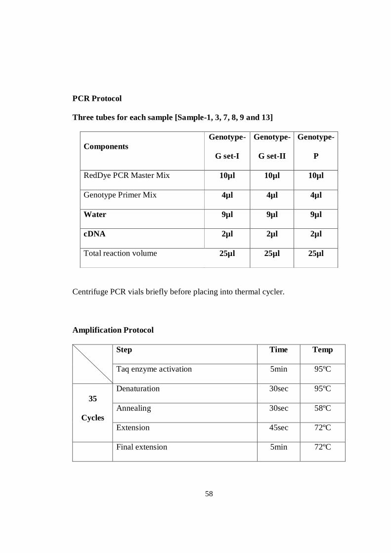
58
PCR Protocol
Three tubes for each sample [Sample-1, 3, 7, 8, 9 and 13]
Components Genotype-
G set-I
Genotype-
G set-II
Genotype-
P
RedDye PCR Master Mix 10µl 10µl 10µl
Genotype Primer Mix 4µl 4µl 4µl
Water 9µl 9µl 9µl
cDNA 2µl 2µl 2µl
Total reaction volume 25µl 25µl 25µl
Centrifuge PCR vials briefly before placing into thermal cycler.
Amplification Protocol
Step Time Temp
Taq enzyme activation 5min 95ºC
35
Cycles
Denaturation 30sec 95ºC
Annealing 30sec 58ºC
Extension 45sec 72ºC
Final extension 5min 72ºC

59
Gel electrophoresis:
Prepare 2% agarose gel as per standard procedure. Loaded entire PCR
amplified product along with 10µl of 100bp DNA Ladder. Run electrophoresis
and visualize in UV Transilluminator.
Agarose gel electrophoresis:
1. Agarose (2%) prepared by adding 2 gms agarose in 100ml of IX TAE buffer
and melted by using micro oven.
2. Added 5µl of Ethidium bromide when the agarose gel temperature reaches
around 60oC.
3. Warm agarose solution poured slowly into the gel platform.
4. Kept the gel set undisturbed till the agarose solidifies.
5. IX TAE buffer poured into submarine gel tank.
6. The gel platform was carefully placed into tank and the tank buffer level 0.5cm
above than the gel was maintained.
7. PCR Samples are loaded after mixed with gel loading dye along with 10µl
HELINI 100bp DNA Ladder.
8. Run electrophoresis at 50V till the dye reaches three fourth distance of the gel.
9. Gel viewed in UV Transilluminator and observed the bands pattern.

FIG 1: IMMUNOCHROMATOGRAPHY KIT WITH ITS CONTENTS
(SD Rotavirus)
Fig 2: IMMUNOCHROMATOGRAPHY CASSETTES (i) Negative Sample and (ii) Positive Sample

Fig 3: ELISA KIT AND ITS CONTENTS
Fig 4: STOOL SAMPLES AND MICROTITRE PLATE

Fig 5:ELISA READER

Fig 6: ELISA MICROTITRE PLATE WITH CONTROLS AND SAMPLES
Negative Control
Positive Control
Positive Control
Negative Control
Negative Control
Positive Control
Plate 1
Plate 2
Plate 3

Fig 7a: RT - PCR ROTAVIRUS: PLATE SETUP AND THERMAL PROFILE

Fig 7b: RT PCR GROUP A ROTAVIRUS AMPLIFICATION PLOTS

Fig 7c: GROUP A ROTAVIRUS GENOTYPING
P - GENOTYPING
G - GENOTYPING

Fig 8a: RT - PCR ROTAVIRUS: PLATE SETUP & THERMAL PROFILE
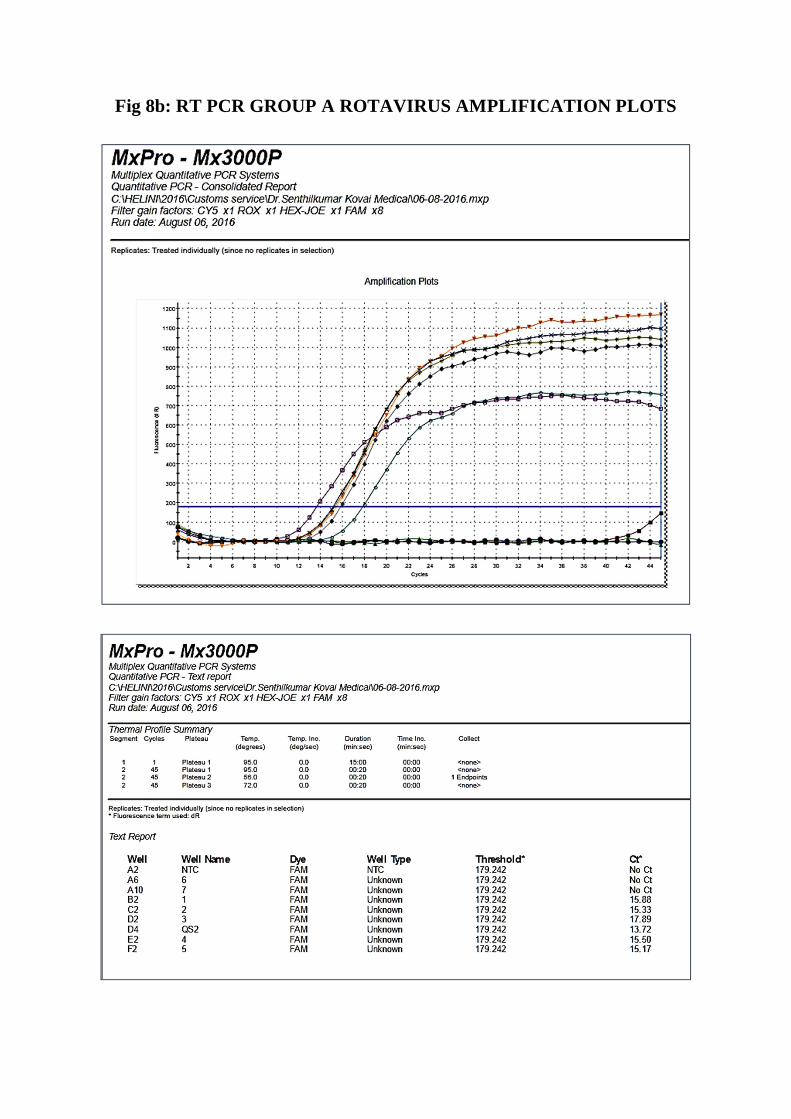
Fig 8b: RT PCR GROUP A ROTAVIRUS AMPLIFICATION PLOTS
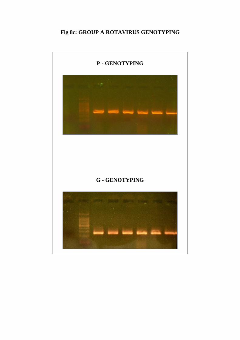
Fig 8c: GROUP A ROTAVIRUS GENOTYPING
P - GENOTYPING
G - GENOTYPING

Fig 9 PCR MACHINE – Aligent MX3000P, USA

RESULTS

60
RESULTS
Prevalence of Rotavirus
During the study period of one year (July 2015 to June 2016), 100
children below five years with acute gastroenteritis were screened for
Rotavirus antigen; of which, 56 (56%) were male and 44 (44%) were female.
Out of 100 samples, 24 were positive for Rotavirus antigen.
Gender distribution of Rotavirus
In this study, out of 100 children 24 were positive for Rotavirus
antigen; of which male children showed 58% (14/24) and female children
showed 42% (10/24) positivity [Chart No. 2].
Age Distribution of Rotavirus
The maximum number of Rotavirus positivity was seen in the age
group of 6 to 12 months (58.3), followed by 13 to 24 months (25%). The
infection rate was found to be lower in the age group of 0 to 6 months
(8.3); likewise after 2 years of age, Rotavirus infection decreases sharply (8.3%)
[Table No. 2] .
61
Seasonal Trends of Rotavirus
Even though the occurrence of Rotavirus was observed throughout the
year, the maximum cases were found during the cooler months of the year
( September to February ) than other months [Chart No. 3].
Geographical Distribution of Rotavirus
Despite Rotavirus being prevalent in all geographical areas, 22% of
cases were from urban whereas 28% from rural areas in this study [Chart
No. 4 and Table No. 4].
Rotavirus infection related with water consumption
In this study higher incidence of Rotavirus positive cases were found in
those using public water supply (25.3% ) than those used bore well water(20.7%).
It indicates there is a higher frequency of Rotavirus infection in those using
public water supply than bore wells [Table No. 5].
Literacy of Parents
Poor literacy of parents is linked with increased rate of diarrhoeal
diseases in young children (76) [Chart No. 5].
Socio Economic Status of Parents
In this study eighty percent of Rotavirus infected children belongs to
low socioeconomic group and 20% from middle class It shows low socio

62
economic status has been linked with higher incidence of rotavirus
gastroenteritis [Chart No. 6] .
Rotavirus Gastroenteritis Related with Feeds
In this study, lower incidence of gastroenteritis were seen among
children who were exclusive breast fed (18%) whereas higher incidence of
gastroenteritis were found in children who were on mixed fed (27%) in
addition to breast fed [Table 6] .
Clinical Features
Vomiting was found in 83% of Rotavirus positive cases while it was
54% in Rotavirus negative cases. Severe dehydration was observed more
consistently with Rotavirus positive patients (79%) whereas it was less in
Rotavirus negative cases (24%). Fever was noted more or less equally in both
Rotavirus positive and negative cases [Table 7].
Diagnosis
Out of 100 samples tested for Rotavirus antigen by ELISA and ICG method,
24 were positive and 76 were negative which indicates both methods are
equally sensitive and specific [Table No. 8].
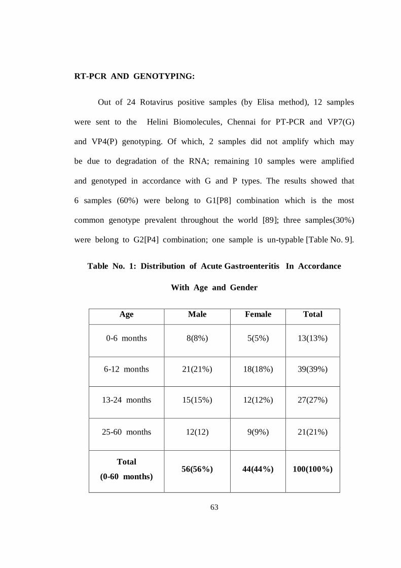
63
RT-PCR AND GENOTYPING:
Out of 24 Rotavirus positive samples (by Elisa method), 12 samples
were sent to the Helini Biomolecules, Chennai for PT-PCR and VP7(G)
and VP4(P) genotyping. Of which, 2 samples did not amplify which may
be due to degradation of the RNA; remaining 10 samples were amplified
and genotyped in accordance with G and P types. The results showed that
6 samples (60%) were belong to G1[P8] combination which is the most
common genotype prevalent throughout the world [89]; three samples(30%)
were belong to G2[P4] combination; one sample is un-typable [Table No. 9].
Table No. 1: Distribution of Acute Gastroenteritis In Accordance
With Age and Gender
Age Male Female Total
0-6 months 8(8%) 5(5%) 13(13%)
6-12 months 21(21%) 18(18%) 39(39%)
13-24 months 15(15%) 12(12%) 27(27%)
25-60 months 12(12) 9(9%) 21(21%)
Total
(0-60 months) 56(56%) 44(44%) 100(100%)

64
Table No. 2: Age Distribution in RVGE Cases
Age No. of Cases No. of positive No. of negative
0-6 months 13 2 (15.4%) 11(84.6%)
6-12 months 39 14 (36%) 25(64%)
13-24 months 27 6 (22.2%) 21(77.8%)
25-60 onths 21 2 (9.5%) 19(90.5%)
Table No. 3: Seasonal Distribution of Rotavirus
Month No. of AGE cases
No. of Rotavirus
positive cases
No. of Rotavirus
negative cases July 4 1 (25%) 3 (75%)
August 5 1 (20%) 4 (80%)
September 7 2 (28.5%) 5 (71.5%)
October 8 2 (25%) 6 (75%)
November 12 3 (25%) 9 (75%)
December 15 4 (26.7%) 11 (73.3%)
January 17 4 (23.5%) 13 (76.5%)
February 9 2 (22.2%) (77.8%)
March 5 1 (20%) 4 (80%)
April 6 1 (16.6%) 5 (83.4%)
May 5 1 (20%) 4 ( 80%)
June 7 2 (28.5%) 5 (71.5%)
Total 100 24 76

65
Table No. 4: Geographical Distribution of Rotavirus:
Geographical area
No. of cases No. of Rotavirus
positive cases
No. of Rotavirus
negative cases
Urban 60 13 (22%) 47 (78%)
Rural 40 11 (28%) 29 (72%)
Total 100 24
Table No. 5: RVGE and water consumption:
Water supply No. of cases No. of Rotavirus positive cases
No. of Rotavirus negative cases
Public water 71 18(25.3%) 53(74.6%)
Bore well water 29 6(20.7%) 23(79.3%)
Table No. 6: Rotavirus Gastroenteritis cases related with feeds:
Feeds No. of cases No. of Rotavirus positive cases
No. of Rotavirus negative cases
Breast feed 33 6 (18%) 27 (82%)
Mixed feed 67 18 (27%) 49 (73%)
Total 100 24 76

66
Table No. 7: Clinical findings in Rotavirus Positive and Negative cases
Clinical features Rotavirus positive cases (%)
Rotavirus negative cases (%)
Vomiting 83 54
Diarrhoea 100 100 Dehydration:
Severe
Mild
79
25
21
75
Fever 88 83
Table No. 8: Comparison of ELISA and ICT in Detection of Rotavirus Antigen
Test No. of specimens
Positive (%) Negative (%)
Elisa 100 24 76
ICT 100 24 76
Table No. 9: Genotypic distribution of Rotavirus
Genotype No of Samples
G1[P8] 6 (60%)
G2[P4] 3 (30%)
Un-typable 1 (10%)

Chart No: 1 Age and Sex Distribution in RVGE Cases.
Chart No.2 Sex Distribution of study population and Rotavirus positive cases.
0
1
2
3
4
5
6
7
8
0-6 months 6-12 months 13-24 months 25-60 months
1
8
4
11
6
2
1
Male
Female
0
10
20
30
40
50
60
MALEFEMALE
56
44
14
10
STUDY POPULATION
POSITIVITY
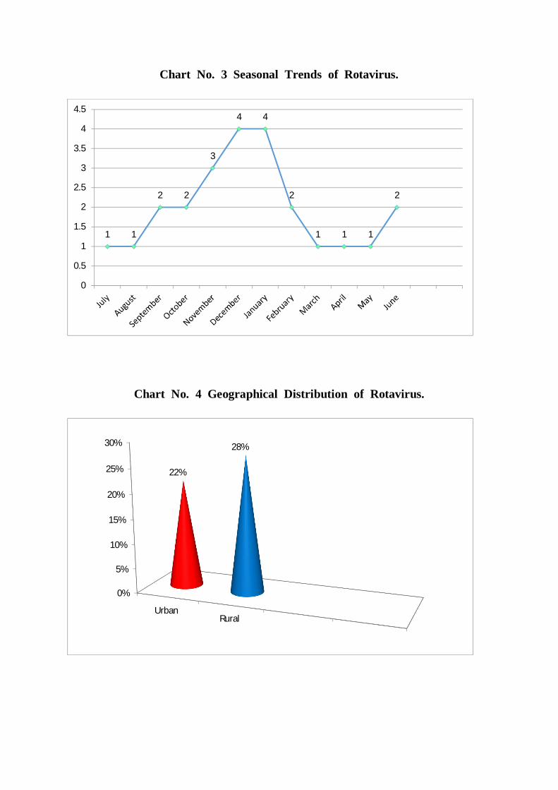
Chart No. 3 Seasonal Trends of Rotavirus.
Chart No. 4 Geographical Distribution of Rotavirus.
1 1
2 2
3
4 4
2
1 1 1
2
0
0.5
1
1.5
2
2.5
3
3.5
4
4.5
0%
5%
10%
15%
20%
25%
30%
UrbanRural
22%
28%
21.7%

Chart No. 5 Educational Status of the Parents.
Chart No. 6 Socioeconomic Status of parents.
55%40%
5%
Secondary education
Primary education
Graduates
0%10%20%30%40%50%60%70%
80%
Low socioeconomic group Middle class
80%
20%

Chart No.7 Rotavirus Gastroenteritis and feeds.
Chart No. 8 Comparison of ELISA and ICT in
Detection of Rotavirus Antigen.
Mixed fed
Exclusive Breast fed
27%
18%
010
20
30
40
50
60
70
80
90
100
ElisaICT
100 100
24 24
76 76
No of specimens
positive
Negative
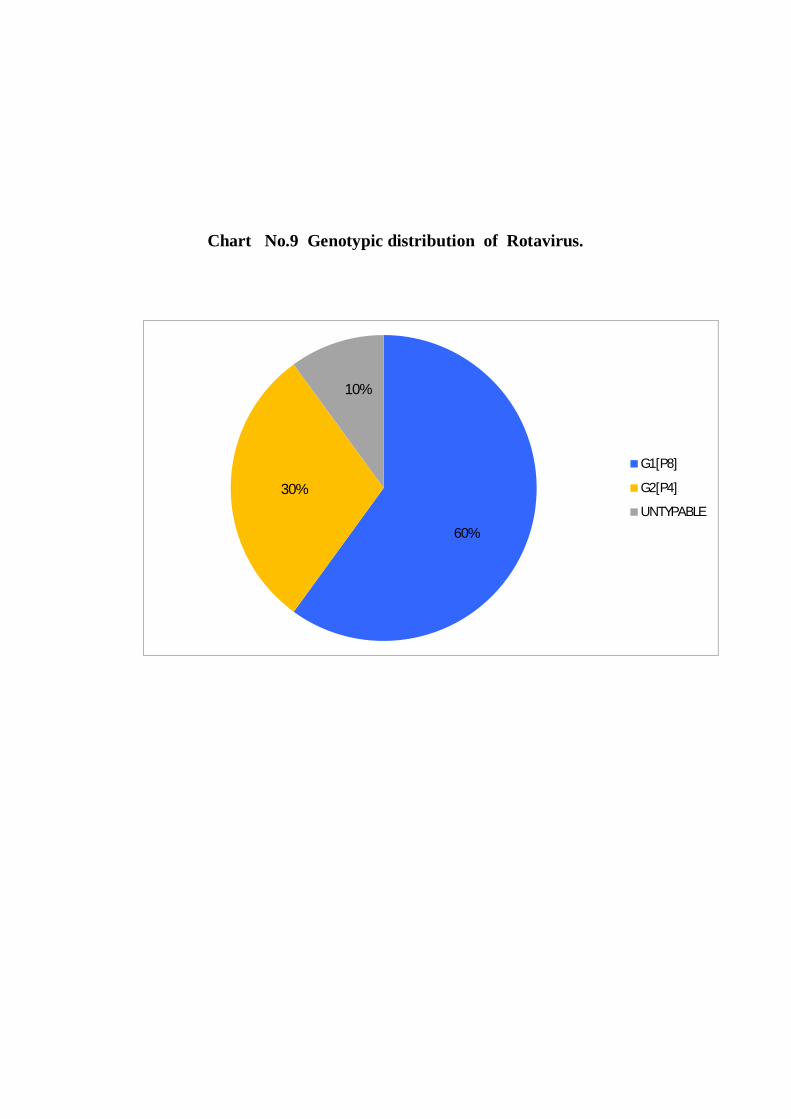
Chart No.9 Genotypic distribution of Rotavirus.
G1[P8]
G2[P4]
UNTYPABLE
60%
30%
10%

DISCUSSION

67
DISCUSSION
Diarrhoea continuous to be the significant cause of mortality and morbidity
in paediatric population worldwide [98,99]; India accounts for one – fourth of all
global diarrhoeal deaths among children less than five years [100]. Even though
diarrhoeal deaths have declined considerably through improved access to medical
facility, the burden of the diarrhoeal disease continues to remain significant. It is
estimated that more than 90% of the diarrhoeal cases occur in developing countries.
Globally Rotavirus diarrhoea alone accounts for 125 million cases and more than
6,00,000 deaths every year; of which, India contributes around 22% of deaths, 30%
of hospitalization and 8.3% of outpatient visits [94] .
Rotavirus diarrhoea also causes significant economic burden to India which
was estimated to be around 4.9 Billion and 5.4 Billion Rupees for hospitalization
and outpatient visits respectively each year , whereas the estimated cost to fund a
Rotavirus immunization program is even less [104]. The average cost of
hospitalization for Rotavirus diarrhoea is approximately from Rs.2000-8400
[105,106] which equates to one or two months of income for an average Indian
family.
Rotavirus belongs to the Reoviridae family. Since 1973, when it was first
demonstrated in children with acute gastroenteritis [29], numerous studies have
been conducted throughout the world regarding the virus and its pathogenesis as
well as epidemiology.

68
Prevalence of Rotavirus:
Global prevalence of Rotavirus gastroenteritis in children below 5 years
varies from 6% to 56% whereas in India the incidence of Rotavirus diarrhoea is
from 5% to 89.9% [92]. The wide difference in the prevalence of Rotavirus
gastroenteritis may be due to difference in the age group, different diagnostic
methods conducted, time of onset of disease and seasonal variation of Rotavirus
and differences in geographical distribution of Rotavirus among various countries
[92].
In this study, the prevalence of Rotavirus infection in children less than five
years was 24% which is in accordance with the study by Rahoni SM et al., who
reported 22.5% positivity for Rotavirus from stool specimen in children from
Brazil [66]. Desai et al., from a hospital based study reported 23.5% of prevalence
in children less than five years. It also correlates with the study conducted by Bahl
et al., from New Delhi (23.5%) and by Saravanan P et al., from Chennai which
showed the prevalence rate of 22.5% in children with acute gastroenteritis.
Previous studies across the world revealed different prevalence rates of Rotavirus
from 6 to 56% [92]. The prevalence rate of the present study (24%) is within the
range of Indian studies, which varies from 5% to 89.9%.
69
In contrast, a low prevalence of Rotavirus has been reported from Bangalore
(16.3%), Kolkata (14.6%) and Vellore (17.8%). This may be related with factors
such as good literacy level, better socioeconomic status and good awareness of the
people regarding hygiene and sanitary measures.
A high prevalence rate of 89.9% was recorded in Manipur. This may be
due to poor literacy level, low socioeconomic status, lack of awareness regarding
hygienic practices and sanitary measures. It is also found that high rain fall and low
humidity as well as low temperature is associated with high incidence of Rotavirus
gastroenteritis [93].
Age distribution of Rotavirus:
In this study, the peak incidence of Rotavirus occurred in the age group of
6 to 12 months (58.3%) which is closely related with the study by Jain V et al.,
who reported the prevalence of Rotavirus as highest in the age group between 7 to
12 months; It also correlates with a study by Bahl R et al., in which the result
showed the peak incidence of Rotavirus in the age group of 9 to 11 months and a
decrease considerably after 18 months of age [71].A hospital based study by
Banerjee I et al., reported that the median age of Rotavirus infection in children is
10months (IQR 7.5-12.5) [69].
The higher frequency of Rotavirus diarrhoea in this age group of 6 to 12
months (58.3%) is because of weaning from breast feed and introduction of

70
artificial feeds. The chance of infection tends to increase when breast feed is
withdrawn since the protective antibodies are lost. Furthermore, introduction of
artificial feed is linked with increased risk of infection unless proper hygienic
measures are strictly followed.
The second most common incidence of Rotavirus was observed in children
in the age group of 13 to 24 months age (25%) as in a study conducted in New
Delhi which reported that 98% of hospitalized Rotavirus gastroenteritis cases were
below 2 years. The peak incidence of Rotavirus infection was found in children
aged 6 to 23 months as reported by Kang G et al., [72]. The higher frequency of
Rotavirus infection in this age group is due to various factors like playing with
elder siblings or neighbours who may have subclinical infection. Transmission of
virus could be possible through fomites such as toys and objects since Rotavirus is
stable in room temperature and highly contagious. Even a low viral load of <100
is enough to cause infection in susceptible children. Malnutrition is another
predisposing factor for these children to develop various infections especially
gastroenteritis.
Lesser frequency of Rotavirus incidence was observed in the babies of 0
to 6 months (8.3%) of age which may be due to protection offered by maternal
antibodies against Rotavirus infection. The IgA anti Rotavirus antibody from
mother passes through the breast milk and offers protection to the babies against
Rotavirus infection; it does not protect reinfection but gives protection against

71
severe infection; baby may get subclinical infection without showing any clinical
features. Asymptomatic infections in newborn nurseries also occur round the year.
The occurrence of Rotavirus declined sharply after 2 years of age (8.3%).
This is because of subsequent infections that usually occur less severely due to the
antibodies formed against previous exposure to Rotavirus. More than 90% of
children aged below 3 years had antibodies against Rotavirus. Every child might
have at least one episode of Rotavirus diarrhoea before reaching 5 years [29].
Sex distribution of Rotavirus:
In this study out of 100 children, 24 were positive for Rotavirus antigen of
which male children showed 58% (14/24) and female 42% (10/24) positivity. The
present study showed that male children were more susceptible for Rotavirus
infection than female children (M: F- 1.4:1) which correlates with the study by
Banerjee I et al., [69]. Another study by Samir et al., from Bahrain reported a ratio
of 1.5:1 in male and female children. A study by Ghazi et al., showed 63% of males
and 33% of females were affected by Rotavirus infection [67].
In contrast, no relationship was observed between Rotavirus infection and
gender preponderance in a study by Saravanan et al., [65]. It is found that male
children excreted a higher rate of Rotavirus in their stool than female children
which could be the reason for higher susceptibility of male children. This was also

72
postulated by Surajudeen A Junaid et al. But the exact reason for susceptibility of
male children than female children to Rotavirus infection is obscure and is still
under study.
Seasonal trends of Rotavirus:
In this present study, Rotavirus peak occurred during the cooler months
(September to February) of the year than other months. This study correlated with
the study done by Bahl R et al., which also showed, there was a distinct peak of
Rotavirus diarrhoea in winter months (November to February) [73]. Another study
by Rahoni SM et al., observed a seasonal pattern of Rotavirus diarrhoea in
association with cooler months [70]. Shariff M et al., from Eastern Nepal reported
that Rotavirus infection occurred round the year with a distinct peak in late winter
(January & February) [72].
In contrast, no significant seasonal trends were noted in a study conducted
in Vellore by Banerjee I et al., [69]. It is postulated that the reason behind seasonal
pattern of Rotavirus infection is because of the possibility of survival of the virus
in the environment and its transmission enhanced by low humidity as well as low
temperature [29]; higher rainfall also enhances the incidence of Rotavirus
infection. It was also noted that, there was no such typical seasonal pattern of
Rotavirus in tropical countries due to high temperature and humidity as well as
high birth rates [87].
73
Rotavirus infection in accordance with feeds (Breast feed/Artificial feed)
In this study, lower incidence of gastroenteritis was seen among children
(18%) who were exclusively breast fed (EBF) whereas higher incidence (27%)
were found in children who were on mixed feed. This indicates that Rotavirus
infection was less frequent in breast-fed than babies on mixed feeds because of
protection afforded by maternal IgA anti Rotavirus antibodies (80,81,82&83). This
is due to the anti-Rotavirus antibody present in the mother because of previous
infection; as persistent subclinical Rotavirus infection is common throughout the
life in adults. In addition, introduction of supplementary feed increases the chance
of infection considerably unless proper hygienic measures such as proper
sterilization of feeding bottles/bowls and washing hands with soap and water
before preparing/ feeding food are strictly followed.
Rotavirus infection in association with socioeconomic status:
Children from low socioeconomic status constitute 80% of the study
population and the rest 20% were from middle class in this study. This indicates
that these children have a higher risk of diarrhoeal diseases than their counterparts
[70,74]. Factors such as, poor sanitation and lack of access to safe drinking water
plays a major role in diarrhoel diseases among children less than five years.
Rotavirus infection and Educational Status of parents:
Literacy of the parents also reflects on the health of the children as
indicated in our study. Children whose parents had only primary and secondary
74
level education seem to be affected more by Rotavirus infections [76,77]. The
incidence was less among children with parents who were graduates. This could
be due to lack of knowledge regarding personal and general hygienic measures as
well as teaching hygienic practices to their children [76].
Geographical Distribution of Rotavirus
Despite Rotavirus being prevalent in all geographical areas, the positivity
rate seems to be higher in rural than urban areas. This is due to lack of awareness
among the rural population regarding hygiene and improper waste disposal which
are considered as the major risk factors for diarrhoel diseases in children below 5
years. Hand washing before preparing food and feeding their children is also
lacking among these population. Open air defecation and vectors such as flies are
important environmental issues to be addressed.
Nowadays, there seems to be increasing prevalence of Rotavirus is being
noted in urban areas as well. This may be due to early weaning from breast feeding
and introduction of artificial feeds, increasing day care centers and play schools
where subclinical infections of Rotavirus occur throughout the year and
transmission through fomites is also a possibility. Another factor could be children
engaging in water sports.

75
Rotavirus Infection and Water supply
There was a higher frequency of Rotavirus infection in those using public
water (25.3%) supply than those used bore wells (20.7%) as evidenced in this
study. In developing countries, use of an unsafe drinking water is one of the most
important and commonest cause of Rotavirus infections [81,84&85]. Group B
Rotavirus is especially known to cause outbreaks of water borne acute
gastroenteritis in children and adults in China, India, Bangladesh and Myanmar
[95].
Clinical Features in Rotavirus Positive Cases:
Diarrhoea, severe dehydration, vomiting and fever are the common
symptoms observed in Rotavirus infected children [8, 86]. In our study, vomiting
was noted in 83 % (20) of Rotavirus positive children whereas it was 54% (13) in
Rotavirus negative cases. Dehydration was a consistent finding in Rotavirus
positive cases in this study. Severe dehydration was found in 79% of Rotavirus
positive cases (19) while 21% (5) of the children had only mild dehydration. In
contrast, severe dehydration was found in 25% (6) and mild dehydration in 75%
(18) of non-Rotavirus gastroenteritis cases. Fever was found more or less equally
in both Rotavirus positive and negative cases.
The above findings from this study indicate that watery diarrhoea,
dehydration, fever and vomiting, are consistent symptoms related with Rotavirus

76
gastroenteritis. Diarrhoea is watery in nature and occurs several times a day which
quickly results in severe dehydration particularly, when it is associated with
vomiting. But the frequency of diarrhoea and the degree of dehydration are much
less in Rotavirus negative diarrhoeal cases. Regarding management, Rotavirus
positive cases need supportive measures which include prompt correction of fluid
and electrolyte imbalance by ORS/IVF. Appropriate antibiotics may be needed in
case of bacterial gastroenteritis whereas it should be avoided in Rotavirus
diarrhoea. So, immense important should be given to diagnose Rotavirus
diarrhoea which helps to avoid indiscriminate use of antibiotics in infants and
young children.
Comparison of ELISA and ICT in Detection of Rotavirus Antigen from
Stool Specimen
Out of 100 stool samples tested for Rotavirus antigen, 24 were positive and
76 were negative in both Enzyme linked immunosorbent assay (ELISA) and
Immunchromatography (ICG) methods which indicate both are equally sensitive
and specific.
Elisa is the method of choice in diagnosing Rotavirus antigen from stool
specimens. It is the most commonly employed method especially when sample
load is heavy [87]. Immunochromatography is an equally sensitive and rapid
method of Rotavirus diagnosis; it is easy to perform; can be used in places where
there is lack of resources and also as a rapid screening test. In addition, it is less

77
time consuming [88] and does not require any technical expertise. Moreover it
seems to be marginally cost effective when compared with ELISA.
Rotavirus Genotyping:
Vast diversity in Rotavirus at genotypic level emphasizes the need for
surveillance of circulating Rotavirus strains. Furthermore, surveillance of
Rotavirus strains in a particular geographical area is essential to assess the
epidemiology of the disease and also to identify the changing pattern following the
introduction of vaccination in the community. This study is aimed to find out the
prevalence of Rotavirus and its molecular characterization to identify the serotypes
which are circulating among the population.
The most common G and P types reported from India are G1,G2,P4,P6 and
P8. In Indian children,G9 strains as well as mixed Rotavirus infections are quiet
common; in some regions of India, bovine origin of P6 strains have been
reported[105]. The unusual Rotavirus strains are formed by re-assortment of
human and bovine Rotaviruses which are promoted by the age old tradition of
calves and humans living in the same household and socio economic conditions in
India. Diversity of Rotavirus strains as well as high prevalence of mixed infections
is a distinct feature of Rotavirus epidemiology in India [105].
Out of 24 Rotavirus positive samples (by Elisa method), only 12 were
chosen randomly for RT-PCR and VP7(G) and VP4(P) genotyping due to resource

78
limited settings. Each sample was put in a separate 2 ml sterile centrifuge vial along
with viral buffer medium and sent to the Helini Biomolecules, Chennai for RT-
PCR and genotyping.
In this study, out of 12 samples tested for RT-PCR, 2samples did not
amplify; this may be due to degradation of RNA while transportation or low viral
load at the time of specimen collection. The remaining 10 samples were amplified
and genotyped in accordance with G (G1,G2,G3,G4 & G9 ) and P (P4,P6&P8)
types. The results showed that G1[P8] was the predominant combination (60%)
followed by G2[P4] (30%). This coincides with the study conducted by Girish
Kumar, C.P., et al., who reported G1[P8] 62.7% [98].A similiar study from
Mumbai by Sushmitha A Shetty et al., reported G1[P8]-33.3% and G2[P4] as
26.7% [99]. It also correlates with results from Karnataka by Ranjitha S. Shetty et
al., G1[P8] and G2[P4] as the most common genotypes[92]. S.Babji et al., from
Nagercoil reported a prevalence of G1[P8]-44.6% and G2[P4]-18.4% genotypes
[100]. The G2[P4] is the predominant combination next to G1[P8] [95,96,97].Out
of 10 samples tested for strain characterization, one is un-typable which may be
due to mixed infections or point mutation at primer binding site [94,100].
In contrast, a rare emerging strain G11P[25] was isolated from Mumbai
during a 2005-2009 study by Sushmitha A. Shetty et al.,[99]. From Pune,G9P[8],
G12P[6] and G12P[8] were isolated in 2009 as reported from a study by Shoba D
et al.,[94]. Another study from Nagercoil by SBabji et al., reported that G2 [P4]

79
was predominant than G1[P8] in 2007 and G12P[6] as well as G12P[8] emerged
only during that period [100].
In Group A rotavirus, G1 is the most common prevalent strain worldwide.
Epidemiologically, Children infected with G1 strain were associated with severe
dehydration than those infected with other strains [89,96,97]. Despite the Rotavirus
incidence being similar in developed as well as developing countries, more
mortalities and health care associated expenses occur in the developing countries
[90] mainly because of poor access to medical care, timely intervention,
predisposing factors like malnutrition and other social as well as environmental
factors.
Rotavirus vaccination:
Interventions such as improved hygiene, sanitary measures and drinking
water do not adequately prevent the Rotavirus diarrhoea. Other than that there is
delay in accessing health care facilities thereby affecting timely intervention and
prognosis.So vaccination is the best way of protecting children from Rotavirus
infection thereby, reducing disease burden and mortality.
The first vaccine for Rotavirus was licensed for use in United States,
Finland and Venezuela in 1998 and it was found to be 80-100% effective in
preventing severe group A Rotavirus diarrhoea in children [54]. But it was
withdrawn in 1999 by the manufacturer due to the issue of suspected

80
intussusception in vaccinated children (1in12000). In 2006, two new vaccines,
Rotarix and Rotateq were introduced for combating group A Rotavirus which were
found to be effective, safe and without risk of intussusception as reported by
clinical trials [58]. Mexico was the first among all other countries in the world to
use Rotavirus vaccine in children (2006) and it was found that diarrhoea associated
deaths decreased by more than 65% [57,58]. Subsequently Rotavirus vaccines were
implemented in more than 80 countries around the world and there was found to
be a decrease in hospitalization due to diarrhoea reported in more than 10 countries
of the developing world[11,12,13,14]. Rotavirus vaccine also significantly reduces
hospitalization among older children who were not immunized. This phenomenon
is called indirect effect or herd protection.
The WHO recommended that Rotavirus vaccination can be integrated in
the National Immunization Program (2009) to prevent infant mortality and
morbidity [57]. At present, two vaccines - Rotarix TM (GlaxoSmithkline Vaccines)
and Rotateq R (Merk) have shown good safety profile and excellent efficacy against
the globally circulating G (G1,G2,G3 & G4) and P types(P[4], P[6], & P[8] ) in
large global studies [58].
Integration of these vaccines in National immunization programs has been
associated with marked reduction in gastroenteritis related mortality and less
frequent diarrhoea related hospitalization as well as outpatient visits in different
parts of the world [90&91].
81
Around 90,000 - 1,53,000 children die from Rotavirus infection each year
in India; around 4% of overall mortality in children under five years could be
saved in India, if Rotavirus vaccines were integrated with National Immunization
Program [Shaun K Morris et al] (Bulletin of the World Health Organization
2012;90:720-727) [59].
India has developed a new vaccine against Rotavirus - Rotavac in May
2013. It was developed under the joint collaboration between India and the United
states in the area of medical research. Integration of a Rotavirus vaccine in the
National Immunization Program would prevent more than 25,000 deaths, nearly
3,00,000 hospitalization and more than 6,00,000 outpatient visits each year in
India[105]. On phase 3 clinical trials, Rotavac has showed effective efficacy and
excellent safety profile and it would be available at Rs.54 per dose [60]. Indian
Academy of Pediatrics has recommended inclusion of Rotavirus vaccine in the
National Immunization Program [112].
It is found that the Rotavac reduced severe diarrhoea by more than 56%
during first year of life with protection continuing into the second year also.
Moreover, the vaccine also exhibited great impact against severe diarrohea of any
etiology[5].
In India, Himachal Pradesh, Andhra Pradesh, Haryana and Odisha have
become the states involved in pilot study to include Rotavirus vaccination in
Universal Immunization Program (UIP). The project was launched in Kangra

82
district (H.P) by the honorable Chief Minister. It is given as Rotavirus
drops(1ml,orally,for two doses-Rotarix and 2ml,orally,for three doses-Rotateq ) to
babies at 6, 10 and 14 weeks after birth. The vaccine is started at 6 weeks of age
and should be completed by 24 weeks of age.

SUMMARY

83
SUMMARY
Rotavirus gastroenteritis is a global health concern which accounts for
around 125 million cases of diarrhea and 6,00,000 deaths in children
under five years worldwide every year.
The present study was aimed to determine the prevalence of Rotavirus and
its molecular characterization in children less than five years with acute
gastroenteritis in Coimbatore.
Overall, 100 children were screened for Rotavirus antigen from stool
specimen by Enzyme linked Immunosorbent Assay and
Immunochromatography.
Out of 100 samples, 24 were positive for Rotavirus antigen; of which, 58
were male children while 42 were females.
From overall 24 positive samples, 12 were tested for RT-PCR. Of these, 10
samples were PCR confirmed, and were analyzed further for strain
characterization and showed G1P[8] to be the predominant strain (60%)
followed by G2P[4] (30%) while one sample(10%) was un-typable.
The Rotavirus peak occurred in the age group of 6 to 12 months (58.3%)
followed by 13 to 24 months (25%).
The majority of cases were observed during the cooler months of the year
September to February) although cases were recorded throughout the year.

84
The risk factors for Rotavirus diarrhea include artificial feeds, low
socioeconomic status and literacy level of parents, open air defecation and
use of unsafe drinking water whereas breast feed appears to give protection
against Rotavirus.
Correction of electrolyte and acid base imbalance by Oral Rehydration
Solution (ORS)/Intravenous Fluid (IVF) is the mainstay in the treatment of
Rotavirus gastroenteritis.
In Rotavirus, vast diversity in its genotypic level emphasizes the
significance of epidemiological surveillance of circulating strains.
The immense importance of molecular characterization of Rotavirus is
to assess the epidemiology and to detect the diversity of the current
circulating strains to prepare an effective vaccine to deal with various
strains.
Rotarix (monovalent) and Rotateq (multivalent) are the effective
vaccines against Rotavirus, recommended by the WHO currently available
worldwide.
Rotavac is the indigenous vaccine of India as recommended by Indian
Academy Of Paediatrics (IAP) which shows good safety and excellent
efficacy in lowering the disease burden and mortality associated with
diarrhea in children less than five years.

CONCLUSION

85
CONCLUSION
o Diarrhea continues to be the third most common cause of childhood
deaths worldwide; Rotavirus remains the leading cause of diarrhea in
children less than five years accounting for 6,00,000 deaths every year
o Out of 100 children tested for Rotavirus antigen from their stool specimens
for a period of one year, 24 were positive which includes 14 male and 10
female children indicating more susceptibility among male children.
o The maximum Rotavirus positivity occurred in the age group of 6 to 12
months.
o The peak incidence was noted during the cooler months of the year.
o On screening of Rotavirus antigen from stool specimen, both ELISA and
ICG showed equal sensitivity and specificity (24 positive and 76 negative).
o ELISA is commonly employed especially when sample load is high while
ICG is particularly useful for rapid screening test especially in resource
limited settings
o Oral rehydration therapy (low osmolarity ORS) is the mainstay of treatment
in Rotavirus Gastroenteritis.
o On molecular characterization of Rotavirus positive samples, G1 P[8]
(60%) and G2 P[4] (30%) were the most common serotypes isolated in
the study.

86
o Rotarix (monovalent) and Rotateq (multivalent) are the universally
accepted vaccines against Rotavirus and also recommended by the WHO.
o Rotavac is the indigenous vaccine of India which showed excellent efficacy
and good safety profile in lowering the illness and death linked with
childhood diarrhoea.
o Inclusion of Rotavirus vaccines in the National Immunization
Program(NIP) schedule has in Himachal Pradesh , Andhra Pradesh ,
Haryana and Odisha will go a long way in preventing Rotavirus
gastroenteritis and its associated mortality and morbidity in paediatric
population.

BIBLIOGRAPHY

BIBILIOGRAPHY
1. Surajudeen A Junaid ,Chijioke Umesh ,Atanda O Olabode and Jim M
Banda : Incidence of RotaVirus infection in children with gastroenteritis
attending Jos University Teaching Hospital,Nigeria.Virology journal
2011;8:233.
2. Estes MK and Kapikian AZ.RotaVirus.In:chapter 53,Field’s
virology,Knipe DM,Howley PM, Ciffin DE,Lamb RA,Martin RA ,
Roizman B,SE Straus(eds) vol.2,section II,5th edition,Wolters
Kluwer,Lippincott Williams and Wilkins
publication,Philadephil,2007:PP.1924.
3. Jawetz E, Malnick JL,Adelberg EA.Reovirus,Rotavirus and
Calcivirus,In:chapter 37,medical microbiology;24th edition,Jancet F,Jim
R,Harriet L,Barbara R(eds).Lange medical publisher,Los
Angeles,California,2007:PP.503-506.
4. De Zoysal,Feachem RG.Intervention for the control of diarrhoeal disease
among young children:RotaVirus and cholera immunization.Bull World
Health Organisation 1985;63:569-83.
5. Bhautik Modi : RotaVirus diarrhoea – current scenario and preventive
strategies:Natinal Journal of Medical Research 2013;4:2:3.
6. Jawetz E,Malnick JL,Adelborg EA:Reovirus In Medica
microbiology,24th edition,edited by Mandell GL,Bennett JE,Dolin
R,Philedelphia,PA.Churchill Livingstone:2000:1699-1707
7. Subitha Lakshmi Narayan and Ramakrishnan Jayalakshmi:Journal of
Natural science,Biology and medicine.J.Nat Sci Bio /Med.2015 Jan-
Jun;6(1):24-28.
8. Anup Kumar Shetty,Faizan Mohammed Kalektan,Sabin Jote
Muthiravalapil,Rekha Boloor,Beena Antony:Detection of RotaVirus and
AdenoVirus diarrhoea in children below five years,In Dakshita Kannada
district,A costal region of Karnataka state,India.
9. Dey RS,Gosh S,Chawala – Sarkar M,Panchalingam S,Natro JP,Sur
D.etal.circulation of novel pattern of infections by entrick AdenoVirus
type 41 among children below five years of age in Kolkata,India.J Clinical
microbiology 2011;49:500-5.
10. Glass RI,Parashorr UD,Bresee JS,Tureios R,Fischer TK,Widdowsen
MA,etal.RotaVirus vaccine:Current prospects and Future
challenges.Lancet 2006;368:323-32.
11. Georicy RV, Dockrel HM, Wakelin D, Roitt IM, Mims C, Cliodine (PL)
ends. GIT infection. In:chapter 22, Mims Medical microbiology, GM
celium, mosby Glsevier Ltd, china,2008:P.300-302.
12. Feigin RD, Cheny JD, Demmlev GJ, Kaplem SL. calcivirus and
Haepetetis E virus In;Chapter 173,Text book of pediatric Infectious
Diseases,5th edition,Volume II,2011:PP.2087-2101.
13. Winn WC,Allen SD,Jinda Win,Konemann EW,preop
GW,Schreekembezer PC,Would GL.Diagnosis Of Infectious Diseases
Caused by viruses,Chlamydia,Rickettsia and related organisms In
:Chapter23,Konemann’s colour Atlas and textbook of Diagnoistic
Microbiology,6th edition,Lippincots,William would Wikkins
Publications,Philedelphia,2006:PP,1394.
14. Forben.BA,Salm DF,Weissfield As,Laboratory methods in basic
virology.In chapter 51,Bailey an Scott’s diagnostic microbiology,12th
edition,mosby elserier,Mussourie:PP.725.
15. Parashar UD,Glass R.Viral gastroenteritis:In:Chapter 174,Harrison’s
principles of medicine 16th edition,Kasper DL,Fauli AS,Longo
DL,Brawn.
16. Chin J:Control of communicable diseases manual.Wash.DC:American
Public Health Association.J Clinical Microbiology 2000,125(17):910-7.
17. Jawetz E,Malnick JL,Adelborg EA:Reovirus In Medica
microbiology,24th edition,edited by Mandell GL,Bennett JE,Dolin
R,Philedelphia,PA.Churchill Livingstone:2000:1696-1703.

18. Rajiv Bahl,Pratima Ray Strati Subosh,Prashant Shambharkar,Manju
Saxena,Umesh Parshar,Jon Gentsch,Roger Glass and M.K.Bhan for the
Delhi Rotavirus study group.Identification of seven Rotavirus diseases in
New Delhi, India and G and P types or the infecting Rotavirus strains.Jid
2005:192.Bahl et al.
19. Ghassan Dhaibo,Marriam Rajab,Adlette I Nati,Raymond Mikhad,Elie
Choueiru,Mohammed Al-Tamir,Omar Salam,Gunasekaran
Ramakrishnan.Rodrigo De Antonio.Hospital based survilence study of
Rotavirus based entities in children under five years of age in
Lebanon.Trails in vaccinology,13th Aug 2013.
20. Alam MM, Khurshid A,Shankar S,Suleman RM,Sharif J etal(2013)
correction:epidemiology and genetic distribution of Rotavirus strain
inchildren with acute gastroenteritis in Lahore,Pacistin.Plos
one(12):10.1371/annotation/68A/d 471- b31 – 45E – 7 – ab81 – d242fic
20 ad.
21. Damme PV,Giaguicuto C,Todd P Wielen MV.On behalf of the reveal
study group distribution of Rotavirus geography in Europe 2004 –
2005.The reveal study.J inf dis 2007:s-17-25.
22. Matthijnessens J, Ciarlet M, Heinom E, Arijis I, Delbeke T, Mc Donald
SM et al. Full genome based classification of Rotaviruses reveals
common origin between human WA like and porcine Rotavirus strain and
human DS-1 like and bovine Rotavirus strains. J Viroligy April/2008;
3204-3219.
23. Cardio DDP, Soaves CMA, Azeredo MSP, Leite Jple, Munford,V and
Racz ml. Serotypes and Subgroups of Rotavirus isolated from children in
central Brazil. J Health Population Nutr June; 18(1): 39-43.
24. Courbas mcu, Beraud Dm, Berads GM, Camphell AD, Genzalez JP,
Georges AJ and Flewtt TH,. Subgroups serotypes and e-pherotypes of
Rotaviruses isolated from children in Bangui, Central Africa Republic. J
clinical Microbiology April 1988; Vol 26(4): 668-671.

25. Eshes MK and Kapikias AZ. RV In: Chapter 53, Field’s virology Knipe
DM, Howler PM, Giffin DE, Lamb RA, Martin MA, Roizman B, SE
Straus (eds) edition, Vol.2, Wolters Kluwer, Lippincott Williasms and
Wilkins Publications, Pheledelphia, 2007: P. 1941-1944.
26. Levinson W,.RNA Non enveloped Viruses. In: Chapter 40, Medical
Micrology and Immunology, 10th edition, McGraw Hill publications,
New York, 2006: P.290-291.
27. Giaguinto C, Damme PV, Muet F, Gotheofors L, Maxwell M, Todd P et
al., On hehel of the Reveal study Clinical consequences of Rotavirus
Acute Gastroenteritis in Europe.2009-2005: The reveal study. J Inf Dis
2007;195:S26-35.
28. Bass CW, Rosey Kn: Rotavirus and other agents of Viral Gastroenteritis.
In: Nelson text book of paed. Edited by Richard E and Beharman F. Raven
press, Pheledelphia; 2004:107-110.
29. Jawetz E, Malnick JL,Adelberg EA.Reovirus,Rotavirus and
Calcivirus,In:chapter 37,medical microbiology;24th edition,Jancet F,Jim
R,Harriet L,Barbara R(eds).Lange medical publisher,Los
Angeles,California,2007:PP.505-506.
30. Gentsch JR, Woods PA, Ramachandran M, et al., Review of G and P
typing reveal from a global collection of strains: Implication for Vcccine
develop. J Inf Dis, 1996; Au (supplement 1) ; S30-6.
31. Nair GB, Ramamoorthy T, Bhattacharya MK, Krishnan T, Ganguly S,
Suha DR et al., Emerging trends in the etiology of enteric pathogens as
evidenced from an active surveillance of hodpitalized diarrhoea patient in
Kolkata, India, Gut Pathog 2010;2:4.
32. Murphy TV, Gargiullo PM, Massondi MS, Nelson PB, Jumaan AO,
Okoro CA, et al. Rotavirus intussusception investigation team.
Intussusception among infants given an Oral Rotavirus vaccine. N Engl J
med. 2001;344:564-72.

33. Kelkar SD, Purohit SG, Simha KV. Prevention Rotavirus diarrhoea
among hospitalized children in Pune, India. Indian journal of Med Res
1999;109:131-5.
34. Chakravan A, Kumar S, Mittal SK, Brcor S. Clinical and epidemiological
features of acute gastroenteritis caused by Human Rotavirus Sub groups,
JDiarrhoeal Dis Res 1992;10: 21-4.
35. Singh V, Brcor S, Mehtha S, Metha SK. Clinical and epidemiological
features of Acute Gastroenteritis associated with hauman Rotavirus Sub
groups I and II in Northern India. Indian Journal Gastroenterol.1989;
8:23-5.
36. Younis Abdul-Redha-AL-Khafasi and Hawraa J. Al-Jihoury: Detection
of rotavirus in diarrhoea stool sample of children with acute
gastroenteritis in Babylon Governorate, Iraq. Intern Reaearch Journal of
Microbiology: (IRJM) ISSN: 2141-5963 Vol.4(3) 84-88. March 2013.
37. Patten JT and Gallegas CO.Rotavirus RNA Replication:SS RNA extends
from the replicate particle.J General Virology 1990;31:1087-1094.
38. Dessel Berger U,Gray J and Ester MK.Rotavirus In:chapter 44,Topley
and Wilsons microbiology and microbial infections,virology.Brain
WJ,Mahy,Meutwn VT(edta).10th edition,vol – II,Hodder Arnold
Publications,London 2005:ID:946 – 958.
39. Rao.M.R:Savarino,S.J;Kim,Y:Wierzha,T.F;Peruski,L;Lee,Y.J;Gentsch,J
.R:Glass,R.I and Demens .J D.Epidermiology of Rotavirus diarrhoea in
Egyptian children and implication for disease control.Am J
epidermiol.1999,150(7):770-7.
40. Bass DM viral infections of intestinal trackt.In : Chap 19.26 Walkers
Pediatric GI disease,Kleinman RE,Sanderson IR,Goulet O,Sherman
PM,Vergani GM,Schneider BL(eds),5th edition,vol.I,BC Decker
INC,Hamilton,2008:PP.434-437.

41. Kang G.Rotavirus vaccines. Ind J Med microb 2006;24(4):252-7.
42. Jawetz E,Malnick JL,Adelborg EA:Reovirus In Medica
microbiology,24th edition,edited by Mandell GL,Bennett JE,Dolin
R,Philedelphia,PA.Churchill Livingstone:2000:1699-1705.
43. Saravanan P,AnanthaRam S,Anantha Subramaniyan N.Rotavirus
infections among infants and young children in Chennai,South India,Ind.J
Medical microbial.2004;22(4):212-21.
44. Jawetz E,Malnick JL,Adelborg EA:Reovirus In Medica
microbiology,24th edition,edited by Mandell GL,Bennett JE,Dolin
R,Philedelphia,PA.Churchill Livingstone:2000:1693-1703
45. Berrnstein D1,Ward RL.In:chapter 176,text book of pediatric infectious
diseases,Leigin RD,Cheny JD,Demmler GJ,Kaplan SL,5th
edition,Vol.2,2004 PP.2110-2133.
46. Englekirk PG,Englekirk JD.Laboratory diagnosis of selected viral
infections.In : Chap 25, Laboratory diagnosis of infectious diseases
Wolters Kluwer,Lippincott,Williams and Wilkins
publications,Pheledelphia,2008:PP.657.
47. Fenner F,White DO.Other family of viruses.In : Chap 22 ,medical
virology,2nd edition,Academic Press,Newyork,1976;PP.406 – 409.
48. Bhatnagar S,Lodha R,Chowdhury P,Sachder HP,Shah N,Narayan S,et
al.IAP Guidelines 2006 on management of adult diarrhoea.Indian
pediatr.2007;44:380/9(Pub Med).
49. Shah D,Choudhury P,Gupta P,Mathew JL,Gera T,Gogia S,et
al.Promoting appropriate management of diarrhoea;a system review of
literature for Advocacy and Action : UNICEF – PHFI series on new born
and child health,India.Indian Pediatr.2012;49:627-49(Pub Med).
50. Bajait C,Thawani V.Role of zinc in pediatric diarrhoea.Indian J
Pharmacal.2011;43:232-50(Pub Med).
51. The situation of children in India - A
profile(http/www.UNICEF.org/India/health).

52. Rotavirus vaccine for the protection of viral gastroenteritis among
children. Recommendation of the Advisory Committee on Immunization
practices (ACIP).MMWR Recomm Reep 48(RR-2):1-
20.1999.PMID102/9046(www.nchi.nlm.nih.gov/Pub Med/10219046).
53. Kapikian AZ(2001)A Rotavirus vaccine for prevention of severe
diarrhoea of infants and young children:development,utilization and
withdrawl.Novartis found synp.238:153-71;discussion 171-
9.doi:10.1007/0470846534 chap 10.
54. Biner JE(2005) Rotavirus vaccine and Intussusception
risk(http/meta.wkhealth.com/pt/pt.core/template
journal/wngaterway/media/landingpage.htmpissm.
55. Dennehy Plt 2008.Rotavirus vaccines:overview.Chin
microbial,Rev.2,(1):198-208.
56. Tate JE,Patel MM,Steele AD,Gentsch JR,Dayne DC,Cortese
MM,Nakagoni O,Cunliffe NA,Jinng B,Neuzil KN,de Oliversa LA,Glass
R,Parashar UD (Apr 2010).Global impact of Rotavirus vaccines.
Expert review of vaccines 9(4):395-407.
57. Vesikari T,Masson DO,Dennehy P.Van.Damme P,Santosh
MM,Rodriguezz Z,et al.Safety and efficacy of pentavalent human –
bovine (WC3) reasons for Rotavirus vaccine.N Engl J Med 2006:354:23-
33 doi:10.1056.
58. Soarej – Weiser K,Madehose H,Bergman H,et al(2012).Vaccine for
preventing Rotavirus diarrhoea:vaccine in use.Cocbrane database syst rev
11:CD0085.
59. Shaun K Morris,Shally Awasthi,Ajay Khera,diegag Bassanis Gagindeep
Kang,Umesh D,Parashar et al.Rotavirus mortality in India : estimates
based on anationally representative survey of diarrhoel deaths.Bulletin of
the WHO 2012,90:720-727.
60. India Unveils first indigenous Rotavirus vaccine. Available
@http://www.thehindu.com/sei-tech/health/medicine and
vaccine/article4714757. ece. Last assest on may 2013.

61. ICMR to launch task force projects on burden of Rotavirus diarrhoea in
India. available@:http;//www.pharmatutor.org/pharma-news/icmr-
project-on-rv-diarr. Last assessed on may,2013.
62. Verma R, Khanna P, Chawk S. Rotavirus vaccine can save millions of
childrens’ lives in develop counts. Hum vaccine Immunother.2012;7:272-
4 (Pub Med).
63. Ramani S, Kana G, Burden of disease and molecular epidemiology of
group A Rotavirus infection in India. Indian J Med Res 2007,125;617-32.
64. Kang G, Arora R, Chitambar SD, Neshpamele J, Gupta MD, Kullikans M
et al; Indian Rotavirus strain surveillance Network. J Inf Dis 2009 Nov
1;200(suppl 1); S147-53. Mendez E and Arias CF.Astrovirus.In : chapter
29,fields virology,Knipe Dm,Howley Pm,Giffin DE,Lamv RA,Martin
MA,Roizman B,SE Straus (eds).5th edition,vol.2,section II,Wolters
Kluwer,Lippincott Williams and Wilkins
Publications,Pheledelphia,2007:PP.982-984.
65. Paniker CK, Mathew S, Martham M, Rotavirus and adult diarrhoea,
diarrhoeal diseases in children at south indian costal town.Bull world
Health Organ 1982;60:123-7.
66. Sengupta PG, Sen D, Saha MR, Niyogis S, Defi BC, Dal SC, et al. An
epidemiology of Rotavirus diarrhoea in Manipur, India. Trans R Soc Trop
Med Hyg 1981;75:521-3.
67. Banerjee I, Ramani S, Prime rose B, Mosis P, Iturriza GM, Gray J et al.
Comparative study of epidemiology of Rotavirus in children from
community based cohort and hospital in South India. J clin Micro 2006
July;44(7):2468-74.
68. Rahoni SM, Nogueira MB, Hakim VM, Torrecilha V TG, Lerner H and
T.Sudhiya LR V. Comparison of Latex agglutination with EIA for
detection of Rotavirus in fecal specimens. AM J Clinical pathol
2002;117:392-394.
69. Jain V, Parashar UD, Glass RI, Bhan MK. Epidemiology of Rotavirus in
India. Ind L Pediatr 2011 Sep;68(9):855-62.
70. Shariff M, Deb M, Singh R. A study of diarrhoea among children in
Eastern Nepal with reference to Rotavirus. Ind Journal of Med Microbiol,
2003;21(2):87-90.
71. Gupta N,Jain Sk,Rathnesh,Chawla U,Hossain S,Venkatesh S.An
evaluation of diarrhoel diseases and acute respiratory infections control
programs in a delhi slum.Indian J Pediatr.2007;74:471-6(Pub Med).
72. Zarani AH,Modarres SH,Jadali F,Sabahi F,Moqsseni SM,Vazirian
F:Role of Rotavirus in children with acute diarrhoea in Teheran,Iran.J
Clin Virol 2004,29:189-93(Pub Med).
73. Park K,Textbook of preventive end social medicine.19th
ed,Jalalpur,India:Banarsidas Bhanot,2007.Epidermiology of
communicable diseases:PP.142-7.
74. Jayalakshmi R,Roy G,Premarajam KL.Incidence risk factors of acute
diarrhoel disease among under five children in urban slums in
Pudhucherry Z – one year follow up study.Indian J matern child health
2011,13:1-11.
75. Ghosh S,Sengupta PG,Mondal,Banu MK,Gupta DN,Sircar BK.Risk
behavioural practices of rural mothers as determinance of childhood
diarrhoea.J commune dis.1997;29:7-14(Pub Med).
76. Abdul – Razzak SH,Hissan,Dawood S,Hameed,Asmaa
H,Hwaid.comparison of latex agglutination with EIA for detection of
Rotavirus in children with acute diarrhoea in Diyala
province,Iraq.American Journal of infectious diseases and microbiology
3.2(2015)70-73.
77. Angel.J:Franco.M.A and Green bergs H.B. Rotavirus immune responses
and correlation of protection.Curr.Opin Virol.2012;2(4):419-25.
78. Panda S;Deb.AK;Chawla- Sarkar.M;Ramamurthy.T;Ganguly S;Pradlow
P;Chakraborty.A,Desai.S,Gupta.MD and Dhese.R.Factors associated
with diarrhoea in young children and incidence of symptomatic Rotavirus
infection in Rural West
Bengal,India.Epidemiol.Infect.2014,142(9):1848-58.

79. Bonkounjou,I J;Sanou,I;Bon.F;Benon,B;Coulibaly,SO;Haukka ;
Traore,A.S and Barro.N.Epidermiology of Rotavirus infection among
children with acute diarrhoea in Burking Faso.BMC Pediatr.2010:10:94.
80. Pleng – Bonig.A ; Soto – Ramirez.A ; Karmaus, W;Petersen,G;Davis,S
and Forster.J.Breast feeding protects against acute gastroenteritis due to
Rotavirus in infants.Eur. J Pediatr.2010;169(12):1471-6.
81. Malek.MA:Teleb.N:AB4 =
Elyazeed.R,Riddle,M.S;Sherif:M.E;Steele,A.N; Glass.RI and Brgel.J.S.
The epidemiology of Rotavirus diarrhoea in countries of the Eastern
Mediterranean Regions. J. Infect Dis.2010:202 suppl:S12-22.
82. Naficy, A.D; AbElyazeed.R;Holmes, J.L; Rao
.M.R:Savarino,S.J;Kim,Y:Wierzha,T.F;Peruski,L;Lee,Y.J;Gentsch,
J.R:Glass,R.I and Demens .J D.Epidermiology of Rotavirus diarrhoea in
Egyptian children and implication for disease control.Am J
epidermiol.1999,150(7):770-7.
83. Stact MA,Azimi PH,Berke T,Robert’s N,Bernstein DI,Ward
RL,Pickering LK,Matson DO:Clinical presentation of Rotavirus infection
among hospitalized children.Pediatr.infect.Dis J 2002,21(3):221-7(Pub
Med)
84. Mendez E and Arias CF.Astrovirus.In : chapter 29,fields virology,Knipe
Dm,Howley Pm,Giffin DE,Lamv RA,Martin MA,Roizman B,SE Straus
(eds).5th edition,vol.2,section II,Wolters Kluwer,Lippincott Williams and
Wilkins Publications,Pheledelphia,2007:PP.981-984.
85. Momenzatdh A,Shahrzad M,Faraji A,Motamoda Radi M,Sohrabi L
A,Modarres S,Azarmoush L,Mirshahahil H.Comparision of EIA,ICT and
RNA- Polyacrylamide – Gel Electrophorosis,for diagnosis of Rotavirus
infection in children with Acute Gastroenteritis.Iran J Med Sci Sep
2008;33(3):173.
86. Altactiol.Aparecido Nunes,Lune Marques DE Mello,Renata Parrode,Joao
Paulo Maneira Bitlor.Revista DA Amrigs,Porto Alegre,54(2):147-151-
abr – Jun.2010.

87. MM Patel.AD Elark.CF Sanderson.J,Tate.UD.Paraskar,Removing the
age restrictions for Rotavirus vaccination:A benefit – risk modeling
analysis.Plos Med 9e(2012)1001330.
88. GM.DO Earmo.C Yen.J.Cortes.A.A.Sikuerira.W.K. DE
Oliveira.J.J.Cortez – Escalarte et al.,Decline in diarrhoea mortality and
admissions after routine childhood Rotavirus immunization in Brazil:a
time – series analysis.Plos Med 8e(2011)1001024.
89. Ranjitha S.Shelly,Veena G.Kanath,Dinesh N.Nayak,Asha Hegde,Tarun
Salnja.Rotavirus associated gastroenteritis among under 5 children
admitted in two secondary care hospitals.In southern
Karnataka,India,Clinical epidermiol Glob Health(2016).
90. A.Sumi,K.Rajendran,T.Ramamoorthy,Murthy,T.Krishnan,G.B Nair
,K.Herigane et al.Effect of temperature,relative humidity and rainfall on
Rotavirus infection in Kolkata,India,Epidemol infec.141(Aug
8)(2013),PP :1652 – 1662.
91. Shoba D.Chitambar,Sujatha S.Ramsingh,Gawri N.Pradhal.Vijay
R.Kalrao.Ram K.Dhongde.Ashish R.Bardekar.Changing trends in
circulatory Rotavirus strain in Poone,Western Induia in2009 -
2012:emergency of a rare G9P(4) Rotavirus strain.Rotavirus in India:An
update on epidemiology and vaccine.
92. Alam MM,Pun SP,Gauchal P,Yokoo M,Doan Yh,Tran TN,et al.The first
identification of the Rotavirus be from children and adults with acute
diarrhoea Kathmandu,Nepal.Trop Med Health.2013;41(3):129 – 34.doi
:10.2149/tmh.2013-15.(Pub Med).
93. Parashar UD,Bresee JS,Gentsch JR,Glass RI.Rotavirus.Emerg Infect
Dis.1998;4(4):561-70.doi:10.3201/eit0404.980406(PubMed).
94. Parry J.New vaccines to Tohoolr Child Care in developing countries.Bull
World Health Organisation.2017;85(6)426-7.(Pub Med).

95. C.P.Girish Kumar,S.Venkata Subramanian,Gagandeep Kang,Rashmi
Arora,Saurjay Mehendale,National Rotavirus Survilence Netwok.Profile
and trends of Rotavirus gastroenteritis in under 5 chikldren in India(2012-
2014):Preliminary report of the Indian National Rotavirus – Survillence
Network.Indian Pediatrics:Jul 2016,Vol 53,Issue 7,PP 619 – 622.
96. Susmitha A.Shetty,Meenakshi Mathur and Jagadeesh M.Desh
Pande:Complete Genome Analysis of a rare group A Rotavirus,G11 P(25)
isolated from a child in Mumbai,India reveals interspecies transmission
and reassortment with human Rotavirus strains:Journal of Medical
Microbiology(2014),63,1220-1227.
97. S.Babji,R Arumugam,A Peters,S.Ramani,G Kang:Detection and
characterization of Rotaviruses from children under five year hospitalized
with acute gastroenteritis in Nagercoil.Indian Journal of Medical
Microbiology.(2013)31(1):69-71.
98. Fischer Walker,C.Rudan I,Liu,L.et al Global Burden of Childhood
Pneumonia and Diarrhoea,Lancet,2013
99. Committing to Child survival : A Promise Renewed Progress Report
2014,UNICEF(2014).Accessed 4 Nov
2014:http://files.unicef.org/publications/files/APR_2014_WEB_15Sept1
4.pdf
100. Liu,L et al ,Global.Regional and National causes of Child Mortality in
2000 – 13 with projections to inform post – 2015 priorities : an Updated
systematic analysis.Lancet (2014),Access 21 November
2014:http://www.thelancet.com/journals/lancet/article/PIISO140-
6736(14)61698-6/fulltext.
101. Kang G,Arora R,Chitambar SD et al at Multicenter Hospital – Based
survillence of Rotavirus Disease and Strains Among Indian Childhood
Aged less than five years.Journal of Infectious
Diseases.2009:200(supplement):5147.S53.

102. Tate JE,Chitambar S,Esposito DH,Sarkar R,Gladstone B,Ramani S,et
al.Disease and Economic Burden of Rotavirus Diarrhoea in
India.vaccine.2009;27 suppl 5:F18-24.
103. Kang G,et al.Diversity of circulating Rotavirus strains in children
hospitalized with diarrhoea in India.2005 –
2009.Vaccine,2013.31:P.2879 – 2883.
104. John J,Sarkar R,Muliyil J,Bhandari N,Bhan MK,Kang G.Rotavirus
Gastroenteritis in India.2011 – 2013:Revised estimates of disease burden
and potential impact of vaccines.Vaccine.2014:32suppl 1 :A5 – 9.
105. Sowmya Narayanan TV,Patel T,Sarkar R,Broor S,Chitambar
SD,Krishnan T,et al.Direct costs of hospitalization for Rotavirus
gastroenteritis in different health facilities in India.Indian J Med
Res.2012;136(1):68-73.
106. Johns Hopkins Bloomberg School of Public Health.Vaccine Information
Management System(VIMS).2014(cited sept 2014);Available
from:http://www.jhsph.edu/research/centers-and-institutes/ivac/vims/
107. Patel MM,Glass R,Desai R,Tate JE,Parashar UD.Fullfilling the promise
of Rotavirus vaccines:how far have we come since licensure?Lancet
Infect Dis.2012:12(7):561-70.
108. Patel MM,Steele D,Gentsch JR,Wecker J,Glass RI,Parashar UD.Real –
world impact of Rotavirus vaccination.Pediatr Infect Dis J.2011;30(1
suppl ):S1-5.
109. Yen C,Tate JE,Patel MM,Cortese M,Lopman B,Flemming J,et
al.Rotavirus vaccines : update on Global Impact and Future
Priorities.Hum Vaccine.2011;7(12):1282-90.
110. Patel MM,Patzi M,Pastor D,Nina A,Roca Y,Alvarez L,et al.Effectiveness
of monovalent Rotavirus vaccine in Bolivia:case – control
study.BMJ.2013;346:f 3726.
111. W.H.O,UNICEF,The Integrated Global Action Plan For Pneumonia And
Diarrhoea(GAPPD)2013.http://defeatdd.org/sites/default/files/node-
images/gapped-full-repiort.pdf(accessed 1 Aug 2013).

112. WHO,Strategic Advisory Group of Experts on
Immunisation(SAGE).2014 Assessment Report of the Global Vaccine
Action
Plan.http://www.who.int/immunization/sage/meetings/2014/oct/1_GVA
P_Assessment_Report_2014_draft_SAGE_discussion.pdf(accessed 24
Nov 2014).

ANNEXURES

STATEMENT OF CONSENT
I, Father/Mother/Guardian of _____________________, do hereby
volunteer and consent to participate my ward in this study being conducted
by Dr.R. Senthilkumar. I have read and understood the consent form (or) it
has been read and explained to me thoroughly. I am fully aware of the study
details as well as aware that I may ask questions to him at any time regarding
this research on my ward.
Signature / Left Thumb Impression of the Father / Mother / Guardian
Station: Coimbatore
Date:
Signature / Left Thumb Impression and Name of the witness
Station: Coimbatore
Date:

CONSENT FORM
You, _________________________, aged ____ years, S/o / D/o /
___________________________, residing at ________________
_______________________________________ are requested to be a
participant in the research study titled “A Study on Rotavirus
Gastroenteritis in Children Under Five Years in Coimbatore.”
conducted by Dr.R. Senthilkumar, one of the post graduate trainees in
the Dept. of Microbiology, Govt. Coimbatore Medical College and
Hospital, Coimbatore. You are eligible for the study as per the inclusion
criteria. You can ask him any question or seek from him any
clarifications about the study which you may have before agreeing to
participate in the study.


ANNEXURE -I
PROF ORMA
A STUDY ON ROTAVIRUS GASTROENTERITIS IN CHILDREN UNDER
FIVE YEARS IN COIMBATORE.
NAME : SAMPLE NO & DATE:
AGE: SEX: M / F IP/OP NO: WEIGHT: Kg
ADDRESS:
SOCIO ECONOMIC STATUS :
WATER SUPPLY :
LITERACY :
TYPE OF FEEDING: BREAST / FORMULA FEEDS / OTHERS
GASTROENTERITIS DATA :
1. VOMITING : YES/NO
2. DIARRHEA : YES/NO IF YES, DURATION –
FREQUENCY
3. DEHYDRATION : YES/NO
4. TEMPERATURE :
5. HOSPITALISED : YES/NO
6. COMPLICATION ( If any) :
FOR LABORATORY USE:
GROSS EXAMINATION : COLOR - CONSISTENCY
BLOOD : YES/ NO MUCUS: YES/NO
INVESTIGATIONS DONE FOR DETECTION OF ROTAVIRUS ANTIGEN
IMMUNOCHROMATOGRAPHY TEST : POSITIVE/ NEGATIVE
ELISA: POSITIVE/NEGATIVE


MASTER CHART

MA
STER
CH
AR
T
Sl N
o. N
ame
Age ( M
onths) Sex
OP/IP N
o
Month of
Illness ELISA
ICT Feeds
Geographical area
Socioeconomic
Status Literacy
level W
ater supply
1 `Varshan
21 M
15713
Jul -15 N
N
M
F U
LS PE
PW
2 H
ariharan 4
M
15718 Jul -15
P P
BF R
LS SE
BW
3 H
arshini 16
F 15728
Jul -15 N
N
M
F U
LS PE
PW
4 Farzan
17 M
15734
Jul -15 N
N
M
F U
LS SE
PW
5 Kiruthika
29 F
15767 Aug -15
N
N
MF
R M
C SE
BW
6 Ajm
al 15
M
15783 Aug -15
N
N
MF
R LS
PE PW
7 Priyanka
13 F
17790 Aug -15
N
N
MF
U LS
PE BW
8 O
mar Farooq
7 M
15820
Aug -15 P
P BF
U LS
SE PW
9 Rinzal
18 M
15834
Aug -15 N
N
M
F R
LS SE
PW
10 B/o U
ma
1 F
15842 Sep -15
P P
BF U
MC
PE BW
11 Sathis Kum
ar 36
M
15851 Sep -15
N
N
MF
R LS
SE PW
12 Lakshitha
16 F
15862 Sep -15
N
N
MF
R LS
PE PW
13 Ajay
60 M
15874 Sep -15
N
N
MF
U LS
SE PW
14 D
ivya Sree 9
F 15879
Sep -15 P
P M
F R
MC
PE BW

15 Poornim
a 35
F 15895
Sep -15 N
N
M
F U
LS SE
PW
16 Aarthi
14 F
15909 Sep -15
N
N
MF
U M
C PE
BW
17 N
aveen Kumar
16 M
15917
Oct -15
N
N
MF
U LS
SE PW
18 Anu Shri
48 F
15924 O
ct -15 N
N
M
F R
LS PE
PW
19 Raghav
7 M
15933
Oct -15
P P
BF U
LS SE
BW
20 Kavin
5 M
15946
Oct -15
N
N
BF R
LS PE
PW
21 Kavya Sree
8 F
15957 O
ct -15 N
N
BF
U M
C PE
PW
22 Sri Ram
10
M
15966 O
ct -15 P
P BF
U LS
SE PW
23 Thanga Pandi
23 M
15974
Oct -15
N
N
MF
U M
C PE
PW
24 Anushya
8 F
15983 O
ct -15 P
P BF
R LS
SE BW
25 Vishnu
43 M
15987
Oct -15
N
N
MF
U LS
PE BW
26 Janani
12 F
16011 N
ov -15 P
P M
F U
LS SE
PW
27 M
anoj 8
M
16017 N
ov -15 P
P BF
R LS
SE BW
28 Rekha
44 F
16022 N
ov -15 N
N
M
F U
MC
PE PW
29 Kam
na 18
F 16036
Nov -15
N
N
MF
U LS
G BW
30 Kam
alesh 5
M
16039 N
ov -15 N
N
BF
U LS
PE PW
31 Kanika
11 F
16044 N
ov -15 P
P M
F R
LS SE
BW
32 M
adhumitha
2 F
16056 N
ov -15 N
N
BF
U M
C SE
PW

33 M
ohamm
ed Khan
56 M
16063
Nov -15
N
N
MF
U LS
PE BW
34 Lakshm
an 12
M
16069 N
ov -15 N
N
M
F R
LS SE
PW
35 M
ukesh Kum
ar 26
M
16074 N
ov -15 N
N
M
F U
LS PE
BW
36 Suganya
12 F
16080 N
ov -15 N
N
M
F U
MC
PE PW
37 Sw
etha 19
F 16097
Nov -15
N
N
MF
R LS
SE PW
38 N
aveen 7
M
16122 Dec -15
P P
BF U
LS SE
BW
39 M
eena 5
F 16119
Dec -15 N
N
BF
U LS
G BW
40 Kiran
32 M
16131
Dec -15 N
N
M
F U
LS PE
PW
41 G
uru 15
M
16145 Dec -15
N
N
MF
R LS
SE BW
42 Anw
ar 19
M
16167 Dec -15
N
N
MF
U M
C SE
43 Saranya
8 F
16178 Dec -15
P P
BF R
LS SE
PW
44 Prasath
11 M
16189
Dec -15 N
N
M
F U
LS PE
PW
45 D
evaraj 10
M
16191 Dec -15
N
N
MF
U M
C SE
BW
46 Shruthi
17 F
16213 Dec -15
N
N
MF
R LS
SE PW
47 Kam
ala Kannan
6 M
16216
Dec -15 N
N
BF
U LS
PE PW
48 Raveena
13 F
16225 Dec -15
P P
MF
U LS
SE PW
49 Lakshm
i 10
F 16237
Dec -15 N
N
M
F U
LS PE
BW

50 Joel Sharon
9 M
16244
Dec -15 N
N
BF
R LS
SE PW
51 Sri N
ithi 7
F 16258
Dec -15 N
N
M
F U
MC
SE PW
52 Raghu
18 M
16267
Dec -15 P
P M
F R
LS PE
PW
53 Ram
raj 2
M
16273 Jan -16
N
N
BV U
LS SE
BW
54 Kalaiselvi
9 F
16289 Jan -16
N
N
BF R
LS SE
PW
55 Krishnan
8 M
16299
Jan -16 N
N
BF
U M
C PE
BW
56 N
isha 16
F 16313
Jan -16 P
P M
F U
LS SE
PW
57 Ram
ar 14
M
16325 Jan -16
N
N
MF
R LS
PE BW
58 Raja
3 M
16332
Jan -16 N
N
BF
U LS
G PW
59 Alageshw
ari 60
F 16347
Jan -16 P
P M
F R
MC
SE PW
60 Ikbal
11 M
16356
Jan -16 N
N
M
F U
LS PE
BW
61 Prem
a 6
F 16364
Jan -16 N
N
BF
U LS
SE PW
62 Rupa
48 F
16377 Jan -16
N
N
MF
R LS
SE PW
63 Ferros Kapoor
20 M
16380
Jan -16 N
N
M
F R
MC
PE PW
64 Abishek
10 M
16389
Jan -16 P
P M
F U
LS SE
BW
65 Rohith
19 M
164o1
Jan -16 P
P M
F U
LS PE
PW
66 Aishw
arya 9
F 16414
Jan -16 N
N
BF
R M
C SE
PW
67 M
ohamm
ed Rafiq
42 M
16425
Jan -16 N
N
M
F R
LS SE
BW

68 D
inesh 9
M
16436 Jan -16
N
N
BF U
LS PE
PW
69 Ananya
4 F
16444 Jan -16
N
N
BF R
LS SE
PW
70 G
okul 48
M
16458 Feb -16
P P
MF
U LS
PE PW
71 Sujitha
21 F
16469 Feb -16
N
N
MF
U LS
SE PW
72 G
owtham
17
M
16483 Feb -16
P P
MF
R LS
SE PW
73 Azar
4 M
16496
Feb -16 N
N
BF
U LS
PE PW
74 Vishal
8 M
16512
Feb -16 N
N
M
F U
MC
SE BW
75 D
eeptha 8
F 16526
Feb -16 N
N
M
F R
LS SE
PW
76 D
eepan 11
M
16534 Feb -16
N
N
MF
U LS
PE BW
77 M
ukil 9
M
16544 Feb -16
N
N
BF U
LS SE
PW
78 M
anoj 14
M
16559 Feb -16
N
N
MF
R LS
G PW
79 Jeevitha
11 F
16567 M
ar -16 N
N
M
F U
MC
PE PW
80 Aadhav
5 M
16573
Mar -16
N
N
BF U
LS SE
BW
81 Vinothini
10 F
16582 M
ar -16 N
N
M
F R
LS SE
PW
82 Pradeepa
36 F
16590 M
ar -16 N
N
M
F R
LS SE
PW
83 Kishore
12 M
16606
Mar -16
P P
MF
U LS
PE PW
84 Ashw
in 40
M
16613 Apr -16
N
N
MF
U LS
PE BW
85 Priya
18 F
16620 Apr -16
N
N
MF
U LS
SE PW

86 Sachin
48 M
16633
Apr -16 N
N
M
F R
LS SE
BW
87 Varun
8 M
16642
Apr -16 N
N
BF
U LS
PE PW
88 Bhoom
ika 7
F 16656
Apr -16 P
P BF
U LS
SE BW
89 Pranav
36 M
16667
Apr -16 N
N
M
F R
LS SE
PW
90 H
ashini 28
F 16678
May -16
N
N
MF
U LS
SE PW
91 Jam
una 10
F 16689
May -16
N
N
BF R
MC
PE BW
92 Yuvraj
7 M
16702
May -16
N
N
BF U
LS SE
PW
93 Vidhya
30 F
16718 M
ay -16 N
N
M
F U
LS PE
BW
94 N
ikhil 23
M
16721 M
ay -16 P
P M
F R
LS SE
PW
95 Bharat
8 M
16734
Jun -16 N
N
BF
R LS
SE BW
96 Poorvi
11 F
16746 Jun -16
N
N
MF
R LS
PE PW
97 Dhivakar
30 M
16759
Jun -16 N
N
M
F U
LS SE
PW
98 Lavanya
8 F
16763 Jun -16
P P
BF R
MC
G BW
99 H
arshitha 15
F 16774
Jun -16 N
N
M
F U
LS SE
PW
100 Ishanth
10 M
16783
Jun -16 N
N
BF
R LS
PE PW
KEY TO MASTER CHART
ELISA Enzyme Linked Immunosorbent Assay
ICG Immunochromatography
N Negative
P Positive
MF Mixed Feed
BF Breast Feed
U Urban
R Rural
LS Low Socio Economic Status
MC Middle Class
PE Primary Education
SE Secondary Education
G Graduates
PW Public Water
BW Bore wells