a comparative evaluation of chemically activated, …...activated teeth bleaching systems.5-14 the...
TRANSCRIPT

A Comparative Evaluation of Chemically Activated, Light-activated and Laser-activated Tooth Bleaching Systems
Journal of Contemporary Dentistry, May-August 2015;5(2):69-75 69
JCD
A Comparative Evaluation of Chemically Activated, Light- activated and Laser-activated Tooth Bleaching Systems on Whitening Efficacy and Effect on Enamel Surface Texture: An in vitro Study 1Aditi Chintamani Sabnis, 2Sabita M Ram
ABSTRACT Background: Tooth bleaching systems can be classified as chemically activated, light-activated and laser-activated depen- ding on activation mechanism. There is no unanimity of opinion with respect to the whitening efficacy and effect on enamel surface texture of these three types of systems. So, the present study was carried out to comparatively evaluate their whitening efficacy and effect on enamel surface texture.Materials and methods: Thirty-three extracted human anterior teeth were stained using coffee decoction for 7 days. Pre-bleach-ing color was evaluated with VITAPAN classical shade guide and a spectrophotometer. Samples were divided in three groups of 11 each namely: group I—chemically activated, group II— light activated, group III—laser activated. Post-bleaching color was evaluated with the same previous methods and diffe- rence in color pre- and postbleaching, dE, was calculated. Three samples, one per group, were subjected to scanning elec-tron microscopic (SEM) evaluation both pre- and post-bleaching to study effect on enamel surface texture. Half the labial surface of these samples was covered using polytetrafluoroethylene tape so that half the surface was bleached and remaining half was unbleached acted at control. Photomicrographs were obtained pre- and post-bleaching. Results: Clinically, improvement of color was observed in all three groups. On statistical analysis, there was significant diffe-rence in dE between group II and III. On SEM evaluation, laser activation produced minimal effects on enamel surface texture. Conclusions: It was found that laser activated system was more effective in terms of whitening efficacy and it produced least effect on enamel surface texture. Keywords: Enamel surface texture, Teeth bleaching, White-ning efficacy.How to cite this article: Sabnis AC, Ram SM. A Comparative Evaluation of Chemically Activated, Light-activated and Laser- activated Tooth Bleaching Systems on Whitening Efficacy and Effect on Enamel Surface Texture: An in vitro Study. J Contemp Dent 2015;5(2):69-75.Source of support: Nil
Conflict of interest: None
1Practising Prosthodontist, 2Dean, Professor and Head1,2Department of Prosthodontics, MGM Dental College and Hospital, Navi Mumbai, Maharashtra, India
Corresponding Author: Aditi Chintamani Sabnis, Practising Prosthodontist, Department of Prosthodontics, 10, Shanti kunj, BJ Deorukhkar Road, Dadar (East), Mumbai-400014, Maharashtra India, Phone: 09920663837, e-mail: [email protected]
JCD
original artiCle10.5005/jp-journals-10031-1110
InTRoduCTIon
Esthetics of teeth has become an essential consideration in the dental treatment. Color of teeth has become a significant factor in attractiveness of smile. Compared with the restorative treatment modalities, like porcelain thinners, laminate veneers, crowns or composite restora-tions available for improving esthetics, teeth whitening, also referred to as teeth bleaching, is one of the most conservative, low-cost, less invasive treatment for dis-colored teeth.1,2 During the teeth bleaching process, free radicals can be generated either by chemical activation, direct heat activation, activation caused by light and laser. Light and laser enhance the decomposition rate of hydrogen per-oxide by the process of thermocatalysis, photolysis and photochemical effect.3,4 However, there is no unanimity of opinion with respect to the whitening efficacy and effect on enamel surface texture of light and laser- activated teeth bleaching systems.5-14 The purpose of the present study was to comparatively evaluate the whitening efficacy of chemically activated, light-activated and laser- activated teeth bleaching system on extracted human teeth and their effect on the enamel surface texture: an in vitro study.
MATERIALS And METhodS
The study included 33 extracted human anterior teeth based on strict inclusion and exclusion criteria. They were debrided, disinfected using 0.2% thymol solution, num-bered and stored in distilled water. They were randomly divided in three groups namely group I—chemically acti-vated, group II—activated by light emitting diode (LED) of wavelength 430 to 475 nm and group III—activated by diode laser of wavelength 810 nm. Teeth were subjected to staining using coffee decoction15 for 7 days. Prebleaching color was evaluated clinically using VITAPAN classical shade guide (Fig. 1) and using a spectrophotometer16 by mounting the teeth on the transparent wax block as transparent wax block would not affect the color of the teeth. After evaluation, the teeth were mounted in a block of type II dental plaster as per their groups for the ease

Aditi Chintamani Sabnis, Sabita M Ram
70
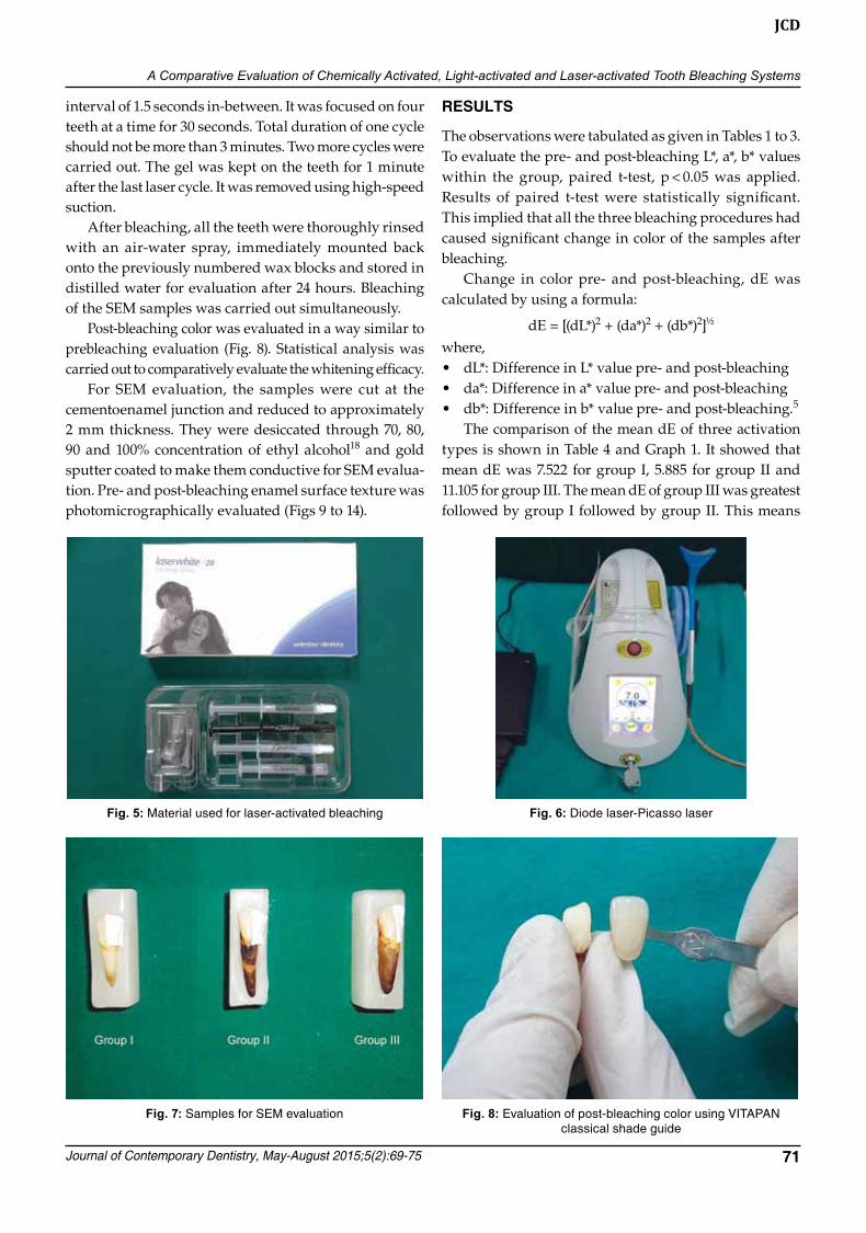
of carrying out further procedure (Fig. 2). Bleaching tray was made using a vacuum formed sheet. The material used for chemical and light activation was Opalescence Boost 40% by Ultradent (Fig. 3) contai-ning 40% hydrogen peroxide and beta carotene which efficiently absorbs the LED radiation.17 eZee White LED light of wavelength 430 to 475 nm (Fig. 4) was used. Laserwhite*20 by Waterlase dentistry is a material spe-cially designed for laser activation (Fig. 5) containing 46% hydrogen peroxide. On activation, the concentration of hydrogen peroxide reduces to less than 40%. Picasso diode laser of wavelength 810 nm (Fig. 6) was used.4
Three samples, one per group, were selected for scan-ning electron microscopic (SEM) evaluation to study the effect on enamel surface texture. Half of the labial surface of these samples was covered using a polytetrafluoro-ethylene sealant tape9 (Fig. 7) and they were subjected to bleaching. The covered unbleached surface acted as a control. All the teeth were cleaned, polished and air dried before bleaching. The flow of the material was verified
on a piece of gauze prior to applying it on the teeth sur-faces. For bleaching of group I, two drops of bleaching gel were placed for each tooth in the bleaching tray. Approximately, 0.5 to 1 mm thick layer of bleaching gel was applied. Bleaching was carried for 20 minutes as per the manufacturer’s instructions. The gel was reap-p lied in areas that had thinned or needed replenishing periodically. The gel was suctioned from the teeth after 20 minutes. The same procedure was repeated two times immediately after the first application. For group II, bleaching gel was applied on the labial surface of the teeth forming a layer of approximately 0.5 to 1 mm thickness. Three cycles of 10 minutes each with 10 minutes of resting time in-between were carried out as per manufacturer’s instructions. After the third cycle, the gel was suctioned from teeth. For bleaching of group III, approximately 1 mm layer of Laserwhite*20 was applied over the labial surfaces of the teeth. The consistency of the Lavender color within the gel was observed for uniform thickness. The laser was set at 7.0 watts with duration of 9.9 second with an
Fig. 1: Evaluation of color after staining and prebleaching using VITAPAN classical shade guide
Fig. 2: Teeth mounted on dental plaster blocks
Fig. 3: Material used for chemical and light-activated bleaching Fig. 4: LED light—eZee white light
A Comparative Evaluation of Chemically Activated, Light-activated and Laser-activated Tooth Bleaching Systems
Journal of Contemporary Dentistry, May-August 2015;5(2):69-75 71
JCD
interval of 1.5 seconds in-between. It was focused on four teeth at a time for 30 seconds. Total duration of one cycle should not be more than 3 minutes. Two more cycles were carried out. The gel was kept on the teeth for 1 minute after the last laser cycle. It was removed using high-speed suction. After bleaching, all the teeth were thoroughly rinsed with an air-water spray, immediately mounted back onto the previously numbered wax blocks and stored in distilled water for evaluation after 24 hours. Bleaching of the SEM samples was carried out simultaneously. Post-bleaching color was evaluated in a way similar to prebleaching evaluation (Fig. 8). Statistical analysis was carried out to comparatively evaluate the whitening efficacy. For SEM evaluation, the samples were cut at the cementoenamel junction and reduced to approximately 2 mm thickness. They were desiccated through 70, 80, 90 and 100% concentration of ethyl alcohol18 and gold sputter coated to make them conductive for SEM evalua-tion. Pre- and post-bleaching enamel surface texture was photomicrographically evaluated (Figs 9 to 14).
RESuLTS
The observations were tabulated as given in Tables 1 to 3. To evaluate the pre- and post-bleaching L*, a*, b* values within the group, paired t-test, p < 0.05 was applied. Results of paired t-test were statistically significant. This implied that all the three bleaching procedures had caused significant change in color of the samples after bleaching. Change in color pre- and post-bleaching, dE was calculated by using a formula:
dE = [(dL*)2 + (da*)2 + (db*)2]½
where,• dL*: difference in L* value pre- and post-bleaching• da*: difference in a* value pre- and post-bleaching• db*: difference in b* value pre- and post-bleaching.5 The comparison of the mean dE of three activation types is shown in Table 4 and graph 1. It showed that mean dE was 7.522 for group I, 5.885 for group II and 11.105 for group III. The mean dE of group III was greatest followed by group I followed by group II. This means
Fig. 8: Evaluation of post-bleaching color using VITAPAN classical shade guide
Fig. 5: Material used for laser-activated bleaching Fig. 6: Diode laser-Picasso laser
Fig. 7: Samples for SEM evaluation

Aditi Chintamani Sabnis, Sabita M Ram
72
Fig. 14: Photomicrograph of treated surface—group III
Fig. 9: Photomicrograph of untreated surface—group I Fig. 10: Photomicrograph of treated surface—group I
Fig. 11: Photomicrograph of untreated surface—group II Fig. 12: Photomicrograph of treated surface—group II
Fig. 13: Photomicrograph of untreated surface—group III
that change in color in group III was maximum followed by group I followed by group II. So as per the means, the whitening efficacy of group III was expected to be more compared to the group I and group II. The results were subjected to the statistical evaluation. One-way analysis of variance (ANOVA) followed by Scheffe’s post hoc test, p < 0.05 was carried out. Results
of these tests were statistically significant. The mean dE for light and laser activation was statistically significant. Photomicrographs of laser activated group showed mini- mal changes on enamel surface post-treatment compared to pretreatment surface. Chemically and light-activated systems caused surface alterations on sample surface post-treatment compared to pretreatment surface.

A Comparative Evaluation of Chemically Activated, Light-activated and Laser-activated Tooth Bleaching Systems
Journal of Contemporary Dentistry, May-August 2015;5(2):69-75 73
JCD
Table 1: Spectrophotometric analysis of group I
Prebleaching Post-bleachingSamples L* a* b* L* a* b* dE1 71.81 1.04 15.72 72.82 –0.72 6.79 9.162 65.87 –1.11 9.3 69.45 –1.22 5.17 5.463 68.96 –0.58 7.39 73.33 –1.39 2.4 6.684 66.11 1.77 13.63 77.57 –0.36 12.06 11.765 65.9 –0.65 6.79 69.28 –1.89 –0.28 7.936 77.13 –1.51 10.09 77.69 –1.41 6.19 3.947 71.03 1.33 11.87 77.76 –0.18 8.18 7.838 76.76 –0.3 16.29 82.02 –1.1 12.46 6.559 67.68 –0.65 9.48 71.4 –1.66 5.88 5.2810 66.43 –0.91 5.49 76.72 –1.69 2.91 10.63Mean 7.522Standard deviation 2.45691
Table 2: Spectrophotometric analysis of group II
Prebleaching Post-bleachingSamples L* a* b* L* a* b* dE11 72.96 –0.99 6.05 73.88 –0.87 2.43 3.7412 66.27 0.68 12.77 72.01 –0.39 8.55 7.2113 74.23 0.46 13.22 79.22 –0.8 8.89 6.7314 63 –0.53 7.88 69.46 –1.06 7.37 6.515 76.62 0.14 11.49 78.09 –0.2 9.37 2.616 68.88 0.55 12.88 75.55 –0.04 9.1 7.6917 69.17 –0.27 10.98 73.25 –1.18 5.99 6.518 68.14 0.72 11.34 75.51 –0.45 10.4 7.5219 79.46 –0.69 14.24 79.03 –1.32 9.94 4.3720 74.28 –0.53 9.91 69.17 –0.55 6.79 5.99Mean 5.885Standard deviation 1.72742
Table 3: Spectrophotometric analysis of group III
Prebleaching Post-bleachingSamples L* a* b* L* a* b* dE21 59.12 –0.8 6.31 69.94 –1.32 2.69 11.4222 59.66 –0.31 8.73 70.16 0.05 11.17 10.7823 60.75 –0.32 12.55 75.5 –1.03 11.86 14.7824 67.83 –0.37 11.91 79.56 –0.97 12.28 11.7525 61.01 0.66 11.45 62.91 –0.64 8.88 3.4526 64.45 0.33 8.82 71.45 0.33 13.02 8.1627 69.14 –1.21 7.59 83.81 –1.3 9.27 14.7728 70.09 –0.82 7.59 72.14 –1.2 5.21 3.1729 63.02 2.19 16.55 84.11 –0.33 8.25 22.830 59.45 –1.46 5.63 67.36 –2.33 –0.38 9.97Mean 11.105Standard deviation 5.73349
dISCuSSIon
In the present study, 40% hydrogen peroxide was used with 810 nm diode laser and 430 to 475 nm LED. There was variation in the concentration of the agents and the wave-length of the LED and diode laser compared to the studies in the past. This study compared all the three activation systems with each other than the past studies comparing any two. Thus, the present study was different.
Wetter NU17 carried out a study using 35% hydrogen peroxide activated with 470 nm LED light and 808 nm diode laser. He concluded that the laser activation was more effective than LED. Dostalova T19 compared dental bleaching efficacy with 790, 970 nm diode laser and 467 nm LED irradiation with 38% hydrogen peroxide gel. He found that, with diode laser activation, the desired shade was obtained in shorter time and slight surface

Aditi Chintamani Sabnis, Sabita M Ram
74
Table 4: descriptive statistics for dE of three activation types
Activation type MeanChemical 7.522Light 5.885Laser 11.105Total 8.1707
Graph 1: Direct comparison of the mean of difference in color of three activation types
modifications were seen. He concluded that laser was the most valuable energy source for power bleaching with simple and short application in the dental office. Stud-ies were carried out using light and no light activation by Ziemba20 using 25% hydrogen peroxide, Ontiveros5 using 25% hydrogen peroxide and Cardoso21 using 25% and 38% hydrogen peroxide. Their studies showed that treatment with light activation showed significantly greater bleaching-dependent changes in color compared to treatment without light. Goharkhay K4 studied the bleaching effects of 810 nm diode laser and 1064 nm neodymium-yttrium, aluminium, garnet laser on 35, 37 and 38% hydrogen peroxide. They concluded that laser-activated bleaching offered an improvement in terms of effectiveness and enamel surface protection. Buchalla W, Thomas A3 mentioned that LED’s emit light distributed across a range of 20 to 80 nm bandwidth and more. They hold a position in between monochromatic lasers and broad band light sources. Wetter NU,17 De Moor RJg22 mentioned that diode lasers emit coherent, well-collimated light, whereas LEDs are much more difficult to collimate usually presenting smaller output power. This reason may have been contributed in the results of this study. A study by Dostalova T19 showed that surface was smoother in case of bleaching with diode laser activation. He found that laser radiation can decrease the time of bleaching without any surface modification. In the study by Goharkhay,4 laser bleaching did not decisively alter the enamel surface. The reason for the changes in the
enamel surface texture could be loss of organic matrix after exposure to hydrogen peroxide as suggested by Markovik.14 Duration of application is one of the factors responsible for enamel surface texture alterations. In this study, the duration of application of bleaching gel for laser activation was in seconds, whereas for the other two systems it was in minutes. That could have contributed to the results. Prerequisites for laser-activated teeth bleaching are a perfect match of the chosen laser wavelength and the bleaching gel.4 This means that laser activation system along with its specially designed bleaching agent would yield better results but with any other gel it may or may not be equally effective. Therefore, the material used for bleaching is much important. As laser activated bleaching produces slight to mini-mal alterations on enamel surface, further staining of the surface may be minimized. It can produce desired effect with safety, in shorter time and with good efficacy. In recent times educated and aware patients are interested in saving time, obtaining quicker results. Studies in the past have shown that diode laser activated bleaching caused less tooth sensitivity.17,23 Diode laser causes crystalliza-tion of dentin inorganic component and coagulation of fluids contained into the dentinal tubules resulting in less sensitivity.24 The same installed laser system can be used for various applications other than teeth bleaching. Small sample size is one limitation of the study. As settling of the shade post-bleaching depends on indi-vidual’s dietary habits, immediately obtained shade was considered. However, desiccation of samples was prevented by storing them in distilled water always as desiccation affects the shade.
ConCLuSIon
Within the limitation of the study, it was found that laser- activated system along with its specially designed bleaching material was more effective in terms of white-ning efficacy and it produced least effect on enamel surface texture. Light-activated system was least effec-tive in terms of whitening efficacy chemically activated system being in between the two. Chemically and light-activated bleaching systems produced alterations on enamel surface texture. It is important to note that the material used for bleaching is of greater importance.
REfEREnCES
1. Kihn PW. Vital tooth whitening. Dent Clin N Am 2007;51: 319-331.
2. Griffiths CE, Bailey JR, Jarad FD, Youngson CC. An investiga-tion into most effective method of treating stained teeth: an in vitro study. Journal of dentistry 2008;36:54-62.

A Comparative Evaluation of Chemically Activated, Light-activated and Laser-activated Tooth Bleaching Systems
Journal of Contemporary Dentistry, May-August 2015;5(2):69-75 75
JCD
3. Buchalla W, Thomas A. External bleaching therapy with activation by heat, light or laser: a systematic review. Dental Materials 2007;23:586-596.
4. Goharkhay K, Schoop U, Wernisch J, Hartl S, De Moor R, Moritz A. Frequency doubled neodymium:yttrium–alu-minum–garnet and diode laser-activated power bleach-ing—pH, environmental scanning electron microscopy, and colorimetric in vitro evaluations. Lasers Med Sci 2009;24: 339-346.
5. Ontiveros JC, Paravina RD. Color change of vital teeth exposed to bleaching performed with and without supple-mentary light. Journal of dentistry 2009;37:840-847.
6. Ernst CP, Marroquin BB, Willershausen-Zönnchen B. Ef-fects of hydrogen peroxide containing bleaching agents on the morphology of human enamel. Quintessence Int 1996; 27(1):53-56.
7. Sulieman M, Addy M, Macdonald E, Rees JS. A safety study in vitro for the effects of an in-office bleaching system on the integrity of enamel and dentine. Journal of dentistry 2004; 32:581-590.
8. Caballero AB, Navarro LF, Lorenzo JA. Dental bleaching and alteration of the enamel surface in vivo evaluation of the ef-fects of 10% carbamide peroxide and 3.5% hydrogen peroxide on the enamel surface. Med Oral Patol Oral Cir Bucal 2007;12: E404-407.
9. Cadenaro M, Breschi L, Nucci C, Antoniolli F, Visintini E, Prati C, Matis BA, Di Lenarda R. Effect of two in-office white-ning agents on the enamel surface in vivo: a morphological and non-contact profilometric study. Operative Dentistry 2008;33(2):127-134.
10. Shannon H, Spencer P, Gross K, Tira D. Characterization of enamel exposed to 10% carbamide peroxide bleaching agents. Quintessence Int 1993;24(1):39-44.
11. Josey AL, Meyers IA, Romaniuk K, Symons AL. The effect of a vital bleaching technique on enamel surface morphology and the bonding of composite resin to enamel. J Oral Rehabil 1996;23:244-250.
12. Zalkind M, Arwaz JR, Goldman A, Rotstein I. Surface mor-phology changes in human enamel, dentine and cementum
following bleaching: a scanning electron microscopy study. Endod Dent Traumatol 1996;12:82-88.
13. Miranda CB, Pagani C, Benetti AR, Matuda FS. Evaluation of the bleached human enamel by Scanning Electron Micros-copy. J Appl Oral Sci 2005 Apr-Jun;13(2):204-211.
14. Markovik L, Jordan RA, Lakota N, Gaengler P. Micromorpho-logy of enamel surface after vital tooth bleaching. Journal of Endodontics 2007 May;33(5):607-610.
15. Pleffken PR, Borges AB, De Paiva Gonçalves SE, Torres CRG. The effectiveness of low-intensity red laser for activating a bleaching gel and its effect in temperature of the bleaching gel and the dental pulp. J Esthet Restor Dent 2012;24:126-134.
16. Chu SJ, Trushkowsky RD, Paravina RD. Dental color matching instruments and systems: review of clinical and research aspects. Journal of dentistry 2010;38s:e2-16.
17. Wetter NU, Barrosso MCS, Pelino JEP. Dental bleaching ef-ficacy with diode laser and LED irradiation: an in vitro study. Lasers in Surgery and Medicine 2004;35:254-258.
18. Janda R. Preparation of extracted natural human teeth for SEM investigations. Biomaterials 1995;16(3):209-217.
19. Dostalova T, Jelinkova H, Housova D, Sulc J, Nemec M, Miyagi M, Brugnera A Jr, Zanin F. Diode Laser-activated bleaching. Braz dent J 2004;15(special issue SI-3-SI-8).
20. Ziemba SL, Felix H, Giniger M, Ward M. Randomized, pro-spective evaluation of the effectiveness of the Zoom2™ dental whitening lamp and light-catalyzed peroxide gel. Discus denta Inc 2005 Feb.
21. Capel Cardoso PE, Muench A, Pinheiro HB. Photo-fenton vs conventional in-office dental whitening treatment; in vitro study dental materials 2011;27(1):76.
22. De Moor RJG, Vanderstricht K. The use of the KTP laser, an added value for tooth bleaching. J Oral Laser Applications 2009;9:219-226.
23. Gurgan S, Cakir FY, Yazici E. Different light-activated in-office bleaching systems: a clinical evaluation. Lasers Med Sci 2010;25:817-822.
24. Umberto R, Claudia R, Gaspare P, Gianluca T, Alessandro DV. Treatment of dentine hypersensitivity by diode laser: a clini-cal study. International Journal of Dentistry 2012;1-8.