a clamp-bearing fungus using stalked adhesive …...a clamp-bearing fungus using stalked adhesive...
TRANSCRIPT
A clamp-bearing fungus using stalked adhesive youngchlamydospores in capturing amoebae
By Charles D r e c h s l e r(Crops Research Division, Agricultural Research Service, United StatesDepartment of Agriculture, Plant Industry Station, Beltsville, Maryland,
U.S.A.)
With Plates XXXIV—XXXVI.
An amoeba-capturing fungus displaying in its predacious appa-ratus remarkable convergence as well as an unusual combination ofvegetative and reproductive functions came to light in several Petriplates of maize-meal agar which after being overgrown with Pylhiummycelium had been further planted on March 11, 1960, with pinchesof crumbly leaf mold gathered on April 16, 1959, in two locations nearLake Alfred, Florida. The fungus attained only rather limiteddevelopment, chiefly because in all cultures the durably pelliculaterhizopod on which it mainly subsisted was killed off early by arobust zoopagaceous form that subsequently became recognizable asa species of Cystopage. Its growth may have been hampered also bysome slime-mold plasmodia which manifestly had originated fromspores that survived desiccation in the leaf mold stored for nearly ayear in an air-dry condition. Here and there, nevertheless, the funguswas observed to capture and consume an abundance of prey, and inthe favorable areas it extended its mycelium for distances of 5 to8 mm from the deposits of partly decayed material.
Although from resemblances with respect to biological rela-tionship and to production of clamp-connections the Florida funguswould seem kindred with the 7 nematode-destroying forms I de-scribed ( D r e c h s l e r , 1941, 1943, 1946, 1949, 1954 a) as species ofNematoctonus the peculiarities shown in its development merit recog-nition in the establishment of a new genus. One of these peculiaritiesmay appropriately be signalized in a generic term compounded oftwo words (Ttayk, genitive TrayiSoc; (mopa) meaning "trap" and"seed", respectively.
Pagidospora gen. nov.Mycelium ramosum, incoloratum; hyphae saepius septato-nodo-
sae, hie illic vel saepius praecipue in apice ramulorum breviumchlamydosporas ferentes; chlamydospora in juventute incolorata,membrana tenui circumdata, maximam partem vel paene omninovel forsitan ex toto in bulla tenaci constans; bulla tenax saepe adanimal inhaerens, id ita capiens, hypham cum ramis assumentibus
246
(haustorium) in captivum intrudens quae protoplasma exhaurit;chlamydospora in maturitate muro crasso circumdata, saepe aliquidflavida. Typus: Pagidospora amoebophilla.
Pagidospora amoebophila spec. nov.Mycelium exigue expansum, inconspicuum; hyphae repentes vel
immersae, incoloratae, mediocriter ramosae, multis nodosis septis etpaucis levibus septis divisae, plerumque 1.5—4 \n latae, in apice eorumsed praecipue in apice ramulorum plerumque i—50 ̂ longorum et 1.2—2.5 \a latorum chlamydosporas singulatim vel quandoque deinceps feren-tes. Chlamydospora juvenis basi septo nodoso semper delimitata, in bullatenaci et pediculo cylindraceo plerumque 0.5—20 (i longo et 1.2—3 ^lato constans; bulla tenax globosa, vulgo 6—12 \i longa, 5.5—10 ^ lata,aliquando brevem fertilem stipulam vel longiorem hypham emittens,saepius ad amoebam inhaerens, ita animal capiens, id primo strictepostea vulgo laxius tenens, mox hypham (interdum 2 hyphas) incaptivum intromittens; haec hypha basi vulgo 1.5—2 |i lata, sursumsaepius usque 2—3.5 ^ latescens, intra animal ramos assumentesemittens itaque haustorium faciens; rami assumentes plerumque2—2.7 n crassi, late curvati, saepe in glomum laxum intertexti, proto-plasma animalis gradatim exhaurientes. Chlamydosporae in maturi-tate aliquid flavidae, saepe globosae vel elongato-ellipsoideae, 6—12 [i longae, 5.5—10 \i latae, membrana 0.7—1.3 ^ crassa circumdatae,protoplasmatis crasse granulosi cum 1—3 corporibus nitidis circa1.4 n latis repletae, pediculo cylindraceo indurato 0.5—20 \i longo et1.2—3 n lato et saepius omnino vel magnam partem vacuo praeditae;quandoque praeterea apice vel a latere i—2 tubulis hypharum vacuisornatae; plerumque discretae sed quandoque cum 1—3 aliis chla-mydosporis conjunctae. Rami assumentes quandoque in corpora per-durantia prave filiformia, saepe 10—30 p, longa, 3—5 u, lata, chlamy-dosporis aliquid similia transeuntes.
Amoebas duarum specierum capiens consumensque habitat inmateriis plantarum putrescentibus prope Lake Alfred, Florida. Ty-pus: Tabulae XXXIV—XXXVI.
When Pagidospora amoebophila develops in maize-meal-agarplate cultures that after being planted with slowly decaying materialshave become overgrown with bacteria, permeated with various fungi,and infested with protozoans and eelworms, it does not usually be-come visible to the naked eye. It extends its moderately branchedmycelium (PI. I, A—O; PI. II, A—J; PI. Ill, A—K) somewhatsparingly both on and under the surface of the substratum. For themost part its hyphae, which commonly vary from 1.5 p. (PI. Ill, H)to 4 n (PI. I, L) in width, are filled with protoplasm of nearly homo-geneous consistency. They are divided by cross-walls (PI. I, A, a—d;B, a—c; H, a—i; I, a—c; PI. II, B—F; PI. Ill, A—I) into segmentsmostly between 5 and 100 ^ long. As by far the larger number of
247
cross-walls are associated with clamp-connections the fungus at firstsight gives the impression that it is wholly devoid of unmodifiedsepta. However, in nearly any considerable tract of mycelium furthermicroscopical scrutiny usually reveals some scattered septa that lackany indication of a clamp (PL I, A, b; H, a; PL HI, J, a). In oneobserved instance (PL I, H, d) formation of a clamp had apparentlybegun in the usual manner, but through a developmental mishapthe elongating lateral process became fused with a filamentousbranch nearby rather than with the adjoining segment.
In the design of its predacious organs Pagidospora amoebophilashows no close parallelism with the 3 nematode-capturing species Idescribed (D r e c h s 1 e r, 1946. 1949, 1954 a) under the binomialsNematoctonus haptocladus, N. concurrens, and N. compylosporus.Contrary to expectations suggested by the abundant display of clampsthese organs consist of stalked adhesive knobs (PI. I, A, e; B, d;C—G; H, j—1; PL II, B, a, b; PL III, D; E, a—d; P, b; G, a) closelysimilar to the stalked knobs by means of which capture of eelwormsis accomplished in several clampless hyphomycetes including Dac-tylella ellipsospora Grove (1886), Dactylella asthenopaga Drechsler(1937), Dactylaria haptotyla Drechsler (1950), and Dactylaria sclero-hypha Drechsler (1950). The similarity in design is sustained bynotable similarity in dimensions, as the adhesive knobs of P. amoebo-phila, typically of globose or prolate ellipsoidal shape, commonlymeasure 6 ^ (PL III, C, g) to 12 n (PL II, B, c) in length and 5.5 ^(PL III, C, g) to 10 n (PL II, D) in greatest width, while the stalkssupporting them vary mostly from 1.5 ̂ (PL III, E, c) to 60 ̂ (PL I, G)in length and from 1.2 y, (PL I, F) to 2.5 p. (PL I, G) or 3 ^ (PL I, LOin width. Sometimes the stalk arises from the crest of a clamp-connection (PL I, D, P) but more usually it originates from an un-modified portion of the parent hypha. In some few instances wherea line of demarcation, commonly more or less indistinct, appears todelimit the stalk basally (PL I, C; PL II, D) it remains uncertainwhether the line represents merely the contour of the parent hyphaor denotes the presence either of an unmodified septum or possiblyof a septum accompanied by a small clamp concealed from view onthe under side. Each stalk, in any case, is provided with at least onecross-wall that is attended by a clamp-connection. This nodose cross-wall, unlike the unmodified septum which in Dactylella asthenopaga,for example, always delimits the adhesive knob accurately in theplane where the globose contour meets the cylindrical contour of thestalk, is variable with respect to its position. It occurs sometimes inthe basal portion (PL I, D; H, b; I, c; PL II, A; B, a; P; PL III, C, g),sometimes in the middle portion (PI. I, A, d;E; PL II, B, b, c; C; D; E;G; PL III, D; E,a; G, a), and some times in the distal portion (PL I,B,c;P; H, i; PL III, E, c, d) of the stalk, so that proximally the adhesive
248
knob remains continuous with a cylindrical part mostly 0.5—20 n longand 1.2—3 ^ wide. This cylindrical part accordingly is treated in thediagnosis as constituting a basal "pediculus", or pedicel, of the youngas well as of the mature chlamydospore, while the proximal portionof the stalk is treated as the sporiferous branch. Consonant with theinterpretation underlying such disposition, the stalk supporting theadhesive knob shown in Plate I, G, is held to consist of a sporepedicel 10 ̂ long and of a sporiferous branch 50 (i long. In stalks thatcontain 2 nodose septa the proximal one would seem to have nogreater significance either in the earlier (PI. I, G) or in the later(PI. HI, B) development of the predacious organ than any similarpartition in the parent hypha.
An Amoeba surrounded by a firm membranous pellicle andmeasuring 30—55 ^ across when drawn into a rounded shape wasmost frequently found serving as prey of Pagidospora amoebophila.Within its protoplast Pythium oospores (PL I, I, e, f) in variousstages of digestion were often visible. Following its capture thisanimal usually continues for hours to operate its contractile vacuole(PI. I, I, v; J, v; PI. II, B—E: v) in proximity to the single elongated-ellipsoidal nucleus (PI. I, I, n; J, n; PI. II, B—E: n), 12—15 ^ longand 4—9 jj, wide, which encloses a slightly darker endosome, mostly5—7 [i long and 3—4.5 ^ wide. The fungus meanwhile proceeds withits attack by extending from the globose knob broadly adhering to theanimal's pellicle an infection tube (PL I, I, d) often about 1.7 \\, indiameter proximally. This tube usually widens as it penetratesfarther into the protoplasm of the captive (PL II, B), with the resultthat on reaching a length of 15—25 ^ it has commonly attained adiameter between 2.5 and 4 p. Thereupon it usually gives off a branchat a wide angle and in a manner often rather strongly simulatingdichotomy. In continuing growth the 2 terminal elements commonlydescribe divergent curves. After some elongation each puts forth abranch, again usually at a wide angle and with an appearance ofdichotomy (PI. I, J; PI. II. C—E). The same sequence of elongationand branching may then be repeated, though owing to the depth ofthe overlying protoplasm the development of the infection tube into asomewhat elaborately ramifying system is nearly always badlyobscured. Later when the animal's protoplasm has been largely ab-sorbed (PL II, F) some branching of a third order can often be madeout. The completed assimilative system, or haustorium, with itscurving hyphal elements intertangled into a loose clew, bears mostresemblance to that of Zoopage thamnospira Drechsler (1938), azygomycete wholly alien with respect to taxonomic affinity butsimilar in subsisting through capture of amoebae. On exhaustion ofthe animal's fleshy substance all the protoplasm in the ramifiedhaustorium is, as a rule, gradually withdrawn backward through the
249
adhesive knob into the parent hypha (PL I, K, b). The emptiedassimilative branches vanish from sight within the minutely wrinkledcollapsed pellicle (PL I, H, N; PL II, G, H, I; PL III, B), but themembrane of the stalk-like part — originally the infective tube —having become markedly thickened and indurated, often remainsclearly visible as a distal appendage of the adhesive knob. Adjacentportions of assimilative apparatus that fail to become emptied in thegeneral withdrawal of haustorial contents may form hyphal cysts(PL III, C, h; X, a) or give rise to chlamydospores (PL III, C, b).
In one of my maize-meal-agar plate cultures Pagidosporaamoebophila was observed capturing and destroying also a conspic-uously globuliferous Amoeba that was surrounded by an ectoplasmicmantle about 0.4 p, thick rather than by the thinner yet firmer mem-branous pellicle of familiar type. The animal in question was evi-dently conspecific with the one earlier found utilized as prey byStylopage ischnospora var. pleacra Drechsler (1959). In capturedindividuals of this protozoan, too, the fungus forms a rather luxurianthaustorium whose curving assimilative branches are intertwined intoa loose clew. Soon after attaining full development the haustoriumhere usually becomes exposed to alien microorganisms as a result ofcomplete disintegration of the animal's peripheral mantle (PL I, K, a;PL III, A). Owing probably to such exposure the ensuing withdrawalof contents from the assimilative branches is often less thorough(PL I, L) than where a membranous pellicle remains as an un-breached protective sheath long after the captive has succumbed toinvasion. Partly because some portions of its assimilative system thatfail to become emptied of their protopolasm manage to survivethrough encystment, P. amoebophila incurs less degeneration ofhaustorial contents than is incurred by S. ischnospora var. pleacra inpreying on the same animal.
Sometimes 2 neighboring adhesive knobs (PI. Ill, A, a, b) ofPagidospora amoebophila are found instrumental in capturing andinvading the same individual amoeba, just as in the clamplesshyphomycetes 2 or more adhesive knobs, or, for that matter 2 or moreconstricting rings, sometimes are found to operate jointly in cap-turing and invading the same eelworm. An adhesive knob ofP. amoebophila not infrequently intrudes 2 infection tubes into acaptured protozoan, though similar double invasion of an eelwormfrom an adhesive knob of a clampless hyphomycete ( D r e c h s l e r ,1950, p. 52, Fig. 13, D, e) would seem to occur only rarely. No instanceappears to have been recorded wherein an adhesive knob of a clamp-less hyphomycete served in capturing more than a single eelworm,but in P. amoebophila it is not unusual to find adhesive knobs thathave extended separate infection tubes into 2 captive amoebae, whichmay or may not (PL I, K, a, b) be of the same species. The greater
250
predacious capabilities are associated with a curious difference in themanner of holding prey. When an eelworm has been capturedthrough adhesion to a knob of Daclylella asthenopaga, for example,the animal remains closely affixed to the globose cell, which therebybecomes much less accessible to other nematodes. An amoeba cap-tured by P. amoebophila is in the beginning likewise kept directlyaffixed to the adhesive knob, but very often this affixture is laterdiscontinued, being then superseded by an adhesive attachment tothe infection tube. Consequently the animal, and finally its emptypellicle or other residual material, is often found tethered on thefilamentous tube in a position 2 to 20 n away (PI. I, H; K, b; L; M;PI. II, F—I; Plate III, A, a, b; B; C), leaving the adhesive knobexposed to encounter with other amoebae suitable as prey. Even ininstances where the knob, besides, has put forth a hypha bearing one(PL I, H, 1) or more new predacious organs its capabilities for furthercapture of animals would not seem seriously impaired.
While the adhesive knobs by means of which several clamplesshyphomycetes capture nematodes can apparently function only aspredacious organs, those of Pagidospora amoebophila serve very im-portantly, besides, as reproductive structures, since they consistentlyundergo conversion into the spores characteristic of the fungus. Theconversion takes place sooner or later, whether the individual knobshave succeeded or have failed in securing prey. As the conversionentails no noticeable change in size the resulting spores (PI. I, P—Y;Z, a, b; PL II, J, a; K—X; PL III, A, b; B; C, a—f; E, e—m; P,a, c; G, b; H, a, b; I a—c; J, b; K, a—d; L—U) have generally the sameoutward dimensions as the adhesive organs from which they wereformed, and like them include a basal cylindrical part, or pedicel, ofvariable length. In mature spores, which are nearly always yellowish,the globose cell is surrounded by a wall mostly 0.7—1.3 n thick andcontains protoplasm often crowded with granules about 0.5 \L wide.Among these coarse granules 1—3 round lustrous bodies, about 1.4 nin diameter, are often discernible. Where the basal pedicel is short(approximately 0.5—2 y, long) it usually is present as a solid protu-berance of the wall (PL I, U—X; PL II, 0, P, S; PL III, B; C, c, e;E, f, g, 1; P, a; G, b; H, a, b; I, b, c; K, a, b), but where it is longer(approximately 2—20 \i in length) it usually retains a tubular charac-ter, though its narrow lumen is often largely or wholly empty ofprotoplasm. Whatever partition may be present between the chamberof the globose cell and the lumen of the empty pedicel, usually isonly indistinctly visible, especially with respect to its proximalcontour.
When on slight disturbance the mature spore of Pagidosporaamoebophila becomes detached at the base of its pedicel it sometimesremains connected with a portion of infection tube, which, though
851

usually empty (PI. 1, Z, a; PL II, X), is found in scattered instancesto have retained some protoplasm and formed a hyphal cyst (PL III,X, a). Usually the more crooked hyphal cysts (PI. Ill, Y, Z), mostly10—30 n long and 3—5 \n wide, that are formed mainly from distalportions of the haustorium (PL I, M) no longer have any visibleconnection with the spore from which they originated. A rather moredurable connection is sometimes found between two catenated spores(PL I, Z, b; PL II, Y, Z; PL III, V; W), of which one, while in thethin-walled predacious stage, gave rise to the other on a short hyphaloutgrowth (PL I, 0; PL II, J, b; PL III, C, a, c; E, a, f ; K, c, d).
As Pagidospora amoebophilus has hitherto not been found pro-ducing basidia and basidiospores, or, indeed, any other propagativebodies except hyphal cysts and the spores resulting from maturationof the stalked adhesive knobs, opinion concerning its taxonomicrelations can be based only on similarities to imperfect stages ofvarious basidiomycetes. Its reproductive apparatus shows strikingparallelism, certainly, with the figures given by D a v i d s o n ,C a m p b e l l , and V a u g h n (1942, p. 24, Pig. 4, F, a, c; p. 32, Fig. 5,H, a, c) to illustrate hyphae and chlamydospores formed in malt-agarcultures of Polyporus compactus Overh. and of Polyporus spragueiBerk. & Curt. Despite the wide difference in biological habit betweenthe microscopic organism subsisting through capture of protozoansand the two fungi causing decay of living oaks, the close morpho-logical correspondence here would seem to reveal a true homology.In conformity, therefore, with the usage of Davidson, Campbell, andVaughn -as well as that of N o b 1 e s (1948) the spores of Pagidosporaamoebophilus are considered in the diagnosis as mature chlamydo-spores, and the adhesive knobs are considered as young chlamydo-spores. There is good reason to believe that while the adhesive knobsof Dactylella asthenopaga, for example, obviously represent purelyvegetative organs wholly without reproductive function, the outwardlysimilar knobs of Pagidospora amoebophila are in a basic sensereproductive bodies, which, during the protracted period when theyare full-grown but still immature, operate habitually as predaciousorgans. A somewhat similar combination of functions has elsewherecome under observation only in a clampless septate nematode-cap-turing fungus whose adhesive networks ultimately are often trans-formed into mycelial complexes of distended indurated cells. In thisclampless fungus, however, the dual-purpose structures are mani-festly designed primarily for a predacious function, and their laterutility as durable propagative apparatus — at no stage can they beregarded as typical spores — appears as a supplementary feature.
The resemblance shown by Pagidospora amoebophila to theasexual reproductive apparatus of at least 2 members of the Poly-poraceae is not shared by any of the 7 clamp-bearing predacious or
252
parasitic fungi described as species of Nematoctonus. While in 2 ofthe 7 species, namely N. lylospora Drechsler (1941) and N. pachysporaDrechsler (1943), chlamydospores are formed, these are elongated-ellipsoidal in shape rather than globose, aerial rather than procum-bent or submerged, verrucose or echinulate rather than smooth, thin-walled rather than thick-walled, and slenderly rather than sturdilyattached at the base. For capturing nematodes N. haplocladus, N.concurrens, and N. campylosporus employ a medially constrictedglandular cell that is enveloped in a droplet of adhesive secretion andis borne slightly aloft on an upcurved hyphal tip or on a short erecthyphal spur — a distictive predacious organ markedly different fromthe young chlamydospore of P. amoebophila. The aerial conidiaproduced by all 7 known species of Nematoctonus have no counter-part in P. amoebophila. Resemblance to the conidia of Corticiumincurustans Hohn. & Litsch. as figured by L y m a n (1907) and byN o b l e s (1937) was noted earlier ( D r e . c h s l e r , 1941) in theconidia of N. tylosporus and is evident also in those of the 6 con-generic species. The general similarity in conidial morphology heremight be held to suggest kinship of Nematoctonus with the Thele-phoraceae, just as similarity in respect to chlamydospore morphologymight be held to suggest kinship of P. amoebophila with the Poly-poraceae.
It seems possible that the unnamed fungus I earlier found( D r e c h s l e r , 1954 b, p. 780, Fig. 6, D—K; p. 781) capturing aspecies of Amoeba by means of sessile adhesive knobs may havebeen a close relative of Pagidospora amoebophila. The protoplasm inits mycelial hyphae as well as in its knobs appeared of even morepronouncedly homogeneous texture than the hyphal contents in mem-bers of the clampless series of predacious hyphomycetes; and in someinstances a captured rhizopod was found not closely affixed to theglobose knob, it being fastened instead to a proximal portion of theinfection tube. No clamp-connections were seen in the fungus, butas has been pointed out by various authors, including Lyman as wellas Davidson, Campbell, and Vaughn, the production of clamps is insome species an inconstant feature. In the small tract of mycelium,again, no conversion of globose knobs into chlamydospores wasobserved, but it is possible that my observations were not carried onlong enough to include the final stages of development. Accordinglysome little consideration was given to the unnamed fungus in fra-ming the diagnosis of the genus Pagidospora.
As the scanty material of Pagidospora amoebpohila depositedunder the number 71635 in the National Fungus Collections, PlantIndustry Station, Beltsville, Maryland, is in several respects unsatis-factory, it has appeared advisable to designate as type the accom-panying figures, which were prepared from living structures in
253
sufficient numbers to allow some scope for random selection. Purecultures of P. amoebophila have not become available, for owing tolack of aerial conidia all attempts to isolate the fungus were unsuc-cessful.
Z u s a m m e n f a s s u n gEin septierter und mil Schnallen versehener Fadenpilz der sich (lurch
den Fang zweier Amoeben-Arten ernahrt, 1st unter den Namen Pagido-spora amoebophila neu beschrieben. Als Fang-Organe dienen wohl diegestielten kugligen Chlamydosporen des Pilzes, aber nur solange als siesich, mit diinner klebriger Merabran umgeben, in verlangertem unreifemZustand befinden. Diese unreifen Sporen sind von den aufrechten kleb-rigen Drusenknopfen der mit Schnallen versehenen Nematoden-fangendenNematoctonus-Arien recht verschieden, aber den klebrigen Knopfenetlicher sehnallenloser Nematoden-fangenden Hyphomyceten der Gattun-gen Dactylella und Dactylaria auffallend ahnlich. Weil tiberdies die Spo-ren im unreifen wie auch im reifen Zustand den Chlamydosporen derholzzerstorenden Arten Polyporus compactus und Polyporus spraguei sehrahnlich sind, scheint es moglieh, dass der Pilz zu den Polyporaceen gehort.
H e f e i - e n c e sD a v i d s o n, R. W., C a m p b e l l , W. A. and Dorothy B. V a u g h n. Fungi
causing decay of living oaks in the eastern United States and theircultural identification. U.S. Dept. Agr. Techn. Bui. 785, 65pp., 3 pi.1942.
D r e c h s l e r , C. Some hyphomycetes that prey on free-living terricolousnematodes. Mycologia 29: 447—552. 1937.
— New Zoopagaceae capturing and consuming soil amoebae. Myco-logia 30. 137—157. 1938.
— Some hyphomycetes parasitic on free-living terricolous nematodes.Phytopathology 3*: 773—802. 1941.
— Two new basidiomycetous fungi parasitic on nematodes. Jour.Washington (D. C.) Acad. Sci. 33: 183—189. 1943.
— A clamp-bearing fungus parasitic and predaceous on nernatode?.Mycologia 3S: 1—23. 1946.
— A nematode-capturing fungus with anastomosing clamp-bearinghyphae. Mycologia M: 369—387. 1949.
— Several species of Dactylella and Dactylaria that capture free-livingnematodes. Mycologia 42: 1—79. 1950.
— A nematode-capturing fungus with clamp-connections and curvedconidia. Jour. Washington (D. C.) Acad. Sci. 44: 82—85. 1954 a.
— Some hyphomycetes that capture eelworms in southern states. Myco-logia 46: 762—782. 1954 H.
— Several Zoopagaceae subsisting on a nematode and on some terri-colous amoebae. Mycologia 51: 1959.
G r o v e , W. B. New or noteworthy fungi: Part III. Jour. Bot. 24: 129—137.197—206. 1886.
L y m a n, G. R. Culture studies on polymorphism of hymeiiomycetes.Proc. Boston Soc. Nat. Hist. 33: 125—209, pi. 18—26. 1907.
N o b l e s , Mildred K. Production of conidia by Corticium incrustana.Mycologia 29: 557—566. 1937.
— Studies in forest pathology VI. Identification of cultures of wood-rotting fungi. Canad. Jour. Research, C, 26: 281—431. 1948.
254
E x p l a n a t i o n of P l a t e XXXIV—XXXV1.
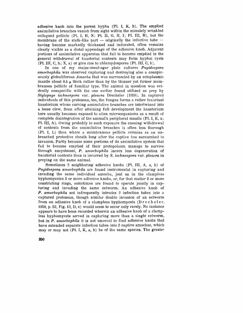
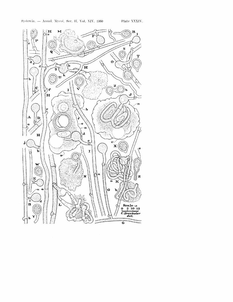
Plate XXXIV. Pagidospora amoebophila in maize-rneal-agar plate cul-tures that had been planted with crumbly leaf mold 29 to 38 days earlier:drawn with the aid of a camera lucida at a uniform magnification; xlOOOthroughout. A, Portion of hypha with 3 cross-walls, a—c, among1 which one, b.is not accompanied by a clamp-connection; a fourth septum, d, which has aclamp, delimits a young chlamydospore, e, from its supporting branch.B, Portion of hypha with 2 septa, a and b, each with a clamp-connection:a third nodose septum, c. delimits basally a young chlamydospore, d. C,D, Young chlamydospores, each with a relatively long pedicel delimitedbasally by a nodose septum from a short branch. E, F, Portions of narrowhyphae, each with a nodose septum and bearing a short branch surmountedby a young chlamydospore. G, Portion of hypha with a long sporiferou-branch containing 2 septa with clamps. H, Portion of mycelial hypha, wit l ia sporiferous branch; a—i, nine septa, including two,"a and d" that arewithout clamp-connections; j—1, three immature chlamydospores, one ofwhich, k, has extended a hypha after capturing an amoeba and expropria-ting its contents. I, Portion of hypha with 2 nodose septa, a and b; a thirdnodose septum, c, delimits basally a young chlamydospore, d, that hasbeen operative in capturing an amoeba surrounded by a firm membranouspellicle within which are shown 2 partly digested Pythium oospores, eand f, as well as an elongated-ellipsoidal nucleus, n, and a contractilevacuole, v. J, Portion of hypha bearing a short branch surmounted by ayoung chlamydospore that has been operative in capturing an amoebaenveloped in a firm membranous pellicle and has intruded into the animul3 recurving assimilative branches; n. nucleus of animal; v, contractilevacuole. K, Portion of hypha with a short branch bearing a youngchlamydospore from which a separate haustorium has been extended intoeach of 2 captured amoebae, a and b: the soft thickish pellicle of onecaptive, a, has disintegrated, leaving exposed a clew of haustorial bran-ches, while the wrinkled membranous pellicle of the other captive, b,continues to protect the assimilative branches still in process of beingevacuated. L, Portion of hypha with :i short branch bearing a youngchlamydospore from which 2 haustoria were intruded into a capturedamoeba whose soft thickish pellicle disintegrated early, so that the con-tents of the assimilative branches were only incompletely withdrawnbackward through the infection tube and adhesive knob. M, Portion ofmycelium showing a mature chlamydospore with an empty infection tubeto which is attached a shrunken membranous pellicle that surroundsisolated living portions of 2 assimilative branches. N, Portion of hyphawith a short branch bearing a mature chlamydospore from which extendan empty membrane of an infection tube and an empty wrinkled mem-branous pellicle. 0, Portion of mycelium showing a young chlamydosporethat has put forth a short hypha bearing a mature chlamydospore; thepedicel of the young chlamydospore is of unusual length. P, Maturechlamydospore with empty pedicel attached to empty branch. Q, Detachedmature chlamydospore with empty pedicel and proximal portion of emptyinfection tube. R—Y, Detached chlamydospores, showing usual variationsin size and shape. Z, Mature chlamydospores: a, one showing unusualshape, extension of protoplasm to base of pedicel, and empty apicalappendage representing proximal portion of infection tube; b, two closelyconnected mature chlamydospores.
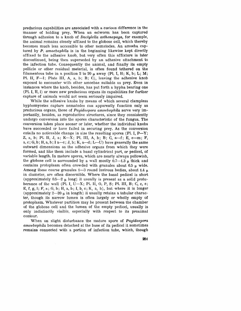
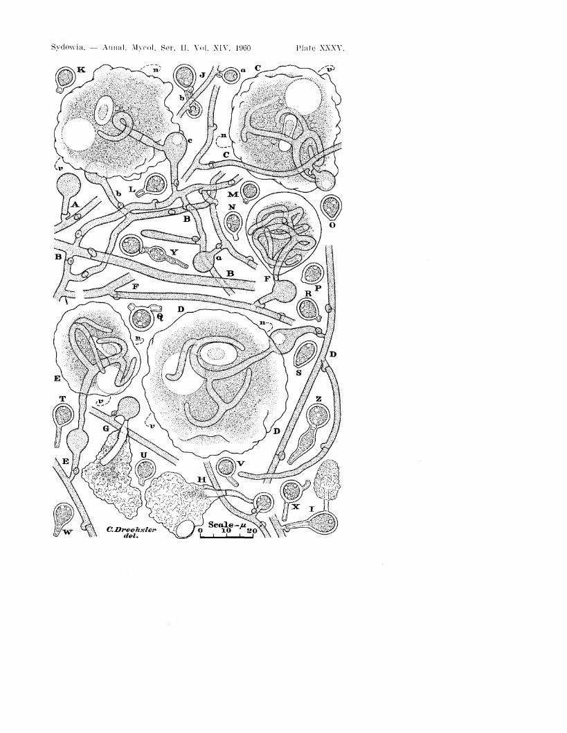
Plate XXXV. Pagidospora amoebophila in maize-meal-agar plate cul-tures that had been planted with crumbly leaf mold 29 to 38 deys earlier;
255
drawn with the aid of a camera lucida at a uniform magnification; xlOOOthroughout. A, Young chlamydospore with pedicel of moderate length sup-ported on short branch. B, Portion of mycelium showing 15 septa with clamp-connections and 3 young chlamydospores, a—c; from one of the youngchlamydospores, c, an infective hypha has been extended into a capturedamoeba that is surrounded by a firm membranous pellicle; n, nucleus ofanimal; v, contractile vacuole. C—E, Smaller portions of mycelium, eachshowing an immature chlamydospore from which an infection tube hasbeen extended into a captured amoeba surrounded by a firm membranouspellicle; within each captive the infection tube has ramified in forminga haustorium with 4 assimilative branches; n, nucleus of animal; v, con-tractile vacuole. F, Portion of mycelium showing a young chlamydosporefrom which an infection tube has been extended into a captured amoebasurrounded by a firm membranous pellicle; within the captive a branchedhaustorium has been produced. G, Portion of hypha with a young chla-mydospore bearing an empty infection tube to which is affixed thewrinkled membranous pellicle of an amoeba. H, I, Portions of hyphae,each with a mature chlamydospore bearing an empty infection tube towhich is affixed the wrinkled membranous pellicle of an amoeba. J, Por-tion of hypha on which is borne a discrete mature chlamydospore, a, andalso 2 rather firmly conjoined chlamydospores, b. K—X, Detached maturechlamydospores showing usual variations in size and shape. Y, Z, Pairs ofconnected mature chlamydospores.
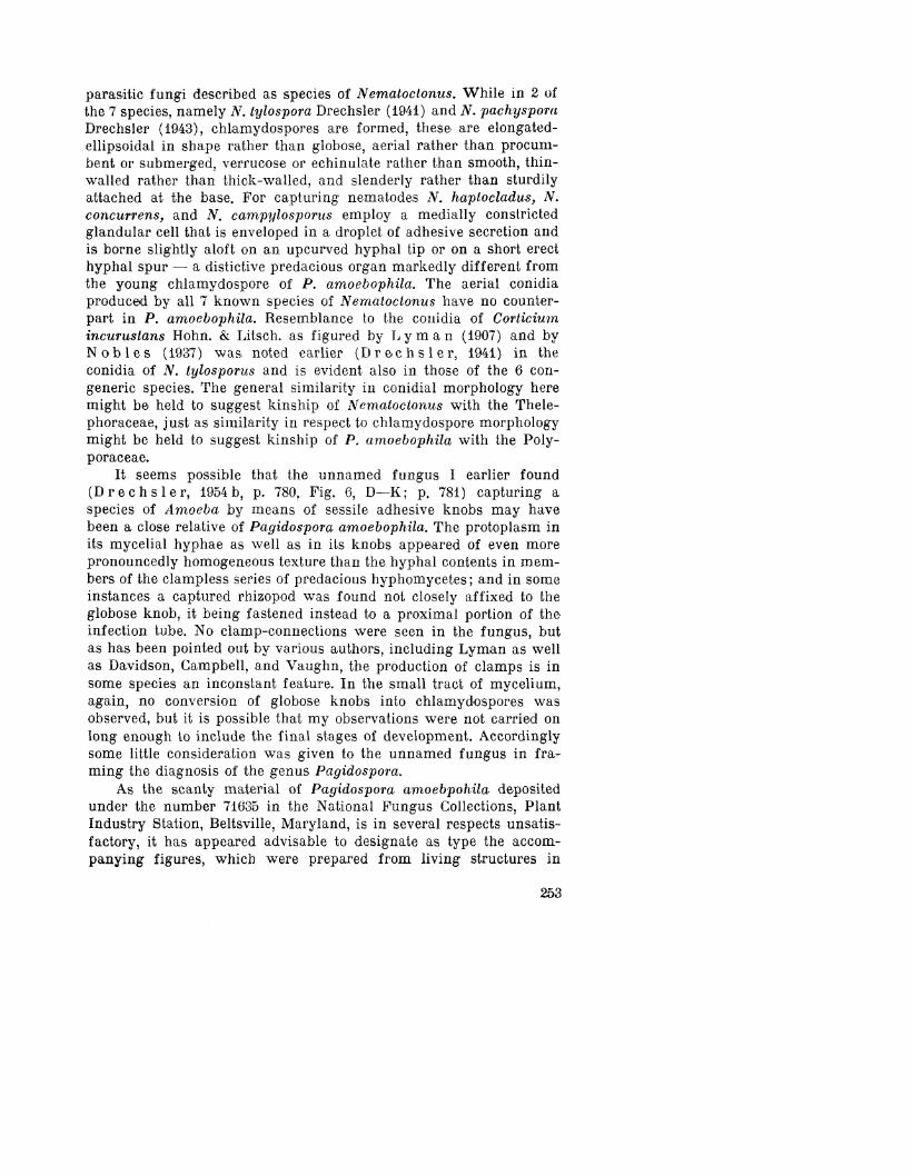
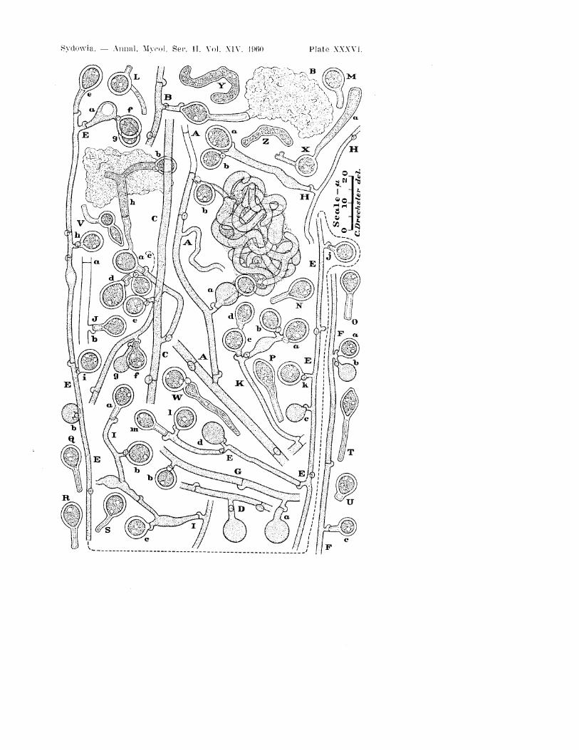
Plate XXXVI. Pagidospora amoebophila in maize-meal-agar plate cul-tures that had been planted with crumbly leaf mold 29 to 38 deys earlier;drawn with the aid of a camera lucida at a uniform magnification; xlOOOthroughout. A, Portion of mycelium on which are borne a young clamydo-sporek a, and a mature chlamydospore, b; infection tubes extended from the 2spores into an amoeba surrounded by a soft thickish pellicle gave rise tohaustoria whose assimilative branches became intertangled into a looseclew, which has been left unprotected on disintegration of the pellicle.B, Portion of hypha with a branch whereon is borne a mature chlamydo-spore bearing distally the thickened membrane of an empty infection tubeto which is attached the wrinkled membranous pellicle of a capturedamoeba. C, Portion of mycelium with a branch supporting a maturechlamydospore, a, which on one side bears a tubular membrane termina-ting in a hyphal cyst, h, and a conjoined mature chlamydospore, b, whileon the other side it bears 2 successively formed mature chlamydospores,c and d; to the hyphal cyst is affixed the wrinkled pellicle of a capturedamoeba; on a secondary hyphal branch are borne 2 mature chlamydospores,e and f, and a young chlamydospore, g. D, Portion of hypha with branchbearing young chlamydospore. E, Terminal portion of branched hypha*bearing 4 young chlamydospores, a—d, and 9 mature chlamydospores, e—m,two of which, f and g, originated laterally from the immature but olderspore a. (Owing to lack of space E is shown in 2 parts whose proper con-nection is indicated by a broken line.) F, Portion of hypha with 3 chla-mydospores, a—c; the immature spore b, was presumably produced laterthan the mature spore a, G, Portion of hypha bearing a young chlamydo-spore, a, and a mature chlamydospore, b. H. Portion of hypha with a ratherlong branch bearing 2 mature chlamydospores, a and b, probably formedin succession. I, Portion of mycelium with 3 mature chlamydospores,a—c, J, Empty portion of hypha showing a clampless septum, a, and amature chlamydospore, b. K, Portion of hypha with a distally ramifiedbranch bearing 2 discrete, successively developed mature chlamydospores,
256
X
Sycknvia. — A n n a l . Mycol . Ser. 11. Vol . X I V . i960 Pla t e XXXV.
Sydowia. — A n n u l . Mynj l . Ser. II. Vol . XIV. i960 Plate XXXVI
a and b, as well as 2 closely connected mature chlamydospores, c and A.L—U, Detached mature chlamydospores, showing usual variations in sizeand shape. \7, W, Pairs of closely connected mature chlamydospores.X, Mature chlamydospore connected with hyphal cyst, a, formed fromdistally widened portion of infection tube. Y, Z, Crooked hyphal cystsformed from assimilative branches.
C o r r e c t i o n :
References to plates No. 1—III in the t e x t should be replacedby numbers XXXIV—XXXVI. (XXXIV instead of I etc.)!
T h e references i n t h e e x p l a n a t i o n t o t h e p l a t e showever are correct!
17 Sydowia, Vol. XIV, 1960, No. 1,6