2 ring enhancing lesions
TRANSCRIPT
2 Ring-Enhancing Lesions
CLINICAL IMAGAGINGAN ATLAS OF DIFFERENTIAL DAIGNOSIS
EISENBERG
DR. Muhammad Bin Zulfiqar PGR-FCPS III SIMS/SHL
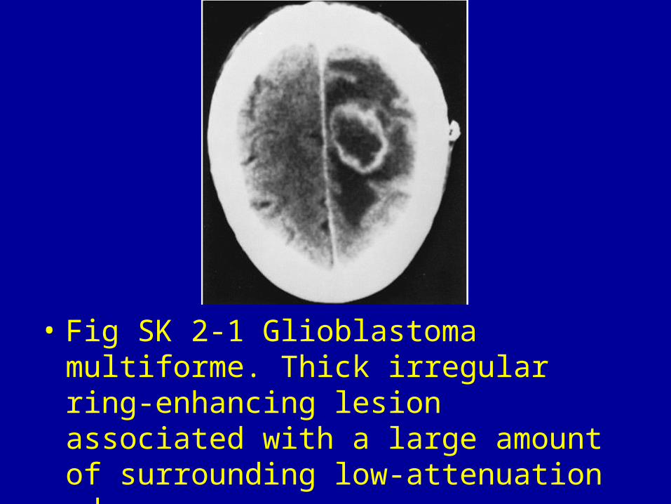
• Fig SK 2-1 Glioblastoma multiforme. Thick irregular ring-enhancing lesion associated with a large amount of surrounding low-attenuation edema.

Fig SK 2-2 Multicentric glioblastoma multiforme. Bilateral, irregular enhancing masses (arrows) with surrounding low-density edema.
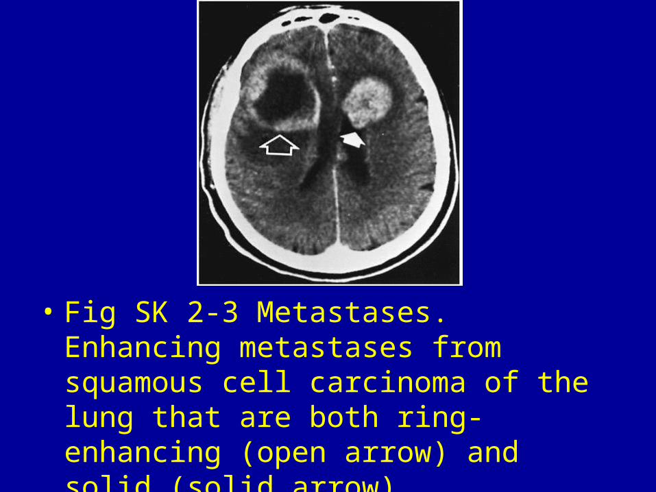
• Fig SK 2-3 Metastases. Enhancing metastases from squamous cell carcinoma of the lung that are both ring-enhancing (open arrow) and solid (solid arrow).

• Fig SK 2-4 Lymphoma developing after renal transplantation. Heart-shaped, peripherally enhancing, central lucent lesion (arrow) situated in the frontoparietal region. There is moderate surrounding edema.2
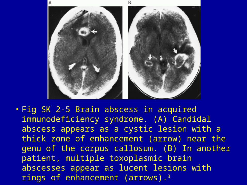
• Fig SK 2-5 Brain abscess in acquired immunodeficiency syndrome. (A) Candidal abscess appears as a cystic lesion with a thick zone of enhancement (arrow) near the genu of the corpus callosum. (B) In another patient, multiple toxoplasmic brain abscesses appear as lucent lesions with rings of enhancement (arrows).3

• Fig SK 2-6 Cysticercosis. (A) Precontrast CT scan shows a primarily low-density area in the right frontoparietal region. The ring of increased density around the lesion is vaguely evident initially, but becomes readily apparent after contrast enhancement (B).4
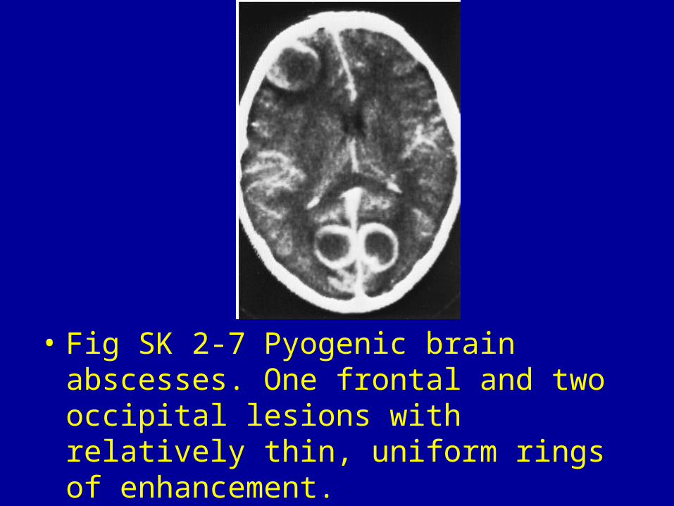
• Fig SK 2-7 Pyogenic brain abscesses. One frontal and two occipital lesions with relatively thin, uniform rings of enhancement.
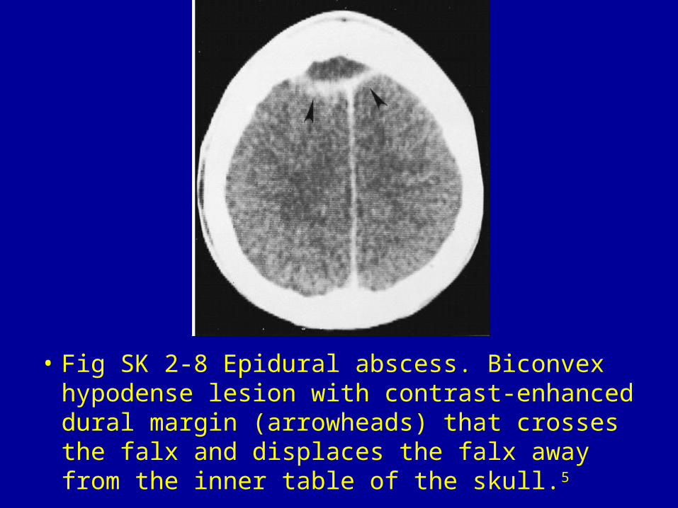
• Fig SK 2-8 Epidural abscess. Biconvex hypodense lesion with contrast-enhanced dural margin (arrowheads) that crosses the falx and displaces the falx away from the inner table of the skull.5
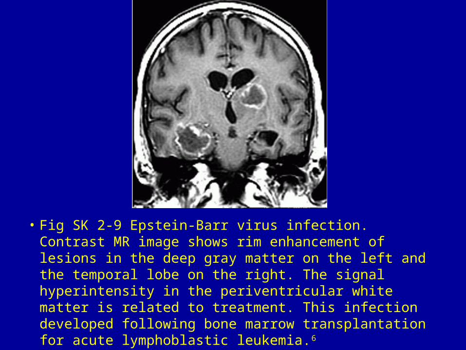
• Fig SK 2-9 Epstein-Barr virus infection. Contrast MR image shows rim enhancement of lesions in the deep gray matter on the left and the temporal lobe on the right. The signal hyperintensity in the periventricular white matter is related to treatment. This infection developed following bone marrow transplantation for acute lymphoblastic leukemia.6

• Fig SK 2-10 Resolving intracerebral hematoma. Five weeks after the initial episode of bleeding, there is peripheral contrast enhancement of the thalamic lesion.

• Fig SK 2-11 Atypical meningioma. The contrast enhancement is predominantly peripheral, with the area of central necrosis remaining relatively nonenhanced. The correct diagnosis is indicated by the origin of the tumor from the thickened tentorium.
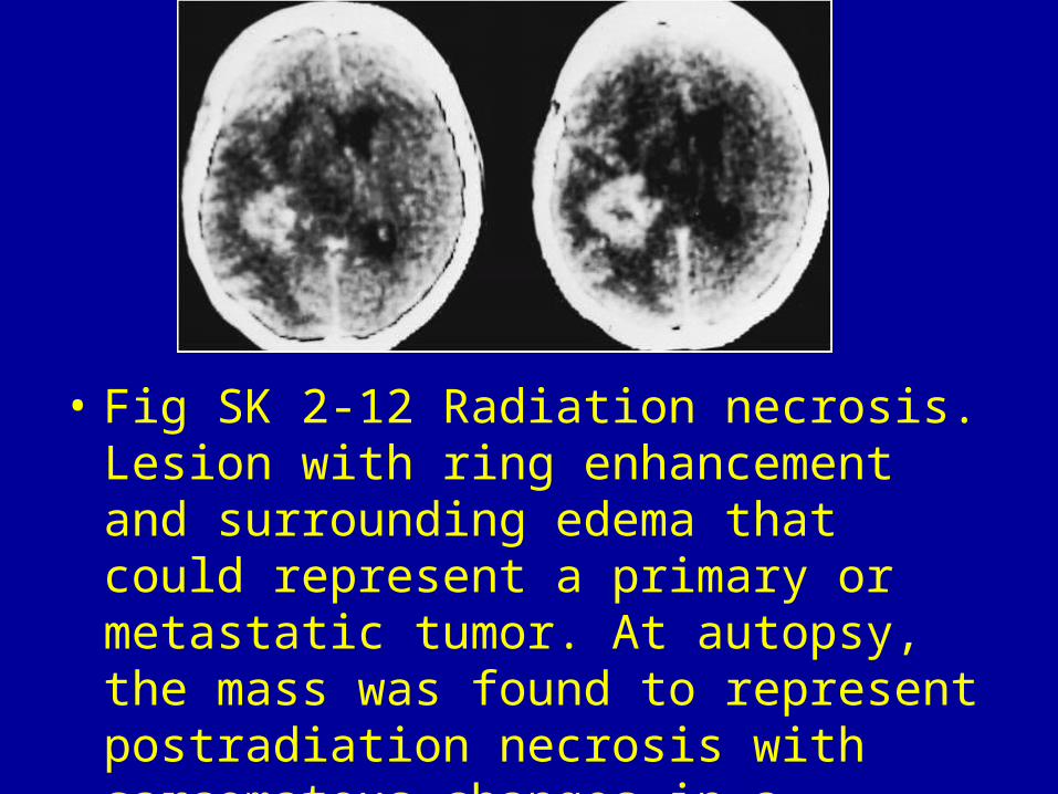
• Fig SK 2-12 Radiation necrosis. Lesion with ring enhancement and surrounding edema that could represent a primary or metastatic tumor. At autopsy, the mass was found to represent postradiation necrosis with sarcomatous changes in a patient who had undergone surgery for a solitary metastasis.5
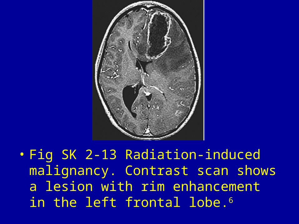
• Fig SK 2-13 Radiation-induced malignancy. Contrast scan shows a lesion with rim enhancement in the left frontal lobe.6
