cns tuberculosis diagnosis and management 2011 · posterior fossa lesion ... well enhancing ring or...
TRANSCRIPT

CNS tuberculosis diagnosis and management
Moderators – Dr MMS Dr SS
Introduction Introduction As old as history of mankind .
Odier, Ford described meningeal TB 1790 .
Sir William McEwen performed first surgery for intracranial tuberculoma in 1983 .
Caused by Mycobacterium tuberculosis (Acid fast bacillus , obligate aerobe)

CNS TB complicate 10% of all tuberculosisp
Always secondary to primary focus elsewhere in body (pulmonary GIT etc)(pulmonary , GIT etc)
Route of dissemination - haematogenous or contagious g gspread
Incidence has increased with emergence of HIV infection Incidence has increased with emergence of HIV infection

CNS tuberculosisCNS tuberculosisIntracranial
Parenchymal Parenchymal Meningeal Osseous
S l Spinal ParenchymalMeninges gArachnoiditis Osseous

Parenchymal lesion Parenchymal lesion Abscess Tuberculoma (Micro ) Tuberculoma (Micro )
Tuberculoma en plaqueTuberculous abscessTuberculous abscessCystic tuberculomaMultiple grape like tuberculomaMultiple grape like tuberculomaMicrotuberculoma Calcified tuberculomaTubercular encephalopathy

Meningeal - meningitis + HCP
Calvarial – osteomyelitis
Spinal - parenchymal – tuberculoma Spinal parenchymal tuberculoma meningeal - arachnoditis vertebral – pott’s spine p p

Diagnosis Diagnosis Hb/ ESRCXRCXRMantoux testELISA ELISA CSF PCRPCRImaging Biopsy p y

Tubercular Meningitis Tubercular Meningitis Most common manifestation of CNS TB. Considered disease of childhood however in India all age Considered disease of childhood , however in India all age groups susceptible . Acute , chronic phase & its sequelae ., p qOf neurosurgery interest are sequelae – HCP , tuberculoma or chiasmal arachnoiditis. Other sequelae - vasculitis , infarcts.
TBM with HCP Invariably occurs after 4-6 weeks .Communicating (mostly) or obstructive .

Diagnosis of TBM Diagnosis of TBM Diagnosis of TBM still pose considerable difficulties.
Supportive - H/O tuberculosis Hgm /ESR Hgm /ESR CXRMantoux test Mantoux test
CSF analysis – Sugar - low y gProtein – high Cells - lymphocytosis y p y

Bacteriological test (CSF)
Method Sensitivity Specificity
Z-N stain 25%
Culture 18-83% 100%
Limitations – CSF should examined before or just after start of ATT Time for growth – 2-4 weeks.

Molecular and Biochemical assay
T S i i i S ifi iTest Sensitivity Specificity
PCR 56% 98%
ELISA (Antigen ) 52-93% 58-98%
ELISA (Antibody) 38-94% 95-100%
Rapid and positive after starting treatment. Drawback – can’t differentiate acute or chronic infection, ,
cross - reactivityoften poor sensitive and specific p p
African Health Sciences Vol 11 No 1 March 2011

CSF ADA level - >5-15 iu/L
Sensitivity Specificity
44-100% 10-100%
High CSF ADA levels- malaria , lymphoma , pyogenic & cryptococcal meningitis brucellosis cryptococcal meningitis , brucellosis . Not recommended as routine diagnostic test
Rock RB, Olin M, Baker CA, Molitor TW, Peterson P K . C e n t r a l n e r vo u s s y s t e m t u b e r c u l o s i :African Health Sciences Vol 11 No 1 March 2011 1 2 7pathogenesis and clinical aspects. Clin Microbiol Rev.
2008 ;21:243-62008 ;21:243 6
Imaging Imaging NCCT:
scans may be normalObliteration of basal cisterns by hypo/ iso dense exudateen plaque dural thickeningPopcorn calcificationHydrocephalus Hydrocephalus Sequelae of chronic meningitis
Infarcts
CECT:Abnormal meningeal enhancementLeptomeningeal enhancemant sylvian fissures, tentorium p g yGranulomas in the basal meningesEpendymitis

Imaging Imaging
Basal exudates
HCP
Infarct

B l d h
Exudates along sylvian fissure
Basal exudates enhancement

Periventricular lucency indicates transependymal flow of CSF –sign of raised ICP however in TBM it could be CSF sign of raised ICP however in TBM it could be spread of inflammatory process making unreliable sign of raised intraventricular pressure.
R Patir, R Bhatia, Tandon PN. Surgical management of tuberculous infections of the nervous system. Schmidek and Sweet operative neurosurgical techniques 5th edition; 1617-1631
MRIMRI
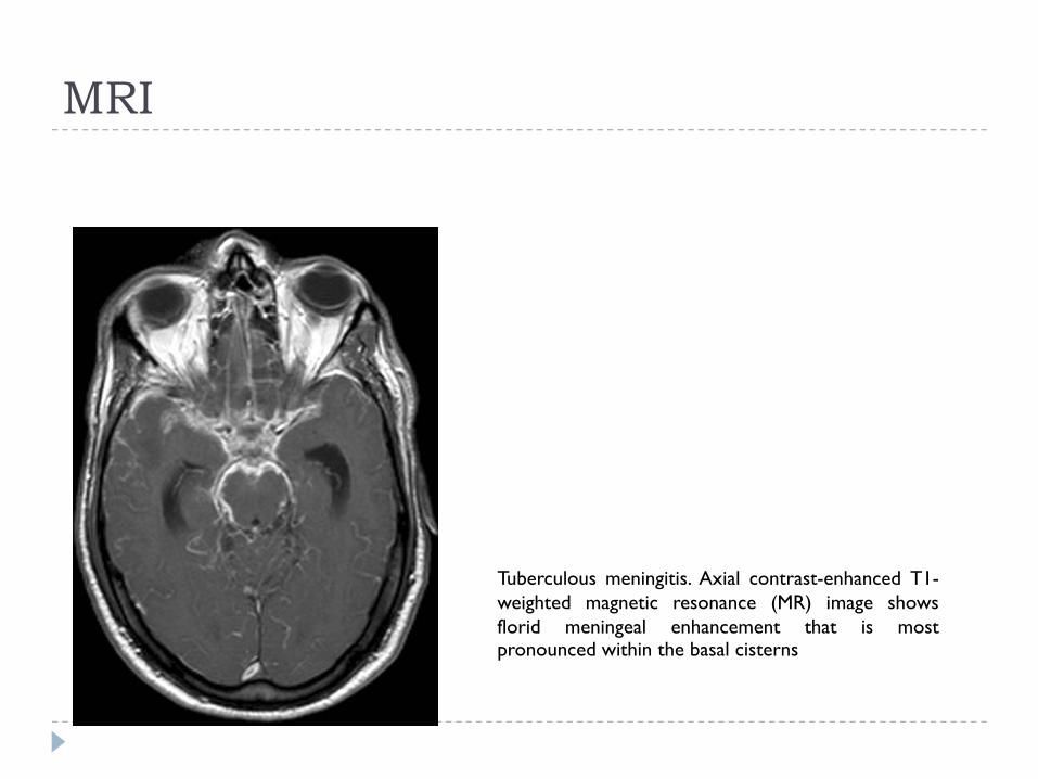
Tuberculous meningitis. Axial contrast-enhanced T1-weighted magnetic resonance (MR) image showsflorid meningeal enhancement that is mostpronounced within the basal cisterns

Ahuja and colleague set criteria for clinical diagnosis of diagnosis of TBM based on diagnosis of TBM based on
Clinical feature CSF CT scan Presence of extra neural tuberculosis .
Definite, Highly probable, Probable and Possible TBM .91% of highly probable & 66%of probable group improved with ATT . Diagnostic criteria for tuberculos meningitis and their validation , Tuber lung dis Vol – 75 , 149-152, 1994 .

Treatment for TBM Treatment for TBM ATT Anti convulsant Anti convulsant Steroids
Role of surgery - V-P shunt or ETVOptico –chiasmatic decompression for arachnoiditsOptico chiasmatic decompression for arachnoidits

ATT ATT
WHO T t t f t b l i id li 4th dWHO Treatment of tuberculosis: guidelines – 4th ed.

Duration of Anti –Tubercular treatment Duration of Anti Tubercular treatment Pulmonary and extra pulmonary disease should be treated with the same regimens. Note that some experts treated with the same regimens. Note that some experts recommend 9–12 months of treatment for TB meningitis given the serious risk of disability and mortality, and 9 months of treatment for TB of bones or joints because of the difficulties of assessing treatment response. Unless drug resistance is suspected adjuvant corticosteroid treatment is resistance is suspected, adjuvant corticosteroid treatment is recommended for TB meningitis and pericarditis. In tuberculous meningitis, ethambutol should be replaced by streptomycin
WHO Treatment of tuberculosis: guidelines – 4th ed.

British infection society recommendation 2009 –12 months 12 months
Indian academy of pediatrics 2010 recommendation –Indian academy of pediatrics 2010 recommendation In patient with TBM on category I Tt, 4 drugs can be
used either HRZE or HRZS , continuation phase of TBM pTt should extend for 6-7 months , extending total duration of treatment for 8-9 months.

American thoracic society and centre for disease control y(2003)recommendation
TBM and tuberculoma is for 12 months if bacterial strain sensitive .
For MDR TB – 24 months .
For patients who do not receive pyrizinamide in first 2 months extend Tt for 18months .

Steroids – Dexamethasone should be given to all irrespective of age and stage . p g g
Prasad K, Singh MB. Corticosteroids for managing tuberculous meningitis. Cochrane Database Syst Rev 2008;1:CD002244.
Role of steroids –Improve survival and intellectual outcome Enhance rate of resolution of basal exudate Enhance rate of resolution of basal exudate.
• Kumar Velu and assoc Randomised control trial of dexamethasone in TBM , Tuber Lung Dis , 5 page 203-207 .
No change in incidence of basal ganglia infarction , ICP . Age >14 - Dexamethasone for 4-6 weeks . Age <14 – Prednisolone for 8 weeks .
African Health Sciences Vol 11 No 1 March 2011African Health Sciences Vol 11 No 1 March 2011

British MRC grading for TBMBritish MRC grading for TBM

Role of surgery Role of surgery TBM with HCP
V-P shunt OR ETV

Role of ETV Role of ETV Success rate of ETV is 77% in 35 patient with 60% had early and 17% delayed recovery . early and 17% delayed recovery .
Minim Invasive Neurosurg. 2005 Feb;48(1):47-52.Endoscopic third ventriculostomy in post-tubercular meningitic hydrocephalus: a preliminary report. Singh D, Sachdev V, Singh AK, Sinha S.
68% benefited from ETV Hussain et al ,neurosurgery review 2005 , role of neuroendoscopy in management of patient with TBM
Success rate for ETV 73.1 % J. Neurosurg.: Pediatrics / Volume 3 / May 2009

Success rate of ETV depend upon –Stage of disease (1 & II)
Success rate of ETV depend upon –Stage of disease (1 & II)Stage of disease (1 & II)Presence of cisternal exudates Duration of pre-op ATT (4 weeks)
Stage of disease (1 & II)Presence of cisternal exudates Duration of pre-op ATT (4 weeks)Duration of pre-op ATT (4 weeks)
Surgical outcome of tuberculous meningitis hydrocephalus treated by endoscopic third ventriculostomy: ti f t d t ti i i f f ti l t f t i l t J
Duration of pre op ATT (4 weeks)
Surgical outcome of tuberculous meningitis hydrocephalus treated by endoscopic third ventriculostomy: prognostic factors and postoperative neuroimaging for functional assessment of ventriculostomy J prognostic factors and postoperative neuroimaging for functional assessment of ventriculostomy J. Neurosurg.: Pediatrics / Volume 3 / May 2009prognostic factors and postoperative neuroimaging for functional assessment of ventriculostomy J. Neurosurg.: Pediatrics / Volume 3 / May 2009

Prognosis of TBM Prognosis of TBM Based on Palur et al (mean follow up 45.6 months)
Grade Mortality
I 20%
II 34.7%
III 51.9%
IV 100%
Grade of TBM at time of admission is most significant factor determine outcome .

CNS Tuberculoma CNS Tuberculoma Mostly cortical and subcortical In children mostly posterior fossa in involved while in In children mostly posterior fossa in involved , while in adult supratentorial compartment is common Can occur at brainstem , thalamus , pitutary gland , , p y g

TuberculomaTuberculoma
SUPRATENTORIAL 78
PARIETAL 28
FRONTAL 26
TEMPORAL 15
BG / THALAMUS 4
SELAR/SUPRASELLAR 4
ORBITAL FISSURE 1ORBITAL FISSURE 1
INFRATENTORIAL 50CEREBELLUM 44
CP ANGLE 3
TENTORIUM 1
BRAINSTEM 2BRAINSTEM 2
R Patir, R Bhatia, Tandon PN. Surgical management of tuberculous infections of the nervous system. Schmidek and Sweet operative neurosurgical techniques 5th edition; 1617-1631

CT appearance of tuberculoma CT appearance of tuberculoma Cerebritis stage – hypodense lesion with out of proportion edema. proportion edema.

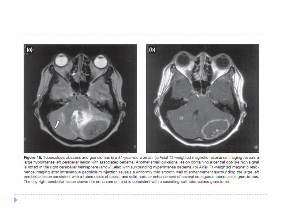
Posterior fossa lesion Posterior fossa lesion
Mature tuberculoma

Immature Tuberculoma -with out contrast – iso to hyper dense area , with
edema With contrast – either ring or nodular or irregular
enhancement
Mature tuberculoma – well enhancing ring or disc shape lesion with perilesional edema , target sign , calcification p g gseen often .

Sensitivity of CT – 100% , specificity – 85.7% and positive predictive value - 33%.predictive value 33%.

Caseating tuberculosis granulomainvolving the left temporal lobe.CECT shows a rim enhancingCECT shows a rim-enhancinglesion in the left temporal lobeconsistent with a caseatingtuberculosis granuloma

Imaging (MRI tuberculoma)Imaging (MRI tuberculoma)T1 : isointenseT2: central hyper with hypo ring T2: central hyper with hypo ring Marked thin rim enhancementHypo on T2: fibrosis gliosis macrophage infiltrationHypo on T2: fibrosis, gliosis, macrophage infiltration
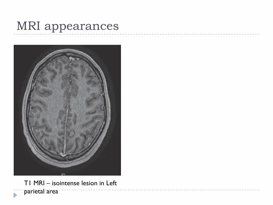
MRI appearances MRI appearances
T1 MRI – isointense lesion in Left parietal area

hypointense lesions in the hypointense lesions in the bilateral gangliathalamicregions
centrally hyperintense granuloma with a peripheral hypointense rim.

Parrenchymal tuberculosis. contrast-enhanced T1-weighted MR image demonstrates multiple enhancingcaseating and non-caseating tuberculomas,predominantly within the left frontal and parietal lobespredominantly within the left frontal and parietal lobes

Milliary CNS tuberculosis. Axial contrast-enhancedT1-weighted MR image shows multiple small high-signal-intensity foci within both cerebral hemispheres

Tubercular abscess Tubercular abscess 4-8% of all patient with CNS TB , and 20 % of all patient with HIV infection. with HIV infection.
MRS for TB abscess – lipid and phosphoserine p p pPyogenic abscess - lactate

Treatment of tuberculomaTreatment of tuberculomaMedical therapy –
ATT ATT Anti epilepticsSteroids Steroids
Role of Surgery-Role of SurgeryVision or life threatened by mass effectFailure of response to medical therapyP d l l h hParadoxical increase in lesion size with therapyDiagnosis in doubt

Anti Tubercular Treatment Anti Tubercular Treatment Intensive phase - HRZE (3-4 months)continuation phase HR (12 16months)continuation phase - HR (12-16months)Pyridoxine

Duration of treatment6 months
van Loenhout-Rooyackers JH, Keyser A, Laheij RJ, Verbeek AL, van der Meer JW. Tuberculousmeningitis: Is a 6-month treatment regimen sufficient? Int JTuberc Lung Dis 2001;5:128-35.
Th it GE H i TT T b l i iti M ti t f L t
12 months
Thwaites GE, Hein TT. Tuberculous meningitis: Many questions, too few answers. Lancet Neurol 2005;4:160-70
18 months or Longer
Santosh Isac Poonnoose, Vedantam Rajashekhar: Rate of Resolution of histologically verified intracranial tuderculomas. Neurosurgery 53:873-879, 2003
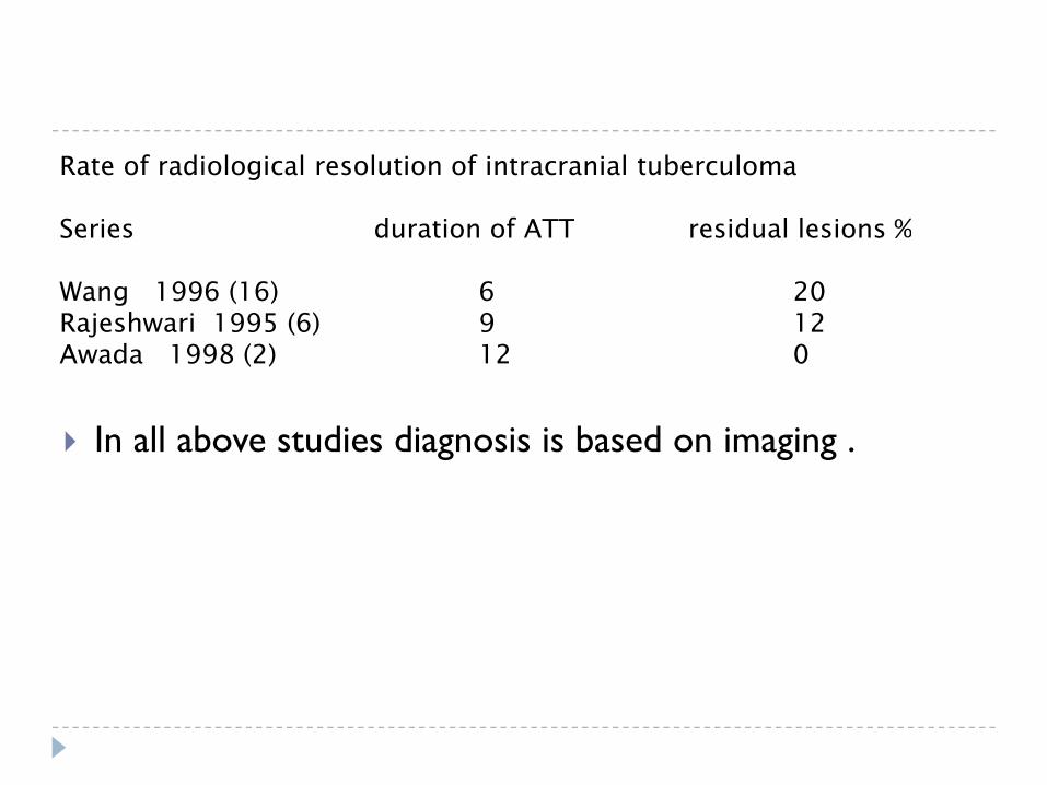
Rate of radiological resolution of intracranial tuberculoma
Series duration of ATT residual lesions %Series duration of ATT residual lesions %
Wang 1996 (16) 6 20Rajeshwari 1995 (6) 9 12
In all above studies diagnosis is based on imaging .
jAwada 1998 (2) 12 0
In all above studies diagnosis is based on imaging .

Poonnoose et al , Neurosurgery VOLUME 53 | NUMBER 4 | OCTOBER 2003
Rate of resolution of histopathologically proven tuberculoma with ATT
Duration of ATT – 9 months - 18.2 % complete resolution Duration of ATT 9 months 18.2 % complete resolution 18 months - 69.2% residual lesion 24 months - 54% complete resolution

Duration of ATT must be tailored to radiological response of lesion to therapy , pt’s clinical status should response of lesion to therapy , pt s clinical status should not govern the discontinuation of drugs . The radiological findings should dictate the continuation or termination of ATT or the administration of alternative drugs
Size of lesion (4 cms) and extent of surgical resection can affect duration of treatment affect duration of treatment .
Santosh Isac Poonnoose, Vedantam Rajashekhar: Rate of Resolution of histologically verified intracranial tuderculomas. Neurosurgery 53:873-879, 2003

Drugs Drugs Drugs Contraindication Side effects
INH Drug induced liver disease Hepatotoxicity , peripheral neuritis , optic neuritis, convulsion , lupus
dsyndrome
Rifampicin Jaundice ,pregnancy Liver toxicity , GI distrubances
Ethambutol Optic neuritis Optic neuritis, color blindness , peripheral neuritis
Pyrizinamide Hepatitis
Streptomycin Pregnancy Ototoxicity renal damage Streptomycin Pregnancy Ototoxicity , renal damage

S d li dSecond line drugs
Use at least 4 drugsUse at least 4 drugs

Role of surgery Role of surgery Life threatening edema Risk of vision loss Risk of vision loss Diagnosis is in doubt No response to drugs clinically and radiologically No response to drugs clinically and radiologically Obstructive HCP

Principles of surgery Principles of surgery Non eloquent areas total excision (small lesion)Subtotal/ partial excision (eloquent cortex)Subtotal/ partial excision (eloquent cortex)Conservative excision around vital structuresEvacuation of central liquifactive portion in deep seated Evacuation of central liquifactive portion in deep seated lesions .

Thank you y