2-1#-22,2?3@+$&-3+$a#.#'&.3'*,?3$2/2 …€¦ · · 2017-01-25......
TRANSCRIPT

Oncotarget51815www.impactjournals.com/oncotarget
www.impactjournals.com/oncotarget/ Oncotarget, Vol. 7, No. 32
E1A-engineered human umbilical cord mesenchymal stem cells as FDUULHUV�DQG�DPSOL¿HUV�IRU�DGHQRYLUXV�VXSSUHVV�KHSDWRFDUFLQRPD�in mice
Zhenzhen Li1,2, Zhou Ye3, Xiaolong Zhang1, Qing Zhang1, Dongmei Fan1, Yanjun Zhang1, Hongbo R. Luo4��;LDQJIHL�<XDQ1,5��=RQJIDQJ�/L2, Dongsheng Xiong1
1State Key Laboratory of Experimental Hematology, Institute of Hematology & Hospital of Blood Diseases, Chinese Academy of Medical Sciences & Peking Union Medical College, Tianjin 300020, China21DWLRQDO�/RFDO� -RLQW� (QJLQHHULQJ� 5HVHDUFK� &HQWHU� RI� %LRGLDJQRVWLFV� � %LRWKHUDS\�� 7KH� 6HFRQG� $I¿OLDWHG� +RVSLWDO�� ;L¶DQ�-LDRWRQJ�8QLYHUVLW\��;L¶DQ���������&KLQD3&HQWUDO�+RVSLWDO�RI�.DUDPD\��.DUDPD\��;LQMLDQJ���������&KLQD4Department of Pathology, Joint Program in Transfusion Medicine, Harvard Medical School, and Department of Laboratory 0HGLFLQH��&KLOGUHQ¶V�+RVSLWDO�%RVWRQ��%RVWRQ��0$��������86$5Tianjin Institute of Integrative Medicine for Acute Abdominal Diseases, Nankai Hospital, Tianjin 300100, China
Correspondence to: Dongsheng Xiong, email: [email protected]
Zongfang Li, email: [email protected]
Xiangfei Yuan, email: [email protected]
Keywords: HUMSC, adenovirus delivery, gene therapy, hepatocellular carcinoma
Received: September 28, 2015 Accepted: May 17, 2016 Published: June 17, 2016
ABSTRACT
*HQH�WKHUDS\�LV�DQ�DWWUDFWLYH�DSSURDFK�IRU�KHSDWRFHOOXODU�FDUFLQRPD��+&&��SDWLHQWV��1HYHUWKHOHVV��HI¿FLHQW�WUDQVJHQH�GHOLYHU\�UHPDLQV�D�FKDOOHQJH��,Q�WKLV�VWXG\��ZH�H[SORUHG�D�QHZ�WDUJHWHG�V\VWHP�EDVHG�RQ�KXPDQ�XPELOLFDO�FRUG�GHULYHG�PHVHQFK\PDO�VWHP�FHOOV��+806&V���ZKLFK�ZHUH�HQJLQHHUHG�WR�GHOLYHU�DGHQRYLUXV�WR�WXPRU�VLWHV��DQG�WR�UHSOLFDWH�DQG�DVVHPEOH�LQWR�QHZ�DGHQRYLUXV�DJDLQVW�+&&��2XU�UHVXOWV�VKRZHG�WKDW�+806&V�LQIHFWHG�E\�$G�K7(57S�,/���IROORZHG�E\�/HQWL5�(�$�LQIHFWLRQ�FRXOG�VSHFL¿FDOO\�PLJUDWH�WR�+HS*��WXPRU�FHOOV�DQG�VXSSRUW�DGHQRYLUDO�UHSOLFDWLRQ�LQ�YLWUR�DQG�LQ�YLYR����K�DIWHU�/HQWL5�(�$�LQIHFWLRQ��$G�K7(57S�,/���VSHFL¿FDOO\�LQKLELWHG�+HS*��FHOOV�JURZWK��DQG�WKLV�LQKLELWRU\�HIIHFW�ZDV�HQKDQFHG�E\�ORZ�GRVHV�RI���ÀXRURXUDFLO����)X���EHFDXVH�WKH�H[SUHVVLRQ�OHYHOV�RI�FR[VDFNLH�DGHQRYLUXV�UHFHSWRU��&$5��DQG�LQWHJULQ�Įnjǃ��RQ�WXPRU�FHOOV�ZHUH�VLJQL¿FDQWO\�LQFUHDVHG��FDXVLQJ�KLJKHU�YLUDO�XSWDNH��&RPSDUHG�ZLWK�WKH�QR�WUHDWPHQW�JURXSV��$G�K7(57S�,/���DQG�/HQWL5�(�$�FR�ORDGHG�+806&V�H[KLELWHG�VLJQL¿FDQW�DQWL�WXPRU�DFWLYLW\�LQ�YLYR��SDUWLFXODUO\�LQ�FRPELQDWLRQ�ZLWK�ORZ�GRVHV�RI���)X��,Q�VXPPDU\��WKLV�VWXG\�SURYLGHV�D�SURPLVLQJ�WDUJHWHG�JHQH�WKHUDSHXWLF�VWUDWHJ\�GHSHQGHQW�RQ�WKH�WXPRU�WURSLVP�RI�+806&V��WR�LPSURYH�WKH�RXWFRPH�RI�YLURWKHUDS\�IRU�WXPRU�SDWLHQWV�HVSHFLDOO\�WKRVH�ZLWK�PHWDVWDWLF�GLVHDVHV�
INTRODUCTION
Gene therapy is an attractive and promising approach to cancer treatment. Currently, adenoviral vectors have been employed for gene therapy due to their low pathogenicity, high titer and lack of integration LQWR� WKH� KRVW� FHOOV¶� JHQRPHV�� 7KH� ³¿UVW� JHQHUDWLRQ´�YHFWRUV�� UHSOLFDWLRQ�GH¿FLHQW� YHFWRUV� EDVHG� RQ� KXPDQ�adenovirus serotypes 5 (Ad5), in which the E1 and E3 regions of the genome are deleted, are the most widely used [1]. Clinical studies have shown that administration
RI�UHSOLFDWLRQ�GH¿FLHQW�DGHQRYLUDO�YHFWRUV�LQWUDWXPRUDOO\��intraperitoneally and intravesically is a safe, feasible and effective antitumor strategy against many types of cancers [2]. Nevertheless, the major concerns over the use of such YHFWRUV�OLH�LQ�WKH�LQHI¿FLHQW�YLUDO�GHOLYHU\�WR�WKH�PHWDVWDWLF�tumor sites due to the fact that the metastatic tumors are often smaller and directly inaccessible. Moreover, systemic administration of high doses of adenovirus is associated with systemic toxicity and rapid elimination of the virus by the immune system before reaching the tumor [3]. Thus, it is absolutely critical to develop an
Research Paper

Oncotarget51816www.impactjournals.com/oncotarget
HI¿FLHQW� DQG� WDUJHWHG� GHOLYHU\� V\VWHP� IRU� LQWUDYHQRXV�administration of adenoviral vectors to improve the clinical outcome of patients with recurrent and metastatic lesions.
Mesenchymal stem cells (MSCs) have been shown to migrate toward malignant tumors and track microscopic metastasis when administered by intravenous injection in vivo [4, 5]. Further, engineered MSCs have been indicated as a potential vehicle to deliver anticancer agents to primary and metastatic tumors [6–8]. At present, scientists have successfully taken advantage of MSCs to deliver antitumor agents, including cytokines [5], interferons [9], pro-drugs [10] and conditionally replicating virus [11]. Human umbilical cord’s Wharton’s jelly (WJ)-derived PHVHQFK\PDO�VWHP�FHOOV��+806&V���ZHUH�¿UVW�GHVFULEHG�by McElreavey et al [12]. HUMSCs share common characteristics of MSCs, such as immunosuppression, H[SUHVVLRQ� RI� D� SKHQRW\SLFDOO\� GH¿QHG� VHW� RI� VXUIDFH�markers (CD90, CD105 and CD73), multi-differentiation potential to the osteogenic, adipogenic and chondrogenic lineages [13], and ability to accumulate at sites of WLVVXH�GDPDJH��LQÀDPPDWLRQ�DQG�WXPRUV�LQ�YLYR�>�@��,Q�addition, HUMSCs are advantageous in term of rapid cell expansion, yield, ease of procedure, lack of ethical problems and are suitability for genetic engineering with viral vectors [14]. These characteristics make HUMSCs to be a promising platform for targeted delivery of anticancer agents for a variety of cancers.
To enhance the transfer of adenovirus to tumor cells, we engineered the HUMSCs to produce an adenovirus HQFRGLQJ� DQWLWXPRU� DJHQWV�� 7KH� UHSOLFDWLRQ�GH¿FLHQW�adenoviral vectors based on Ad-5 can be propagated in complementing human cell lines that provide the E1 proteins [15]. Therefore, if E1A proteins, which are essential for the replication of the adenovirus, were supplied in HUMSCs, the HUMSCs would permit UHSOLFDWLRQ�GH¿FLHQW� DGHQRYLUXVHV� WR� EH� UHSOLFDWHG� DQG�SDFNDJHG�>��@��7R�WKLV�HQG��+806&V�ZHUH�¿UVW�LQIHFWHG�E\�UHSOLFDWLRQ�GH¿FLHQW�DGHQRYLUXVHV�KDUERULQJ�WKH�DQWLWXPRU�JHQH��DQG�WKHQ�PRGL¿HG�E\�OHQWLYLUXVHV�H[SUHVVLQJ�(�$�proteins. The engineered HUMSCs not only delivered adenoviral vehicles to tumor or metastatic tumor sites but also supported the adenoviral replication. Ultimately, the UHFRPELQDQW�DGHQRYLUXV�ZDV�DPSOL¿HG�DQG�SDFNDJHG��DQG�released, allowing the infection of the surrounding tumor cells to express the target therapeutic proteins.
,Q� WKLV� VWXG\�� PHODQRPD� GLIIHUHQWLDWLRQ� DVVRFLDWHG�JHQH���LQWHUOHXNLQ���� �PGD���,/����� ZDV� FKRVHQ� DV� WKH�target therapeutic protein carried by the adenoviral vectors. ,/�����D�PHPEHU�RI�WKH�,/����IDPLO\��FDQ�VHOHFWLYHO\�LQGXFH�apoptosis in a variety of cancer cells without affecting the normal cells in vitro [17, 18], in vivo [17, 19, 20], and in D�SKDVH�,�FOLQLFDO� WULDO� >������@��0RUHRYHU��XQWUDQVIHFWHG�neighboring cancer cells can be killed by the bystander HIIHFW�RI�PGD���,/����>������@��7R�HQKDQFH�WKH�VHFRQGDU\�DQWL�WXPRU� VSHFL¿FLW\�� WKH� H[SUHVVLRQ� RI� ,/���� LV� GULYHQ�
by the human telomerase reverse transcriptase (hTERT) promoter, which is highly active in over 85% of human cancer cells but inactive in most somatic cells. The hTERT promoter has shown great potential in regulating the cell-VSHFL¿F�H[SUHVVLRQ�RI�H[RJHQRXV�WKHUDSHXWLF�JHQHV�LQ�WXPRU�FHOOV�ZLWKRXW�LQÀXHQFLQJ�WKH�QRUPDO�WLVVXHV�>������@�
:H� HYDOXDWHG� WKH� HI¿FDF\� RI� WKLV� GXDO� WDUJHWLQJ�therapeutic system in vitro and in vivo in hepatocarcinoma models. Our results showed that adenovirus-loaded HUMSC.lentiR.E1A could support adenoviral replication and viral particle release to infect the tumor cells. Moreover, virus-loaded HUMSCs were still capable of migrating to hepatocellular carcinoma. The tumor suppressive effect of this dual targeted therapeutic system was also observed in vitro and in vivo. Furthermore, we investigated the synergistic antitumor effect of this dual targeted therapeutic system in combination with ��ÀXRURXUDFLO����)X��
RESULTS
&XOWXUH�DQG�LGHQWL¿FDWLRQ�RI�+806&V
The HUMSCs, obtained from the WJ of human umbilical cord with informed consents, had a typical VSLQGOH�VKDSH�DQG�UHVHPEOHG�¿EUREODVWV��FRQVLVWHQW�ZLWK�the morphology reported by others [12, 27]. Although D� VSHFL¿F� DQWLJHQ� SUR¿OH� RI� +806&V� KDV� QRW� EHHQ�GH¿QHG�� IRU� HDFK� LVRODWLRQ� DQG� FXOWXUH�� ZH� YHUL¿HG� E\�ÀRZ�F\WRPHWU\�WKDW�WKH�LVRODWHG�FHOOV�ZHUH�SRVLWLYH�IRU�WKH�mesenchymal markers CD73, CD90 and CD105 (Figure 1A) and negative for typical hematopoietic antigens CD34, CD45 and CD19 (Figure 1B) as previously described [13]. Moreover, the HUMSCs were able to undergo adipogenesis (Figure 1C) and osteogenesis (Figure 1D) XQGHU�VSHFL¿F�GLIIHUHQWLDWLQJ�FRQGLWLRQV�LQ�YLWUR�
7KH�VSHFL¿F�DFWLYLW\�RI�K7(57�SURPRWHU�DQG�WKH�WUDQVIHFWLRQ�HI¿FLHQF\�RI�YLUXV
:H� VXFFHVVIXOO\� FORQHG� WKH� VSHFL¿F� K7(57�SURPRWHU� VHTXHQFH� ����ES��� WKH� ,/���� JHQH� DQG� WKH�(�$�JHQH��ZKRVH�QXFOHRWLGH�VHTXHQFHV�ZHUH�YHUL¿HG�E\�'1$�VHTXHQFLQJ��7R�H[DPLQH�WKH�VSHFL¿F�WUDQVFULSWLRQDO�activity of the hTERT promoter, we performed a dual-OXFLIHUDVH�UHSRUWHU�DVVD\�XVLQJ�D�S*/��K7(57S�UHSRUWHU�SODVPLG��)LJXUH��(���/XFLIHUDVH�DFWLYLW\�LQ�S*/��K7(57S�was expressed as a percentage of the positive control SODVPLG��S*/��&RQWURO��GULYHQ�E\� WKH�69���HQKDQFHU�SURPRWHU��$�S*/��%DVLF�SODVPLG�ZLWKRXW�WKH�HQKDQFHU�promoter sequence was used for negative control. HepG2, BJAB and K562 cells exhibited high transcriptional activity, whose values were 49.73±5.49%, 64.52±5.53%, and 134.59±16.25% of the positive control activity, UHVSHFWLYHO\� �)LJXUH� �)��� ,Q� FRQWUDVW�� QHLWKHU� QRUPDO�human embryonic lung MRC-5 cells nor HUMSCs

Oncotarget�����www.impactjournals.com/oncotarget
)LJXUH����,GHQWL¿FDWLRQ�RI�+806&V��WKH�VSHFL¿F�DFWLYLW\�RI�K7(57�SURPRWHU�DQG�DGHQRYLUDO�WUDQVIHFWLRQ�HI¿FLHQF\� $� Representative histogram overlays of FACS analysis showed the isolated HUMSCs were positive for CD73, CD90 and CD105. %� Representative histogram overlays of FACS analysis showed the negative antigens of HUMSCs: CD34, CD19 and CD45. &� The representative microscope image showed differentiation of HUMSCs into the adipogenic lineages in vitro. Cells were stained with Oil-red O. '� The representative microscope image showed differentiation of HUMSCs into the osteogenic lineages in vitro. Cells were VWDLQHG�ZLWK�$OL]DULQ�UHG���&��'��VFDOH�EDU� �����ȝP��(� The schematic representation displayed the vectors involved in the study. )� The VSHFL¿F�WUDQVFULSWLRQDO�DFWLYLW\�RI�K7(57�SURPRWHU�LQ�GLIIHUHQW�FHOO�OLQHV�ZDV�GHWHFWHG�E\�D�GXDO�OXFLIHUDVH�UHSRUWHU�DVVD\��*� Adenoviral WUDQVIHFWLRQ�HI¿FLHQFLHV�DW�GLIIHUHQW�02,�DIWHU����KRXUV�LQ�WKUHH�LQGHSHQGHQW�+806&�VDPSOHV�ZHUH�HYDOXDWHG�E\�)$&6�DQG�H[SUHVVHG�DV�mean ± SD. +��7KH�UHSUHVHQWDWLYH�LPDJHV�GHSLFWHG�WKH�WUDQVIHFWLRQ�HI¿FLHQF\�RI�06&V�ZLWK�����02,�$G�7UDFN�����KRXUV�DIWHU�LQIHFWLRQ��+806&V�FDUU\LQJ�*)3�ZHUH�REVHUYHG�XQGHU�ÀXRUHVFHQW�¿HOG��OHIW�SDQHO��DQG�EULJKW�¿HOG��ULJKW�SDQHO���VFDOH�EDU� �����ȝP�

Oncotarget51818www.impactjournals.com/oncotarget
showed evident transcriptional activity, with values of 1.73±0.71% and 2.99±1.07% of the positive control DFWLYLW\�� UHVSHFWLYHO\��7KHVH�¿QGLQJV�VXJJHVWHG� WKDW� WKH�K7(57�SURPRWHU�ZH�FORQHG�KDG�D�VSHFL¿F�WUDQVFULSWLRQDO�activity in cancer cells but not in normal cells.
1H[W�� WKH� OHQWLYLUDO� H[SUHVVLRQ� YHFWRUV� S/HQWL5�E1A and the adenoviral expression vectors pAd-hTERTp-,/���ZHUH�VXFFHVVIXOO\�GHYHORSHG��7KH�02,�RI�OHQWLYLUXV�RI�+806&V�ZDV�VHW�DW���02,�DV�SUHYLRXVO\�GHVFULEHG�>��@��:H�GHWHFWHG�WKH�DGHQRYLUDO�LQIHFWLRQ�HI¿FLHQF\�RI�HUMSCs using AdTrack at 48 h after infection. Flow F\WRPHWU\� UHYHDOHG� WKH� HI¿FLHQFLHV� ZHUH� ���������������������DQG�����������DW�02,����������DQG�������respectively (Figure 1G). Based on these results, for WKH�IROORZLQJ�H[SHULPHQWV�ZH�FKRVH�����02,��DW�ZKLFK�+806&V�FRXOG�EH�WUDQVIHFWHG�HI¿FLHQWO\�ZLWKRXW�DIIHFWLQJ�their growth (Figure 1H).
5HSOLFDWLRQ�GH¿FLHQW�DGHQRYLUXVHV�DPSOL¿HG�LQ�+806&�/HQWL5�(�$�DQG�ZHUH�HYHQWXDOO\�UHOHDVHG�IURP�WKH�FHOOV
To directly verify whether the replication-GH¿FLHQW� DGHQRYLUXV� FDQ� EH� DPSOL¿HG� LQ� +806&�/HQWL5�(�$�� ZH� GHWHFWHG� WKH� FRQFHQWUDWLRQV� RI� WKH�
hexon gene, a late adenoviral gene, intracellularly and in the supernatant of virus-loaded HUMSCs at the LQGLFDWHG� WLPH� SRLQWV� DIWHU�$G7UDFN� DQG�/HQWL5�(�$�co-infection. Hexon gene expression was measured by quantitative PCR to determine the copy number of DGHQRYLUDO�'1$��7KH�VWDUW�RI�/HQWL5�(�$�LQIHFWLRQ�ZDV�set as 0 h. As the expression of E1A increased gradually (Figure 2A), the total concentration of adenoviral DNA (intracellularly and in the supernatant) in the HUMSC./HQWL5�(�$�JURXS�LQFUHDVHG�UDSLGO\�DIWHU����K��ZKHUHDV�the adenoviral DNA was not detected in the control JURXS�RI�+806&�/HQWL5��)LJXUH��%���,Q�SDUWLFXODU��WKH�intracellular concentration of adenoviral DNA increased synchronously and sharply between 36 and 48 h after infection, and lasted until 72 h, while the supernatant concentration of viral DNA increased gradually and UHDFKHG� SHDN� OHYHOV� DW� ��� K� LQ� WKH� +806&�/HQWL5�E1A group (Figure 2C). Further, electron microscopy YHUL¿HG�WKH�SUHVHQFH�RI�LQWUDFHOOXODU�YLUDO�SDUWLFOHV�LQ�the HUMSCs 48 h after the infection (Figure 2D). These results indicated that viral DNA was synthesized and assembled into new viral particles in the HUMSCs co-LQIHFWHG�E\�$G7UDFN�DQG�/HQWL5�(�$��)ORZ�F\WRPHWU\�ZDV�XVHG�WR�GHWHFW�WKH�LQIHFWLRQ�HI¿FLHQF\�RI�+HS*�����h after co-culture of virus-loaded HUMSCs and HepG2.
)LJXUH����5HSOLFDWLRQ�GH¿FLHQW�DGHQRYLUXVHV�DPSOL¿HG�LQ�+806&�/HQWL5�(�$�DQG�ZHUH�HYHQWXDOO\�UHOHDVHG�IURP�WKH�FHOOV� $��7KH�P51$�H[SUHVVLRQ�OHYHO�RI�(�$�DW�LQGLFDWLQJ�WLPH�SRLQWV�DIWHU�/HQWL5�(�$�LQIHFWLRQ��%� The total concentration (intracellularly and in the supernatant) of viral DNA was measured at different time points in in three independent samples of HUMSC sequentially infected E\�$G�7UDFN�DQG�/HQWL5�(�$���/HQWL5��7KH�VWDUW�RI�/HQWL5�(�$�LQIHFWLRQ�ZDV�VHW�DV���K��&� The intracellular and supernatant concentration of viral DNA were detected at different time points respectively. '� Electron micrographs show viral particles in the HUMSC sequentially LQIHFWHG�E\�$G�7UDFN�DQG�/HQWL5�(�$��$GHQRYLUXV�SDUWLFOHV�ZHUH�VKRZQ�E\�WKH�EODFN�DUURZ��/HIW�SDQHO��DW�ORZ�PDJQL¿FDWLRQ��5LJKW�SDQHO��DW�KLJK�PDJQL¿FDWLRQ��(��7KH�LQIHFWLRQ�HI¿FLHQF\�RI�+HS*��ZDV�GHWHFWHG�E\�)$&6����K�DIWHU�FR�FXOWXUH�RI�YLUXV�ORDGHG��$G�7UDFN�DQG�/HQWL5�(�$��+806&V�DQG�+HS*��ZLWK�GLIIHUHQW�UDWLRV�LQ�WKUHH�LQGHSHQGHQW�VDPSOHV�

Oncotarget51819www.impactjournals.com/oncotarget
7KH�LQIHFWLRQ�HI¿FLHQFLHV�ZHUH�����������������������55.5±3.5% and 72.4±3.9% at HUMSCs: HepG2 ratio of 1:20, 1:10, 1:4 and 1:1, respectively (Figure 2E). These observations indicated that the released viral particles have the ability to infect cells.
0LJUDWLRQ�FDSDFLW\�RI�YLUXV�ORDGHG�+806&V�WR�KHSDWRFDUFLQRPD�LQ�YLWUR�DQG�LQ�YLYR
+806&V�DQG�PRGL¿HG�+806&V�ZHUH�SUHYLRXVO\�shown to have a homing predisposition to tumor cells in vitro and to the tumor site in hepatocarcinoma models, and their migration capacity presents a concentration–dependent pattern [28]. However, in this study, the virus-loaded HUMSCs would be lysed after 36 h of /HQWL5�(�$� LQIHFWLRQ� EHFDXVH� DGHQRYLUXV� UHSOLFDWLRQ�and packaging was activated by the expression of
E1A, as described in Figure 2B. To test the migration capacity of virus-loaded HUMSCs within 36 h after infection, in vitro migration assays using Transwell SODWHV� ZHUH� SHUIRUPHG�� 8QPRGL¿HG� +806&V� ZHUH�XVHG� DV� D� SRVLWLYH� FRQWURO�� :H� FRQ¿UPHG� WKDW� YLUXV�loaded HUMSCs migrated towards HepG2 cultures in D�VLPLODU�SDWWHUQ�DV�XQPRGL¿HG�+806&V�ZLWKLQ����K�after infection (Figure 3A, 3B). These results indicated that human hepatocellular carcinoma HepG2 cells were capable of stimulating the migration of HUMSCs and that the migration ability of HUMSCs was not affected by adenoviral and lentiviral co-infection.
Next, to investigate the homing capability of virus-loaded HUMSCs in vivo, we designed an adenoviral YHFWRU��S$G�/XF��KDUERULQJ�D�¿UHÀ\�OXFLIHUDVH�UHSRUWHU�JHQH��)LJXUH��(���5HSUHVHQWDWLYH�%LROXPLQHVFHQW�,PDJLQJ��%/,��DQDO\VLV�LQ�YLYR�UHYHDOHG�WKDW���GD\�DIWHU�LQMHFWLRQ�
)LJXUH� ���0LJUDWLRQ� FDSDFLW\� RI� YLUXV�ORDGHG�+806&V� WR� KHSDWRFDUFLQRPD� LQ� YLWUR� DQG� LQ� YLYR� $� Representative photographs showed the migrated HUMSCs stained with crystal violet in vitro migration assays using Transwell plates. %� The numbers of migrated HUMSCs in three independent samples were expressed as mean ± SD. &��7KH�KRPLQJ�FDSDELOLW\�RI�+806&�/HQWL5�$G�/XF�WR�tumor sites was monitored by bioluminescence imaging using Xenogen imaging system at indicated times after tail vein injection. '� The KRPLQJ�FDSDELOLW\�RI�YLUXV�ORDGHG�+806&�/HQWL5�(�$�$G�/XF�WR�WXPRU�VLWHV�ZDV�PRQLWRUHG�LQ�YLYR�
Oncotarget51820www.impactjournals.com/oncotarget
RI� +806&�/HQWL5�$G�/XF�� LQWHQVH� ¿UHÀ\� OXFLIHUDVH�signal was detected at the tumors site. 2 days after the injection, the signal further increased, and it lasted until ��GD\V��)LJXUH��&���,Q�WKH�+806&�/HQWL5�(�$�$G�/XF�injection group, the intense imaging signal could also be detected at the tumor site 1 day after the injection, and disappeared after 4 days. The signal was discovered once again 7 days after the injection, indicating that the UHFRPELQDQW� $G�/XF� ZDV� DPSOL¿HG� DQG� SDFNDJHG� LQ�+806&�/HQWL5�(�$�� DQG� WKHQ� UHOHDVHG� WR� LQIHFW� WKH�surrounding tumor cells (Figure 3D).
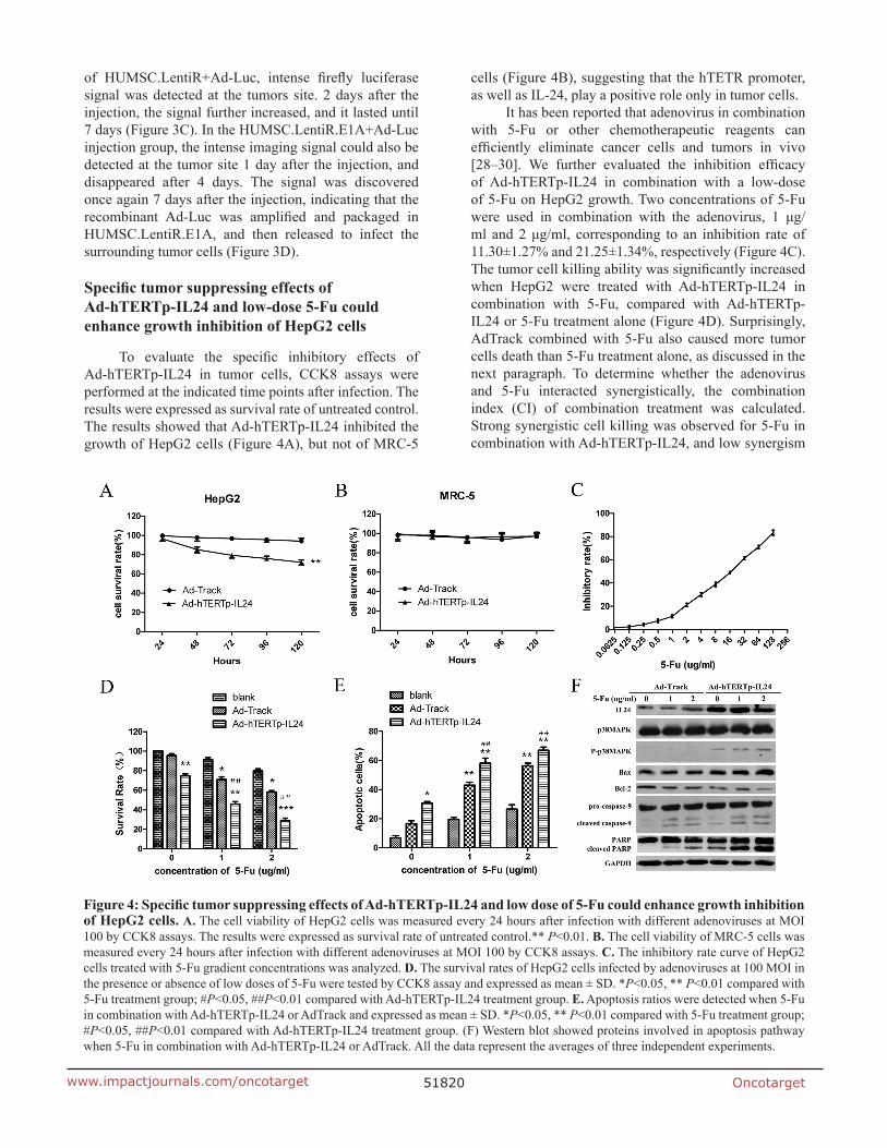
6SHFL¿F�WXPRU�VXSSUHVVLQJ�HIIHFWV�RI�$G�K7(57S�,/���DQG�ORZ�GRVH���)X�FRXOG�HQKDQFH�JURZWK�LQKLELWLRQ�RI�+HS*��FHOOV
7R� HYDOXDWH� WKH� VSHFL¿F� LQKLELWRU\� HIIHFWV� RI�$G�K7(57S�,/��� LQ� WXPRU� FHOOV�� &&.�� DVVD\V� ZHUH�performed at the indicated time points after infection. The results were expressed as survival rate of untreated control. 7KH�UHVXOWV�VKRZHG�WKDW�$G�K7(57S�,/���LQKLELWHG�WKH�growth of HepG2 cells (Figure 4A), but not of MRC-5
cells (Figure 4B), suggesting that the hTETR promoter, DV�ZHOO�DV�,/�����SOD\�D�SRVLWLYH�UROH�RQO\�LQ�WXPRU�FHOOV�
,W�KDV�EHHQ�UHSRUWHG�WKDW�DGHQRYLUXV�LQ�FRPELQDWLRQ�with 5-Fu or other chemotherapeutic reagents can HI¿FLHQWO\� HOLPLQDWH� FDQFHU� FHOOV� DQG� WXPRUV� LQ� YLYR�>��±��@�� :H� IXUWKHU� HYDOXDWHG� WKH� LQKLELWLRQ� HI¿FDF\�RI� $G�K7(57S�,/��� LQ� FRPELQDWLRQ� ZLWK� D� ORZ�GRVH�of 5-Fu on HepG2 growth. Two concentrations of 5-Fu ZHUH� XVHG� LQ� FRPELQDWLRQ� ZLWK� WKH� DGHQRYLUXV�� �� ȝJ�PO� DQG���ȝJ�PO�� FRUUHVSRQGLQJ� WR� DQ� LQKLELWLRQ� UDWH�RI�11.30±1.27% and 21.25±1.34%, respectively (Figure 4C). 7KH�WXPRU�FHOO�NLOOLQJ�DELOLW\�ZDV�VLJQL¿FDQWO\�LQFUHDVHG�ZKHQ� +HS*�� ZHUH� WUHDWHG� ZLWK� $G�K7(57S�,/��� LQ�combination with 5-Fu, compared with Ad-hTERTp-,/���RU���)X�WUHDWPHQW�DORQH��)LJXUH��'���6XUSULVLQJO\��AdTrack combined with 5-Fu also caused more tumor cells death than 5-Fu treatment alone, as discussed in the next paragraph. To determine whether the adenovirus and 5-Fu interacted synergistically, the combination LQGH[� �&,�� RI� FRPELQDWLRQ� WUHDWPHQW� ZDV� FDOFXODWHG��Strong synergistic cell killing was observed for 5-Fu in FRPELQDWLRQ�ZLWK�$G�K7(57S�,/����DQG�ORZ�V\QHUJLVP�
)LJXUH����6SHFL¿F�WXPRU�VXSSUHVVLQJ�HIIHFWV�RI�$G�K7(57S�,/���DQG�ORZ�GRVH�RI���)X�FRXOG�HQKDQFH�JURZWK�LQKLELWLRQ�RI�+HS*��FHOOV� $��7KH�FHOO�YLDELOLW\�RI�+HS*��FHOOV�ZDV�PHDVXUHG�HYHU\����KRXUV�DIWHU�LQIHFWLRQ�ZLWK�GLIIHUHQW�DGHQRYLUXVHV�DW�02,�100 by CCK8 assays. The results were expressed as survival rate of untreated control.** P<0.01. %� The cell viability of MRC-5 cells was PHDVXUHG�HYHU\����KRXUV�DIWHU�LQIHFWLRQ�ZLWK�GLIIHUHQW�DGHQRYLUXVHV�DW�02,�����E\�&&.��DVVD\V��&� The inhibitory rate curve of HepG2 cells treated with 5-Fu gradient concentrations was analyzed. '��7KH�VXUYLYDO�UDWHV�RI�+HS*��FHOOV�LQIHFWHG�E\�DGHQRYLUXVHV�DW�����02,�LQ�the presence or absence of low doses of 5-Fu were tested by CCK8 assay and expressed as mean ± SD. *P<0.05, ** P<0.01 compared with ��)X�WUHDWPHQW�JURXS���P���������P������FRPSDUHG�ZLWK�$G�K7(57S�,/���WUHDWPHQW�JURXS��(� Apoptosis ratios were detected when 5-Fu LQ�FRPELQDWLRQ�ZLWK�$G�K7(57S�,/���RU�$G7UDFN�DQG�H[SUHVVHG�DV�PHDQ���6'�� P<0.05, ** P������FRPSDUHG�ZLWK���)X�WUHDWPHQW�JURXS���P���������P������FRPSDUHG�ZLWK�$G�K7(57S�,/���WUHDWPHQW�JURXS���)��:HVWHUQ�EORW�VKRZHG�SURWHLQV�LQYROYHG�LQ�DSRSWRVLV�SDWKZD\�ZKHQ���)X�LQ�FRPELQDWLRQ�ZLWK�$G�K7(57S�,/���RU�$G7UDFN��$OO�WKH�GDWD�UHSUHVHQW�WKH�DYHUDJHV�RI�WKUHH�LQGHSHQGHQW�H[SHULPHQWV�
Oncotarget51821www.impactjournals.com/oncotarget
with AdTrack (Table 1). A similar synergistic effect was discovered in the apoptosis assays when 5-Fu was used in FRPELQDWLRQ�ZLWK�$G�K7(57S�,/���RU�$G7UDFN��)LJXUH��(���:HVWHUQ�EORWWLQJ�DVVD\V�VKRZHG�WKDW�,/����H[SUHVVLRQ�OHYHOV� ZHUH� JUHDWO\� LQFUHDVHG� LQ� WKH�$G�K7(57S�,/���group, and promoted p38MAPK phosphorylation (Figure 4F), correlating cell killing with the activation of the p38MAPK pathway. Furthermore, Bax protein expression levels increased, while Bcl-2 protein expression decreased, and other apoptosis-related proteins such as PARP, caspase-3 and 9, were cleaved and activated. All of these FKDQJHV�ZHUH�VLJQL¿FDQWO\�PRUH�UREXVW�ZKHQ�$G�K7(57S�,/���DQG���)X�ZHUH�FRPELQHG��)LJXUH��)��
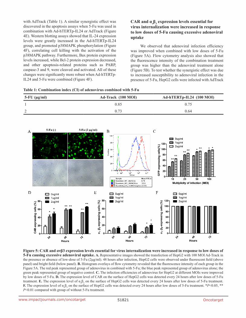
&$5�DQG�ĮȞȕ3�H[SUHVVLRQ�OHYHOV�HVVHQWLDO�IRU�
YLUXV�LQWHUQDOL]DWLRQ�ZHUH�LQFUHDVHG�LQ�UHVSRQVH�WR�ORZ�GRVHV�RI���)X�FDXVLQJ�H[FHVVLYH�DGHQRYLUDO�XSWDNH
:H� REVHUYHG� WKDW� DGHQRYLUDO� LQIHFWLRQ� HI¿FLHQF\�was improved when combined with low doses of 5-Fu (Figure 5A). Flow cytometry analysis also showed that WKH�ÀXRUHVFHQFH� LQWHQVLW\�RI� WKH�FRPELQDWLRQ� WUHDWPHQW�group was higher than the adenoviral treatment alone (Figure 5B). To test whether the synergistic effect was due to increased susceptibility to adenoviral infection in the presence of 5-Fu, HepG2 cells were infected with AdTrack
7DEOH����&RPELQDWLRQ�LQGH[��&,��RI�DGHQRYLUXV�FRPELQHG�ZLWK���)X
��)8��ȝJ�PO� $G�7UDFN�������02,� $G�K7(57S�,/���������02,�
1 0.85 0.75
2 0.73 0.64
)LJXUH����&$5�DQG�ĮȞȕ��H[SUHVVLRQ�OHYHOV�HVVHQWLDO�IRU�YLUXV�LQWHUQDOL]DWLRQ�ZHUH�LQFUHDVHG�LQ�UHVSRQVH�WR�ORZ�GRVHV�RI���)X�FDXVLQJ�H[FHVVLYH�DGHQRYLUDO�XSWDNH� $��5HSUHVHQWDWLYH�LPDJHV�VKRZHG�WKH�WUDQVIHFWLRQ�RI�+HS*��ZLWK�����02,�$G�7UDFN�LQ�WKH�SUHVHQFH�RU�DEVHQFH�RI�ORZ�GRVH�RI���)X���ȝJ�PO������KRXUV�DIWHU�LQIHFWLRQ��+HS*��FHOOV�ZHUH�REVHUYHG�XQGHU�ÀXRUHVFHQW�¿HOG��DERYH�SDQHO��DQG�EULJKW�¿HOG��EHORZ�SDQHO���%��+LVWRJUDP�RYHUOD\V�RI�ÀRZ�F\WRPHWU\�UHYHDOHG�WKDW�WKH�ÀXRUHVFHQFH�LQWHQVLW\�RI�HDFK�JURXS�LQ�WKH�)LJXUH��$��7KH�UHG�SHDN�UHSUHVHQWHG�JURXS�RI�DGHQRYLUXV�LQ�FRPELQHG�ZLWK���)X��WKH�EOXH�SHDN�UHSUHVHQWHG�JURXS�RI�DGHQRYLUXV�DORQH��WKH�green peak represented group of negative control. &��7KH�LQIHFWLRQ�HI¿FLHQFLHV�RI�DGHQRYLUXV�IRU�+HS*��DW�GLIIHUHQW�02,V�ZHUH�LPSURYHG�by low doses of 5-Fu. '� The expression level of CAR on the surface of HepG2 cells was detected every 24 hours after low doses of 5-Fu treatment. (��7KH�H[SUHVVLRQ�OHYHO�RI�ĮȞȕ3 on the surface of HepG2 cells was detected every 24 hours after low doses of 5-Fu treatment. )��7KH�H[SUHVVLRQ�OHYHO�RI�ĮȞȕ5 on the surface of HepG2 cells was detected every 24 hours after low doses of 5-Fu treatment. *P<0.05, ** P<0.01 compared with group of without 5-Fu treatment.

Oncotarget51822www.impactjournals.com/oncotarget
DW�GLIIHUHQW�02,V�LQ�WKH�SUHVHQFH�RU�DEVHQFH�RI�ORZ�GRVHV�of 5-Fu. Dose-dependent increases in viral uptake from ���WR�����02,�ZHUH�REVHUYHG��DQG�VLJQL¿FDQW�LQFUHDVHV�LQ�YLUDO�XSWDNH�ZHUH�GHWHFWHG�DW�DOO�02,V�LQ�WKH�SUHVHQFH�of low doses of 5-Fu (Figure 5C). Next, we investigated whether the expression levels of the viral attachment receptor CAR and the major internalization receptors ĮȞȕ3�DQG�ĮȞȕ5integrins were increased in response to 5-Fu WUHDWPHQW��FDXVLQJ� WKH�KLJKHU�YLUDO�XSWDNH�� ,Q�XQWUHDWHG�HepG2 cells, the baseline CAR membrane levels reduced gradually with the increase of time in culture, and they were 17.60±4.38%, 13.95±4.45%, 6.53±3.31% at 24, 48 and 72 hours, respectively. While in the presence of 5-Fu, WKH� FHOO� VXUIDFH� H[SUHVVLRQ� OHYHOV� RI�&$5�VLJQL¿FDQWO\�LQFUHDVHG�LQ�+HS*��FHOOV��)LJXUH��'���)RU�LQWHJULQ�ĮȞȕ3, baseline expression was low (<10%), and it slightly increased after 5-Fu treatment (Figure 5E). For integrin ĮȞȕ5, baseline expression was as high as 100%, and 5-Fu treatment did not affect its expression levels (Figure 5F). All these observations might explain the excessive adenoviral uptake and the enhanced cytotoxicity when the adenovirus was combined with low doses of 5-Fu.
$QWLWXPRU�SRWHQWLDO�RI�$G�K7(57S�,/���ORDGHG�06&�/HQWL5�(�$�LQ�FRPELQDWLRQ�ZLWK���)X�DJDLQVW�+HS*��[HQRJUDIW�WXPRUV
We further investigated the antitumor potential of DGHQRYLUXV�ORDGHG�06&�/HQWL5�(�$�LQ�FRPELQDWLRQ�ZLWK���)X� LQ� %$/%�&� QXGH�PLFH� WUDQVSODQWHG� ZLWK� +HS*��human hepatocellular carcinoma cells. Compared with the mice in the control group (untreated), the mice treated ZLWK�06&�/HQWL5�(�$�$G�K7(57S�,/���ZLWK�RU�ZLWKRXW�5-Fu all exhibited evident tumor regression, especially in WKH�06&�/HQWL5�(�$�$G�K7(57S�,/���SOXV���)X�JURXS��the tumor inhibition rate of which reached 71.21% 21 GD\V�DIWHU�WUHDWPHQW��)LJXUH��$���$QLPDOV�ZHUH�VDFUL¿FHG�when tumors had grown to approximately 2000 mm3 in VL]H��7XPRUV�ZHUH�GLVVHFWHG�DQG�¿[HG�LQ�����IRUPDOLQ�IRU�SDUDI¿Q�HPEHGGLQJ�
,PPXQRKLVWRFKHPLVWU\� VKRZHG� PDUNHG� ,/����expression (red) around the tumor cells (green) in the JURXS�WUHDWHG�ZLWK�06&�/HQWL5�(�$�$G�K7(57S�,/����)LJXUH��(���,Q�FRQWUDVW��ZH�FRXOG�REVHUYH�*)3�H[SUHVVLQJ�FHOOV�QHJDWLYH�IRU�,/����H[SUHVVLRQ�DPRQJ�WXPRU�WLVVXHV�LQ�WKH�JURXS�RI�06&�/HQWL5�$G�K7(57S�,/����)LJXUH��&���,Q�WKH�06&�/HQWL5�(�$�$G7UDFN�JURXS��*)3�H[SUHVVLQJ�WXPRU�FHOOV� ODFNLQJ� ,/����H[SUHVVLRQ�ZHUH�REVHUYHG�DV�expected (Figure 6D). Previous research has shown that a portion of transplanted HUMSCs are entrapped when injected intravenously [28, 31]. To investigate the fate of HUMSCs trapped by pulmonary capillaries in the lung, ZH�DOVR�GHWHFWHG�WKH�H[SUHVVLRQ�RI�,/����DQG�*)3�LQ�OXQJ�WLVVXHV�WUHDWHG�ZLWK�06&�/HQWL5�(�$�$G�K7(57S�,/����1HLWKHU�,/����QRU�*)3�H[SUHVVLQJ�FHOOV�ZHUH�GHWHFWHG�LQ�the lung (Figure 6F). The trapped HUMSCs had probably
died rapidly due to the unsuitable environment, and they had released sporadic adenoviral particles in lung, LQGLFDWLQJ� WKH� WLVVXH�VSHFL¿FLW\� RI� WKLV� GXDO� WDUJHWHG�system.
)XUWKHU��781(/�VWDLQLQJ�ZDV�SHUIRUPHG�WR�HYDOXDWH�the apoptosis-inducing ability of this targeted system LQ� YLYR�� 7XPRUV� WUHDWHG� ZLWK� 06&�/HQWL5�(�$�$G�K7(57S�,/���ZLWK�RU�ZLWKRXW���)X�SUHVHQWHG�VLJQL¿FDQWO\�apoptosis, which was limited to the tumor mass (Figure �.�/���7KH� DSRSWRWLF� DFWLYLW\� GHWHFWHG� LQ� RWKHU� WUHDWHG�JURXSV�ZDV�PXFK�ORZHU�WKDQ�LQ�WKH�06&�/HQWL5�(�$�$G�K7(57S�,/���SOXV���)X�JURXS��)LJXUH��*±�-���LQGLFDWLQJ�WKDW�06&�/HQWL5�(�$�$G�K7(57S�,/���DQG���)X�ZHUH�synergistically induced tumor cell death in vivo.
DISCUSSION
We successfully established a new targeted treatment system based on HUMSCs, which were engineered to GHOLYHU�D� UHSOLFDWLRQ�GH¿FLHQW� DGHQRYLUDO�YHFWRU�� DQG� WR�enable its replication and assembling into new viruses to tumor sites in a mouse model of hepatocellular carcinoma. Our results showed that engineered HUMSCs have the ability to incorporate into tumors and release cancer-killing DGHQRYLUDO� SDUWLFOHV�� 06&�/HQWL5�(�$�� $G�K7(57S�,/���WUHDWPHQW�H[KLELWHG�VLJQL¿FDQW�DQWL�WXPRU�HIIHFWV�RQ�the transplanted hepatocarcinoma, which were mediated E\�$G�K7(57S�,/����$GGLWLRQDOO\��WKLV�WXPRU�VXSSUHVVLQJ�effect was greatly strengthened when combined with low doses of 5-Fu, due to the increased expression of CAR DQG�ĮȞȕ3 on tumor cells in response to low doses of 5-Fu. Previously, a similar targeted therapeutic strategy has been reported. Ando and his colleagues adapted a suicide system based on an inducible caspase-9 (iC9) protein WKDW� LV� DFWLYDWHG� XVLQJ� D� VSHFL¿F� FKHPLFDO� LQGXFHU� RI�GLPHUL]DWLRQ��&,'��IRU�DGHQRYLUDO�EDVHG�GHOLYHU\�WR�OXQJ�WXPRUV�WKURXJK�06&V�>��@��7KHLU�¿QGLQJV�VXJJHVWHG�WKDW�WKLV�DSSURDFK�FRXOG�EH�XVHG�WR�NLOO�16&/&�LQ�YLWUR�DQG�in an orthotopic mouse xenograft model of lung cancer. Compared with this study, our results not only showed the effectively anti-tumor activity of this strategy but also provided more evidence of adenoviral replication in vitro and in vivo (Figures 2 and 3).
The tumor-migrating tropism of MSCs has been acknowledged in recent years. Due to its applications to tumor treatment, increasingly frequent investigations have been performed on the kinetic distribution of systemically administered MSCs in vivo and on the time required for MSCs to the tumor site [11, 33, 34]. Our laboratory previously demonstrated by luciferase bioluminescence in vivo that MSCs migrate and selectively accumulated at the tumor site at 24 h after intravenous injection [34]. Further, Xi Xia [33] investigated the distribution pattern RI� 06&V� LQ� D� VSHFL¿F� WLPH� SHULRG�� UDQJLQJ� IURP� ���h to 72 h after injection, and found that the number of 06&V� LQ� WKH� WXPRU� LQFUHDVHG� RYHU� WLPH�� ,Q� WKLV� VWXG\��

Oncotarget51823www.impactjournals.com/oncotarget
)LJXUH����7XPRU�VXSSUHVVLQJ�HIIHFWV�RI�$G�K7(57S�,/���ORDGHG�06&�/HQWL5�(�$�LQ�FRPELQDWLRQ�ZLWK���)X�DJDLQVW�+HS*��[HQRJUDIW�WXPRUV� $� The tumor volumes of different groups were measured every 3 days after treatment. Points indicate the mean values (n ����EDUV�LQGLFDWH�6'�� P<0.05, ** P<0.01 compared with PBS group. %�)� The target protein expression was detected LQ� GLIIHUHQW� JURXSV�E\� FRQIRFDO�PLFURVFRS\��7KH� UHG�ÀXRUHVFHQFH� UHSUHVHQWHG� ,/����SURWHLQV�� WKH�JUHHQ�ÀXRUHVFHQFH� UHSUHVHQWHG� WKH�FHOOV�LQIHFWHG�E\�DGHQRYLUXV��7KH�EOXH�ÀXRUHVFHQFH�VKRZHG�WKH�QXFOHL���%��3%6���&��06&�/HQWL5�$G�K7(57S�,/�����'��06&�/HQWL5�(�$�$G�7UDFN�� �(��06&�/HQWL5�(�$�$G�K7(57S�,/���� �)��7KH� OXQJ� WLVVXH� RI� WKH� JURXS� RI�06&�/HQWL5�(�$�$G�K7(57S�,/��� *�/��781(/�VWDLQLQJ�UHYHDOHG�DSRSWRVLV�LQ�WKH�WXPRUV�RI�GLIIHUHQW�WUHDWPHQW�JURXSV��&\��ODEHOHG�781(/�SRVLWLYH�FHOOV�RQ�WKH�VHFWLRQV�ZHUH�GHWHFWHG�E\�FRQIRFDO�PLFURVFRS\���*��3%6���+����)X���,��06&�/HQWL5�$G�K7(57S�,/�����)X���-��06&�/HQWL5�(�$�$G�7UDFN���)X� �.��06&�/HQWL5�(�$�$G�K7(57S�,/�����/��06&�/HQWL5�(�$�$G�K7(57S�,/�����)X�

Oncotarget�����www.impactjournals.com/oncotarget
the therapeutic adenoviruses were delivered to tumor VLWHV�DQG�DPSOL¿HG�E\�+806&V�VHTXHQWLDOO\�LQIHFWHG�E\�$G�K7(57S�,/��� DQG� /HQWL5�(�$��:H� IRXQG� WKDW� WKH�adenovirus could extensively amplify in HUMSCs 36 K�DIWHU� WKH� WUDQVGXFWLRQ�RI�/HQWL5�(�$�LQ�YLWUR��+HQFH��the lentiviral expression of E1A, which is necessary IRU�DGHQRYLUDO�DPSOL¿FDWLRQ��UHTXLUHV�DW�OHDVW����K��7KH�increased expression of E1A was accompanied by massive DGHQRYLUDO�DPSOL¿FDWLRQ��DQG�UHDFKHG�SHDN�OHYHOV�DW����h. Furthermore, in vivo experiments demonstrated that it took no longer than 48 h for most of the virus-loaded +806&V�WR�UHDFK�WKH�WXPRU�VLWHV��7KHVH�¿QGLQJV�VXJJHVW�that, taking advantage of the delay of lentiviral gene H[SUHVVLRQ��D�VXI¿FLHQW�QXPEHU�RI�+806&V�FRXOG�PLJUDWH�to tumor sites before being lysed.
Recently, MSCs have been demonstrated to deliver conditionally replicative adenovirus (CRAd) to various malignant tumors. These viruses are able to destroy tumor cells by replication and consequent oncolysis, which enables the newly produced virus to be released to the surrounding tumor tissues to prevent tumor growth [6, 11, 33, 35]]. However, a considerable number of CRAd-loaded MSCs were also found in the lung, liver, and spleen, with the exception of the tumor site after systemic administration [33]. These ectopic CRAd-loaded MSCs would produce virus to injury normal tissues as well, GXH�WR�WKHLU�ODFN�RI�DEVROXWH�VSHFL¿FLW\��ZKLFK�OLPLWV�WKH�HIIHFWLYH� XVH� RI� &5$G� LQ� FOLQLFDO� DSSOLFDWLRQV�� ,Q� RXU�study, we used E1A-engineered HUMSCs to deliver a UHSOLFDWLRQ�GH¿FLHQW�DGHQRYLUXV��ZKLFK�FDQ�UHSOLFDWH�DQG�assemble into new viruses to tumor sites. The replication-GH¿FLHQW� DGHQRYLUXV� UHSOLFDWHG� RQO\� LQ� +806&V� EXW�not tumor cells or other normal cells because of the complementary expression of E1A in HUMSCs.
Combining adenoviral constructs with chemotherapeutics has represented an appealing strategy to increase their potency [36]. Several studies have presented combinatory cytotoxic effects in esophageal carcinoma by Ad-delE1B55 in combination with 5-Fu [37], in pancreatic adenocarcinoma model by Ad-dl922-947 in combination with 5-Fu or gemcitabine [29], and in patients with recurrent head and neck cancer by intratumoral ONYX-015 in combination with cisplatin or 5-Fu [30]. The reasonable explanation for combinatory cytotoxic effects on tumors was that chemotherapeutic agents increased adenoviral LQIHFWLYLW\� GHSHQGHQW� RQ� &$5�� ĮȞȕ3� DQG� ĮȞȕ5 [29] or associated with morphological changes in lipid membranes >��@��,Q�WKLV�VWXG\��ZH�LQYHVWLJDWHG�WKH�HIIHFWV�RI���)X��DV�LW�is frequently used for hepatocellular carcinoma treatment. Synergistic tumor cell inhibition was observed for 5-Fu in FRPELQDWLRQ�ZLWK�$G�K7(57S�,/����DV�ZHOO�DV�$G7UDFN�LQ�vitro and in vivo. Two explanations might account for the V\QHUJ\��¿UVW��WKH�H[SUHVVLRQ�OHYHO�RI�&$5�ZDV�LQFUHDVHG�in response to the 5-Fu treatment, causing the higher YLUDO�XSWDNH��VHFRQG��RYHU�H[SUHVVHG�,/����FRXOG�HQKDQFH�sensitivity of cancer cells to 5-Fu [39].
,Q� FRQFOXVLRQ�� WKLV� ZRUN� LQYHVWLJDWHV� D� SURPLVLQJ�targeted therapeutic strategy using E1A-engineered +806&V� WR� GHOLYHU� DQG� SURGXFH� UHSOLFDWLRQ�GH¿FLHQW�adenovirus against hepatocarcinoma to tumor sites. The therapeutic strategy provides a new, effective and safe DGPLQLVWUDWLRQ� URXWH� IRU� UHSOLFDWLRQ�GH¿FLHQW� DGHQRYLUXV�WR� UHVROYH� WKH� SUREOHP� RI� LQHI¿FLHQW� YLUXV� GHOLYHU\� WR�inaccessible and/or metastatic tumor sites. Meanwhile, it also solves the potential safety hazard of HUMSCs. However, this therapeutic system retains some problems that will need to be improved in the future. For example, the expression of E1A can be controlled by an inducible promoter, which would ensure more HUMSCs migrate to tumor sites before lysis, enhancing the tumor-suppressing effect. Additional studies are warranted to demonstrate the superiority of this therapeutic strategy in the tumor metastasis models, which would make it highly appealing for the treatment of tumor patients with metastatic diseases.
0$7(5,$/6�$1'�0(7+2'6
&HOO�FXOWXUH
The human hepatocellular carcinoma cell line +HS*�� DQG� WKH� KXPDQ� HPEU\RQLF� OXQJ� ¿EUREODVW� FHOO�line MRC-5 were obtained from the Cell Resource Center, Peking Union Medical College (which is the KHDGTXDUWHU� RI� WKH�1DWLRQDO� ,QIUDVWUXFWXUH�RI�&HOO�/LQH�5HVRXUFH��167,��RQ�0D\���WK��������&HOOV�ZHUH�WHVWHG�for the absence of mycoplasma contamination by PCR DQG� FXOWXUH�� &HOO� VSHFLHV� ZDV� FRQ¿UPHG� E\� 3&5�� 7KH�identity of the cell lines was authenticated with STR SUR¿OLQJ��)%,��&2',6���$OO�WKH�UHVXOWV�FDQ�EH�YLHZHG�RQ�the website (http://cellresource.cn). Cells were maintained LQ� '0(0� VXSSOHPHQWHG� ZLWK� ���� )%6� DQG� Į�0(0�supplemented with 1×MEM Non-Essential Amino Acids and 10% FBS, respectively. The human embryonic renal FHOO�OLQH����$��,QVWLWXWH�RI�+HPDWRORJ\��%ORRG�'LVHDVHV�+RVSLWDO�&KLQHVH�$FDGHP\�RI�0HGLFDO�6FLHQFHV��3HNLQJ�Union Medical College, PUMC), and human embryonic kidney cell derived 293T cell line (kindly provided by Professor Cheng Tao, PUMC) were maintained in DMEM supplemented with 10% FBS. Cells were cultured in a cell incubator containing 5% CO2 at 37°C.
+806&V�SUHSDUDWLRQ
HUMSCs were isolated from human umbilical cord Wharton’s jelly (WJ) as previously described [27]. HUMSCs were seeded at a density of 8×10³ cells/cm2 in ')����VXSSOHPHQWHG�ZLWK���PPRO�O�/�JOXWDPLQH�DQG�����)%6��:KHQ�FHOOV�UHDFKHG���a����FRQÀXHQFH��WKH\�ZHUH�detached using a 0.125% trypsin/1 mM EDTA solution, and re-seeded using the same growth media for subsequent passages. Cells at passage number 3-5 were used for the following experiments.

Oncotarget51825www.impactjournals.com/oncotarget
5HFRPELQDQW�DGHQRYLUXV�UHOHDVH�IURP�+806&�/HQWL5�(�$�LQ�YLWUR
HUMSCs were seeded in 6-well plates at a density of 1×10⁵ cells/well and incubated overnight at 37°C. On the next day, the HUMSCs were infected with Ad-K7(57S�,/���DW�PXOWLSOLFLW\�RI�LQIHFWLRQ��02,������IRU�6 hours. Then, the culture medium was replaced with IUHVK�PHGLXP�FRQWDLQLQJ�OHQWLYLUDO�VXSHUQDWDQWV��/HQWL5�RU�/HQWL5�(�$��DW�02,���ZLWK��ȝJ�PO�SRO\EUHQH��6LJPD��Santa Clara, CA). Twelve hours later, the medium was replaced. HUMSCs and supernatants were harvested after the indicated periods of lentiviral infection, and used for DGHQRYLUDO�'1$�SUHSDUDWLRQ�XVLQJ� WKH�+LJK�3XUH�9LUDO�Nucleic Acid Extraction Kit (Roche, Basel, Switzerland). 4XDQWLWDWLYH� UHDO�WLPH� 3&5� ZDV� SHUIRUPHG� XVLQJ�$%,�35,60� ����� UHDO� WLPH� 3&5� V\VWHP�� 6<%5� JUHHQ�technology was used to detect a 286-bp-long amplicon (nucleotides 21049-21334) within the conserved region of the Ad5 hexon gene. The primers were designed as IROORZV�� �ƍ�**7**&&$77$&&777*$&7&77&��ƍ�DQG��ƍ�&&$&&7*77**7$*7&&77*7$777$*7$7&$7&��ƍ��3&5�F\FOHV�ZHUH�SURJUDPPHG�DFFRUGLQJ�WR�WKH�manufacturer’s instructions for SYBR Premix Ex Taq reagent (Takara, Dalian, China). The standard curve for DGHQRYLUXV�TXDQWL¿FDWLRQ�ZDV�JHQHUDWHG�E\�VHULDO�GLOXWLRQV�of pAdTrack plasmid. The experiments were repeated for three times.
0LJUDWLRQ�DVVD\�RI�YLUXV�ORDGHG�+806&V�LQ�YLWUR�DQG�LQ�YLYR
The migration of virus-loaded HUMSCs was GHWHUPLQHG�XVLQJ���ȝP�SRUH�PHPEUDQH�LQVHUWV�ZLWK����PP�diameter (BD Falcon, New York, USA). 12 hours after co-infection, 1×10⁵ HUMSCs were plated in the top chamber LQ�����ȝO�RI�VHUXP�IUHH�PHGLXP��7KH�SUHYLRXV�GD\��+HS*��cells were seeded at a density of 5×10⁴ cells/well in the lower chamber in fresh medium containing 2% FBS. After 20 h incubation at 37°C, cells that had not migrated from the upper side of the membrane were scraped off with a cotton swab, and membranes were stained with 0.1% crystal violet at 37°C for 45 min. Cells that had migrated WR�WKH�ORZHU�VLGH�RI�WKH�PHPEUDQH�ZHUH�TXDQWL¿HG��7KH�QXPEHU�RI�FHOOV�ZDV�GHWHUPLQHG�LQ�¿YH�UDQGRPO\�VHOHFWHG�KLJK�SRZHU��î�����PLFURVFRSH�¿HOGV��([SHULPHQWV�ZHUH�performed in triplicate.
For the in vivo HUMSC migration assays, we GHYHORSHG� DQ� DGHQRYLUDO� YHFWRU� FRQWDLQLQJ� D� ¿UHÀ\�OXFLIHUDVH� UHSRUWHU� JHQH� �S$G�/XF��� 6HYHQ� GD\V� DIWHU�HepG2 cells inoculation into the mouse right armpits, when the solid tumors reached 100-200 mm3 in size, 1×10⁶�+806&V�ZHUH�LQIHFWHG�E\�$G�/XF�DW�02,������IROORZHG� E\� /HQWL5�(�$� LQIHFWLRQ� IRU� �� KRXUV�� DQG�injected into mice via the tail vein to detect cell migration. %LROXPLQHVFHQFH� LPDJLQJ� ZDV� SHUIRUPHG� XVLQJ� ,9,6�
;HQRJHQ�����V\VWHP��&DOLSHU�/LIHVFLHQFHV��86$��DW�WKH�indicated times, as previously described [28].
*URZWK�LQKLELWLRQ�RI�KHSDWRFHOOXODU�FDUFLQRPD��+HS*���[HQRJUDIWV�LQ�YLYR
All animal procedures were approved by the Committee on the use and care of animals, Chinese Academy of Medical Science. 5-6-weeks-old female %$/%�F� QXGH� PLFH� ZHUH� LQRFXODWHG� VXEFXWDQHRXVO\�with 5×10⁶ HepG2 cells into right armpits. 7 days after tumor inoculation, when solid tumors reached 100-200 mm3 in size, mice were randomized into 9 groups (7 PLFH� IRU� HDFK� JURXS�� DV� IROORZV�� ���� 3%6� FRQWURO�� ����+806&��������)X������06&�/HQWL5�$G�K7(57S�,/��������06&�/HQWL5�$G�K7(57S�,/���DQG���)X������06&�/HQWL5�(�$�$G7UDFN�� ���� 06&�/HQWL5�(�$�$G7UDFN�DQG� ��)X�� ���� 06&�/HQWL5�(�$�$G�K7(57S�,/�������� 06&�/HQWL5�(�$�$G�K7(57S�,/��� DQG� ��)X��The HUMSCs were co-infected as described above and injected intravenously with 1×10⁶ cells/mouse. 5-Fu was i.p. injected at a dose of 10 mg/kg for 5 continuous days starting 3 days after HUMSCs injection. Growing tumors were measured every three days using a vernier caliper in two perpendicular dimensions. The tumor volumes were FDOFXODWHG� XVLQJ� WKH� IROORZLQJ� IRUPXOD�� 9 �/î:������ZKHUH� /� UHSUHVHQWV� WKH� ORQJHVW� D[LV� RI� WKH� WXPRUV� �LQ�PP�� DQG�:� UHSUHVHQWV� WKH� D[LV� SHUSHQGLFXODU� WR�/� �LQ�PP���0LFH�ZHUH�VDFUL¿FHG�E\�FHUYLFDO�GLVORFDWLRQ�XQGHU�anesthesia when the tumors reached 2000 mm3 in size, and tumor tissues were harvested and weighed.
,PPXQRKLVWRFKHPLFDO�DQDO\VLV�IRU�WKH�H[SUHVVLRQ�RI�,/���
7XPRU� WLVVXHV� ZHUH� ¿[HG� LQ� ���� IRUPDOLQ� DQG�HPEHGGHG� LQ� SDUDI¿Q� EORFNV�� �� ȝP� VHFWLRQV� ZHUH�REWDLQHG� IRU� +(� VWDLQLQJ� DQG� VXEVHTXHQW� DQDO\VLV��7KH�H[SUHVVLRQ�RI�,/����LQ�WKH�WXPRUV�WUHDWHG�E\�06&�/HQWL5�(�$�� $G�K7(57S�,/��� ZDV� GHWHFWHG� E\�LPPXQRKLVWRFKHPLVWU\�� ,Q� EULHI�� WKH� SDUDI¿Q� VHFWLRQV�ZHUH�GHSDUDI¿QL]HG� LQ�[\OHQH� DQG� UHK\GUDWHG� WKURXJK�a series of graded-ethanol and PBS solutions. Antigen retrieval was performed by heated in a hot bath. Sections were then treated with goat serum for 30 min at room temperature followed by incubation with rabbit anti-,/����PRQRFORQDO�DQWLERG\��$EFDP��&DPEULGJH��8.��and mouse anti-GFP monoclonal antibody (Abbkine, CA) at 4°C overnight. On the following day, sections were incubated with secondary polyclonal donkey DQWL�UDEELW� '\/LJKW� ����FRQMXJDWHG� DQWLERG\� DQG�VHFRQGDU\�SRO\FORQDO�UDEELW�DQWL�PRXVH�),7&�FRQMXJDWHG�antibody for 30 min. 4, 6-diamidino-2-phenylindole �'$3,��6LJPD��6DQWD�&ODUD��&$��ZDV�XVHG�IRU�QXFOHDU�staining. The stained sections were imaged by confocal PLFURVFRS\��/HLFD�7&6�63���*HUPDQ\��

Oncotarget51826www.impactjournals.com/oncotarget
,Q�VLWX�DQDO\VLV�RI�DSRSWRWLF�FHOOV
Apoptotic cells in the tumors were detected by terminal deoxynucleotidyl transferase dUTP nucleotide QLFN�HQG�ODEHOLQJ��781(/���%ULHÀ\��D�FRPPHUFLDO�RQH�VWHS�781(/�DSRSWRVLV�DVVD\�NLW� �%H\RWLPH�,QVWLWXWH�RI�Biotechnology, Shanghai, China) was used according WR�WKH�PDQXIDFWXUHU¶V�LQVWUXFWLRQV��&\��ODEHOHG�781(/�positive cells on the sections were detected by confocal microscopy.
6WDWLVWLFDO�DQDO\VLV
Data were analyzed using an independent sample t-tests and were represented as the mean ± SD. P<0.05 ZDV�FRQVLGHUHG�WR�EH�VWDWLVWLFDOO\�VLJQL¿FDQW��DQG�P<0.01 ZDV�FRQVLGHUHG�WR�EH�KLJKO\�VWDWLVWLFDOO\�VLJQL¿FDQW�
$&.12:/('*0(176
This work was supported by grants from the Natural Science Foundation of China (Grant No. 81572993 and 81400176), the major projects of the ministry of science and technology (Grant No. 2012ZX09102301-015) and the Natural Science Foundation of Tianjin (Grant No.05YFZGX02800). We thank Prof. Tao Cheng for generously providing the human embryonic kidney cell derived 293T cell line.
CONFLICTS OF INTEREST
7KH�DXWKRUV�GHFODUH�QR�SRWHQWLDO�FRQÀLFW�RI�LQWHUHVW�
$EEUHYLDWLRQV
+&&�� +HSDWRFHOOXODU� FDUFLQRPD�� 7$&(��7UDQVFDWKHWHU�DUWHULDO�FKHPRHPEROL]DWLRQ��$G���$GHQRYLUXV�VHURW\SHV����06&V��0HVHQFK\PDO�VWHP�FHOOV��+806&V��+XPDQ�XPELOLFDO�FRUG�GHULYHG�PHVHQFK\PDO�VWHP�FHOOV��0GD���,/����0HODQRPD� GLIIHUHQWLDWLRQ� DVVRFLDWHG� JHQH���LQWHUOHXNLQ����� K7(57�� +XPDQ� WHORPHUDVH� UHYHUVH�WUDQVFULSWDVH�� ��)X�� ��ÀXRURXUDFLO��:-��:KDUWRQ¶V� MHOO\��&$5��&R[VDFNLH�DGHQRYLUXV�UHFHSWRU��781(/��7HUPLQDO�deoxynucleotidyl transferase dUTP nucleotide nick end ODEHOLQJ��02,��0XOWLSOLFLW\�RI�LQIHFWLRQ��&,��&RPELQDWLRQ�LQGH[��&5$G��&RQGLWLRQDOO\�UHSOLFDWLQJ�DGHQRYLUXV�
REFERENCES
1. Douglas JT. Adenoviral vectors for gene therapy. Molecular ELRWHFKQRORJ\�����������������
2. Tazawa H, Kagawa S and Fujiwara T. Advances in adenovirus-mediated p53 cancer gene therapy. Expert RSLQLRQ�RQ�ELRORJLFDO�WKHUDS\���������������������
3. Worgall S, Wolff G, Falck-Pedersen E and Crystal RG. ,QQDWH� LPPXQH� PHFKDQLVPV� GRPLQDWH� HOLPLQDWLRQ� RI�
adenoviral vectors following in vivo administration. Human JHQH�WKHUDS\����������������
4. 6WXGHQ\� 0�� 0DULQL� )&�� 'HPELQVNL� -/�� =RPSHWWD� &��Cabreira-Hansen M, Bekele BN, Champlin RE and Andreeff M. Mesenchymal Stem Cells: Potential Precursors IRU� 7XPRU� 6WURPD� DQG� 7DUJHWHG�'HOLYHU\� 9HKLFOHV� IRU�$QWLFDQFHU�$JHQWV��-1&,�-RXUQDO�RI�WKH�1DWLRQDO�&DQFHU�,QVWLWXWH���������������������
5. 0RKU�$��$OEDUHQTXH�60��'HHGLJDQ�/��<X�5��5HLG\�0��)XOGD�6�DQG�=ZDFND�50��7DUJHWLQJ�RI�;,$3�FRPELQHG�with systemic mesenchymal stem cell-mediated delivery RI�V75$,/�OLJDQG�LQKLELWV�PHWDVWDWLF�JURZWK�RI�SDQFUHDWLF�FDUFLQRPD�FHOOV��6WHP�FHOOV���������������������
6. 2]DZD�.��6DWR�.��2K�,��2]DNL�.��8FKLERUL�5��2EDUD�<��.LNXFKL�<�� ,WR�7��2NDGD�7��8UDEH�0��0L]XNDPL�+�DQG�Kume A. Cell and gene therapy using mesenchymal stem FHOOV��06&V���-RXUQDO�RI�DXWRLPPXQLW\�������������������
7. )ULW]� 9� DQG� -RUJHQVHQ� &�� 0HVHQFK\PDO� VWHP� FHOOV�� DQ�emerging tool for cancer targeting and therapy. Current VWHP�FHOO�UHVHDUFK��WKHUDS\����������������
8. /L� =�� )DQ� '� DQG� [LRQJ� '�� 0HVHQFK\PDO� VWHP� FHOOV�as delivery vectors for anti-tumor therapy. Stem cell ,QYHVWLJDWLRQ����������
9. 6WXGHQ\�0��0DULQL�)&��&KDPSOLQ�5(��=RPSHWWD�&��)LGOHU�,-�and Andreeff M. Bone marrow-derived mesenchymal stem cells as vehicles for interferon-beta delivery into tumors. &DQFHU�UHVHDUFK���������������������
10. Martinez-Quintanilla J, Bhere D, Heidari P, He D, 0DKPRRG�8�DQG�6KDK�.��7KHUDSHXWLF�HI¿FDF\�DQG�IDWH�RI�bimodal engineered stem cells in malignant brain tumors. 6WHP�FHOOV���������������������
11. Stoff-Khalili MA, Rivera AA, Mathis JM, Banerjee NS, Moon AS, Hess A, Rocconi RP, Numnum TM, Everts M, &KRZ�/7��'RXJODV�-7��6LHJDO�*3��=KX�=%��%HQGHU�+*��Dall P, Stoff A, et al. Mesenchymal stem cells as a vehicle for targeted delivery of CRAds to lung metastases of breast FDUFLQRPD��%UHDVW�&DQFHU�5HVHDUFK�DQG�7UHDWPHQW��������105:157-167.
12. 0F(OUHDYH\�.'��,UYLQH�$,��(QQLV�.7�DQG�0F/HDQ�:+��,VRODWLRQ�� FXOWXUH� DQG� FKDUDFWHULVDWLRQ� RI� ¿EUREODVW�OLNH�cells derived from the Wharton’s jelly portion of human XPELOLFDO� FRUG�� %LRFKHPLFDO� 6RFLHW\� WUDQVDFWLRQV�� ������19:29S.
13. 'RPLQLFL� 0�� %ODQF� ./�� 0XHOOHU� ,�� 6ODSHU�&RUWHQEDFK�,��0DULQL�)��.UDXVH�'��'HDQV�5�DQG�.HDWLQJ�$��0LQLPDO�FULWHULD�IRU�GH¿QLQJ�PXOWLSRWHQW�PHVHQFK\PDO�VWURPDO�FHOOV��7KH� ,QWHUQDWLRQDO� 6RFLHW\� IRU� &HOOXODU� 7KHUDS\� SRVLWLRQ�VWDWHPHQW��&\WRWKHUDS\������������������
14. Kim DW, Staples M, Shinozuka K, Pantcheva P, Kang SD DQG�%RUORQJDQ�&9��:KDUWRQ¶V�-HOO\�'HULYHG�0HVHQFK\PDO�Stem Cells: Phenotypic Characterization and Optimizing Their Therapeutic Potential for Clinical Applications. ,QWHUQDWLRQDO� MRXUQDO� RI� PROHFXODU� VFLHQFHV�� ������14:11692-11712.

Oncotarget�����www.impactjournals.com/oncotarget
15. *UDKDP� )/�� 6PLOH\� -�� 5XVVHOO� :&� DQG� 1DLUQ� 5��Characteristics of a human cell line transformed by DNA from human adenovirus type 5. The Journal of general YLURORJ\�����������������
16. 6XEUDPDQLDQ� 7�� 9LMD\DOLQJDP� 6� DQG� &KLQQDGXUDL�*�� *HQHWLF� LGHQWL¿FDWLRQ� RI� DGHQRYLUXV� W\SH� �� JHQHV�WKDW� LQÀXHQFH� YLUDO� VSUHDG�� -RXUQDO� RI� YLURORJ\�� ������80:2000-2012.
17. &KHQ�:<��&KHQJ�<7��/HL�+<��&KDQJ�&3��:DQJ�&:�DQG�&KDQJ�06��,/����LQKLELWV�WKH�JURZWK�RI�KHSDWRPD�FHOOV�LQ�YLYR��*HQHV�DQG�LPPXQLW\������������������
18. Chada S, Mhashilkar AM, Ramesh R, Mumm JB, Sutton RB, Bocangel D, Zheng M, Grimm EA and Ekmekcioglu S. Bystander activity of Ad-mda7: human MDA-7 protein NLOOV�PHODQRPD�FHOOV�YLD�DQ�,/����UHFHSWRU�GHSHQGHQW�EXW�67$7��LQGHSHQGHQW�PHFKDQLVP��0ROHFXODU�WKHUDS\��������10:1085-1095.
19. Tamai H, Miyake K, Yamaguchi H, Takatori M, Dan K, ,QRNXFKL�.�DQG�6KLPDGD�7��$$9��YHFWRU�H[SUHVVLQJ�,/���HI¿FLHQWO\�VXSSUHVVHV�WXPRU�JURZWK�PHGLDWHG�E\�VSHFL¿F�PHFKDQLVPV� LQ� 0//�$)��SRVLWLYH� $//� PRGHO� PLFH��%ORRG������������������
20. Saeki T, Mhashilkar A, Swanson X, Zou-Yang XH, Sieger .��.DZDEH�6��%UDQFK�&'��=XPVWHLQ�/��0H\Q�5(��5RWK�-$�� &KDGD� 6� DQG� 5DPHVK� 5�� ,QKLELWLRQ� RI� KXPDQ� OXQJ�cancer growth following adenovirus-mediated mda-7 gene H[SUHVVLRQ�LQ�YLYR��2QFRJHQH���������������������
21. Tong AW, Nemunaitis J, Su D, Zhang Y, Cunningham C, Senzer N, Netto G, Rich D, Mhashilkar A, Parker K, Coffee K, Ramesh R, Ekmekcioglu S, Grimm EA, van Wart Hood -��0HUULWW�-��HW�DO��,QWUDWXPRUDO�LQMHFWLRQ�RI�,1*1������D�nonreplicating adenovector expressing the melanoma-GLIIHUHQWLDWLRQ� DVVRFLDWHG� JHQH��� �PGD���,/����� ELRORJLF�outcome in advanced cancer patients. Molecular therapy. �����������������
22. Cunningham CC, Chada S, Merritt JA, Tong A, Senzer N, =KDQJ�<��0KDVKLONDU�$��3DUNHU�.��9XNHOMD�6��5LFKDUGV�D, Hood J, Coffee K and Nemunaitis J. Clinical and local biological effects of an intratumoral injection of mda-7 �,/����,1*1������LQ�SDWLHQWV�ZLWK�DGYDQFHG�FDUFLQRPD��D�SKDVH�,�VWXG\��0ROHFXODU�WKHUDS\�������������������
23. Chada S, Bocangel D, Ramesh R, Grimm EA, Mumm JB, 0KDVKLONDU�$0�DQG�=KHQJ�0��PGD���,/���NLOOV�SDQFUHDWLF�FDQFHU� FHOOV� E\� LQKLELWLRQ� RI� WKH� :QW�3,�.� VLJQDOLQJ�SDWKZD\V�� LGHQWL¿FDWLRQ� RI� ,/���� UHFHSWRU�PHGLDWHG�bystander activity against pancreatic cancer. Molecular WKHUDS\�������������������
24. Sauane M. Melanoma Differentiation Associated Gene-7/,QWHUOHXNLQ���� 3URPRWHV� 7XPRU� &HOO�6SHFL¿F� $SRSWRVLV�through Both Secretory and Nonsecretory Pathways. Cancer UHVHDUFK���������������������
25. -DFRE� '�� 'DYLV� -�� =KX� +�� =KDQJ� /�� 7HUDLVKL� )��:X� 6��Marini FC, 3rd and Fang B. Suppressing orthotopic SDQFUHDWLF�WXPRU�JURZWK�ZLWK�D�¿EHU�PRGL¿HG�DGHQRYHFWRU�H[SUHVVLQJ� WKH�75$,/�JHQH� IURP� WKH�KXPDQ� WHORPHUDVH�
reverse transcriptase promoter. Clinical cancer research. �������������������
26. Takakura M, Kyo S, Kanaya T, Hirano H, Takeda J, <XWVXGR�0�DQG� ,QRXH�0��&ORQLQJ�RI�KXPDQ� WHORPHUDVH�FDWDO\WLF�VXEXQLW��K7(57��JHQH�SURPRWHU�DQG�LGHQWL¿FDWLRQ�of proximal core promoter sequences essential for transcriptional activation in immortalized and cancer cells. &DQFHU�UHVHDUFK�������������������
27. 0D�/��)HQJ�;<��&XL�%/��/DZ�)��-LDQJ�;:��<DQJ�/<��;LH�QD and Huang TH. Human umbilical cord Wharton’s Jelly-derived mesenchymal stem cells differentiation into nerve-OLNH�FHOOV��&KLQHVH�PHGLFDO�MRXUQDO����������������������
28. <DQ� &�� <DQJ�0�� /L� =�� /L� 6�� +X� ;�� )DQ� '�� =KDQJ� <��Wang J and Xiong D. Suppression of orthotopically implanted hepatocarcinoma in mice by umbilical cord-GHULYHG� PHVHQFK\PDO� VWHP� FHOOV� ZLWK� V75$,/� JHQH�H[SUHVVLRQ�GULYHQ�E\�$)3�SURPRWHU��%LRPDWHULDOV��������35:3035-3043.
29. %KDWWDFKDU\\D�0�� )UDQFLV� -�� (GGRXDGL� $�� /HPRLQH� 15�and Hallden G. An oncolytic adenovirus defective in pRb-ELQGLQJ� �GO��������� FDQ� HI¿FLHQWO\� HOLPLQDWH� SDQFUHDWLF�cancer cells and tumors in vivo in combination with 5-FU RU�JHPFLWDELQH��&DQFHU�JHQH�WKHUDS\�������������������
30. .KXUL�)5��1HPXQDLWLV�-��*DQO\�,��$UVHQHDX�-��7DQQRFN�,)��5RPHO�/��*RUH�0��,URQVLGH�-��0DF'RXJDOO�5+��+HLVH�&��Randlev B, Gillenwater AM, Bruso P, Kaye SB, Hong WK and Kirn DH. a controlled trial of intratumoral ONYX-015, a selectively-replicating adenovirus, in combination with FLVSODWLQ�DQG���ÀXRURXUDFLO�LQ�SDWLHQWV�ZLWK�UHFXUUHQW�KHDG�DQG�QHFN�FDQFHU��1DWXUH�PHGLFLQH������������������
31. Schrepfer S, Deuse T, Reichenspurner H, Fischbein MP, Robbins RC and Pelletier MP. Stem cell transplantation: WKH� OXQJ� EDUULHU�� 7UDQVSODQWDWLRQ� SURFHHGLQJV�� ������39:573-576.
32. $QGR�0��+R\RV�9��<DJ\X�6��7DR�:��5DPRV�&$��'RWWL�*��%UHQQHU�0.�DQG�%RXFKLHU�+D\HV�/��%RUWH]RPLE�VHQVLWL]HV�non-small cell lung cancer to mesenchymal stromal cell-delivered inducible caspase-9-mediated cytotoxicity. &DQFHU�JHQH�WKHUDS\�������������������
33. ;LD�;��-L�7��&KHQ�3��/L�;��)DQJ�<��*DR�4��/LDR�6��<RX�/��;X�+��0D�4��:X�3��+X�:��:X�0��&DR�/��/L�.��:HQJ�<��HW�DO��0HVHQFK\PDO�VWHP�FHOOV�DV�FDUULHUV�DQG�DPSOL¿HUV�LQ� &5$G� GHOLYHU\� WR� WXPRUV�� 0ROHFXODU� FDQFHU�� ������10:134.
34. <DQ�&��/L�6��/L�=��3HQJ�+��<XDQ�;��-LDQJ�/��=KDQJ�<��Fan D, Hu X, Yang M and Xiong D. Human umbilical cord mesenchymal stem cells as vehicles of CD20-VSHFL¿F� 75$,/� IXVLRQ� SURWHLQ� GHOLYHU\�� D� GRXEOH�WDUJHW�therapy against non-Hodgkin’s lymphoma. Molecular SKDUPDFHXWLFV�������������������
35. <RQJ�5/��6KLQRMLPD�1��)XH\R�-��*XPLQ�-��9HFLO�**��0DULQL�)&�� %RJOHU� 2�� $QGUHHII�0� DQG� /DQJ� ))�� +XPDQ� ERQH�marrow-derived mesenchymal stem cells for intravascular delivery of oncolytic adenovirus Delta24-RGD to human JOLRPDV��&DQFHU�UHVHDUFK���������������������

Oncotarget51828www.impactjournals.com/oncotarget
36. Bressy C and Benihoud K. Association of oncolytic adenoviruses with chemotherapies: an overview and IXWXUH� GLUHFWLRQV�� %LRFKHPLFDO� SKDUPDFRORJ\�� ������90:97-106.
37. 0D� *�� .DZDPXUD� .�� /L� 4�� 2NDPRWR� 6�� 6X]XNL� 1��.RED\DVKL�+��/LDQJ�0��7DGD�<��7DWVXPL�.��+LURVKLPD�K, Shimada H and Tagawa M. Combinatory cytotoxic effects produced by E1B-55kDa-deleted adenoviruses and chemotherapeutic agents are dependent on the agents LQ� HVRSKDJHDO� FDUFLQRPD�� &DQFHU� JHQH� WKHUDS\�� ������17:803-813.
38. &DEUHOH�&��9RJHO�0��3LVR�3��5HQWVFK�0��6FKURGHU�-��-DXFK�KW, Schlitt HJ and Beham A. 5-Fluorouracil-related enhancement of adenoviral infection is Coxsackievirus-adenovirus receptor independent and associated with morphological changes in lipid membranes. World journal RI�JDVWURHQWHURORJ\���������������������
39. ;X� -��0R�<��:DQJ�;��/LX� -��=KDQJ�;��:DQJ� -��+X�/��<DQJ�&��&KHQ�/�DQG�:DQJ�<��&RQGLWLRQDOO\� UHSOLFDWLYH�DGHQRYLUXV�EDVHG� PGD���,/���� H[SUHVVLRQ� HQKDQFHV�VHQVLWLYLW\� RI� FRORQ� FDQFHU� FHOOV� WR� ��ÀXRURXUDFLO� DQG�GR[RUXELFLQ��-RXUQDO�RI�JDVWURHQWHURORJ\�������������������