zhejiang univeristy -...
TRANSCRIPT

Pathology(for international students)
Zhou Ren M.D., Ph.D
Institute of Pathology & Forensic Medicine
Department of Pathology & PathoPatho--physiologyphysiology
Centre of Centre of Forensic Science & Technology ServicesTechnology Services
Zhejiang Univeristy

PARASITIC DISEASE

AMOEBIASISDefinition: a human parasitosiscaused by Entamoeba histolytica.

INTESTINAL AMOEBIASIS (AMOEBIC DYSENTERY, or AMOEBIC COLITIS)
1. Etiology and Pathogenesis

It is caused by Entamoeba histolytica excystation
cysts──→(small) trophozoites──→ cysts
↓
(large) trophozoites

Pathogenicity(1)Contact lysis of (large) trophozoites(2)Mechanical damage and phagecytosis(3)enterotoxin(4) The effect of enterobacteria(5)The susceptiblity of adult male and female(6) Decreased host defense function
Morphology
Location: Cecum and ascending colon are often affected at beginning.

(1)In acute stage
– Scattered foci of flask shaped ulceration(a narrow neck and a broad base) surrounded by edematous mucosa.
– Normal-looking mucosa between the ulcers
Grossly:

– undermining ulceration (flask-shaped)– Relative absence of inflammatory
infiltration– Ameboid trophozoites(that often have
phagocytized red cells) in the margins of ulcers
Microscopically:

ulceration (flask-shaped)

(large) trophozoites

Clinicopathologic Association
mild cramps, which increases gradually in severity, diarrhea, and, occasionally, melena or sauce-like feces

(2) In chronic stage
Proliferation of mucosal epithelium and formation of polypsProliferation of fibrous connective tissue and scar formation made the lumen narrowed.amoeboma : focal masses formed by excessive proliferation of granulation tissue in chronic amoebiasis


flask-shaped ulcer
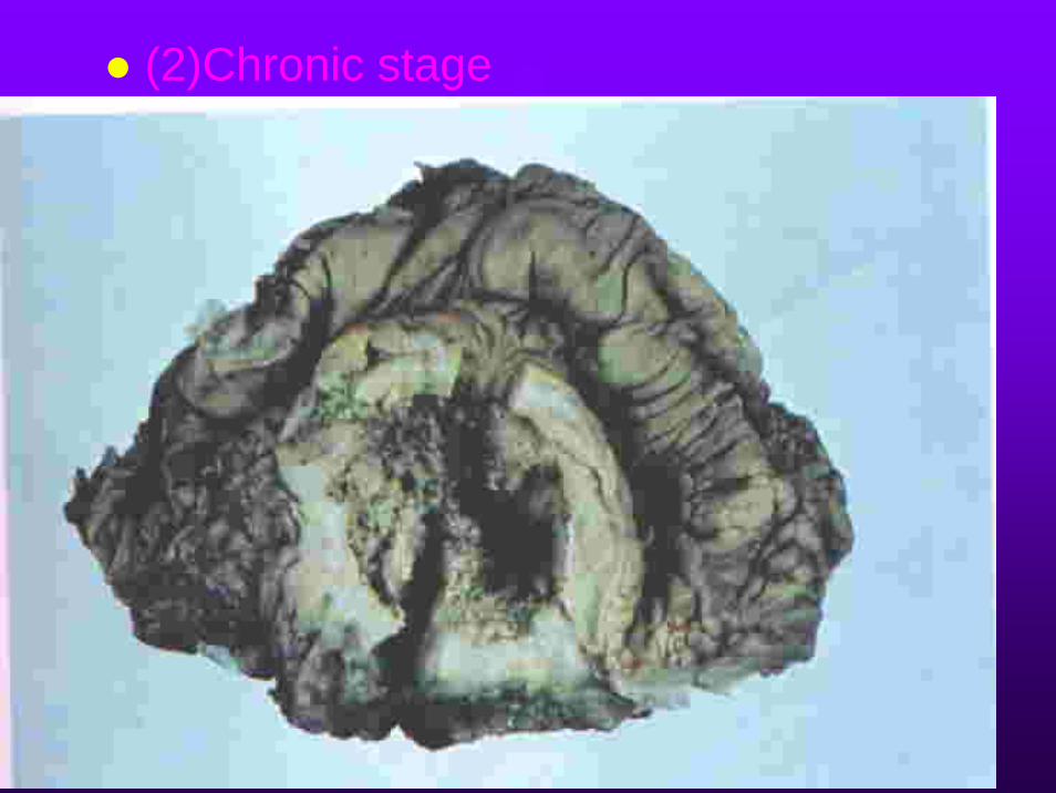
(2)Chronic stage

The differentiation between amoebic dysentery and
bacillary dysentery

Extraintestinal amoebiasis1. amoebic abscess of liverA complication of amebic dysentery , brought about by amebic entering colonic venules and passing by the portal vein to the liver.liquefaction necrosis, not a true abscess.

MacroscopicallyUsually a single ‘abscess’,most often in the upper part of the right lobe.The lesion has a compressed fibrous capsule, with an irregular shaggy necrotic inner wall and the cavity contains thick paste-like material rather than pus, often chocolate-colored or showing admixture of blood.

Microscopically
The contents include necrotic liver cell debris and a varying number of red cells.Trophozoites may be numerous or scanty in the wall of the cavity Only a few neutrophils are present

Amoebic abscess of liver

Amoebic abscess of liver

Complications include rupture and spread beyond the liver.

2).amoebic pulmonary abscess

Schistosomiasis

Three species:a. Schistosoma hematobium b. Schistosom mansonic. Schistosom japonicum
Oncomelania snailEgg ─→ miracidium ─────→ cercaria ─→
schistosomulum ─→adult schistosome
Etiology and transmission route
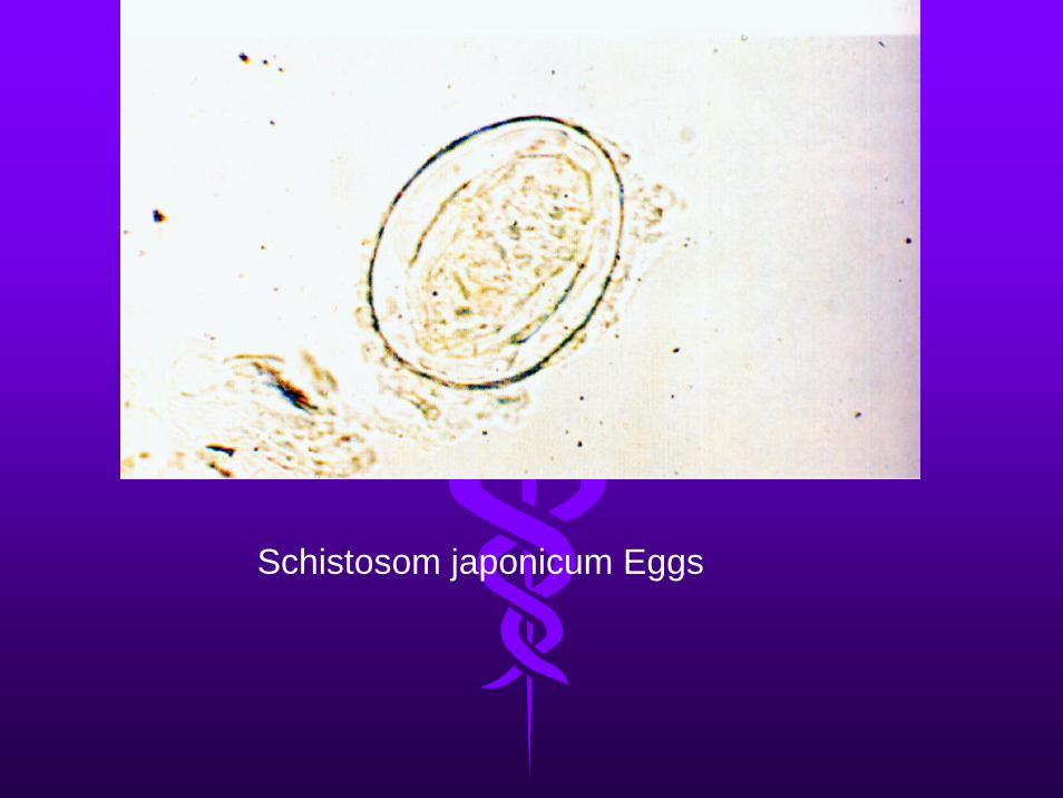
Schistosom japonicum Eggs

cercaria

Basic lesions and pathogenesis
1.Penetration of the skin by cercariae caused a transient local inflammatory reaction: Cercarial dermatitis

Cercarial dermatitis

2. Lesions caused by adult schistosome and their metabolic products
3. Lesions caused by eggs: the main lesion of Schistosomiasis
(1) Acute egg nodule (eosinophilic abscess) : In schistosomiosis, eggs surrounded by many
eosinophils and like abscesses. Hoeppli phenomenonCharcot-Leyden crystal

(2) Chronic egg nodule (pseudotubercle):
In schistosomiosis, eggs surrounded by epithelioid cell, multinuclear giant cell; eggs are often necrotic or calcified. This structure is like that of a tubercle. It is a granuloma.

In schistosomiosis, eggs surrounded by many eosinophils and like abscesses.
Hoeppli phenomenonCharcot-Leyden crystal

In schistosomiosis, eggs surrounded by epithelioid cell, multinuclear giant cell; eggs are often necrotic or calcified. This structure is like that of a tubercle. It is a granuloma.

In schistosomiosis, eggs surrounded by epithelioid cell, multinuclear giant cell; eggs are often necrotic or calcified. This structure is like that of a tubercle. It is a granuloma.
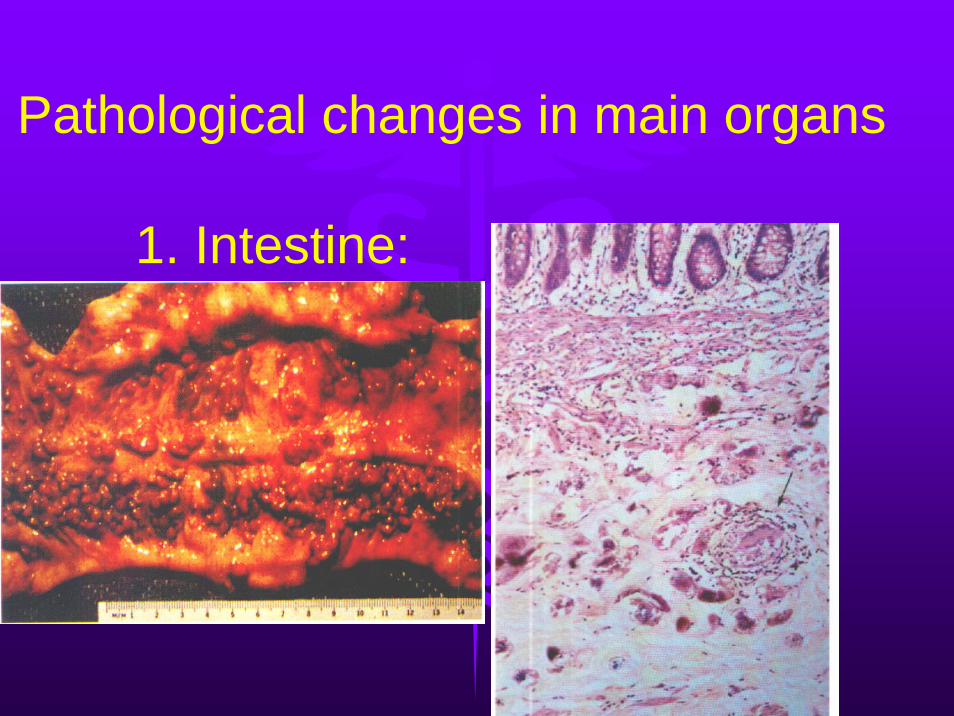
1. Intestine:
Pathological changes in main organs

2. Liver: pipe stem cirrhosis(massive fibrosis around major portal tracts)

3.Spleen: splenomegaly
4. Heterotopic Schistosomiasis(1)lung (2)cerebrum (3)other organs

Infectious DiseasesEPIDEMIC CEREBROSPINAL MENIGITIS/MENI-
GOCOCCAL MENIGITISType B Epidemic EncephalitisBacillary dysenteryTyphoid Fever
Sexually Transmitted Diseases Acquired Immunodificiency Syndrome (AIDS)Condyloma acuminatumSyphilisGonorrhea
Parasitic DiseasesAMOEBIASISSchistosomiasis
inflammations
Briefly summary: