wnt and vg1 interactions in the chick - ucl
TRANSCRIPT
INTRODUCTION
Unlike amphibian embryos, where polarity is established bythe third cleavage division through the localisation of maternaldeterminants (reviewed in Harland and Gerhart, 1997; Arendtand Nübler-Jung, 1999), polarity in the chick embryo remainsplastic until the beginning of gastrulation, when the embryoalready has 20,000-60,000 cells (Spratt and Haas, 1960b). Upto the time of appearance of the primitive streak, theblastoderm can be cut into several fragments, each of whichcan spontaneously initiate the formation of a complete axis(Spratt and Haas, 1960b). In the cut fragments, the new axistends to arise from the edge of the area pellucida, a regioncalled the marginal zone. Furthermore, the frequency of axisformation decreases in a posterior-to-anterior direction aroundthe marginal zone (Spratt and Haas, 1960b).
Subsequent investigators have established that a smallposterior domain within the marginal zone (the posteriormarginal zone; PMZ) is particularly important. When thisdomain is transplanted to an ectopic site of the marginal zoneof a host embryo, it induces the formation of a second axis(Eyal-Giladi and Khaner, 1989; Khaner and Eyal-Giladi,
1989). In fact, the PMZ can act in a manner analogous to theNieuwkoop centre of amphibians, by inducing a complete axisthat includes the organiser, but without making a direct cellularcontribution to it (Bachvarova et al., 1998).
Misexpression of the TGFβ family member chick Vg1 canmimic the activity of the PMZ: when misexpressed in theanterior marginal zone, it will also induce a completeembryonic axis and organiser (Seleiro et al., 1996, Shah et al.,1997). cVg1 misexpression, like grafts of the PMZ, onlyinduce such an axis if they are placed within the marginal zoneof the host embryo, and not in the area pellucida. What makesthe marginal zone unique?
In Xenopus(Sokol and Melton, 1992; Steinbeisser et al.,1993; Cui et al., 1995; Watabe et al., 1995; Kessler, 1997;Crease et al., 1998; Zorn et al., 1999), as well as during laterstages of chick development (Joubin and Stern, 1999), theVg1/Activin and Wnt signalling pathways can synergise toinduce organiser genes, which has prompted us to investigatewhether differences in Wnt activity could explain the specialproperties of the marginal zone. We have found that Wnt8Cisexpressed in the marginal zone, where it describes a gradientthat is highest posteriorly. When cVg1 is misexpressed in the
2915Development 128, 2915-2927 (2001)Printed in Great Britain © The Company of Biologists Limited 2001DEV3224
The posterior marginal zone (PMZ) of the chick embryohas Nieuwkoop centre-like properties: when transplantedto another part of the marginal zone, it induces a completeembryonic axis, without making a cellular contribution tothe induced structures. However, when the PMZ isremoved, the embryo can initiate axis formation fromanother part of the remaining marginal zone. Chick Vg1can mimic the axis-inducing ability of the PMZ, but onlywhen misexpressed somewhere within the marginal zone.We have investigated the properties that define themarginal zone as a distinct region. We show that thecompetence of the marginal zone to initiate ectopicprimitive streak formation in response to cVg1 isdependent on Wnt activity. First, within the Wnt family,only Wnt8C is expressed in the marginal zone, in agradient decreasing from posterior to anterior. Second,misexpression of Wnt1 in the area pellucida enables this
region to form a primitive streak in response to cVg1.Third, the Wnt antagonists Crescent and Dkk-1 block theprimitive streak-inducing ability of cVg1 in the marginalzone. These findings suggest that Wnt activity defines themarginal zone and allows cVg1 to induce an axis. We alsopresent data suggesting some additional complexity: first,the Vg1 and Wnt pathways appear to regulate theexpression of downstream components of each other’spathway; and second, misexpression of different Wntantagonists suggests that different classes of Wnts maycooperate with each other to regulate axis formation in thenormal embryo.
Key words: Marginal zone, Induction, Primitive streak, Gastrulation,Nieuwkoop centre, Vg1, Wnt, Lef1, β-catenin, JNK, Crescent,Dickkopf, Frizzled 8, Chick
SUMMARY
Interactions between Wnt and Vg1 signalling pathways initiate primitive
streak formation in the chick embryo
Isaac Skromne* and Claudio D. Stern ‡,§
Department of Genetics and Development, Columbia University, 701 West 168th Street, New York, NY 10032, USA*Present address: Department of Molecular Biology, Moffett Building, Princeton University, Princeton, NJ 08544, USA‡Present address: Department of Anatomy and Developmental Biology, University College London, Gower Street, London WC1E 6BT, UK§Author for correspondence (e-mail: [email protected])
Accepted 30 April 2001

2916
anterior marginal zone together with the Wnt antagonistsCrescent or Dkk1, axis induction is inhibited. Furthermore,ectopic Wnt expression is able to overcome the inability ofcVg1 to induce an axis in the area pellucida. Based on theseresults, we propose that Wnt8Cdefines the marginal zone andlimits the ability of regions of the embryo to respond to cVg1.We also present data to suggest that the Vg1 and Wnt pathwaysregulate the expression of downstream components of eachother’s pathway. Finally, we show that different Wntantagonists have distinct effects, suggesting that differentclasses of Wnts cooperate to regulate axis formation in thenormal embryo.
MATERIALS AND METHODS
Embryo cultureFertile White Leghorn hens’ eggs (SPAFAS, CT) were incubated for1-20 hours to obtain embryos between stage X EG&K (Romannumbers for pre-primitive streak stages; Eyal-Giladi and Kochav,1976) and stage 5 HH (Arabic numerals for later stages; Hamburgerand Hamilton, 1951). Embryos were explanted in modified Newculture as described previously (New, 1955; Stern and Ireland, 1981).Aggregates of COS cells or RatB1a fibroblasts were grafted onto themarginal zone or area pellucida of the embryo using a micropipette.The posterior end of the embryo was marked with carbon particles(Carbon Lampblack, Fisher). Embryos were incubated at 38°C in ahumidified chamber for different periods of time, fixed, and processedfor whole-mount in situ hybridisation and immunohistochemistry.
Whole-mount in situ hybridisationEmbryos were fixed in 4% formaldehyde, 2 mM EGTA in phosphate-buffered saline (PBS, pH 7.0) for 1 hour at room temperature orovernight at 4°C, and then stored in 100% methanol at −20°C. In situhybridisation using digoxigenin-labelled riboprobes was performed at68°C as described previously. The molecular markers studied in thiswork are cBra (Ch-T, a gift from J. C. Smith; Kispert et al., 1995,Knezevic et al., 1997), chordin(Streit et al., 1998), cFGF8(a gift fromJ. C. Izpisúa-Belmonte), goosecoid(Izpisúa-Belmonte et al., 1993),cLef1 (a gift from J. C. Izpisúa-Belmonte; Kengaku et al., 1998),cNodal(a gift from M. Kuehn; Jones et al., 1995),cVg1(Shah et al.,1997), Wnt8C (a gift from J. Dodd; Hume and Dodd, 1993), andcWnt1, cWnt3, cWnt3a, cWnt4, cWnt5a, cWnt5b, cWnt6, cWnt7b,cWnt8, cWnt10and cWnt11(gifts from A. McMahon; Hollyday et al.,1995).
Myc or HA immunohistochemistryAfter in situ hybridisation, embryos were processed forimmunohistochemistry as described previously (Streit et al., 1997)using monoclonal anti-Myc (9E10, Evans et al., 1985) or anti-HA(Mono HA.11, Berkeley Antibody Co.) antibodies at a finalconcentration of 1:4 or 1:1000, respectively. A goat anti-mouse IgGHRP-linked secondary antibody (Jackson Immunoresearch) was usedat a final concentration of 1:2000 and the peroxidase reaction carriedout using 3,3′-diaminobenzidine (Sigma, MO) as a substrate (Streit etal., 1997). After in situ hybridisation and immunohistochemistry,embryos were transferred to a chamber slide and photographed usingbright field optics on Fuji 64T film. Some embryos were embeddedin Paraplast and sectioned at 10 µm.
β-catenin immunohistochemistry and confocalmicroscopyDetection of β-catenin followed a previously described protocol(Schneider et al., 1996) using a rabbit anti β-catenin antiserum (a giftof R. Moon). Embryos were fixed for 10 minutes in 4% formaldehyde
in PBS and stored overnight in 80% methanol:20% DMSO at −20°C.After rehydrating the embryos in 50% methanol:50% PBS, embryoswere washed in PBS and incubated in blocking buffer (BB; 20%bovine serum albumen in PBS) for 1 hour at 4°C. Rabbit antiserumwas then added to a final concentration of 1:500 and the embryosincubated overnight at 4°C. After five 1 hour washes in PBS at roomtemperature, embryos were incubated in BB for 1 hour at 4°C. Cy5-conjugated goat anti-rabbit IgG antibody (Jackson Immunoresearch)was then added to a final concentration of 1:500 and the embryosincubated overnight at 4°C. After five 1 hour washes in PBS at roomtemperature, embryos were incubated for 5 minutes in 10 µg/mlpropidium iodide (Sigma) in PBS and then washed extensively in PBSbefore being transferred to a chamber slide and mounted in Gelvatol(Stern and Holland, 1993). The epiblast layer of the embryos wasexamined using a Zeiss LSM 410 confocal laser-scanning system witha 15 mW Argon-Krypton laser attached to a Zeiss Axiovert 100TVinverted microscope, and a 63× water immersion objective with anumerical aperture of 1.2. Propidium iodide and Cy5 were excited at568 and 647 nm, respectively. Optical sections 1 and 5 µm apart weretaken and analysed individually and as a stack using Scion Image 1.62and Adobe Photoshop 5.5.
DNA constructsTo facilitate detection of Crescent using an anti-Myc monoclonalantibody (9E10; Evans et al., 1985), a full-length Crescent (Pfeffer etal., 1997) cDNA (a gift from J. C. Izpisúa-Belmonte) was digestedwith NotI and NaeI, removing the last 13 amino acids of the protein.A 944 bp fragment was purified and cloned in frame intopcDNA3.1(−)/Myc-His A (Invitrogen) that had been digested withNotI and EcoRV. The integrity of the construct was confirmed bysequencing.
Isolation of Wnt cDNA clonesA RT-PCR approach was used to identify novel Wnt factors expressedin the pre-primitive streak stage chick embryo. The degenerateprimers designed to recognise conserved amino acid stretches presentin multiple Wnt family members were fWnt (amino acid sequence,CKCHG), 5′-TGY AAR TGY CAY GGN NT-3′; and bWnt (aminoacid sequence, CRFHWC), 5′-CAC CAR TGR AAN NBR CA-3′(Gavin et al., 1990). A third internal degenerate primer was designedto recognise an amino acid stretch conserved in all Wnts except theWnt8and Wnt10subclasses (Gavin et al., 1990) – iWnt (amino acidsequence, LL/MCCGRG), 5′-CCN CKN CCR CAR CAN A-3′.
Total RNA was isolated from chick embryos at stages X-XIII(Ausubel et al., 1995). First-strand cDNA was synthesized from 1 µgof total RNA using 10 pmoles of bWnt primer and Superscript II RT(Gibco/BRL; Ausubel et al., 1995). After phenol:chloroformextraction and ethanol precipitation, the cDNA was PCR-amplifiedusing 2 pmoles of bWnt and fWnt primers and Taq polymerase(Promega) in the presence of 1.5 mM MgCl2. The conditions for PCRamplification were 25 cycles of 90°C for 1 minute, 55°C for 2minutes, 72°C for 2 minutes, with a final extension of 10 minutes at72°C. A second PCR was carried out using 0.5 µl of the initial PCRreaction and fWnt and iWnt primers, under identical amplificationconditions. PCR products were cloned into pGEM-T (Promega) andsequenced. All products isolated encoded Wnt family members withsignificant homology to Xenopusand Zebrafish Wnt3, Wnt5a andWnt5b genes. Probes generated from these clones gave expressionpatterns identical to cWnt3a, cWnt5aand cWnt5b, respectively, by insitu hybridisation.
Cell culture and protein production1.5×105 COS cells on a 35 mm dish were transfected with 1 µg ofplasmid and Lipofectamine Plus (Gibco BRL), according to themanufacturers’ instructions. Constructs used for cell transfection weredsl-myc-cVg1(Shah et al., 1997) in pMT23, Crescent-myc-hisinpcDNA3.1, hDkk1 in pCS2++ (gift from E. Laufer) and XFz-N8 in
I. Skromne and C. D. Stern

2917Wnt and Vg1 interactions in the chick
pCS2++ (gift of P. Klein; Deardorff et al., 1998). As negative controls,transfections were carried out without including DNA (‘mock’). Forconditioned media, transfected cells were grown in 1 ml of serum-free medium for 2 days and the medium then collected as describedpreviously (Shah et al., 1997). To make cell aggregates, 24 hours aftertransfection, cells were trypsinised and counted. Aliquots of 1000cells were allowed to aggregate overnight in hanging 20 µl drops ofmedium (Shah et al., 1997). These small aggregates were rinsed inserum-free medium and grafted to different regions in the embryo.The efficiency of cell transfection and protein secretion wasmonitored by immunoblot and immunohistochemistry as describedabove for whole embryos, except that antibody incubation times werereduced to 1 hour. On average, about 75% of the cells expressed highlevels of the protein 24 hours after transfection.
A stable RatB1a fibroblast cell line transfected with mWnt1-HAinpLNCX was used to misexpress Wnt1 (gift of J. Kitajewski; Shimizuet al., 1997). For immunoblotting, the cells were lysed as describedpreviously (Shimizu et al., 1997). To make cell aggregates, 3000RatB1a cells were allowed to aggregate overnight in hanging 20 µldrops. The medium was supplemented with 1 mM Sodium Butyrate(Sigma) to induce high levels mWnt1 expression (Shimizu et al.,1997). RatB1a cells stably transfected with empty pLNCX vectorwere used as negative controls (‘mock’).
ImmunoblotsSecretion of processed cVg1 (Shah et al., 1997), Wnt1 (Shimizu etal., 1997) and Crescent protein was monitored by immunoblot (notshown). The protein concentration present in supernatants of cellsexpressing dsl-myc-cVg1, Crescent-mycor mWnt1-HAwas initiallydetermined using a modified Bradford assay (BioRad). Samplescontaining 40 µg of protein were electrophoresed in 10% SDS-PAGE.Gels were electroblotted onto PROTRAN nitrocellulose (Schleicher& Schüll). Blots were probed with either anti-Myc (9E10, Evans et al.,1985) or anti-HA (Mono HA.11, Berkeley Antibody Co.) antibody.Goat HRP-coupled anti-mouse IgG (Jackson Immunoresearch) wasused as secondary antibody. Blots were developed using LumiGlochemiluminescence substrate (Kirkegaard & Perry).
RESULTS
The area pellucida can respond to cVg1, but doesnot form an ectopic primitive streakIt has previously been reported that cVg1 induces an ectopicaxis when misexpressed in the marginal zone, but not in thearea pellucida (Shah et al., 1997). This is surprising, becausecVg1 has the ability to induce organiser markers in the area
pellucida of much later embryos (Joubin and Stern, 1999).Does the young area pellucida not yet have a competence torespond to cVg1, or does cVg1 misexpression in this regioninduce only some of the required downstream events? Toaddress this question, we misexpressed cVg1 in the anteriorthird of the area pellucida and analysed the time-course ofexpression (6, 15, 24 and 36 hours) of a series of molecularmarkers for embryonic polarity before and during gastrulation(cVg1, cNodal, cFGF8, Wnt8C, chordin, goosecoidand cBra).No ectopic expression of any of these genes was detected after6 hours. Between 15-24 hours, cVg1, cNodal, chordin(Fig. 1)and goosecoid(not shown) appeared, but no expression ofcFGF8, Wnt8Cor cBra was seen (Fig. 1), even after 36 hours(cBra; Fig. 1). These results suggest that, although the areapellucida cannot form a primitive streak in response to cVg1misexpression, it does respond to cVg1 by expressing severalmarkers of embryonic polarity.
Misexpression of cVg1+Wnt1 in the area pellucidainduces an ectopic primitive streakThe above result suggests that some property of the marginalzone is required for cVg1 to induce a primitive streak. Onecandidate is the Wnt pathway, because: (1) Wnt8Cis expressedthroughout the marginal zone at pre-streak stages (Fig. 2A;Hume and Dodd, 1993), and (2) activation of the Wnt pathwayhas been shown to synergise with Vg1/Activin signalling in theinduction of organiser markers in Xenopus(Sokol and Melton,1992; Steinbeisser et al., 1993; Cui et al., 1995; Watabe et al.,1995; Kessler, 1997; Crease et al., 1998; Zorn et al., 1999) andchick (Joubin and Stern, 1999).
Is the inability of the area pellucida to form a streak inresponse to cVg1 misexpression due to the absence of Wntactivity in this region? We were unable to achieve efficientsecretion of Wnt8C from transfected COS cells. However, asWnt1 and Wnt8 belong to the same functional subclass of Wntproteins in several different Xenopusassays (the tests includemesoderm dorsalisation, alterations in gap junctionalcommunication and direct binding to Frzb-1; Olson et al.,1991; Sokol et al., 1991; Torres et al., 1996; Leyns et al., 1997;Wang et al., 1997a; Wang et al., 1997b), we used instead astable fibroblast cell line secreting Wnt1 (Shimizu et al., 1997).We grafted cVg1-secreting COS cells together with Wnt1-secreting fibroblasts in the anterior third of the area pellucidaof pre-streak embryos. Embryos were allowed to develop for
Table 1. Effects of misexpression of cVg1 and Wnt1 in different regions of the early chick embryoRegion Stage Factor Ectopic streaks/total (%)
Anterior area pellucida X-XIII cVg1 1/21 (5)Wnt1 1/24 (4)cVg1 + RatB1 cells 1/26 (4)Wnt1 + COS cells 0/17 (0)cVg1 + Wnt1 29/58 (50)**
Anterior marginal zone X-XIII cVg1 38/62 (61)Wnt1 3/49 (6)cVg1 + Wnt1 14/34 (41)
2-3 cVg1 1/31 (4)Wnt1 0/14 (0)cVg1 + Wnt1 0/15 (0)
The frequency of induction of an ectopic streak by a graft of cells secreting the factors indicated is shown on the right. Note that misexpression of cVg1+Wnt1in the anterior area pellucida of pre-streak embryos (X-XIII) generates an ectopic axis, while neither cVg1 alone nor Wnt1 alone does so (**, P<0.004). Bycontrast, cVg1, Wnt1 and a combination of the two failed to induce a primitive streak in older (stage 2-3) embryos.

2918
24, 36 or 48 hours and the expression of cVg1, cNodal, cFGF8,Wnt8C, chordinand cBra analysed. Misexpression of neithercVg1 nor Wnt1 alone induced an ectopic streak (Table 1), andWnt1 did not induce expression of any of the markers testedat 24 or 36 hours (Fig. 1). By contrast, when cVg1 and Wnt1were misexpressed together, an ectopic primitive streakdeveloped (29/58, 50%; Table 1, Fig. 1). At 24 hours,expression of cVg1(6/6), cNodal(2/2), chordin (4/5), cFGF8(2/2), Wnt8C(2/2) and cBra (5/7) was induced in the host. At36 hours, the induced streak expressed cBra and gsc (Fig. 1and not shown), suggesting that it contains axial mesoderm.Some embryos were grown for 48 hours to assess whether theyformed a complete axis; none of these produced later axialderivatives, as assessed by morphology and the expression ofcBra and gsc(not shown).
These results suggest that the difference between the areapellucida and the marginal zone in their responses to cVg1misexpression may be due to differences in Wnt signalling inthe two regions.
I. Skromne and C. D. Stern
Fig. 1.Comparison of the inducing ability of cVg1, Wnt1 and cVg1+Wnt1 in the area pellucida of stage X-XIII embryos. Cells secreting thefactors indicated on the left were grafted into the anterior third of the area pellucida of host embryos, and these were cultured for 24 or 36 hours(right-hand column) before in situ hybridisation (purple) for the genes indicated at the top. Anti-Myc (top row) or anti-HA (middle and bottomrows) immunohistochemistry (brown) was used to identify the cell aggregates and confirm protein synthesis. Red arrows indicate ectopicexpression of the gene in the host. The probe used to detect expression of cVg1in the embryo also hybridises with the cVg1-transfected COScells in the top and bottom rows of the first column.
Fig. 2.Wnt signalling in the early chick embryo. (A) Expression ofWnt8Cat stage XII. Expression is seen all around the marginal zone(MZ) and is strongest posteriorly. Some expression is also seen in theperipheral area opaca. (B) Expression of Wnt5aat stage XIII. Weakexpression is detected throughout the outer half of the area opaca(AO), but none in the area pellucida or marginal zone (MZ).(C) Expression of Wnt11at stage XII. Weak expression is detectedthroughout the epiblast and hypoblast of the area pellucida and in theepiblast and deep layers of the marginal zone and area opaca.(D) Wnt3ais not expressed at stage XIII or at any other stage thatprecedes primitive streak formation. Its expression starts to bedetected by stages 3+-4 (not shown).

2919Wnt and Vg1 interactions in the chick
Expression of Wnts and members of their pathwaybefore primitive streak formationThe above findings implicate Wnt signalling in the difference
between the marginal zone and area pellucida in their reactivityto Vg1. Which members of the Wnt family are expressedappropriately to explain this difference in reactivity? We
Fig. 3.Expression of cLef1. (A) At stage XI,cLef1is expressed in Koller’s sickle (ks) andposterior marginal zone (which is partlyobscured by the overlying germ wall, see sectionshown in I). (B) At stage XIII, cLef1expressioncontinues in the posterior marginal zone andKoller’s sickle. (C) By stage 2+, cLef1transcriptsare restricted to the primitive streak. (D) At stage3+, expression is restricted to the posterior two-thirds of the primitive streak. (E) In stage 4embryos, expression accompanies the emerginglateral mesoderm (see section shown in J). F. Bystage 4+, cLef1expression is strong in theprimitive ridges and in mesoderm emerging fromthe streak. (G) At stage 6, cLef1transcripts arefound in the forming paraxial mesoderm andprimitive streak. (H) At stage 11, cLef1expression is seen in the remnants of the streak,the anterior segmental plates and the rostral halfof the forming somite. cLef1 is rapidlydownregulated from the somites after theirformation, but in older (more rostral) somites itappears in the caudal half. cLef1 is alsoexpressed in the heart and brain (see Kengaku etal., 1998). (I) Sagittal section of the embryo in Aat the level indicated, showing expression ofcLef1in Koller’s sickle (ks) and in the epiblast ofthe posterior marginal zone. (J) Transversesection of the stage 4 embryo shown in E at thelevel indicated, showing expression in the primitive streak and lateral mesoderm.
Fig. 4.Regulation of intracellularβ-catenin and induction of cLef1by cVg1+Wnt1. (A-D)Subcellular localisation of β-catenin. In all panels, the greensignal corresponds to β-cateninimmunofluorescence and the redsignal to propidium-iodidestained nuclei. (A) Throughoutthe area pellucida, most of theimmunoreactivity is concentratedat cell membranes. (B) Aftergrafting cVg1-secreting cells(position outlined by brokenline), downregulation of β-catenin expression is seen nearthe graft (outlined). (C) A graftof Wnt1-secreting cells (outlined)increases the level of β-cateninthroughout the cell; where itoverlaps with the nucleus, thesignal appears yellow. (D) Whena pellet of Wnt1-secreting cells(indicated on top of left outline)is grafted together with a pellet ofcVg1-secreting cells (indicated on top of right outline), the pattern seen is a combination of those in B and C. β-catenin is upregulated in theproximity of the Wnt1 cells and downregulated near the cVg1 cells. Scale bar: 25 µm for A-D; 5 µm for E-H. Expression of cLef1aftertransplantation of cells secreting cVg1 (E,F), Wnt1 (G) or cVg1+Wnt1 (H) in the anterior marginal zone (E) or area pellucida (F-H). (E) cVg1induces cLef1when misexpressed in the anterior marginal zone (arrow). Note that induction of cLef1is restricted to the marginal zone and areaopaca. In the area pellucida, neither cVg1 (F) nor Wnt1 (G) induces cLef1, but a combination of the two factors does (arrow, H).
2920
examined the expression of cWnt1, cWnt3, cWnt3a, cWnt4,cWnt5a, cWnt5b, cWnt6, cWnt7b, cWnt8, cWnt8C, cWnt10andcWnt11by in situ hybridisation at stages X-4 (Fig. 2). Wefound that Wnt8Cwas expressed in the marginal zone, moststrongly in posterior regions and decreasing laterally andanteriorly (Fig. 2A; Hume and Dodd, 1993), Wnt5a wasexpressed weakly in area opaca and in edge cells of the epiblast(which attach the embryo to the vitelline membrane; New,1959; Downie and Pegrum, 1971; Downie, 1976; Fig. 2B) andWnt11was expressed ubiquitously at low levels (Fig. 2C). Noother Wnts were detectable before gastrulation by in situhybridisation (Fig. 2D and data not shown). As acomplementary approach, we analysed the expression of otherWnts in pre-streak embryos by RT-PCR using degenerateprimers designed to amplify fragments of all known Wntfamily members except Wnt8, Wnt8C and Wnt10 (seeMaterials and Methods). Using this approach we cloned newfragments of Wnt3a, Wnt5a and Wnt5b (see Materials andMethods); however, neither Wnt3a(Fig. 2D) nor Wnt5b(notshown) was detectable by in situ hybridisation.
It is also possible that the marginal zone and area pellucidadiffer in their sensitivity to cVg1 because of differentialexpression of downstream members of the Wnt pathway. Wetherefore examined the distribution of the Wnt pathwaycomponents, cLef1and β-catenin, in pre-streak- and primitive-streak-stage embryos. cLef1 was expressed in the posteriormarginal zone and Koller’s sickle between stages X and XIII(Fig. 3A,B,I); occasionally, very low levels of expression weredetected throughout the marginal zone of stage X embryos (notshown). cLef1was also expressed at later stages in a variety ofsites (Fig. 3 and Kengaku et al., 1998). We analysed thesubcellular distribution of β-catenin by immunofluorescence.In agreement with previous studies (Roeser et al., 1999), wefound that β-catenin was concentrated in the nucleus of cellsin the germ wall and hypoblast of embryos at stages X-XIII(not shown). Throughout the epiblast, most β-cateninimmunoreactivity was associated with cell membranes (Fig.4A). However, we did not observe either any marked nuclearlocalisation in any region of the epiblast or the downregulationof β-catenin expression in anterior regions at stages XII-XIII,which have been reported previously (Roeser et al., 1999).
Together, our results suggest that only Wnt8C, Wnt5aandWnt11are expressed at significant levels in the pre-streakstage blastoderm, and of these, only Wnt8C is expressedappropriately throughout the marginal zone (Fig. 2A; Humeand Dodd, 1993) to account for the difference in cVg1-reactivity between marginal zone and area pellucida. The Wntpathway components cLef1and β-catenin are both expressedat appropriate stages and locations to be involved in primitivestreak initiation.
Both cVg1 and Wnt1 regulate intracellular levels ofβ-catenin and the expression of cLef1The above results point to synergism between Wnt activityand cVg1 in initiating primitive streak formation. As Wnt1misexpression in the area pellucida does not have anyconsequence unless cVg1 is also present (Fig. 1, Table 1), doesthis imply that cVg1 is required for cells to respond to Wntprotein? To address this question, we examined the subcellularlocalisation of β-catenin in the epiblast of embryos exposed tocVg1, Wnt1 or cVg1+Wnt1 for 3 hours. In untreated embryos,
β-catenin was predominantly associated with the cellmembrane (Fig. 4A). After implantation of cVg1-secretingcells, a pronounced downregulation of β-catenin expressionwas seen in the vicinity of the graft (Fig. 4B). When Wnt1 wasmisexpressed alone, a substantial increase in the intensity ofstaining was seen in the epiblast, not only in the membrane butthroughout the cell (Fig. 4C). This suggests that epiblast cellsdo respond directly to Wnt1, even when cVg1 is not supplied.When cVg1 and Wnt1 were misexpressed together, acombination of the two results was obtained: in the vicinity ofthe Wnt pellet, the level of β-catenin was increased throughoutthe cell; near the cVg1-pellet, β-catenin was predominantlymembrane associated (Fig. 4D). These findings suggest notonly that epiblast cells can respond to Wnt in the absenceof exogenous cVg1, but also that Vg1 may dampen theupregulation of β-catenin caused by Wnt1.
The finding that cVg1 misexpression affects intracellularlevels of β-catenin raises the possibility that other componentsof the Wnt pathway may also be regulated by Vg1/Activins.We turned our attention to cLef1because it is expressed in theposterior marginal zone (PMZ; Fig. 3A,I), a region thatexpresses cVg1 (Seleiro et al., 1996; Shah et al., 1997). We
I. Skromne and C. D. Stern
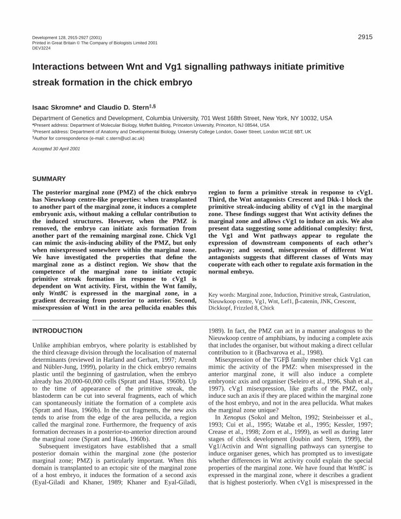
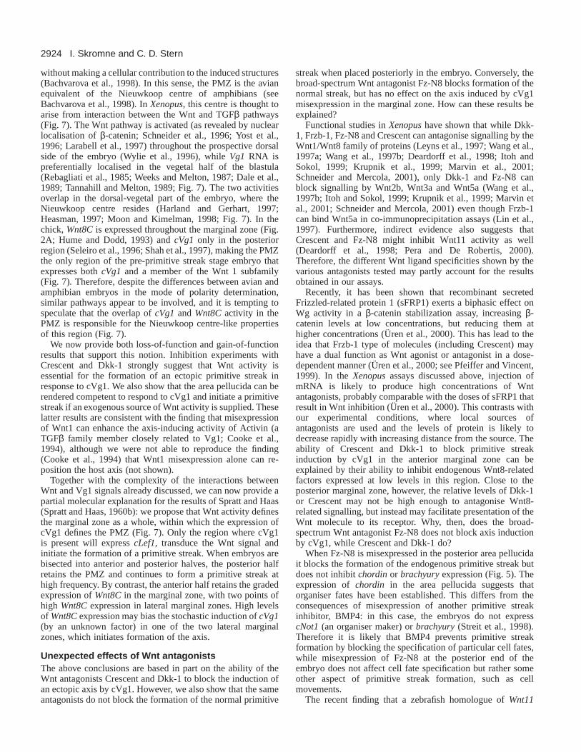
Fig. 5.The broad-spectrum Wnt antagonist Fz-N8 impairs primitivestreak formation in stage X (A,B) but not stage XIII (C,D) embryos.(A,B) When cell aggregates secreting Fz-N8 are grafted in theposterior border of the area pellucida of stage X embryos, primitivestreak formation is inhibited (A) or seriously impaired (B). After 15hours’ incubation, cBra (A) and chordin(B) expression are stronglydownregulated (white arrowheads, compare with C,D). (C,D) StageXIII embryos receiving four Fz-N8-secreting cells in the posteriorborder of the area pellucida develop a normal primitive streak,expressing cBra (C) and chordin(D). (Stage X embryos receivingmock-transfected cell aggregates develop a normal primitive streakidentical to those in C,D.) Black outlines indicate the position of thegrafted cell aggregates; black arrowheads indicate cBraand chordinexpression.
2921Wnt and Vg1 interactions in the chick
misexpressed cVg1 in the anterior marginal zone and analysedthe expression of cLef1at 3 hour intervals for the next 15 hours.Ectopic cLef1 expression was first seen 6 hours aftertransplanting the cVg1-secreting cells (4/5 embryos, Fig. 4E),in the marginal zone and area opaca but not in the areapellucida (Fig. 4E). Therefore cVg1 can regulate theexpression of cLef1.
The finding in this experiment that cVg1 misexpressioninduces cLef1 in the marginal zone and area opaca (whichexpress Wnt8C and Wnt5A, respectively; Fig. 2A,B), couldindicate that cLef1expression may also be regulated by Wntsignalling. To test this, we misexpressed cVg1, Wnt1 orcVg1+Wnt1 in the anterior area pellucida and analysed theexpression of cLef1 after 9, 15 and 24 hours’ incubation.Neither cVg1 nor Wnt1 induces cLef1expression in the areapellucida at any time point (Fig. 4F,G), but a combination ofcVg1+Wnt1 does so (3/4 embryos at 24 hours; Fig. 4H). Thisfinding is consistent with the posterior restriction of cLef1expression at stages XI-2 (Fig. 3A-C) in regions that expressboth cVg1and Wnt8C.
Wnt signals are essential for cVg1 to initiateprimitive streak formationThe results presented above, in the context of previous findings(Shah et al., 1997), suggest that the difference in reactivity ofthe marginal zone and area pellucida to cVg1 misexpressioncould be due to activation of the Wnt pathway in the marginalzone. If the Wnt pathway is indeed required for cVg1 to initiateprimitive streak formation, we would expect that inhibition ofWnt signalling should block the ability of cVg1 to induce anectopic streak. To test this, we used the secreted Wntantagonists Crescent (Frzb family member) and Dkk1, whichcan specifically bind Wnt8/-8C and block their activity(Krupnik et al., 1999; Marvin et al., 2001; Schneider andMercola, 2001), as well as a truncated form of the XenopusFrizzled-8 receptor, Fz-N8, which is a broad-spectrum Wntinhibitor (Deardorff et al., 1998; Itoh and Sokol, 1999). WhencVg1- and Crescent-secreting cells were grafted together in theanterior marginal zone, the frequency of primitive streakinduction by cVg1 dropped from 38/62 (61%) with cVg1 aloneto 5/20 (25%) with cVg1+Crescent (P<0.004; Table 2). Asimilar reduction in frequency of primitive streak induction bycVg1 was observed when Dkk-1 was used instead of Crescent(2/12, 17%, P<0.004; Table 2). Surprisingly, however,misexpression of the broad-spectrum inhibitor Fz-N8 did notsignificantly inhibit ectopic streak formation by cVg1 (10/17,59%; Table 2).
These results suggest that Wnt signals sensitive to inhibitionby Crescent and Dkk1 are essential for cVg1 to initiate theformation of an ectopic primitive streak, and accounts for thedifference in cVg1-reactivity between the area pellucida(where Wnt8Cis not expressed) and marginal zone.
Primitive streak formation in the normal embryorequires Wnt signalsThe conclusion that Wnt signals are required to mediate theinduction of an ectopic primitive streak after misexpression ofcVg1 leads to the question of whether Wnt activity is similarlyrequired for initiation of the endogenous primitive streak innormal embryos. To test this, we misexpressed the three Wntinhibitors described above in the posterior area pellucida, close
to the site of initiation of primitive streak formation at stagesX-XIII. We found that neither Crescent nor Dkk-1 affected thedevelopment of the primitive streak at any stage (Table 2). Bycontrast, the broad-spectrum inhibitor Fz-N8 impairedprimitive streak formation when misexpressed at stages X-XI(16/21, 76%, P<0.04; Table 2; Fig. 5A,B) but not at later stages(20/20, 100%; Table 2; Fig. 5C,D). Fz-N8-treated embryos inwhich the primitive streak failed to form were analysed forchordin and cBra expression. All the embryos examinedexpressed these markers at reduced levels (chordin 6/6; cBra,8/8; Fig. 5A,B). Together, these results indicate that Wntsignalling is involved in primitive streak initiation in normaldevelopment, but that the Wnts involved may be more sensitiveto Fz-N8 than to Crescent or Dkk1.
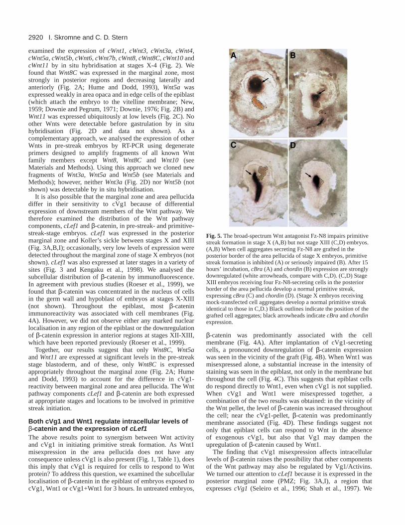
Fig. 6.The competence of the marginal zone to respond tocVg1+Wnt is lost at the primitive streak stage. Cell aggregatessecreting cVg1 were grafted alone (A-F) or in combination withWnt1-secreting cells (G,H) into the anterior margin of stage 2-3 hostembryos. After 6 (A-D) or 24 (E-H) hours of incubation, embryosstained for cVg1(A), cLef1(B), cNodal(C), cFGF-8(D), chordin(E,G) or cBra (F,H) by in situ hybridisation. Anti-Myc (A-F) or anti-HA (G,H) immunohistochemistry identifies the cell aggregate andconfirms synthesis of the factor. Both cVg1 (A-F) and cVg1+Wnt1(G,H) are unable to induce any of the markers tested (the dark colourin the grafts in panels A and B is nonspecific).

2922
Changes in the Wnt pathway do not account for theloss of competence to cVg1 misexpression atprimitive streak stagesIn the previous section, we showed that the ability of cVg1 toinduce an ectopic primitive streak requires Wnt activity. Bystage 2, cVg1 misexpression in the anterior marginal zone canno longer induce an ectopic primitive streak (Shah et al., 1997).At this stage, expression of Wnt8Chas become restricted to theforming primitive streak (Hume and Dodd, 1993). Is the lossof competence to cVg1 misexpression due to the disappearanceof Wnt activity from the marginal zone? To address thisquestion, we misexpressed cVg1, Wnt1 or cVg1+Wnt1 in theanterior marginal zone of stage 2-3 embryos. We found thatnone of these treatments could induce an ectopic axis, nor theexpression of any marker tested (cVg1, cLef1, cNodal, cFGF8,chordin or cBra) (Table 1; Fig. 6). These results suggest thatthe loss of competence to form a streak in response to cVg1misexpression at stages 2-3 is not due to the absence of Wntprotein.
DISCUSSION
Synergism between Vg1 and Wnt pathways duringinduction of an ectopic primitive streakThe ability of cVg1 to induce an ectopic primitive streak islimited to the marginal zone (Shah et al., 1997), a region thatexpresses Wnt8C(Hume and Dodd, 1993). We now show thatthe area pellucida can also form an ectopic primitive streak inresponse to cVg1, provided that a source of Wnt is alsosupplied. In addition, inhibition of endogenous Wnt activitywith Crescent or Dkk1 in the marginal zone blocks the axis-inducing ability of cVg1. However, both cVg1 and Wnt1 aloneelicit molecular responses even in regions where neither caninduce an ectopic streak. How do the Vg1 and Wnt pathwaysinteract to induce a primitive streak? This synergism may occurat the level of transcriptional activation of target genes or atother levels in the two pathways.
Synergism at the level of transcriptional activationAnalysis of the promoter region of genes induced by membersof the Wnt and TGFβ families in several species has led to theidea that synergism between the Wnt and TGFβ pathways canoccur at the transcriptional level. In Drosophila, expression ofthe homeotic/selector gene Ultrabitorax (Ubx) in posteriorendoderm is regulated by the Wnt and TGFβ family memberswingless (wg) and decapentaplegic (dpp), respectively (Bienz,1996), via separate Wg and Dpp response elements in the Ubxpromoter (Riese et al., 1997). In amphibians, high levels ofsiamois, twin and goosecoid expression also depend oncooperation between these two pathways acting throughseparate Wnt and TGFβ response elements (Sokol and Melton,1992; Steinbeisser et al., 1993; Cui et al., 1995; Watabe et al.,1995; Laurent et al., 1997; Crease et al., 1998; Nishita et al.,2000). Molecular characterisation of the mouse goosecoidpromoter has also revealed the presence of Wnt and TGFβ-regulatory elements similar to those found in Xenopusgoosecoid, suggesting that the mechanisms of goosecoidregulation have been conserved during evolution (Watabe et al.,1995; Labbe et al., 1998). The chick goosecoidpromoter hasnot yet been analysed and it will be interesting in future todetermine whether the promoter regions of this and otherorganiser markers contain comparable regulatory elements.
More complex interactions between Vg1- and WntpathwaysThe Wnt and TGFβ signalling pathways have beencharacterised extensively in Drosophila, Xenopus andmammals (reviewed in Cadigan and Nusse, 1997; Massaguéand Chen, 2000). Activation of the ‘canonical’ Wnt pathwayleads to the stabilisation of β-catenin (Cadigan and Nusse,1997; Moon and Kimelman, 1998), which then forms acomplex with Lef1/TCF transcription factors to activate targetgene expression (Behrens et al., 1996; Molenaar et al., 1996).TGFβ family members, on the other hand, lead to the assemblyof a Smad complex, which then recruits co-activators or co-repressors to regulate the expression of target genes (Massaguéand Chen, 2000). At first sight, therefore, the TGFβ and Wntpathways are quite distinct. However, recent experiments inXenopusled to the surprising finding that Lef1 and β-catenininteract directly with Smad4 to activate twin transcription(Nishita et al., 2000), suggesting that some components of the
I. Skromne and C. D. Stern
A. Xenopus B. Chick
Dorsovegetal region
Animal pole
Posterior marginal zone
Anterior
Vg1
Wnt11
Wnt8C
Nuclear β-catenin
Lef1/TCF3
Fig. 7.Distribution of Vg1 and various Wnt signalling pathwaycomponents in amphibian and avian pre-gastrula embryos. (A)Lateral view (animal pole up) of a stage 9 (Nieuwkoop and Faber,1967) Xenopusembryo. Vg1 protein is localised to the vegetal poleof the embryo (yellow; Tannahill and Melton, 1989), whereas β-catenin is concentrated in the nucleus of prospective dorsal cells (red;Schneider et al., 1996), even though cLef1/TCF3is expressedthroughout the embryo (blue; Molenaar et al., 1996; Molenaar et al.,1998). Wnt11transcripts (green; Ku and Melton, 1993) are restrictedto the marginal zone, in a dorsal-to-ventral gradient. (B) Ventral viewof a stage X (Eyal-Giladi and Kochav, 1976) chick embryo (anteriorto the top). Wnt8Cexpression in the marginal zone describes agradient highest posteriorly (purple; Fig. 2A; Hume and Dodd,1993), where cVg1(yellow; Seleiro et al., 1996; Shah et al., 1997)and cLef1(blue, Fig. 3) transcripts are also detected. In contrast toXenopus, chick Wnt11is ubiquitously expressed at low levelsthroughout the embryo (green, Fig. 2C). In both species, the regionwhere Vg1 and Wnt activities overlap (the dorsal-vegetal part inXenopusand the posterior marginal zone in the chick embryo) iswhere the organiser-inducing centre resides (Bachvarova et al., 1998;Harland and Gerhart, 1997, Heasman, 1997, Moon and Kimelman,1998).
2923Wnt and Vg1 interactions in the chick
signal transduction machinery in the Wnt and TGFβ pathwayscan interact directly. Our finding that cVg1 regulates theexpression of components of the Wnt pathway reveals someadditional complexity. First, cVg1 misexpression in thearea pellucida (where Wnt8C is not expressed) causes adownregulation of β-catenin levels (Fig. 4B). Second,misexpression of cVg1 in the marginal zone (a region that doesexpress Wnt8C) induces cLef1 expression (Fig. 4E). Third,when cVg1 is misexpressed together with Wnt1 in the areapellucida, cLef1expression is induced, which is not seen wheneither factor is misexpressed alone (Fig. 4F-H). Together, thesefindings suggest that many components of the Vg1/TGFβ andWnt pathways may be intertwined.
In addition, both TGFβ and Wnt signals can be transducedby mechanisms that do not involve the ‘canonical’ Smad andβ-catenin pathways. A growing body of evidence suggests thatTGFβ family proteins can regulate expression of target genesusing the JNK pathway (reviewed by Massagué and Chen,2000) via TAK1 (TGFβ activated kinase) and NLK (Nemo-likekinase), inducing a variety of responses depending on the celltype (Afti et al., 1997; Shibuya et al., 1998; Hocevar et al.,1999; Yamaguchi et al., 1999). Interestingly, in both Xenopusand C. elegans, activation of the TAK-NLK-JNK pathwaycauses phosphorylation of Lef1/TCF/POP-1 and subsequentinhibition of β-catenin-mediated responses (Ishitani et al.,1999; Meneghini et al., 1999); this mechanism could explainthe downregulation of β-catenin levels caused by cVg1misexpression in our experiments (Fig. 4B).
Likewise, Wnt signals can be transduced using pathwaysother than β-catenin, and two such pathways are known (Coxand Peifer, 1998; Boutros and Mlodzik, 1999; Noselli andAgnes, 1999). One involves activation of the Rho/Rac/Cdc42family of small GTPases and JNK-type kinases (Strutt et al.,1997; Boutros et al., 1998; Boutros and Mlodzik, 1999; Noselliand Agnes, 1999). The second alternative pathway involvesactivation of the phosphoinositol (PI) pathway and increase inintracellular Ca2+ (Slusarski et al., 1997a; Slusarski et al.,1997b). The β-catenin, Rho/JNK and PI/PKC/Ca2+ pathwaysmay serve different functions: activation of the β-cateninpathway in Xenopusand zebrafish causes axial duplications
(reviewed in Moon and Kimelman, 1998), whereas activationof the PI/PKC/Ca2+ pathway disrupts morphogeneticmovements (Moon et al., 1993; Du et al., 1995; Torres et al.,1996) and the Rho/Rac/JNK pathway has been implicated inthe regulation of cell movements and cell shape through theactin cytoskeleton (Nobes and Hall, 1995; Nobes and Hall,1999).
Taken together, these considerations suggest that the cVg1and Wnt pathways can interact at several different levels, andthat the choice of alternative transduction pathways mayregulate different aspects of cell behaviour that are crucial togastrulation, such as cell fate decisions and morphogeneticmovements.
The marginal zone as a special region of the earlyembryo, defined by Wnt activityUnlike amphibians, where polarity is determined bylocalisation of maternal determinants before the beginning ofzygotic transcription (Harland and Gerhart; 1997; Heasman,1997; Moon and Kimelman, 1998; Arendt and Nübler-Jung,1999), avian embryos can re-establish their polarity up untilthe time when primitive streak formation begins, when theembryo may already contain as many as 20,000-60,000 cells(Spratt and Haas, 1960b). Spratt and Haas showed thatwhen embryos are cut into fragments, each fragment canspontaneously generate an embryonic axis, provided both thatthe fragment contains a portion of the marginal zone and thatthey are obtained from embryos younger than the primitivestreak stage (Spratt and Haas, 1960b). The frequency ofprimitive streak formation in these fragments decreases in aposterior-to-anterior direction, but even the most anteriorfragments are capable of initiating an axis. These observationsled them to propose that ‘the marginal zone of an unincubatedblastoderm exhibits a gradient in embryo formingpotentiality…the highest point of which is in the prospectiveposterior median position’ (Spratt and Haas, 1960b).
Within the marginal zone, only the posterior part (the PMZ)can induce an axis (including Hensen’s node) whentransplanted to the anterior side of a host embryo fragment(Eyal-Giladi and Khaner, 1989; Khaner and Eyal-Giladi, 1989)
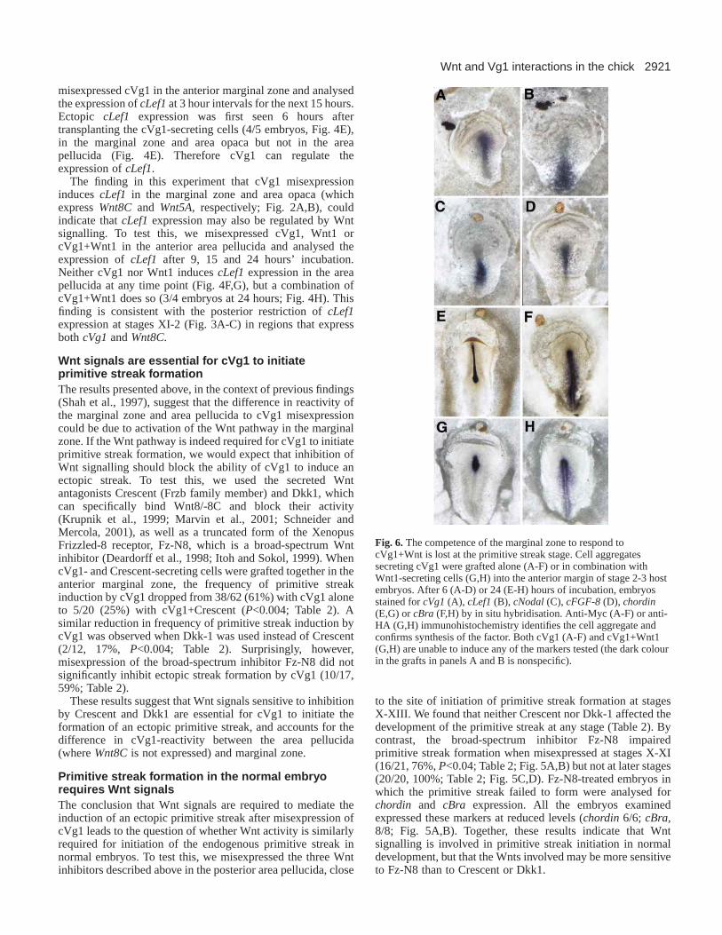
Table 2. Effects of misexpression of the Wnt antagonists Crescent, Dkk1 and Fz-N8 on normal and cVg1-inducedprimitive streak development
Region Stage Factor Primitive streaks/total (%)
Anterior marginal zone X-XIII cVg1 38/62 (61)Crescent 0/20 (0)Dkk1 0/10 (0)Fz-N8 0/16 (0)cVg1+COS cells 9/17 (56)cVg1+Crescent 5/20 (25)**cVg1+Dkk1 2/17 (17)**cVg1+Fz-N8 10/17 (59)
Posterior area pellucida X-XI COS cells 16/16 (100)Crescent 22/22 (100)Dkk1 5/5 (100)Fz-N8 16/21 (76)*
XII-XIII Fz-N8 20/20 (100)
The frequency of induction of an ectopic streak by a graft of cells secreting cVg1 (top section) or the development of a normal primitive streak by the hostembryo (bottom) in the presence of the factors indicated is shown on the right. Note that misexpression of Crescent or Dkk1, together with cVg1 reduces thefrequency of induction in the marginal zone of stage X-XIII embryos from 61% to 25% or 17%, respectively (**, P<0.004). In this assay, Fz-N8 does not haveany effect. However, Fz-N8 impairs normal primitive streak formation when misexpressed at the posterior border of the area pellucida in stage X embryos (*,P<0.04).
2924
without making a cellular contribution to the induced structures(Bachvarova et al., 1998). In this sense, the PMZ is the avianequivalent of the Nieuwkoop centre of amphibians (seeBachvarova et al., 1998). In Xenopus, this centre is thought toarise from interaction between the Wnt and TGFβ pathways(Fig. 7). The Wnt pathway is activated (as revealed by nuclearlocalisation of β-catenin; Schneider et al., 1996; Yost et al.,1996; Larabell et al., 1997) throughout the prospective dorsalside of the embryo (Wylie et al., 1996), while Vg1 RNA ispreferentially localised in the vegetal half of the blastula(Rebagliati et al., 1985; Weeks and Melton, 1987; Dale et al.,1989; Tannahill and Melton, 1989; Fig. 7). The two activitiesoverlap in the dorsal-vegetal part of the embryo, where theNieuwkoop centre resides (Harland and Gerhart, 1997;Heasman, 1997; Moon and Kimelman, 1998; Fig. 7). In thechick, Wnt8Cis expressed throughout the marginal zone (Fig.2A; Hume and Dodd, 1993) and cVg1 only in the posteriorregion (Seleiro et al., 1996; Shah et al., 1997), making the PMZthe only region of the pre-primitive streak stage embryo thatexpresses both cVg1 and a member of the Wnt 1 subfamily(Fig. 7). Therefore, despite the differences between avian andamphibian embryos in the mode of polarity determination,similar pathways appear to be involved, and it is tempting tospeculate that the overlap of cVg1and Wnt8Cactivity in thePMZ is responsible for the Nieuwkoop centre-like propertiesof this region (Fig. 7).
We now provide both loss-of-function and gain-of-functionresults that support this notion. Inhibition experiments withCrescent and Dkk-1 strongly suggest that Wnt activity isessential for the formation of an ectopic primitive streak inresponse to cVg1. We also show that the area pellucida can berendered competent to respond to cVg1 and initiate a primitivestreak if an exogenous source of Wnt activity is supplied. Theselatter results are consistent with the finding that misexpressionof Wnt1 can enhance the axis-inducing activity of Activin (aTGFβ family member closely related to Vg1; Cooke et al.,1994), although we were not able to reproduce the finding(Cooke et al., 1994) that Wnt1 misexpression alone can re-position the host axis (not shown).
Together with the complexity of the interactions betweenWnt and Vg1 signals already discussed, we can now provide apartial molecular explanation for the results of Spratt and Haas(Spratt and Haas, 1960b): we propose that Wnt activity definesthe marginal zone as a whole, within which the expression ofcVg1 defines the PMZ (Fig. 7). Only the region where cVg1is present will express cLef1, transduce the Wnt signal andinitiate the formation of a primitive streak. When embryos arebisected into anterior and posterior halves, the posterior halfretains the PMZ and continues to form a primitive streak athigh frequency. By contrast, the anterior half retains the gradedexpression of Wnt8C in the marginal zone, with two points ofhigh Wnt8C expression in lateral marginal zones. High levelsof Wnt8Cexpression may bias the stochastic induction of cVg1(by an unknown factor) in one of the two lateral marginalzones, which initiates formation of the axis.
Unexpected effects of Wnt antagonistsThe above conclusions are based in part on the ability of theWnt antagonists Crescent and Dkk-1 to block the induction ofan ectopic axis by cVg1. However, we also show that the sameantagonists do not block the formation of the normal primitive
streak when placed posteriorly in the embryo. Conversely, thebroad-spectrum Wnt antagonist Fz-N8 blocks formation of thenormal streak, but has no effect on the axis induced by cVg1misexpression in the marginal zone. How can these results beexplained?
Functional studies in Xenopushave shown that while Dkk-1, Frzb-1, Fz-N8 and Crescent can antagonise signalling by theWnt1/Wnt8 family of proteins (Leyns et al., 1997; Wang et al.,1997a; Wang et al., 1997b; Deardorff et al., 1998; Itoh andSokol, 1999; Krupnik et al., 1999; Marvin et al., 2001;Schneider and Mercola, 2001), only Dkk-1 and Fz-N8 canblock signalling by Wnt2b, Wnt3a and Wnt5a (Wang et al.,1997b; Itoh and Sokol, 1999; Krupnik et al., 1999; Marvin etal., 2001; Schneider and Mercola, 2001) even though Frzb-1can bind Wnt5a in co-immunoprecipitation assays (Lin et al.,1997). Furthermore, indirect evidence also suggests thatCrescent and Fz-N8 might inhibit Wnt11 activity as well(Deardorff et al., 1998; Pera and De Robertis, 2000).Therefore, the different Wnt ligand specificities shown by thevarious antagonists tested may partly account for the resultsobtained in our assays.
Recently, it has been shown that recombinant secretedFrizzled-related protein 1 (sFRP1) exerts a biphasic effect onWg activity in a β-catenin stabilization assay, increasing β-catenin levels at low concentrations, but reducing them athigher concentrations (Üren et al., 2000). This has lead to theidea that Frzb-1 type of molecules (including Crescent) mayhave a dual function as Wnt agonist or antagonist in a dose-dependent manner (Üren et al., 2000; see Pfeiffer and Vincent,1999). In the Xenopusassays discussed above, injection ofmRNA is likely to produce high concentrations of Wntantagonists, probably comparable with the doses of sFRP1 thatresult in Wnt inhibition (Üren et al., 2000). This contrasts withour experimental conditions, where local sources ofantagonists are used and the levels of protein is likely todecrease rapidly with increasing distance from the source. Theability of Crescent and Dkk-1 to block primitive streakinduction by cVg1 in the anterior marginal zone can beexplained by their ability to inhibit endogenous Wnt8-relatedfactors expressed at low levels in this region. Close to theposterior marginal zone, however, the relative levels of Dkk-1or Crescent may not be high enough to antagonise Wnt8-related signalling, but instead may facilitate presentation of theWnt molecule to its receptor. Why, then, does the broad-spectrum Wnt antagonist Fz-N8 does not block axis inductionby cVg1, while Crescent and Dkk-1 do?
When Fz-N8 is misexpressed in the posterior area pellucidait blocks the formation of the endogenous primitive streak butdoes not inhibit chordinor brachyuryexpression (Fig. 5). Theexpression of chordin in the area pellucida suggests thatorganiser fates have been established. This differs from theconsequences of misexpression of another primitive streakinhibitor, BMP4: in this case, the embryos do not expresscNot1 (an organiser maker) or brachyury(Streit et al., 1998).Therefore it is likely that BMP4 prevents primitive streakformation by blocking the specification of particular cell fates,while misexpression of Fz-N8 at the posterior end of theembryo does not affect cell fate specification but rather someother aspect of primitive streak formation, such as cellmovements.
The recent finding that a zebrafish homologue of Wnt11
I. Skromne and C. D. Stern

2925Wnt and Vg1 interactions in the chick
(Silberblick) is required for the proper convergence/extensionmovements of gastrulation, acting through a β-catenin-independent pathway (Heisenberg et al., 2000, see also Tadaand Smith, 2000; Makita et al., 1998) is consistent with thisidea. Misexpression of Fz-N8 in the posterior area pellucidacould prevent primitive streak formation by inhibiting Wnt11signalling that disrupts the Polonaise movements (Gräper,1929; Spratt and Haas, 1960a; Stern, 1990; Hatada and Stern,1994) that precede gastrulation, without blocking theexpression of organiser markers.
The molecular basis of competence for primitivestreak initiation by cVg1When cVg1 is misexpressed in the marginal zone of embryosthat have initiated gastrulation, neither chordin nor cBraexpression is induced, and no ectopic primitive streakdevelops. Although Wnt8C is downregulated in the marginalzone at the start of gastrulation (Hume and Dodd, 1993), thisis not responsible for the loss of competence that occurs at thisstage, because misexpression of Wnt1 does not rescue theability of these older embryos to respond to cVg1. Possiblecandidates to explain this loss of competence are the‘ventralising’ factors of the TGFβ family BMP-2, BMP-4, andBMP-7, which are appropriately expressed at the periphery ofthe embryo (Streit et al., 1998; Joubin and Stern, 1999).Misexpression of the BMP antagonist Noggin at the edge ofthe area pellucida allows cVg1+Wnt1 to induce expression oforganiser markers at stages 3+-4 (Joubin and Stern, 1999).Therefore, before gastrulation, the competence of the marginalzone to respond to cVg1 requires Wnt activity. Oncegastrulation is initiated, other signalling molecules present atthe periphery, such as BMPs, may make the marginal zonerefractory to cVg1 and Wnt signals.
However, some aspects of the competence for ectopicprimitive streak formation cannot be explained by cVg1 andWnt activity alone. When a PMZ is transplanted to the anteriorhalf of a host embryo (Bachvarova et al., 1998) or to the lateralside of embryos that have been deprived of their own PMZ(Eyal-Giladi and Khaner, 1989; Khaner and Eyal-Giladi,1989), a primitive streak is induced, but only if the host embryois at stage XI or younger. By contrast, cVg1 can induce acomplete axis even if misexpressed in the marginal zone ofstage XIII embryos (Seleiro et al., 1996; Shah et al., 1997), andcan induce an ectopic organiser when misexpressed with Wnt1in the area pellucida up to stage 3+ (Joubin and Stern, 1999).The molecular mechanisms that underlie this early window ofcompetence to the PMZ remain unknown.
Likewise, mechanisms other than Wnt signalling seem to berequired for the primitive streak to continue its developmentand give rise to an axis. In the area pellucida of pre-streakembryos, cVg1 alone can induce organiser markers but not anectopic streak, while cVg1+Wnt induce expression oforganiser markers as well as a primitive streak. However, eventhese ectopic primitive streaks do not give rise to axialderivatives like a notochord or prechordal mesoderm. Thisresult is reminiscent of the finding that local misexpression ofChordin in the marginal zone or area pellucida induces anectopic streak containing an organiser, but again no furtheraxial derivatives are formed (Streit et al., 1998; Streit andStern, 1999). Together, these observations suggest that thecompetence of the area pellucida to give rise to axial structures
derived from the streak is limited either by the lack of someadditional permissive signals, or by the presence of inhibitors.
ConclusionsWe have shown that the ability of cVg1 to induce an ectopicprimitive streak in the anterior marginal zone of the chickembryo requires Wnt activity of the Wnt1/Wnt8-related class.However, our results also point to an involvement of otherWnts in axis formation, perhaps members of the Wnt11 class.Our gain- and loss-of-function results explain why an ectopicaxis is induced when cVg1 is misexpressed in the marginalzone but not in the area pellucida. The differences in sensitivityto Wnt antagonists between the normal axis-forming regionand of the ectopic axes induced by Vg1 suggest that there maybe a diversity of cVg1- and Wnt transduction pathways, whichappear to interact with each other in complex ways. Wepropose that different transduction pathways are responsiblefor controlling different aspects of primitive streak formation,such as the acquisition of cell fates and the regulation of cellmovements and adhesion.
This study was funded by the National Institutes of Health(GM53456 and GM56656) and the Medical Research Council (UK).I. S. was also supported by the DGAPA, UNAM, Mexico. Theconfocal microscopy facility was established and supported byNational Institutes of Health shared instrumentation grants (1S10-RR10506 and 5-P30-CA13696) as part of the Herbert Irving CancerCenter at Columbia University. We are grateful to Jane Dodd, Juan-Carlos Izpisúa-Belmonte, Jan Kitajewski, Peter Klein, MichaelKuehn, Ed Laufer, Andy McMahon, Randy Moon, Peter Pfeffer andJim Smith for generous gifts of cells, probes and reagents; MarthaMarvin and Andrew Lassar for sharing unpublished information; andTheresa Swayne for assistance with the confocal microscope. We arealso indebted to Rosemary Bachvarova, Federica Bertocchini, AnnFoley and Andrea Streit for their insightful comments on themanuscript.
REFERENCES
Afti, A., Djelloul, S., Chastre, E., Davis, R. and Gespach, C.(1997).Evidence for a role of Rho-like GTPases and stress-activated proteinkinase/c-Jun N-terminal kinase (SAPK/JNK) in transforming growth factorβ-mediated signaling. J. Biol. Chem.272, 1429-1432.
Arendt, D. and Nübler-Jung, K. (1999). Rearranging gastrulation in the nameof yolk: evolution of gastrulation in yolk-rich amniote eggs. Mech. Dev.81,3-22.
Ausubel, F. M., Brent, R., Kingston, R. E., Moore, D. D., Seidman, J. G.,Smith, J. A. and Struhl, K. (1995). Current Protocols in Molecular Biology(ed. V. B. Chanda). New York: John Wiley.
Bachvarova, R. F., Skromne, I. and Stern, C. D.(1998). Induction ofprimitive streak and Hensen’s node by the posterior marginal zone in theearly chick embryo. Development125, 3521-3534.
Behrens, J., von Kries, J. P., Kuhl, M., Bruhn, L., Wedlich, D., GrosschedlR. and Birchmeier, W. (1996). Functional interaction of β-catenin with thetranscription factor LEF-1. Nature382, 638-642.
Bienz, M. (1996). Induction of the endoderm in Drosophila. Semin. Cell Dev.Biol. 7, 113-119.
Boutros, M. and Mlodzik, M. (1999). Dishevelled: at the crossroads ofdivergent intracellular signaling pathways. Mech. Dev.83, 27-37.
Boutros, M., Paricio, N., Strutt, D. I. and Mlodzik, M. (1998). Dishevelledactivates JNK and discriminates between JNK pathways in planar polarityand wingless signaling. Cell 94, 109-118.
Cadigan, K. M. and Nusse, R.(1997). Wnt signaling: a common theme inanimal development. Genes Dev.11, 3286-3305.
Cooke, J., Takada, S. and McMahon, A.(1994). Experimental control ofaxial pattern in the chick blastoderm by local expression of Wnt and activin:the role of HNK-1 positive cells. Dev. Biol.164, 513-527.
2926
Cox, R. T. and Peifer, M. (1998). Wingless signaling: the inconvenientcomplexities of life. Curr. Biol. 8, R140-R144.
Crease, D. J., Dyson, S. and Gurdon, J. B.(1998). Cooperation between theactivin and Wnt pathways in the spatial control of organizer gene expression.Proc. Natl. Acad. Sci. USA95, 4398-4403.
Cui, Y., Brown, D. J., Moon, R. T. and Christian, J. L.(1995). Xwnt-8b: amaternally expressed Xenopus Wnt gene with a potential role in establishingthe dorsoventral axis. Development121, 2177-2186.
Dale, L., Matthews, G., Tabe, L. and Colman, A.(1989). Developmentalexpression of the protein product of Vg1, a localized maternal mRNA in thefrog Xenopus laevis. EMBO J.8, 1057-1065.
Deardorff, M. A., Tan, C., Conrad, L. J. and Klein, P. S.(1998). Frizzled-8 is expressed in the Spemann organizer and plays a role in earlymorphogenesis. Development125, 2687-2700.
Downie, J. R. (1976). The mechanism of chick blastoderm expansion. J.Embryol. Exp. Morphol.35, 559-575.
Downie, J. R. and Pegrum, S. M.(1971). Organization of the chickblastoderm edge. J. Embryol. Exp. Morphol.26, 623-635.
Du, S. J., Purcell, S. M., Christian, J. L., McGrew, L. L. and Moon, R. T.(1995). Identification of distinct classes and functional domains of Wntsthrough expression of wild-type and chimeric proteins in Xenopus embryos.Mol. Cell Biol.15, 2625-2634.
Evans, G. I., Lewis, G. K., Ramsay, G. and Bishop, J. M.(1985). Isolationof monoclonal antibodies specific for human c-myc protooncogene product.Mol. Cell Biol.5, 3610-3616.
Eyal-Giladi, H. and Khaner, O. (1989). The chick’s marginal zone andprimitive streak formation. II. Quantification of the marginal zone’spotencies – temporal and spatial aspects. Dev. Biol.134, 215-221.
Eyal-Giladi, H. and Kochav, S. (1976). From cleavage to primitive streakformation: a complementary normal table and a new look at the first stagesof the development of the chick. I. General morphology. Dev. Biol.49, 321-337.
Gavin, B. J., McMahon, J. A. and McMahon, A. P.(1990). Expression ofmultiple novel Wnt1/int-1-related genes during fetal and adult mousedevelopment. Genes Dev.4, 2319-2332.
Gräper, L. (1929). Die Primitiventwicklung des Hühnchens nachstereokinematischen Untersuchungen, kontrolliert durch vitaleFarbmarkierung und verglichen mit der Entwicklung anderer Wirbeltiere.Wilhelm Roux. Arch. Entwmech. Org.116, 382-429.
Hamburger, V. and Hamilton, H. L. (1951). A series of normal stages in thedevelopment of the chick. J. Morphol. 88, 49-92.
Harland, R. and Gerhart., J. (1997). Formation and function of Spemann’sorganizer. Annu. Rev. Cell Dev. Biol.13, 611-667.
Hatada, Y. and Stern, C. D.(1994). A fate map of the epiblast of the earlychick embryo. Development120, 2879-2889.
Heasman, J.(1997). Patterning the Xenopus blastula. Development124, 4179-4191.
Heisenberg, C. P., Tada, M., Rauch, G. J., Saude, L., Concha, M. L.,Geisler, R., Stemple, D. L., Smith J. C. and Wilson, S. W.(2000).Silberblick/Wnt11 mediates convergent extension movements duringzebrafish gastrulation. Nature405, 76-81.
Hocevar, B. A., Brown, T. L. and Howe, P. H.(1999). TGF-β inducesfibronectin synthesis through a c-Jun N-terminal kinase-dependent, Smad4-independent pathway. EMBO J.18, 1345-1356.
Hollyday, M., McMahon J. A. and McMahon, A. P. (1995). Wntexpression patterns in chick embryo nervous system. Mech. Dev.52, 9-25.
Hume, C. R. and Dodd, J.(1993). CWnt-8C: a novel Wnt gene with apotential role in primitive streak formation and hindbrain organization.Development119, 1147-1160.
Ishitani, T., Ninomiya-Tsuji, J., Nagai, S., Nishita, M., Meneghini, M.,Barker, N., Waterman, M., Bowerman, B., Clevers, H., Shibuya H. andMatsumoto, K. (1999). The TAK1-NLK-MAPK-related pathwayantagonizes signalling between β-catenin and transcription factor TCF.Nature399, 798-802.
Itoh, K. and Sokol, S. (1999). Axis determination by inhibition of Wntsignaling in Xenopus. Genes Dev.13, 2328-2336.
Izpisúa-Belmonte, J. C., De Robertis, E. M., Storey, K. G. and Stern, C.D. (1993). The homeobox gene goosecoid and the origin of organizer cellsin the early chick blastoderm. Cell 74, 645-659.
Jones, C. M., Kuehn, M. R., Hogan, B. L., Smith J. C. and Wright, C. V.(1995). Nodal-related signals induce axial mesoderm and dorsalizemesoderm during gastrulation. Development121, 3651-3662.
Joubin K. and Stern, C. D. (1999). Molecular interactions continuously
define the organizer during the cell movements of gastrulation. Cell 98, 559-571.
Kengaku, M., Capdevila, J., Rodríguez-Esteban, C., De La Pena, J.,Johnson, R. L., Izpisúa-Belmonte, J. C. and Tabin, C. J.(1998). DistinctWnt pathways regulating AER formation and dorsoventral polarity in thechick limb bud. Science280, 1274-1277.
Kessler, D. S. (1997). Siamois is required for formation of Spemann’sorganizer. Proc. Natl. Acad. Sci. USA94, 13017-13022.
Khaner, O. and Eyal-Giladi, H. (1989). The chick’s marginal zone andprimitive streak formation. I. Coordinative effect of induction and inhibition.Dev. Biol.134, 206-214.
Kispert, A., Ortner, H., Cooke, J. and Herrmann, B. G.(1995). The chickBrachyury gene: developmental expression pattern and response to axialinduction by localized activin. Dev. Biol.168, 406-415.
Knezevic, V., De Santo, R. and Mackem, S.(1997). Two novel chick T-boxgenes related to mouse Brachyury are expressed in different, non-overlapping mesodermal domains during gastrulation. Development124,411-419.
Krupnik, V. E., Sharp, J. D., Jiang, C., Robison, K., Chickering, T. W.,Amaravadi, L., Brown, D. E., Guyot, D., Mays, G., Leiby, K. et al.(1999). Functional and structural diversity of the human Dickkopf genefamily. Gene238, 301-313.
Ku, M and Melton, D. A. (1993). Xwnt-11: a maternal expressed XenopusWnt gene. Development119, 1161-1173.
Labbe, E., Silvestri, C., Hoodless, P. A., Wrana J. L. and Attisano, L.(1998). Smad2 and Smad3 positively and negatively regulate TGFβ-dependent transcription through the forkhead DNA-binding protein FAST2.Mol. Cell 2, 109-120.
Larabell, C. A., Torres, M., Rowning, B. A., Yost, C., Miller, J. R., Wu,M., Kimelman D. and Moon, R. T. (1997). Establishment of the dorso-ventral axis in Xenopus embryos is presaged by early asymmetries in β-catenin that are modulated by the Wnt signaling pathway. J. Cell Biol.136,1123-1136.
Laurent, M. N., Blitz, I. L., Hashimoto, C., Rothbacher U. and Cho, K. W.(1997). The Xenopus homeobox gene twin mediates Wnt induction ofgoosecoid in establishment of Spemann’s organizer. Development124,4905-4916.
Leyns, L., Bouwmeester, T., Kim, S., H., Piccolo, S. and De Robertis, E.M. (1997). Frzb-1 is a secreted antagonist of Wnt signaling expressed in theSpemann organizer. Cell 88, 747-756.
Lin, K., Wang, S., Julius, M. A., Kitajewski, J., Moos, M. and Luyten, F.P. (1997) The cysteine-rich frizzled domain of Frzb-1 is required andsufficient for modulation of Wnt signaling. Proc. Natl. Acad. Sci. USA94,11196-11200.
Makita, R., Mizuno, T., Koshida, S., Kuroiwa, A. and Takeda, H. (1998).Zebrafish wnt11: pattern and regulation of the expression by the yolk celland No tail activity. Mech. Dev.71, 165-176.
Marvin, M. J., DiRocco, G., Gardiner, A., Bush, S. M., Sive H. L. andLassar A. B. (2001). Inhibition of Wnt activity induces heart formationfrom posterior mesoderm. Genes Dev.15, 316-327.
Massagué, J. and Chen, Y. G.(2000). Controlling TGF-β signalling. GenesDev.14, 627-644.
Meneghini, M. D., Ishitani, T., Carter, J. C., Hisamoto, N., Ninomiya-Tsuji, J., Thorpe, C. J., Hamill, D. R., Matsumoto, K. and Bowerman,B. (1999). MAP kinase and Wnt pathways converge to downregulatean HMG-domain repressor in Caenorhabditis elegans. Nature399, 793-797.
Molenaar, M., van de Wetering, M., Oosterwegel, M., Peterson-Maduro,J., Godsave, S., Korinek, V., Roose, J., Destree, O. and Clevers, H.(1996). XTcf-3 transcription factor mediates β-catenin-induced axisformation in Xenopus embryos. Cell 86, 391-399.
Molenaar, M., Roose, J. Peterson, J., Venanzi, S., Clevers, H. and Destree,O. (1998). Differential expression of the HGM box transcription factorsXTcf-3 and XLef-1 during early Xenopus development. Mech. Dev.75, 151-154.
Moon, R. T., Campbell, R. M., Christian, J. L., McGrew, L. L., Shih, J.and Fraser, S.(1993). Xwnt-5A: a maternal Wnt that affects morphogeneticmovements after overexpression in embryos of Xenopus laevis.Development119, 97-111.
Moon, R. T. and Kimelman, D. (1998). From cortical rotation to organizergene expression: toward a molecular explanation of axis specification inXenopus. BioEssays20, 536-545.
New, D. A. T.(1955). A new technique for the cultivation of the chick embryoin vitro. J. Embryol. Exp. Morphol.3, 326-331.
I. Skromne and C. D. Stern

2927Wnt and Vg1 interactions in the chick
New, D. A. T. (1959). The adhesive properties and expansion of the chickblastoderm. J. Embryol. Exp. Morphol.7, 146-164.
Nieuwkoop, P. D. and Faber, J.(1967). Normal Table ofXenopus laevis(Daudin). Amsterdam: North Holland Publishing Company.
Nishita, M., Hashimoto, M. K., Ogata, S., Laurent, M. N., Ueno, N.,Shibuya H. and Cho, K. W.(2000). Interaction between Wnt and TGF-βsignalling pathways during formation of Spemann’s organizer. Nature403,781-785.
Nobes, C. D. and Hall, A.(1995). Rho, rac, and cdc42 GTPases regulate theassembly of multimolecular focal complexes associated with actin stressfibers, lamellipodia, and filopodia. Cell 81, 53-62.
Nobes, C. D. and Hall, A.(1999). Rho GTPases control polarity, protrusion,and adhesion during cell movement. J. Cell Biol.144, 1235-1244.
Noselli, S. and Agnes, F.(1999). Roles of the JNK signaling pathway inDrosophila morphogenesis. Curr. Opin. Genet. Dev.9, 466-472.
Olson, D. J., Christian, J. L. and Moon, R. T.(1991). Effect of Wnt-1 andrelated proteins on gap junctional communication in Xenopus embryos.Science252, 1173-1176.
Pera, E. M. and De Robertis, E. M.(2000). A direct screen for secretedproteins in Xenopus embryos identifies distinct activities for the Wntantagonists Crescent and Frzb-1. Mech. Dev.96, 183-196.
Pfeffer, P. L., De Robertis, E. M. and Izpisúa-Belmonte, J. C.(1997).Crescent, a novel chick gene encoding a Frizzled-like cysteine-rich domain,is expressed in anterior regions during early embryogenesis. Int. J. Dev. Biol.41, 449-458.
Pfeiffer, S. and Vincent, J.-P.(1999). Signalling at a distance: transport ofWingless in the embryonic epidermis of Drosophila. Semin. Cell Dev. Biol.10, 303-309.
Rebagliati, M. R., Weeks, D. L., Harvey, R. P. and Melton, D. A.(1985).Identification and cloning of localized maternal RNAs from Xenopus eggs.Cell 42, 769-777.
Riese, J., Yu, X., Munnerlyn, A., Eresh, S., Hsu, S. C., Grosschedl, R. andBienz, M. (1997). LEF-1, a nuclear factor coordinating signaling inputsfrom wingless and decapentaplegic. Cell 88, 777-787.
Roeser, T., Stein, S. and Kessel, M.(1999). Nuclear β-catenin and thedevelopment of bilateral symmetry in normal and LiCl-exposed chickembryos. Development126, 2955-2965.
Schneider, S. Steinbeisser, H., Warga, R. M. and Hausen, P.(1996). β-catenin translocation into nuclei demarcates the dorsalizing centers in frogand fish embryos. Mech. Dev.57, 191-198.
Schneider, V. A. and Mercola, M. (2001) Wnt antagonism initiatescardiogenesis in Xenopus laevis. Genes Dev.15, 304-315.
Schroeder, K. E., Condic, M. L., Eisenberg, L.M. and Yost, H. J. (1999).Spatial regulated translation in embryos: asymmetric expression of maternalWnt11 along the dorsal-ventral axis in Xenopus. Dev. Biol.214, 288-297.
Seleiro, E. A., Connolly, D. J. and Cooke, J.(1996). Early developmentalexpression and experimental axis determination by the chicken Vg1 gene.Curr. Biol. 6, 1476-1486.
Shah, S. B., Skromne, I., Hume, C. R., Kessler, D. S., Lee, K. J., Stern, C.D. and Dodd, J. (1997). Misexpression of chick Vg1 in the marginal zoneinduces primitive streak formation. Development124, 5127-5138.
Shibuya, H., Iwata, H., Masuyama, N., Gotoh, Y., Yamaguchi, K., Irie, K.,Matsumoto, K., Nishida, E. and Ueno, N.(1998). Role of TAK1 and TAB1in BMP signaling in early Xenopus development. EMBO J.17, 1019-1028.
Shimizu, H., Julius, M. A., Giarre, M., Zheng, Z., Brown, A. M. andKitajewski, J. (1997). Transformation by Wnt family proteins correlateswith regulation of β-catenin. Cell Growth Differ.8, 1349-1358.
Slusarski, D. C., Corces, V. G. and Moon, R. T.(1997). Interaction of Wntand a Frizzled homologue triggers G-protein-linked phosphatidylinositolsignalling. Nature390, 410-413.
Slusarski, D. C., Yang-Snyder, J., Busa, W. B. and Moon, R. T.(1997).Modulation of embryonic intracellular Ca2+ signaling by Wnt5A. Dev. Biol.182, 114-120.
Sokol, S., Christian, J. L., Moon, R. T. and Melton, D. A.(1991). InjectedWnt RNA induces a complete body axis in Xenopus embryos. Cell 67, 741-752.
Sokol, S. and Melton, D. A.(1992). Interaction of Wnt and activin in dorsalmesoderm induction in Xenopus. Dev. Biol.154, 348-355.
Spratt, N. T. and Haas, H.(1960a). Morphogenetic movements in the lower
surface of the unincubated and early chick blastoderm. J. Exp. Zool.144,139-158.
Spratt, N. T. and Haas, H.(1960b). Integrative mechanisms in developmentof the early chick blastoderm. I. Regulative potentiality of separated parts.J. Exp. Zool.145, 97-137.
Steinbeisser, H., De Robertis, E. M., Ku, M., Kessler, D. S. and Melton, D.A. (1993). Xenopus axis formation: induction of goosecoid by injectedXwnt-8 and activin mRNAs. Development118, 499-507.
Stern, C. D. (1990). The marginal zone and its contribution to the hypoblastand primitive streak of the chick embryo. Development.109, 667-682.
Stern, C. D. and Holland, P. W. H.(1993). Essential Developmental Biology:A Practical Approach. Oxford: Oxford University Press.
Stern, C. D. and Ireland, W. H.(1981). An integrated experimental study ofendoderm formation in avian embryos. Anat. Embryol.163, 245-263.
Streit, A. and Stern, C. D.(1999). Mesoderm patterning and somite formationduring node regression: differential effects of chordin and noggin. Mech.Dev.85, 85-96.
Streit, A., Sockanathan, S., Pérez, L., Rex, M., Scotting, P. J., Sharpe, P.T., Lovell-Badge, R. and Stern, C. D.(1997). Preventing the loss ofcompetence for neural induction: HGF/SF, L5 and Sox-2. Development124,1191-1202.
Streit, A., Lee, K. J., Woo, I., Roberts, C., Jessell, T. M. and Stern, C. D.(1998). Chordin regulates primitive streak development and the stability ofinduced neural cells, but is not sufficient for neural induction in the chickembryo. Development125, 507-519.
Strutt, D. I., Weber, U. and Mlodzik, M. (1997). The role of RhoA in tissuepolarity and Frizzled signalling. Nature387, 292-295.
Tada, M. and Smith, J. C.(2000). Xwnt11 is a target of Xenopus Brachyury:regulation of gastrulation movements via Dishevelled, but not through thecanonical Wnt pathway. Development127, 2227-2238.
Tannahill, D. and Melton, D. A. (1989). Localized synthesis of the Vg1protein during early Xenopus development. Development106, 775-785.
Torres, M. A., Yang-Snyder, J. A., Purcell, S. M., DeMarais, A. A.,McGrew, L. L. and Moon, R. T. (1996). Activities of the Wnt1 class ofsecreted signaling factors are antagonized by the Wnt5A class and by adominant negative cadherin in early Xenopus development. J. Cell Biol.133,1123-1137.
Üren, A., Reichsman, F., Anest, V., Taylor, W. G., Muraiso, K., Bottaro,D. P., Cumberledge, S. and Rubin, J. S.(2000) Secreted Frizzled-relatedprotein-1 binds directly to wingless and is a biphasic modulator of Wntsignaling. J. Biol. Chem.275, 4374-4382.
Wang, S., Krinks, M., Lin, K., Luyten, F. P. and Moos, M., Jr (1997a).Frzb, a secreted protein expressed in the Spemann organizer, binds andinhibits Wnt8. Cell 88, 757-766.
Wang, S,. Krinks, M. and Moos, M., Jr (1997b). Frzb-1, an antagonist ofWnt1 and Wnt8, does not block signaling by Wnts -3A, -5A, or -11.Biochem. Biophys. Res. Commun.236, 502-504.
Watabe, T., Kim, S., Candia, A., Rothbacher, U., Hashimoto, C., Inoue K.and Cho, K. W. (1995). Molecular mechanisms of Spemann’s organizerformation: conserved growth factor synergy between Xenopus and mouse.Genes Dev.9, 3038-3050.
Weeks, D. L. and Melton, D. A.(1987). A maternal mRNA localized to thevegetal hemisphere in Xenopus eggs codes for a growth factor related toTGF-β. Cell 51, 861-867.
Wylie, C., Kofron, M., Payne, C., Anderson, R., Hosobuchi, M., Joseph,E. and Heasman, J.(1996). Maternal β-catenin establishes a ‘dorsal signal’in early Xenopus embryos. Development122, 2987-2996.
Yamaguchi, K., Nagai, S., Ninomiya-Tsuji, J., Nishita, M., Tamai, K., Irie,K., Ueno, N., Nishida, E., Shibuya, H. and Matsumoto, K.(1999). XIAP,a cellular member of the inhibitor of apoptosis protein family, links thereceptors to TAB1-TAK1 in the BMP signaling pathway. EMBO J.18, 179-187.
Yost, C., Torres, M., Miller, J. R., Huang, E., Kimelman, D. and Moon, R.T. (1996). The axis-inducing activity, stability, and subcellular distributionof β-catenin is regulated in Xenopus embryos by glycogen synthase kinase3. Genes Dev.10, 1443-1454.
Zorn, A. M., Butler, K. and Gurdon, J. B. (1999). Anterior endomesodermspecification in Xenopus by Wnt/β-catenin and TGF-β signalling pathways.Dev. Biol.209, 282-297.