what is new in burn care? - university of colorado denve · outline 1. superficial burns –...
TRANSCRIPT

Michael Schurr, M.D.Professor of Surgery
What is New in Burn Care?

Outline1. Superficial burns
– Biobrane– Research focus #1
2. Large TBSA burns– Integra versus cadaver skin– Epicel
3. Burn survival statistics4. Stratagraft skin substitute
– Research focus #25. Genetic engineering of Stratagraft

Superficial burns• Clean and moist
– Bacitracin or biologic dressing (Biobrane)• Heal in 2 to 3 weeks• Encourage ROM• Pain control

The problem with silvadene

•
Burns are painful

Biologic dressing: Biobrane

Silicone Sheeting
Nylon Fibers Collagen Peptides
BIOBRANE®
Silicone outer membrane
Nylon fabric, trifilament thread with chemically bound collagen

Advantages• Excellent adherence to a superficial burn • Decreases pain • Easy to store with long shelf life • Relative inexpensive Disadvantages• Difficult to remove if left in place over 2 weeks • Infection in 15% requires removal• >50 % infection if placed on deep dermal burns• Therefore limited to clean superficial burns only
Biobrane clinical use

Functionalization of Biobrane® using Silver-Nanoparticle
Polymer Films
Research Focus #1

PAHPAA
Charged substrate
Ag+
Ag NPs
Add AgNO3
Reducing agent
-Reduces Ag+ to Ag0
-Regenerates -COO-
More Ag+ can be added to regenerated -COO-
PAH, pKa ~ 9poly(allylamine hydrochloride)
PAA, pKa ~ 5poly(acrylic acid)
Assembly of polymeric thin films of silver nanoparticles
< 100 nm

Elastomeric stamp
Biobrane®
Polymer Film with Silver Nanoparticles
Silicone Sheeting
Nylon Fibers Collagen Peptides
A
Stamp removed
Silver Loaded Polymer Film
B
Mechanical transfer of films onto Biobrane®
Figures Not to Scale

Modified Biobrane® provides sustained release of silver ions
0123456789
0 24 48 72 96 120 144 168 192 216 240
Cum
ulat
ive
[Ag+
] ug/
cm2
Time (hr)
1L AgNP2L AgNP
Modified Biobrane® release less than 1 µg/cm2 of Ag+ per day in milliQ waterComparison: Silver dressing Acticoat® release ~100 µg/cm2 of Ag+ per day*

Silver-nanoparticle polymer film on Biobrane® causes 6Log10 reduction in bacterial counts in vitro
012345678
Tissue Culture Polystyrene
Biobrane Biobrane/ Silver-coating
Log 1
0C
FU o
f S. e
pide
rmid
is
24 hr48 hr
Similar response against Staph. aureus and Pseud. aeruginosa

Splinted-wound-infection model
A B C D
•Heterozygous wild mice •Full-thickness cranial wounds (6 mm diameter) splinted•Wounds inoculated with Staph. aureus and covered with Biobrane®•Secured with Tegaderm•Wounds harvested on day 3 and homogenized for bacterial counts

0
1
2
3
4
5
Untreated Biobrane Biobrane/ Silver coating
Log 1
0CF
U o
f P.
aeru
gino
sa
*
*
Biobrane® modified with silver-nanoparticle films prevents infection in full-thickness mice wounds
•Heterozygous wild mice •Wounds inoculated with 3x10⁴
CFU on day 1•Wounds harvested on day 4
n=12 wounds p<0.01, t-test
Similar response against Staph. aureus

3
4
5
6
Biobrane Biobrane/Ag
Log 1
0CF
U/g
tiss
ueBiobrane® modified with silver-nanoparticle films reduces bacteria in partial-thickness pig wounds
Statistically significant difference (p<0.001)n=12 wounds
•Wounds inoculated with 3x10⁶
CFU on Day 0•Control wounds treated with unmodified Biobrane®•Wounds harvested at day 3
*
*
Similar response against Staph. aureus

Conclusions
• Biobrane® integrated with silver-nanoparticle films significantly reduced bacterial growth in vitro and in skin wounds in mice and pigs
• Future directions– Other antibacterial agents (silver nanoparticles,
chlorhexidine)– Growth factors– Integra– Biologics used for abdominal wall reconstruction
• In discussion with UDL to obtain IND– Avoid Biobrane associated infection– Expand indication for use to deeper dermal burns

Large TBSA burns

<20% of burns are large TBSA

Burn shock correlates with TBSA
• Loss of vascular integrity• Increased metabolic rate• Catabolic - futile substrate cycling• Depressed immune function• Cardiac depression• Shortened RBC life• Renal and MSOF

Resuscitation with Parkland formula
• 4 x (%TBSA) x (kg body weight)• total fluid needs for first 24 hours• 1/2 vol. during first 8 hours• 1/2 vol. during next 16 hours

Burn resuscitation
0
0.1
0.2
0.3
0.4
0.5
2 6 10 14 18 22 26
Res
usci
tatio
n vo
l. (m
l/kg/
%bu
rn/h
r)
Time post-burn (hours)
calculated volumerequirements (4 ml/kg/%burn)

Other controversies in resuscitation
• Use of colloid• Hypertonic saline• FFP as resuscitation fluid• Vitamin C, zinc and other adjuncts
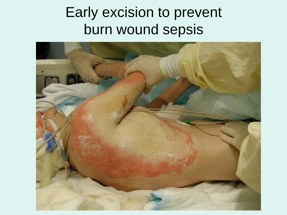
Early excision to prevent burn wound sepsis

Meshed skin greatly expands coverage
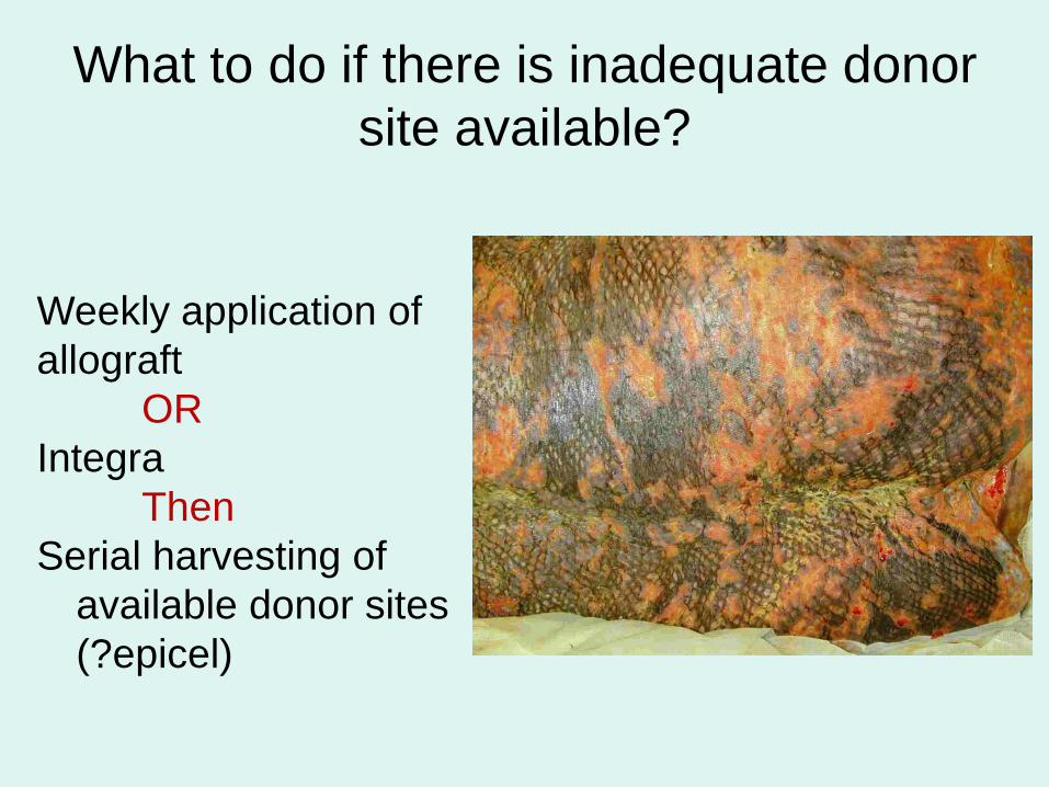
What to do if there is inadequate donor site available?
Weekly application of allograft
ORIntegra
ThenSerial harvesting of
available donor sites(?epicel)

Integra technique

Integra technique

Integra versus allograft• Allograft
– Theoretic infection risks– Must be replaced weekly, multiple operations– Inexpensive and available
• Integra– Expensive– 3 weeks to revascularization– Prone to infection– Able to use thin and widely meshed autografts– May improve cosmesis– May improve function

Epicel - cultured epidermal autografts
• Cultured keratinocytes - 1983• 2-8 cell layers thick• neodermal formation after one year• Two 6 X 2 cm skin biopsies• 15 day lag time
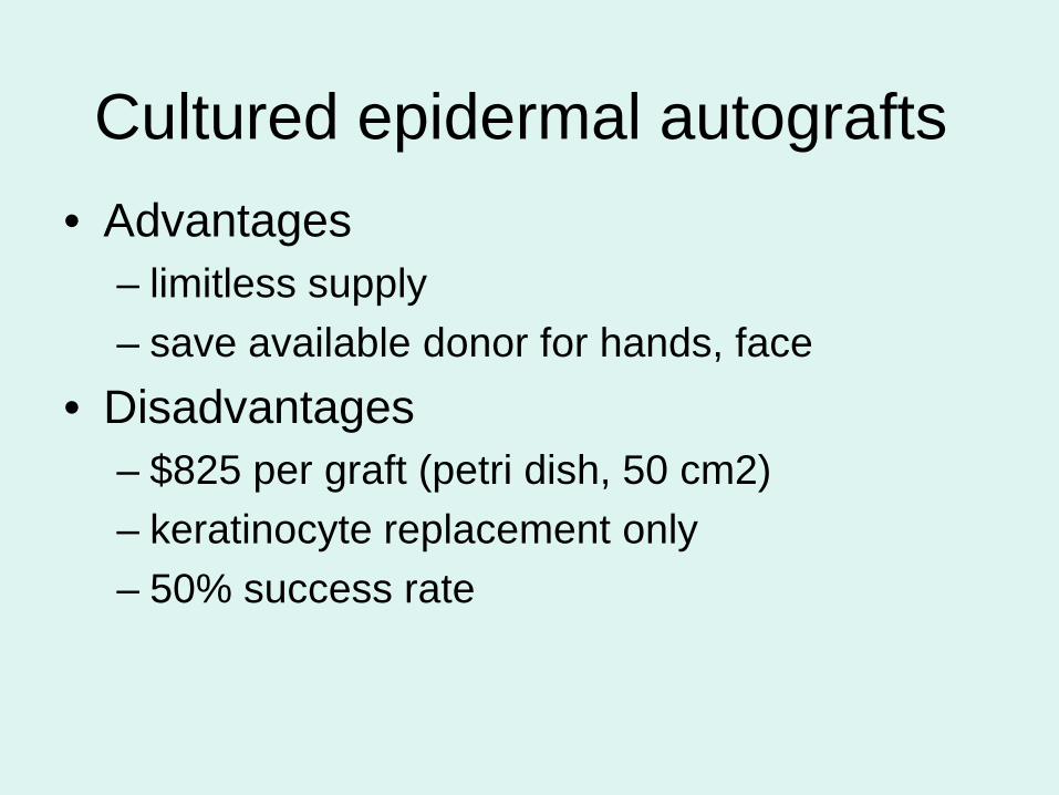
Cultured epidermal autografts• Advantages
– limitless supply– save available donor for hands, face
• Disadvantages– $825 per graft (petri dish, 50 cm2)– keratinocyte replacement only– 50% success rate

Epicel

Epicel

The art of burn surgery
• Timing of excision• Timing of first STSG• Treatment of hands and faces• Coverage of large open areas• Donor site utilization• 4th degree burns with exposed
tendon/bone• Life/Function/Cosmesis

Outcomes

Mortality versus TBSA

Mortality by Baux score
• Burn Size• Age• Inhalation Injury
Age + % TBSA
2

Rapid decline in mortality over time

Mortality by age group - decreased
Age 0–14 15–44 45–64 >65 All
1980s
Mortality % 1.2 6.1 18.7 35.4 7.2
1990s
Mortality % 0.3 3.4 5.5 20 2.6
2000s
Mortality % 0.2 1.8 5.1 17.7 2.3

LA50 has increased
LA50 for Age Groups and Time
1982–1991 1992–2000 2000–2008Age 0–14 55.3 74.3 100
15–44 55.7 49.6 76.4
45–64 32.6 50.9 58.6
>65 13.6 28.1 27.8

Research focus #2

The unique problem of deep partial thickness burns
• Will heal without STSG
• But, scarring can be significant
• Lack of donor site availability in those with large TBSA burns

Allogeneic keratinocytes improve healing of deep dermal burns
• Rab et al, Burns 2005: Should dermal scald burns in children be covered with autologous skin grafts or with allogeneic cultivated keratinocytes

Allogeneic keratinocytes improve healing of deep dermal burns

• Living tissue interacts with the wound bed• Immediate wound coverage• Barrier function• Controls pain• Allogeneic cells are replaced with
autologous keratinocytes by 3 months• There is no consistent source of allogeneic
keratinocytes with full barrier function
Cultured skin substitutes in deep dermal burns

Stratagraft core technologyEpidermal layer:
NIKS® cells
Dermal layer:human fibroblasts
and collagen
+
NIKS® cellsProprietary human epidermal progenitorsFDA CMC testing completeMultiple cell banks in place
Cultured under proprietary conditions
Fully developed multi-layered human skinPhysical barrier presentBiologically activeStrong, durable, suturable
StrataGraft®

Phase I clinical trial
• NIH/NIAMS R44-AR47499 Translational Clinical Grant
• Full-thickness wounds ≥5% TBSA requiring serial debridement and grafting
• Temporary coverage for 7 days with StrataGraft® and cadaver allograft
• Primary efficacy outcome was autograft take at 2 weeks

Phase I clinical trial results
Cadaver allograft, day 7 StrataGraft, day 7
Cadaver allograft, 2 weeks p STSG StrataGraft, 2 weeks p STSG

Phase IIb clinical trial
• Stratagraft can be used as a temporary wound cover in patients with deep dermal burns after operative debridement– Clinical trial grant funded by DOD (AFIRM)– Randomized trial of Stratagraft versus STSG– Multicenter trial 7/20 patients– First patient enrolled at UCH– 100% success in first 7

Genetic engineering of Stratagraft
• Expressgraft– Bacterial bioburden– Poor vascular ingrowth– Abnormal proteases
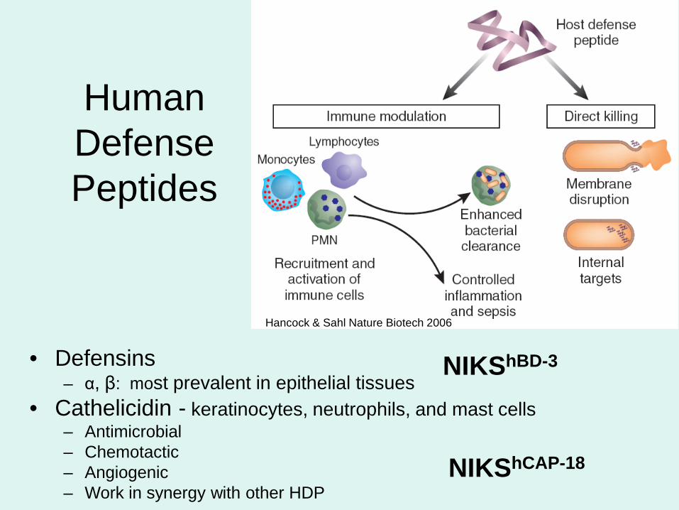
• Defensins– α, β: most prevalent in epithelial tissues
• Cathelicidin - keratinocytes, neutrophils, and mast cells– Antimicrobial– Chemotactic– Angiogenic– Work in synergy with other HDP
Human Defense Peptides
Hancock & Sahl Nature Biotech 2006
NIKShBD-3
NIKShCAP-18

Stable tissue-specific expression of hCAP-18 mRNA
Molecular Therapy, In Press

Murine burn infection model provides method for in vivo assessment of
NIKShBD-3 tissue.
24 hr
72 hr after skin grafting:
Quantitative culture of skin tissue
and underlying muscle
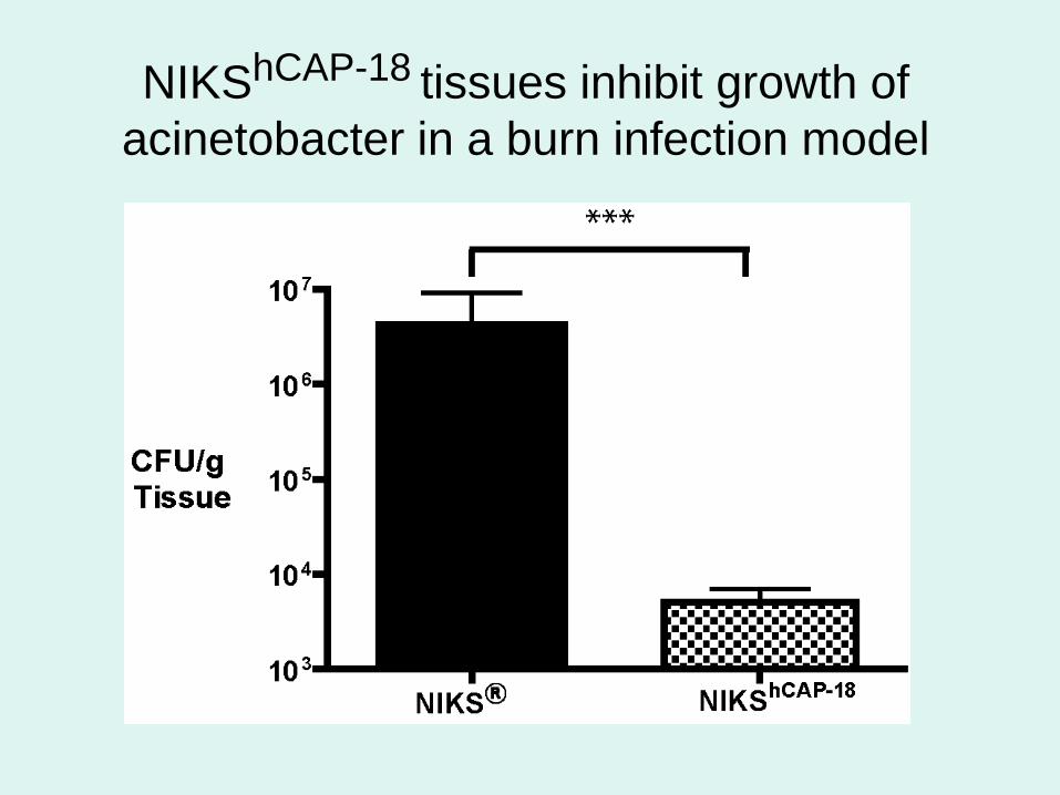
NIKShCAP-18 tissues inhibit growth of acinetobacter in a burn infection model
NIKShCAP-18
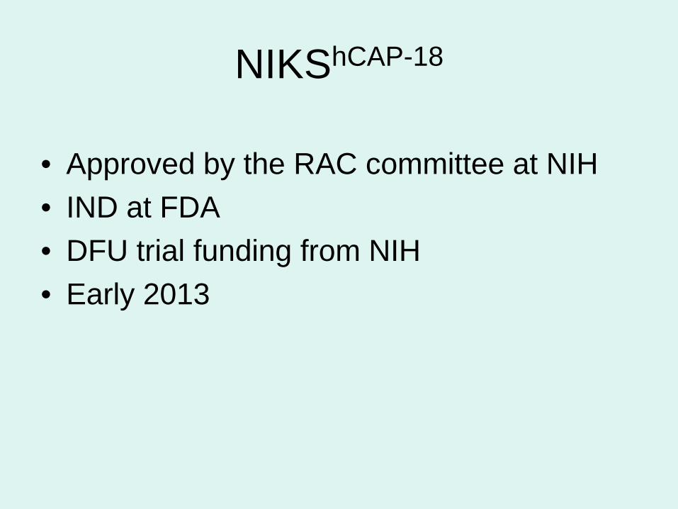
• Approved by the RAC committee at NIH• IND at FDA• DFU trial funding from NIH• Early 2013
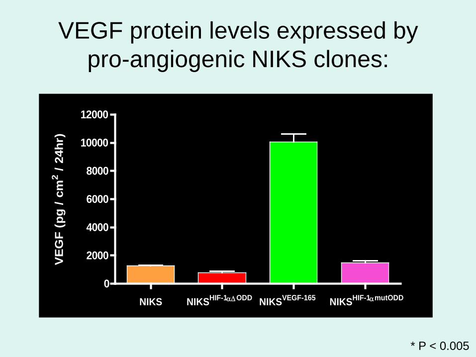
VEGF protein levels expressed bypro-angiogenic NIKS clones:
* P < 0.005
*
NIKS NIKSHIF-1α∆ODD NIKSVEGF-165 NIKSHIF-1αmutODD0
2000
4000
6000
8000
10000
12000
VE
GF
(pg
/ cm
2 / 24
hr)

Visualizing FITC-stained vessels within the CMA
(-) Media Control
NIKSVEGF-165
(+) bFGF Control
NIKSHIF-1αΔODD