wednesday slide conference 2011-2012 … · their most common localization is the gonads.7,11 in...
TRANSCRIPT
CASE I: CS-1 (JPC 4002461).
Signalment: A 5-year-old spayed female Rhodesian Ridgeback dog (Canis familiaris).
History: The dog was presented for a large (approximately 7 cm diameter), subcutaneous moveable mass over the left shoulder. Two weeks after surgical excision, the dog presented with neurologic
signs and blindness. Euthanasia was elected because of poor prognosis.
Gross Pathology: There is a 14 cm diameter, fluid-filled, ulcerated subcutaneous mass present over the left shoulder (seroma). The abdominal lymph nodes are enlarged (2x2x2 to 2x3x1.5 cm diameter). On cut section, the architecture of lymph nodes is effaced by
1
J o i n t P a t h o l o g y C e n t e rVe t e r i n a r y P a t h o l o g y S e r v i c e s
WEDNESDAY SLIDE CONFERENCE 2011-2012
C o n f e r e n c e 1 0 16 November 2011
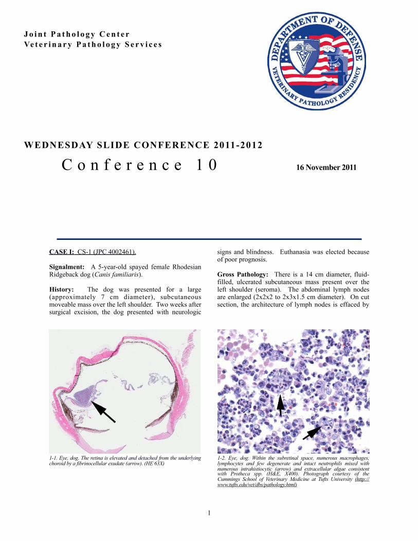
1-1. Eye, dog. The retina is elevated and detached from the underlying choroid by a fibrinocellular exudate (arrow). (HE 63X)
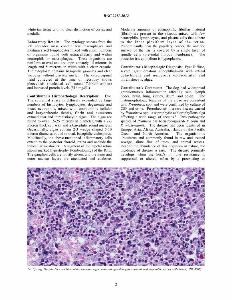
1-2. Eye, dog. Within the subretinal space, numerous macrophages, lymphocytes and few degenerate and intact neutrophils mixed with numerous intrahistiocytic (arrow) and extracellular algae consistent with Protheca spp. (H&E, X400). Photograph courtesy of the Cummings School of Veterinary Medicine at Tufts University (http://www.tufts.edu/vet/dbs/pathology.html)
white-tan tissue with no clear distinction of cortex and medulla.
Laboratory Results: The cytology smears from the left shoulder mass contain few macrophages and medium sized lymphocytes mixed with small numbers of organisms found both extracellularly and within neutrophils or macrophages. These organisms are reniform to oval and are approximately 15 microns in length and 5 microns in width with a clear capsule. The cytoplasm contains basophilic granules and clear vacuoles without discrete nuclei. The cerebrospinal fluid collected at the time of necropsy shows pleocytosis (nucleated cell count-17,600/microliter) and increased protein levels (516 mg/dL).
Contributor’s Histopathologic Description: Eye. The subretinal space is diffusely expanded by large numbers of histiocytes, lymphocytes, degenerate and intact neutrophils, mixed with eosiniophilic cellular and karyorrhectic debris, fibrin and numerous extracellular and intrahistiocytic algae. The algae are round to oval, 15-25 microns in diameter, with a 2-3 micron thick cell wall and a basophilic round nucleus. Occasionally, algae contain 2-3 wedge shaped 5-10 micron diameter, round to oval, basophilic endospores. Multifocally, the above-mentioned inflammatory cells extend to the posterior choroid, retina and occlude the trabecular meshwork. A segment of the tapetal retina shows marked hypertrophy (tomb-stoning) of the RPE. The ganglion cells are mostly absent and the inner and outer nuclear layers are attenuated and coalesce.
Moderate amounts of eosinophilic fibrillar material (fibrin) are present in the vitreous mixed with few neutrophils, lymphocytes, and plasma cells that adhere to the inner plexiform layer of the retina. Predominantly near the papillary border, the anterior surface of the iris is covered by a single layer of spindle cells (pre-iridal fibrous membrane). The posterior iris epithelium is hyperplastic.
Contributor’s Morphologic Diagnosis: Eye: Diffuse, severe, granulomatous endophthalmitis with retinal detachment and numerous extracellular and intrahistiocytic algae.
Contributor’s Comment: The dog had widespread granulomatous inflammation affecting skin, lymph nodes, brain, lung, kidney, ileum, and colon. The histomorphologic features of the algae are consistent with Prototheca spp. and were confirmed by culture of CSF and urine. Protothecosis is a rare disease caused by Prototheca spp., a saprophytic achlorophyllous alga affecting a wide range of species.1 Two pathogenic species of Protheca has been recognized- P. zopfi and P. wickerhamii. The disease has been identified in Europe, Asia, Africa, Australia, islands of the Pacific Ocean, and North America. The organism is ubiquitous and commonly found in raw and treated sewage, slime flux of trees, and animal wastes. Despite the abundance of this organism in nature, the incidence of disease is rare. The disease primarily develops when the host’s immune resistance is suppressed or altered, often by a preexisting or
WSC 2011-2012
2
1-3. Eye dog. The subretinal exudate contains numerous algae, some endosporulating (arrowhead), and some collapsed cell walls (arrow). (HE 400X)

concurrent disease. No preexisting inflammatory, neoplastic or metabolic disease was detected in this dog.
Protothecosis can present either as systemic or localized cutaneous infection. In the alimentary tract, Prototheca is thought to be an opportunistic invader of existing mucosal lesions. Skin infections are thought to result from traumatic inoculation.2 In the present case, the dog was initially presented for dermatitis rather than diarrhea and therefore skin was considered to be the primary site of infection in this dog. Cattle, horses, and wild pigs may pass Prototheca in the feces without apparent clinical disease.
The intestine and the eye are the most common sites in protothecosis of dogs. P. zopfii is almost always isolated from dogs with systemic infections and P. wickerhamii has been isolated from dogs and cats with cutaneous infections. Grossly, white to tan, 0.2-2 mm diameter, granulomatous lesions can be seen within the affected organs. There is mild inflammation with few macrophages, lymphocytes, plasma cells, and neutrophils mixed with large numbers of algal organisms.2,3
Protothecal organisms can be identified on rectal or colonic scrapings if the large intestine is involved. Prototheca sp. are round, oval or angular cells that are 8-20 micron diameter, have a refractile wall and contain granular cytoplasm. The organism reproduces by endosporulation and can be identified histologically as morula of 2-20 daughter cells within a single organism. Prototheca and Chlorella spp. are morphologically indistinguishable in tissues stained with H&E. Chlorella spp. contain periodic acid-Schiff-positive cytoplasmic starch granules that are PAS-negative following diastase digestion, but Prototheca spp. do not contain these granules. Ultrastructurally, chloroplasts consist of a highly organized, twisted lamellar component associated with amorphous, electron dense vacuolated material (starch). Prototheca spp. may contain visible starch granules (plastids) on electron microscopy, but these are not associated with a lamellar component. Prototheca organisms must be differentiated from other organisms that undergo endosporulation, such as Coccidioides immitis and Rhinosporidium seeberi. Rhinosporidium spp. have large sporangia, filled with uniformly round, 2-10 micron endospores. Coccidioidomyces spp. have spherules that measure up to 50 micron in diameter containing small, round endospores.
Molecular characterization and speciation of the Protheca spp. can be performed by 18S rDNA sequencing, genotype-specific PCR, restriction
fragment length polymorphism, or qPCR followed by DNA Resolution Melting Analysis.4
J P C D i a g n o s i s : E y e : E n d o p t h a l m i t i s , pyogranulomatous, diffuse, severe with numerous algae -- etiology consistent with Prototheca sp.
Conference Comment: Ocular protothecosis is only described as part of a systemic infection and never as a primary, solitary lesion. In the eye, it presents as a bilateral granulomatous panuveitis. The primary differential in this case is mycotic endophthalmitis, caused either by Cryptococcus or Blastomyces, which produce similar histologic lesions. Conference participants discussed the likely route of infection in this dog being hematogenous, and the necessity of the algae to overcome the blood-ocular barrier via induction of inflammatory cytokines.2 The blood-ocular barrier is composed of the blood-aqueous barrier (which consists of tight junctions in the ciliary body and iridal capillaries and the phagocytic function of the ciliary epithelium) and the blood-retinal barrier (formed by tight junctions in the non-fenestrated capillary endothelium and between the retinal pigment epithelial cells).
Contributor: Cummings School of Veterinary Medicine at Tufts UniversitySection of Pathology200 Westboro RdNorth Grafton, MA 01536http://www.tufts.edu/vet/dbs/pathology.html
References: 1. Greene CE. Infectious Diseases of the Dog and Cat. 3rd ed. St. Louis, MO: Saunders-Elsevier; 2006: 656-662.2. Wilcock BP. The eye and ear. In: Maxie MG, ed. Jubb, Kennedy, and Palmer's Pathology of Domestic Animals. 5th ed. vol 1. Philadelphia, PA: Elsevier Saunders; 2007:503-504.3. Brown CC, et al. Alimentary System. In: Maxie MG, ed. Jubb, Kennedy, and Palmer's Pathology of Domestic Animals. 5th ed. vol 1. Philadelphia PA, Elsevier Saunders; 2007:231-232.4. Ricchi M, et al. A rapid real-time PCR/DNA resolution melting method to identify Prototheca species. J Appl Micro. 2010;110(1):27-34.
WSC 2011-2012
3

CASE II: PV 1650/10 (JPC 4003047).
Signalment: Common kestrel, (Falco tinnunculus).
History: The wild bird was treated in a center of wildlife rehabilitation because of the presence of periocular swelling and exophthalmos. No additional history was available.
Gross Pathology: Swelling around the eye.
Contributor’s Histopathologic Description: In the retrobulbar space, there is an irregular unencapsulated tumor, composed of tissue types arising from all three primordial germ cell lines. There are numerous cystic areas containing pale basophilic material, surrounded by solid areas of cartilage, bone, neural and adipose tissue. The cysts vary in size, up to 3 mm in diameter, and are lined by cuboidal to columnar epithelial cells, multifocally ciliated or with distended vacuolated cytoplasm (goblet cells), (respiratory epithelium, endoderm). Peripheral to these cysts are occasional, much smaller cysts, filled with concentrically laminated keratin, with a wall of keratinizing squamous epithelium (ectoderm).
Multifocally, solid areas are composed of moderately cellular neoplastic neural tissue with neurons and glial cells embedded in neuropil (ectoderm). There are multifocal areas of formation of bone with marrow and cartilage (mesoderm). In a few of the sections, there are small lobules of zymogen-filled acini, resembling pancreas (endoderm). Mitoses in all cell populations are not prominent (fewer than 1 per HPF). In the small area of eye present, there is evidence of retinal detachment, hypertrophy of the retinal pigment epithelium, and retinal degeneration.
Contributor’s Morphologic Diagnosis: Retrobulbar mass: Teratoma.
Contributor’s Comment: Teratomas are defined as germ cell origin neoplasms composed of at least two, and often three, germinal layers.7
They are uncommon in domestic animals, but have been reported mostly in the bitch, sow, mare and cow; their most common localization is the gonads.7,11 In avian species, involvement of the testes appears to be more common than the ovary9 and they are most commonly observed in chickens.6,10
There are two reports of retrobulbar teratomas in wild birds.6,10 One of the tumors was poorly differentiated and interpreted as malignant.6 Incidentally, old literature reports describe experimental induction of teratomas in testicles of fowl by local injection of metal ions (zinc) in young animals 2,3,9, which was initially intended as a method of chemical castration.2,3
Teratomas in animals, in contrast to those in humans, are almost always benign.7 In humans, classification of a teratoma as benign or malignant is based largely on the identification of primitive, undifferentiated cells and tissues within the tumor.1 In general, the presence of a germ cell component worsens the prognosis, and teratomas with incompletely differentiated tissues
WSC 2011-2012
4
2-1. Retrobulbar space, common kestrel. An unencapsulated neoplasm composed of tissues from three cell lines (ectodermal, mesodermal, and endodermal) compresses the globe (upper portion of image) and the brain (lower right). (HE 63X)
2-2. Retrobulbar space, common kestrel. A variety of well-differentiated tissues are present within the neoplasm, including numerous tortuous cysts lined by columnar epithelium which are occasionally surrounded by glands composed of goblet cells (large arrowhead) ,bone with marrow (large arrow), cartilage (small arrows), and fat (small arrowheads). (HE 160X)

should be considered potentially malignant.1,5
In this particular case, the eye was submitted as an enucleation and the kestrel was presumed alive at the time of biopsy. Unfortunately, follow-up evaluation was not available.
JPC Diagnosis: Eye: Retrobulbar teratoma.
Conference Comment: Tera tomas , l ike dysgerminomas, are germ cell neoplasms; they usually originate from totipotent cells, such as those normally present in the ovary and testis (and sometimes abnormally present in sequestered midline embryonic rests). Such cells have the capacity to differentiate into any of the cell types found in the adult body.12 Most extragonadal teratomas affect juvenile or young adult animals, develop from germinal elements, and occur along the migration pathway of the germ cells along the body midline, in the mediastinum, or in the central neuraxis. The sacrococcygeal region is the most frequent extragonadal location; other sites include the mediastinum and retroperitoneum.6
Several conference participants were unable to identify ectodermal differentiation to haired skin due to slide variability; however, this does not affect the diagnosis of teratoma, since only 2 germ cell lines are required to make the diagnosis. Although most reports define teratomas as having at least 2 of the 3 germ cell lines, rare variants (in humans) have only 1 line (monodermal variant).
Contributor: Weizmann InstituteDepartment of Veterinary ResourcesRehovot 76100Israelhttp://www.weizmann.ac.il/vet/
References: 1. Crum CP. The female genital tract. Germ cell tumors. In: Cotran RS, Kumar V, Collins T, eds. Robbins Pathologic Basis of Disease. 6th ed. Philadelphia, PA:Saunders;1999;1073-1075.2. Guthrie J. Specificity of the metallic ion in the experimental induction of teratomas in fowl. Br J Cancer. 1967;21:619-22.3. Guthrie J. Zinc induction of testicular teratomas in Japanese quail (Coturnix coturnix Japonica) after photoperiodic stimulation of testis. Br J Cancer. 1971;25:311-4.4. Hooper CC. Teratoma in the cerebrum of a fantail pigeon. Avian Pathol. 2008;37:141-3.5. Klein MK. Tumors of the female reproductive system. Germ cell tumors. In: Withrow SJ, MacEwen EG, eds. Small Animal Clinical Oncology. 3rd edition. St. Louis, MO: Saunders; 2001:446.
6. López RM, Múrcia DB. First description of malignant retrobulbar and intracranial teratoma in a lesser kestrel (Falco naumanni). Avian Pathol. 2008;37:413-4.7. MacLachlan NJ, Kennedy PC. Tumors of the genital systems, Teratoma. In: Meuten DJ ed. Tumors in Domestic Animals. 4th ed. Ames, Iowa: Iowa State University Press, 2002:554. 8. Petrak ML, Gilmore CE. Neoplasms. In: Petrak ML, ed. Diseases of Caged and Aviary Birds. 2nd ed. Philadelphia, PA: Lea and Febiger; 1982:606-637.9. Reece RL. Tumors of unknown etiology. In: Calnek BW, ed. Disease of Poultry. 10th ed. Ames, Iowa: Iowa State University Press, 1997:496-497.10. Schelling SH. Retrobulbar teratoma in a great blue heron (Ardea herodias). J Vet Diagn Invest. 1994;6:514-6.11. Shlafer DH, Miller RB. Female genital system. In: Maxie MG, ed. Jubb, Kennedy, and Palmer’s Pathology of Domestic Animals. 5th ed. vol 3. St. Louis, MO; Saunders Elsevier; 2007:453-454.12. Stricker TP, Kumar V. Neoplasia. In: Kumar V, Abbas AK, Fausto N, Aster JC, eds. Robbins and Cotran Pathologic Basis of Disease. 8th ed. Philadelphia, PA: Saunders Elsevier; 2010:261-2.
WSC 2011-2012
5

CASE III: 18124-09 (JPC 3164804).
Signalment: 5-year-old spayed female domestic shorthair cat, Felis domesticus, feline.
History: Five-year-old spayed female cat whose pupils began to dilate and ears to twitch after application of “Bio Spot for cats” on 05/27/2009. Cat returned to clinic with bilateral enlarged eyeballs with normal tonometry on 7/7/09. Conjunctiva slightly enlarged and oozy. Treated with antimicrobial eye ointment and dexamethasone. 7/23/09 and began treatment with metacam, doxycyclin, prednisone, antirobe, and strongid. Vaccination status unknown. Lesions progressively worsened over next few months. Animal euthanized and submitted for necropsy 09/08/09.
Additional History after diagnosis was made:
The owner moved into her present premises several months ago. The previous owner used one of the stalls in the barn as a chicken coop. Owner reported that all her animals (horses, dogs, and cats) began to lose weight with no response to deworming attempts. All other animals, except this cat, appeared to slowly recover on their own. At time of euthanasia all other animals were in good condition, even the other cats which had been treated with “Bio Spot for cats”.
Gross Pathology: The cat is extremely cachectic. All bones can be easily palpated and the skin moves easily over the bones due to a scant amount of subcutaneous adipose tissue. There is no fat around the kidney, in the mesentery, or in the coronary groove. The eyes bulge forward. A portion of the cloudy cornea can be seen on the left eye. The sclera of both eyes is composed of bulging proliferative white tissue. Similar tissue is found in the frontal sinus, bulging through the calvarium at the bridge of the nose and extending down, around, and behind both eyes.
Laboratory Results: 07/23/09: CBC, Chemistry: WNL; FeLV/FIV negative. Toxoplasmosa negative. Hemobartonella positive.
Contributor’s Histopathologic Description: Cross sections of the eyes reveal that all layers of the globe, including the iris, portions of the cornea, and portions of the eyelids are effaced by sheets of macrophages distended with Histoplasma capsulatum and necrotic debris. The tissue removed from the nasal cavity and frontal sinus are found to be distorted in a histologically similar manner.
Contributor’s Morphologic Diagnosis: Chronic, severe, bilateral granulomatous panophthalmitis and
blepharitis with intracellular Histoplasma capsulatum, feline.
Contributor’s Comment: Dogs are very susceptible hosts for histoplasmosis and they are considered sentinel animals.3,4 However, while cats appear less susceptible to the disease, ocular manifestations of histoplasmosis are more commonly encountered in the cat.4,5 Histoplasma capsulatum is a soil-borne dimorphic fungus.2 It grows best in moist soil containing nitrogen-rich organic matter such as bird or bat feces. The fungus can grow in moist soil associated with house plants, so even indoor-only cats can be at risk. Similar to blastomycosis, the disease is acquired by inhalation of fungal microconidia. The incubation period is 12 to 16 days. The microconidia in the lung convert to the yeast form which is
WSC 2011-2012
6
3-1. Eye, cat. 5-year-old cachectic female cat with bilateral exophthalmos, bulging corneas and scleras, and corneal opacity. Photograph courtesy of C.E. Kord Animal Disease Diagnostic Laboratory, Dept. of Agriculture, Ellington Agricultural Center, 440 Hogan Road, Nashville, TN 37220. http://www.tennessee.gov/agriculture/regulatory/kord.html
3.2. Eye, cat. Closer view of head showing the degree of infiltration of the sclera and overlying conjunctiva bilaterally. Photograph courtesy of C.E. Kord Animal Disease Diagnostic Laboratory, Dept. of Agriculture, Ellington Agricultural Center, 440 Hogan Road, Nashville, TN 37220. http://www.tennessee.gov/agriculture/regulatory/kord.html

phagocytized by macrophages, where they undergo further intracellular replication. The organism is then disseminated via the lymphatic and circulatory system. Occasionally, histoplasmosis infects the eye causing con junc t iv i t i s , g r anu loma tous b l epha r i t i s , granulomatous chorioretinitis, retinal detachment, and optic neuritis.
Histoplasmosis is an important zoonotic disease. People do not contract the disease from animals, but
may contract the disease if they are exposed to the same environmental source; consequently, anyone in the household experiencing respiratory disease or nonspecific clinical symptoms should consult a physician as soon as possible. Laboratory workers can also contract the disease from fungal cultures containing mycelial growth of Histoplasmosis capsulatum.
J P C D i a g n o s i s : E y e : P a n o p h t h a l m i t i s , pyogranulomatous, focally extensive, severe, with numerous intrahistiocytic and extracellular yeasts consistent with Histoplasma capsulatum.
Conference Comment: Other common systemic mycoses in animals in North America can typically be readily differentiated from histoplasmosis in tissue section. Blastomyces dermatitidis, the most frequently reported cause of intraocular mycosis in dogs6, appear as in tissue as 5-15 µm round nonencapsulated yeast with a 1 µm thick wall and broad-based budding. Yeasts of Cryptococcus neoformans have a thick capsule and often form a “soap bubble” appearance, frequently extend from brain infection via the optic nerves and generally cause little tissue reaction. Spherules of Coccidioides immitis and C. posadasii, which are generally the most destructive and the most limited in geographical distribution, range from 20-200 µm in diameter and contain endospores.1 Protozoal endophthalmitis caused by Toxoplasma gondii, Encephalitozoon spp., Neospora spp., or Leishmania
WSC 2011-2012
7
3-3. Eye, cat. This sagittal section of the globe shows a marked cellular exudate that expands and effaces the sclera and choroid. (HE 63X)
3-4. Eye, cat. The pyogranulomatous infiltrate is composed of numerous histiocytes and neutrophils with fewer lymphocytes and macrophages which dissect between scleral collagen fibers. Large numbers of 2-4 µm round yeasts are present both within macrophages and neutrophils, as well as extracellularly. (HE 400X)

spp. is more difficult to differentiate histologically and may require additional diagnostic modalities such as immunohistochemistry and electron microscopy.6 Toxoplasma gondii causes necrotizing granulomatous or lymphoplasmacytic ophthalmitis with tachyzoites in intracellular pseudocysts or cysts. Neospora caninum may or may not reside in a parasitophorous vacuole and requires EM to distinguish it from Toxoplasma. The amastigote stage of Leishmania spp. are present in macrophages and are 2-4 µm in diameter with a rod-shaped kinetoplast that is perpendicular to the nucleus. Encephalitozoon cuniculi are intracellular, gram-positive, 1-2 µm protozoa present in large (up to 120 µm) pseudocysts and may induce periarteritis within the uvea and retina. Of these protozoal conditions, only Toxoplasma gondii and Neospora caninum specifically cause intraocular lesions and should be considered in the differential diagnosis for this case.6
Contributor: C.E. Kord Animal Disease Diagnostic LaboratoryDept. of AgricultureEllington Agricultural Center440 Hogan RoadNashville, TN 37220http://www.tennessee.gov/agriculture/regulatory/kord.html
References: 1. Caswell JL, Williams KJ. Respiratory system. In: Maxie MG, ed. Jubb, Kennedy, and Palmer's Pathology of Domestic Animals 5th ed. vol. 1, Philadelphia, PA: Saunders Elsevier; 2007:642.2. Green CE. Histoplasmosis. In: Greene CE, ed. Infectious Diseases of the Dog and Cat. ,3rd ed. St. Louis, MO: Elsevier Saunders; 2006;577-584.3. Gwin RM, Makley TA, Wyman M, et al. Multifocal ocular histoplasmosis in a dog and a cat. J Am Vet Med Assoc. 1980;176:638-642.4. Percy DH. Feline histoplasmosis with ocular involvement. Vet Pathol. 1981;18:163-169.5. Stiles J. Ocular Infections. In Infectious Diseases of the Dog and Cat, , 3rd ed., pp. 974-991. Elsevier Saunders, St. Louis, Missouri, 2006.6. Wilcock BP. Eye and ear. In: Greene CE, Maxie MG, eds. Jubb, Kennedy, and Palmer's Pathology of Domestic Animals. 5th ed. vol. 1. Philadelphia, PA: Saunders Elsevier; 2007:502-3.
WSC 2011-2012
8

CASE IV: 1009-235 (JPC 4002756).
Signalment: 8-year-old intact female Golden Retriever dog, Canis familiaris.
History: A female, golden retriever dog presented for abnormality in the left eye. The intraocular proliferative lesion was confirmed by ultrasonography. No other symptoms were found at clinical examination. Because tumor lesion was suspected in globe, left eye enucleation was performed.
Gross Pathology: The left eye was swollen and approximately 2.5 cm in diameter. The lesion was mainly located in the anterior and posterior uvea and was gray-white or black in color. The lens was dislocated in the globe. The optic nerve had no remarkable change.
Contributor’s Histopathologic Description: The neoplastic tissue effaced the iris and choroid in the globe and was composed of large pleomorphic mononuclear cells and multinucleated giant cells. In addition, some lesions include spindle cell forms, which resemble spindle cell sarcomas such as malignant melanoma. The tumor cells were loosely arranged in sheets, and were partially dispersed. Neutrophils multifocally infiltrated the neoplasm, and there were areas of necrosis and hemorrhage. Tumor cells were predominantly mononuclear cells with marked pleomorphism. The cytoplasm was eosinophilic and varied from scant to abundant. Cytoplasmic vacuolation was frequently encountered. Nuclei were ovoid, indented or folded and extremely variable in size. The chromatin pattern was coarsely granular. Nucleoli were large and frequently multiple. The mitotic index was high and bizarre mitotic figures were frequently shown. Tumor cells often engulfed erythrocytes, melanin granules, nutrophils and mononuclear cells such as lymphocytes. These tumor cells were moderately immunoreactive with Iba-1, lysozyme, MHC-class II and vimentin. However, tumor cells were negative for cytokeratin AE1/AE3, CD3, CD18, CD20, CD79cy, E-cadherin, Melan-A, neurofilament, and S-100 protein. In electron microscopy, the predominant large irregular cells with abundant cytoplasm had no discernable junctional complexes or basal lamina. The cytoplasm contained numerous organelles, including rough endoplasmic reticulum, and a prominent Golgi apparatus. Mitochondria varied in size and shape. However, the number of lysosomes was small, and phagolysosomes and secondary lysosomes were sometimes seen. Tumor cells also contained small lipid droplets.
Contributor’s Morphologic Diagnosis: Eye: Histiocytic sarcoma.
Contributor’s Comment: The initial presentation of our case is an intraocular lesion. The first presenting sign is an ocular abnormality with no other apparent proliferative lesions in clinical examination. After one month, a contralateral globe lesion and disseminated cutaneous masses, which are also diagnosed as histiocytic sarcoma, are identified. Because histiocytic sarcoma is capable of widespread metastasis, it is not always possible to differentiate true multicentric origin as malignant histiocytosis from widespread metastasis of disseminated histiocytic sarcoma. However, as the lesions of eye and cutis do not occur simultaneously and the ocular neoplasm is firstly recognized, our case can be regarded as disseminated histiocytic sarcoma likely to be intraocular in origin.
Histiocytic sarcoma, which is identified as a single lesion of histiocytic neoplasia, is due to proliferation of dendritic antigen presenting cells (DC). The majority occurs in the subcutis, but other primary locations have been observed. Disseminated histiocytic sarcoma, which spreads to distant sites beyond the local lymph nodes, is an aggressive multisystem disease characterized by presence of multiple tumor masses in several organ systems. Primary sites are spleen, lung, and bone marrow. Secondly, lesions are observed in lymph nodes and liver, and subsequently other organs can be affected. When histiocytic neoplasia occurs in multiple sites simultaneously, the disease is termed malignant histiocytosis.1
Histiocytic sarcoma is an uncommon primary intraocular neoplasm, whereas life expectancy following diagnosis is very short compared to other ocular tumors. It is important that malignant melanoma, irido-ciliary epithelial tumor, and malignant
WSC 2011-2012
9
4-1. Eye, dog – The left eye was swollen to approximately 2.5 cm in diameter. The anterior and posterior uvea contained a grey to black neoplasm with dislocation of the lens. The sclera and the lens were dislocated in globe. The optic nerve and sclera had no gross changes. Optic nerve had no remarkable change. Photo courtesy of: Department of Pathology, Faculty of Pharmaceutical Science, Setsunan University, 45-1 Nagaotohge-cho, Hirakata, Osaka 573-0101, Japan [email protected]
lymphoma were eliminated from the differential diagnosis by histomorphologic examination. Melanocytic tumors arising from the uvea are the most commonly detected ocular tumors in dogs, and approximately 20% of these tumors are malignant melanoma. Irido-ciliary epithelial tumor is the second-most-common primary uveal tumor in dogs, accounting 12.5% of canine ocular tumors. Primary malignant lymphoma is the third-most-common intraocular tumor in dogs, representing 3.3% of canine ocular tumors.9
Primary intraocular histiocytic sarcoma has a strong breed association. Approximately 70% of these tumors occur in Retriever breeds accounting for Golden Retrievers and Labrador Retrievers.9 Among other breeds Rottweilers are the third-most-common breed. On the other hand, histiocytic sarcoma and malignant histiocytosis is best recognized in the Bernese mountain dog, in which a familial association is apparent.5 Other breeds are predisposed to histiocytic sarcoma and include Rottweilers, Golden Retrievers, and Flat-coated Retrievers. Because histiocytic sarcoma is characterized by rapid dissemination, it should be listed in differential diagnoses of intraocular tumors of Retriever breeds, especially Golden Retrievers and Labrador Retrievers.
Histiocytes differentiate from CD34+ myeloid stem cells into monocyte/macrophages and several dendritic antigen presenting cells (DC) lineages, which include epithelial DC or Langerhans cells, interstitial DC, plasmacytoid DC, and interdigitating DC of the paracortical area of the lymph node. Histiocytic sarcomas express leukocyte surface molecules characteristic of DC such as CD1, CD11c and MHC II.1
Diffuse expression of E-cadherin, CD4 and Thy-1 has not been observed in histiocytic sarcoma. On the other hand, the phenotype of histiocytoma, which is a benign tumor of non-activated Langerhans cells, is quite similar to that of histiocytic sarcoma except for the expression of E-cadherin, which occurs in histiocytoma especially in the cellular infiltrate immediately adjacent to the epidermis. Reactive histiocytosis, which is a proliferative disease of activated interstitial DC, consistently expresses CD4 and Thy-1.1
CD1, CD11c and CD4 antibodies could not be used on formalin fixed paraffin embedded specimens. So, we need to use the antibody panel for CD3 (T cells), CD20 (B cells), CD79cy (B cells), CD18 (macrophage, l ymphocy te , g r anu locy te ) , MHC-I I , I ba -1 (macrophage, microglia). MHC-II, Iba-1 and lysozyme expression with the absence of CD3, CD20 and CD79cy is evidence of macrophage and DC differentiation. Macrophages, which are specialized scavengers, have more numerous lysosomes than dendritic antigen presenting cell (DC). In the present case, the neoplastic cells have small numbers of lysosomes under electron microscopic examination. Thus, these neoplastic cells are regarded as DC rather than macrophages. In addition, neoplastic cells are negative for E-cadherin, which intra-epithelial DC such as Langerhans cells express. The exact sublineages of histiocytes and dendritic cells involved in HS have not been determined in most instances, but our present case is highly suggestive of interstitial DC origin.
JPC Diagnosis: Eye: Histiocytic sarcoma.
WSC 2011-2012
10
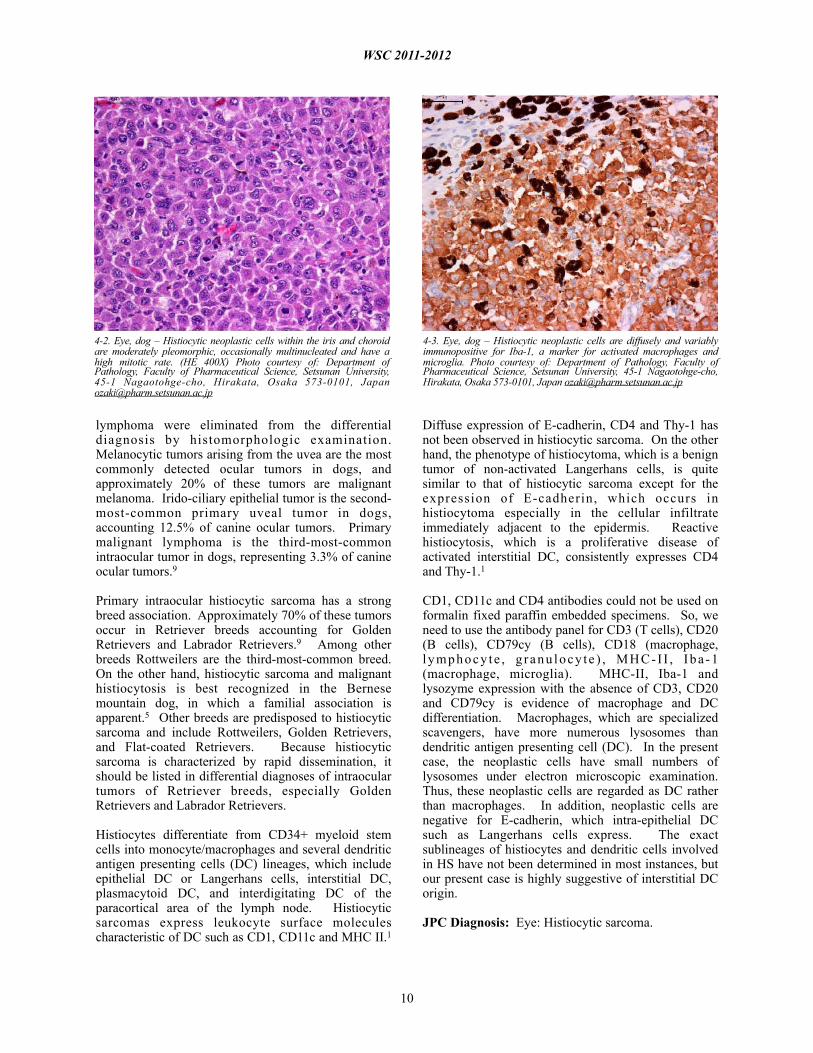
4-2. Eye, dog – Histiocytic neoplastic cells within the iris and choroid are moderately pleomorphic, occasionally multinucleated and have a high mitotic rate. (HE 400X) Photo courtesy of: Department of Pathology, Faculty of Pharmaceutical Science, Setsunan University, 45-1 Nagaotohge-cho, Hirakata, Osaka 573-0101, Japan [email protected]
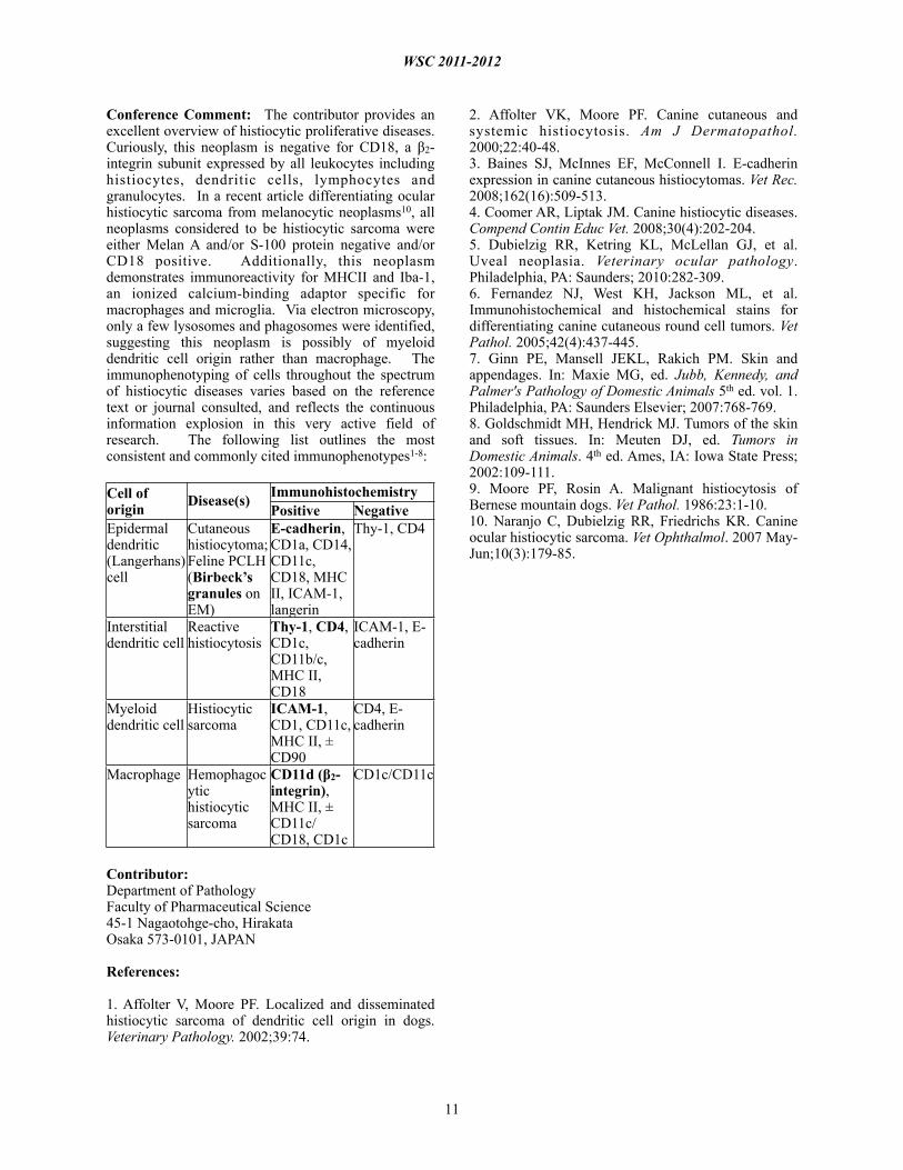
4-3. Eye, dog – Histiocytic neoplastic cells are diffusely and variably immunopositive for Iba-1, a marker for activated macrophages and microglia. Photo courtesy of: Department of Pathology, Faculty of Pharmaceutical Science, Setsunan University, 45-1 Nagaotohge-cho, Hirakata, Osaka 573-0101, Japan [email protected]
Conference Comment: The contributor provides an excellent overview of histiocytic proliferative diseases. Curiously, this neoplasm is negative for CD18, a β2-integrin subunit expressed by all leukocytes including histiocytes, dendritic cells, lymphocytes and granulocytes. In a recent article differentiating ocular histiocytic sarcoma from melanocytic neoplasms10, all neoplasms considered to be histiocytic sarcoma were either Melan A and/or S-100 protein negative and/or CD18 positive. Additionally, this neoplasm demonstrates immunoreactivity for MHCII and Iba-1, an ionized calcium-binding adaptor specific for macrophages and microglia. Via electron microscopy, only a few lysosomes and phagosomes were identified, suggesting this neoplasm is possibly of myeloid dendritic cell origin rather than macrophage. The immunophenotyping of cells throughout the spectrum of histiocytic diseases varies based on the reference text or journal consulted, and reflects the continuous information explosion in this very active field of research. The following list outlines the most consistent and commonly cited immunophenotypes1-8:
Contributor: Department of PathologyFaculty of Pharmaceutical Science45-1 Nagaotohge-cho, HirakataOsaka 573-0101, JAPAN
References:
1. Affolter V, Moore PF. Localized and disseminated histiocytic sarcoma of dendritic cell origin in dogs. Veterinary Pathology. 2002;39:74.
2. Affolter VK, Moore PF. Canine cutaneous and systemic histiocytosis. Am J Dermatopathol. 2000;22:40-48.3. Baines SJ, McInnes EF, McConnell I. E-cadherin expression in canine cutaneous histiocytomas. Vet Rec. 2008;162(16):509-513. 4. Coomer AR, Liptak JM. Canine histiocytic diseases. Compend Contin Educ Vet. 2008;30(4):202-204.5. Dubielzig RR, Ketring KL, McLellan GJ, et al. Uveal neoplasia. Veterinary ocular pathology. Philadelphia, PA: Saunders; 2010:282-309.6. Fernandez NJ, West KH, Jackson ML, et al. Immunohistochemical and histochemical stains for differentiating canine cutaneous round cell tumors. Vet Pathol. 2005;42(4):437-445.7. Ginn PE, Mansell JEKL, Rakich PM. Skin and appendages. In: Maxie MG, ed. Jubb, Kennedy, and Palmer's Pathology of Domestic Animals 5th ed. vol. 1. Philadelphia, PA: Saunders Elsevier; 2007:768-769.8. Goldschmidt MH, Hendrick MJ. Tumors of the skin and soft tissues. In: Meuten DJ, ed. Tumors in Domestic Animals. 4th ed. Ames, IA: Iowa State Press; 2002:109-111.9. Moore PF, Rosin A. Malignant histiocytosis of Bernese mountain dogs. Vet Pathol. 1986:23:1-10.10. Naranjo C, Dubielzig RR, Friedrichs KR. Canine ocular histiocytic sarcoma. Vet Ophthalmol. 2007 May-Jun;10(3):179-85.
WSC 2011-2012
11
Cell of origin Disease(s) Immunohistochemistry
Positive NegativeEpidermal dendritic (Langerhans) cell
Cutaneous histiocytoma; Feline PCLH (Birbeck’s granules on EM)
E-cadherin, CD1a, CD14, CD11c, CD18, MHC II, ICAM-1, langerin
Thy-1, CD4
Interstitial dendritic cell
Reactive histiocytosis
Thy-1, CD4, CD1c, CD11b/c, MHC II, CD18
ICAM-1, E-cadherin
Myeloid dendritic cell
Histiocytic sarcoma
ICAM-1, CD1, CD11c, MHC II, ± CD90
CD4, E-cadherin
Macrophage Hemophagocytic histiocytic sarcoma
CD11d (β2- integrin), MHC II, ± CD11c/CD18, CD1c
CD1c/CD11c