brain histology and mouse models 2018 springmousepheno.ucsd.edu/pdfs/neu.pdf · it acts as a...
TRANSCRIPT

Brain Histology and mouse models
2018 Spring

Brain and Spinal Cord
Central Nervous System
Peripheral Nervous System Human!

Brain:
Cerebrum
Cerebellum
Brain Stem
In mice:
Olfactory lobes
Cerebrum
Cerebellum
Brain Stem

Human! Monkey! Cat! Rat!
Frog!What do each of these lobes do?!!* !Frontal Lobe- associated with reasoning, planning, parts of speech, movement, emotions, and problem solving !* !Parietal Lobe- associated with movement, orientation, recognition, perception of stimuli !* !Occipital Lobe- associated with visual processing !* !Temporal Lobe- associated with perception and recognition of auditory stimuli, memory, and speech


The organs (the "viscera") of our body, such as the heart, stomach and intestines, are regulated by a part of the nervous system called the autonomic nervous system (ANS). The ANS is part of the peripheral nervous system and it controls many organs and muscles within the body. In most situations, we are unaware of the workings of the ANS because it functions in an involuntary, reflexive manner. For example, we do not notice when blood vessels change size or when our heart beats faster. !
The ANS regulates:!• Muscles in the skin (around hair follicles; smooth muscle) !
!around blood vessels (smooth muscle), in the eye (the iris; smooth !muscle) in the stomach, intestines and bladder (smooth muscle) !!of the heart (cardiac)!
*Glands !!The ANS is divided into three parts:!* !The sympathetic nervous system !* !The parasympathetic nervous system !* !The enteric nervous system.


Axial, coronal, sagi.al
www-psych.stanford.edu/~kalina/BB
Three directional planes of the brain: rostral/caudal, dorsal/ventral, and medial / lateral. When sectioning (cutting) the brain, which planes are visible is determined by the type of section. !
In the sagittal section (which is made parallel to the midline, dorsal to ventral) the rostral/caudal and dorsal/ventral planes can be seen. In the coronal or cross section (made perpendicular to the midline, as if you're slicing a loaf of bread) the medial/lateral and dorsal/ventral planes can be seen. The image below shows the 3 different planes (axial, coronal, and sagittal) in which a brain can be sectioned:

Weights and measurements may become important
Examina6on of one set of organs is NOT sufficient
Need to analyze at least 6 sets of organs
To determine if differences observed are sta6s6cally significant
We generally examine organs from a
FULLY back-‐crossed set of animals, minimum of:
6 males, wild type, liKermate controls
6 females, wild type, liKermate controls
6 males, gene6cally altered
6 females, gene6cally altered
12 ini6al and then 12 more follow up


Important points to remember for mouse brain histology :
--Perfusion fixation is important to avoid artefacts, such as “dark neurons”
--Do not leave in 70% alcohol for longer than 24 hours, to avoid vacuole artefact
--both erythrocytes and degenerating neurons are autofluorescent.
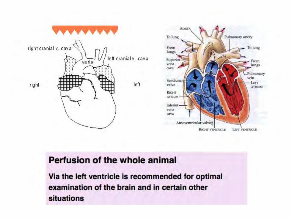
--To avoid hypostatic congestion in multiple tissues and to improve the quality of exsanguination, animals should be anesthetized as closely to the time of euthanasia as is practical.
Preparation and analysis of the central nervous system
http://tpx.sagepub.com/content/39/1/58.full

Examination of the mouse Brain
Planes of section
Cell types within the brain
Special stains
Peripheral Nervous System


Frozen tissues:
1. must be frozen using the correct conditions
2. Must be stored at minus 80 or at liquid nitrogen temperatures
3. Morphology is not the best
4. Usually better for immuno assays
Fixed tissues:
1. Must be fixed as thin slices
2. In at least 10 volumes of fixative
3. Must be processed after 24 hours of fixation if immunoassays are to be done on the processed paraffin sections
4. If fixed and processed optimally, results in the best morphology
5. Immunoassays are a little challenging

Human Brain
With gyri and sulci
Mouse Brain
lissencephalic


http://www.hms.harvard.edu/research/brain/annotations/355N.gif

What are the cells that are found in the brain?
SUPPORTING CELLS or GLIA:
--ASTROGLIA or astrocytes
--OLIGODENDROGLIA
--MICROGLIA
NEURONS
BLOOD VESSELS
MENINGEAL CELLS on the surface of the brain
EPENDYMAL CELLS line the ventricles, which make cerebrospinal fluid (CSF)
Grey matter
White matter

The meninges is the system of membranes which envelopes the central nervous system.
The meninges consist of three layers: the dura mater, the arachnoid mater, and the pia mater. The primary function of the meninges and of the cerebrospinal fluid is to protect the central nervous system.

Pathology terms:
Sub-dural hemorrhage
Sub-arachnoid hemorrhage
Meningitis
Meningioma

Meninges on the outside of paraffin sections of Mouse Brain with H&E stain

h.p://library.med.utah.edu/WebPath/CNSHTML/CNS116.html
Meningioma MRI (magneIc resonance imaging and gross appearance


The Ventricles These four spaces are filled with cerebrospinal fluid and protect the brain by cushioning it and supporting its weight. The two lateral ventricles extend across a large area of the brain. The anterior horns of these structures are located in the frontal lobes. They extend posteriorly into the parietal lobes and their inferior horns are found in the temporal lobes. The third ventricle lies between the two thalamic bodies. The massa intermedia passes through it and the hypothalamus forms its floor and part of its lateral walls. The fourth ventricle is located between the cerebellum and the pons. The four ventricles are connected to one another.

Hydrocephalus: pathologic dilatation of ventricles

Cerebrospinal fluid is a clear liquid produced within spaces in the brain called ventricles. Like saliva it is a filtrate of blood. It is also found inside the sub-arachnoid space of the meninges which surrounds both the brain and the spinal chord. In addition, a space inside the spinal cord called the central canal also contains cerebrospinal fluid. It acts as a cushion for the neuraxis, also bringing nutrients to the brain and spinal cord and removing waste from the system.

Choroid Plexus All of the ventricles contain choroid plexuses which produce cerebrospinal fluid by allowing certain components of blood to enter the ventricles. The choroid plexuses are formed by the fusion of the pia mater, the most internal layer of the meninges, and the ependyma, the lining of the ventricles.

Choroid Plexus in paraffin sections of Mouse Brain with H&E stain

What is the Blood brain barrier?
Over 100 years ago it was
discovered that if blue dye
was injected into the
bloodstream of an animal,
that tissues of the whole
body EXCEPT the brain and
spinal cord would turn blue.

The blood-brain barrier (BBB) is the specialized system of capillary endothelial cells that protects the brain from harmful substances in the blood stream, while supplying the brain with the required nutrients for proper function.
Unlike peripheral capillaries that allow relatively free exchange of substance across / between cells, the BBB strictly limits transport into the brain through both physical (tight junctions) and metabolic (enzymes) barriers.
Thus the BBB is often the rate-limiting factor in determining permeation of therapeutic drugs into the brain.
http://users.ahsc.arizona.edu/davis/bbb.htm

The blood-‐brain barrier acts very effecIvely to
protect the brain from many common bacterial
infecIons. Thus, infecIons of the brain are very
rare. However, since anIbodies are too large to
cross the blood-‐brain barrier, infecIons of the
brain which do occur are oOen very serious and
difficult to treat.
The blood brain barrier becomes more permeable
during inflammaIon however, meaning some
anIbioIcs can get across. Viruses easily bypass the
blood-‐brain barrier by a.aching themselves to
circulaIng immune cells.
h.p://faculty.washington.edu/chudler/bbb.html


COMMUNICATING: or Gap junctions: in Cardiac and Smooth muscle cells
Cell Junctions
Adherent junctions and focal contacts, link with the actin filament network;
Desmosomes and hemidesmosomes, link with the intermediate filament network
Desmosomes: provide mechanical stability in
squamous epithelial cells. Example: E-‐cadherin Hemidesmosomes: anchor
cells to basement membrane
ANCHORING
OCCLUDING: Also known as tight junctions


Functions of the BBB The BBB has several important functions: 1.Protects the brain from "foreign substances" in the blood that may injure the brain. 2.Protects the brain from hormones and neurotransmitters in the rest of the body. 3.Maintains a constant environment for the brain. General Properties of the BBB 1.Large molecules do not pass through the BBB easily. 2.Low lipid (fat) soluble molecules do notpenetrate into the brain. However, lipid soluble molecules, such as barbituate drugs, rapidly cross through into the brain. 3.Molecules that have a high electrical charge to them are slowed. The BBB can be broken down by: 1.Hypertension (high blood pressure): high blood pressure opens the BBB 2.Development: the BBB is not fully formed at birth. 3.Hyperosmolitity: a high concentration of a substance in the blood can open the BBB. 4.Microwaves: exposure to microwaves can open the BBB. 5.Radiation: exposure to radiation can open the BBB. 6.Infection: exposure to infectious agents can open the BBB. 7.Trauma, Ischemia, Inflammation, Pressure: injury to the brain can open the BBB
http://faculty.washington.edu/chudler/introb.html

Nissl stain (e.g., cresyl violet, thionin, azure) stains nuclei acids (DNA and RNA). This stain is useful for viewing cell sizes and numbers.

http://www.neuroscienceassociates.com/Stains/weil_myelin.htm
Myelin stain of Human brain

Markers for CELL TYPES FOUND IN THE BRAIN AND SPINAL CORD!--NEURONS (NeuN)
--GLIA: 3 types of supporting GLIA:
Astrocytes: the principle cells that respond in a non-specific way
to injuries of the nervous system. --marker: GFAP
Oligodendroglia: major function: to produce myelin. marker: MBP
Microglia: are members of the mononuclear phagocyte system. Marker: CD68
--EPENDYMAL cells--- lining the ventricles and the choroid plexus,
(which makes CSF)
--MENINGEAL cells ---on the outside of the brain
--ENDOTHELIAL cells (CD31)
--SCHWANN cells--peripheral nervous system

Neuronal nuclei identified using anti-NeuN (brown)

Arbitrary layers of the cerebral cortex


Cerebellum histology


Astrocytes identified using anti-GFAP (glial fibrillary acidic protein)
Astrocytoma – benign tumor Glioblastoma multiforme--malignant

Cells that are responsible for Myelin production
Oligodendroglia: in the central nervous system
Schwann cells: in the peripheral nervous system
Histochemical stain: Luxol Fast Blue (LFB)
Immunohistochemistry using markers for Myelin Basic Protein (MBP)


Abnormality seen on nuclei in
the mutant hippocampus after
LFB was followed with nuclear
crystal violet stain

Comparison of different stains on mouse brain sections

Microglia: macrophages in the brain
Microglia may be identified using: anti-CD-68 (frozen or paraffin sections)
or anti-Iba1 (paraffin sections) with spleen control

Double label immunofluorescence to show co-localization with macrophage marker

X63 (neg) 4C4 (Siglec11) CD68(macrophages)
Chi
mp
B
rain
Hum
an B
rain

4C
4
C
D68
Hu Brain Alzheimer’s HuBrainHIVencephalopathy
Increased levels of microglia and clustering

Co-localization and detection of similar epitopes on the same tissue section, using fluorescent markers!

Neuroectoderm: after recruitment from the
Ectoderm, this differentiates into the brain
and spinal cord and cells from the neural
crest migrate to:
Skin—melanocytes
Neuroendocrine cells
Adrenal medulla
Retina

Melanocytes in basal layer of skin revealed with a silver stain
The difference in skin color between people of different pigmentation: number (quantity) of melanocytes is the same, but activity level is different (quantity and relative amounts of eumelanin and pheomelanin). This process is under hormonal control, including the MSH and ACTH peptides that are produced from the precursor proopiomelanocortin. Tyrosinase, is required for melanocytes to produce melanin from the amino acid tyrosine.


Abn
orm
al re
tina
in m
utan
t C
ontro
l

Implanted stem cells forming a teratoma—made up of cells from the 3 different compartments of embryonic tissue
Ectoderm (includes neuroectoderm) Endoderm Mesoderm

Teratoma

Teratoma

Although we usually study the spinal cord as a series of cross sections, it is important to remember that it is in fact a column, with continuous tracts and cell columns.

http://www.pathology.vcu.edu/WirSelfInst/glialpath.html
In the peripheral nervous system there are NO glia. There are Schwann cells which share one property in common with oligodendroglia, namely the production of myelin. Interestingly, the Schwann cells also become phagocytes, devouring the debris from injured peripheral nerves, and this property is not shared by the oligodendroglia

In the figures, note the differences in the shape and size of the spinal cord at different levels. The dark gray color in each segment represents "gray matter." If you use your imagination, you can see that the gray matter looks similar to an H or a butterfly. Nerve cell bodies are located in the gray matter. Surrounding the gray matter is white matter (lighter color shading) - this is where the axons of the spinal cord are located.
Only the ventral roots are coming out of the cord - the dorsal roots are actually going in. Throughout the cord, the dorsal grey matter (dorsal horns) deals with sensory perception, and receives information from the periphery through the dorsal root. The ventral horns contain the α-motor neurons, whose axons exit the cord via the ventral roots and travel directly to the muscles.


Spinal cord cross-sections to compare and determine if there are inflammatory infiltrates in the “treated” animal.!
H&E x40

Metabolism changes and Behavioral analyses in mice
Metabolism and Behavior Core The core performs a battery of tests on the live animals over a period of weeks. The tests include analysis and interpretation of: 3D Activity Levels, Food & water Consumption, Oxygen consumption, Carbon Dioxide Production, Circadian Rhythm changes, Measurements of Heart rate, Blood Pressure, Pulmonary Function, Fear Conditioning. Neurological Screen including Eyeblink, Tail Suspension, Rotorod tests, Balance Beam Stability and Wire Hang, Pole Test, Open Field, Tail Flick, Hot Plate Test, tests of Hearing, Prepulse Inhibition of Startle, tests for Social Dominance, Approaching Object, and Passive Avoidance.


Use labeled cassettes, to fix thin slices of organs or rolls of intestine, for at least 24 hours, before transferring to 70% alcohol, for processing into paraffin blocks
Do not leave brain samples in 70% alcohol for longer than 24 hours to avoid dehydration artefact in paraffin sections


Design controls for secondary and/or terIary reagents
A. Reagent controls should include: 1. Slide that receives diluIng buffer alone 2. IgG control at the same concentraIon as the test anIbody 3. PosiIve control reagent, same species as primary being tested
B. Tissue / or cells controls should include: 1. Tissue or cells not expected to be posiIve 2. Tissue or cells expected to be posiIve 3. Blocking reagent to delete posiIve reacIon, to demonstrate specific binding by the test anIbody

Tissue secIon: Frozen or Paraffin embedded
Unfixed or Fixed-‐
Acetone, Paraformaldehyde, . Deparaffinize
AnIgen retrieval
No anIgen retrieval
When tes6ng a new an6body, one needs to know:
Species of origin of the primary an6body: mouse, rabbit, rat, hamster, chicken, goat, sheep, horse……...
Primary an6bodies may be Polyclonal (rabbit, sheep, goat,chicken) Or Monoclonal (rat, mouse)
In order to : -‐-‐-‐-‐ design what cells or 6ssues will be used as posi6ve and nega6ve controls
-‐-‐-‐design secondary and ter6ary detec6ng reagents
-‐-‐-‐-‐design reagents to block non-‐specific binding

Primary anIbody : maybe used, already labeled with fluoresceinated compounds or with an enzyme label
Tissue secIon: Frozen or deParaffinized
Unbound an6body washed off before applica6on of secondary reagent
Block nonspecific binding sites before adding primary an6body / reagent
At each step of the immuno assay , if using new reagents,
one needs to determine the op6mum working dilu6on

Block Non-‐Specific Binding sites in 6ssues, because of the large variety of cells present
-‐-‐Block non-‐specific binding to extra cellular matrix components, usually use bovine serum albumin
if using HRP conjugates Block endogenous peroxidases in RBCs present in all 6ssues
If using alkaline phosphatase conjugates , endogenous alkaline phosphatase in 6ssues will contribute to annoying background binding eg: within sec6ons of frozen intes6ne , Bone marrow, placenta. This may be removed using heat or O.1M glycine
if using bio6nylated secondary reagents Block endogenous bioIn There is endogenous bio6n in most 6ssues other than spleen or thymus
-‐-‐Treat one set with block to prevent binding of first reagent

Tissue secIon: Frozen or deParaffinized
Secondary reagent,:
Unbound reagent is washed off before applicaIon of next reagent
Dilute secondary reagent with normal serum of species being tested to block nonspecific binding of secondary
may be used already labelled with fluoresceinated compounds or with an enzyme label OR it may be conjugated with Bio6n, digoxygenin, etc.

Tissue secIon: Frozen or deParaffinized
Ter6ary reagent is used usually labeled with :
fluoresceinated compounds
or with an enzyme
Remove endogenous binding sites in 6ssue to prevent nonspecific binding
Wash off unbound ter6ary before adding substrate or before moun6ng to view or before further amplifica6on
CY2 , FITC
AMCA
PE, CY3
HRP Alk.Phos
DAB, AEC, red , SG, VIP Blue, Red (also fluoresces)
Primary
Secondary
Ter6ary

Using double stain methods to determine if two an6bodies
recognize the same epitope on cells in a 6ssue sec6on
Tissue sec6on: Frozen or deParaffinized
One an6body may bind with higher affinity
or recognize an epitope within cells that are more abundant
The second an6body may bind to the same epitope
within cells that are less abundant
May use an6bodies sequen6ally and detect each with different fluoresceinated tags so that if the same epitope is recognized a new color will be visualized
Need to use lower dilu6ons of an6bodies when doing a double stain to compensate for addi6ve effect

Bio6nyl tyramide uses the HRP enzyme
to deposit many bio6n molecules that “cover”
the an6gen an6body complexes
already formed over the epitope on the 6ssue sec6on
An example of further Amplifica6on to detect low abundance epitopes in 6ssue
Tissue sec6on: Frozen or deParaffinized
Incuba6on with more labeled streptavidin then detects the addi6onally generated bio6n,
thus enhancing detec6on levels by a factor of 100-‐1000
Wash off unbound before developing or moun6ng