wave-caipi for highly accelerated 3d imagingberkin/bilgic_2014_wave_mrm.pdfwave-caipi for highly...
TRANSCRIPT

FULL PAPER
Wave-CAIPI for Highly Accelerated 3D Imaging
Berkin Bilgic,1 Borjan A. Gagoski,2,4 Stephen F. Cauley,1 Audrey P. Fan,1,3
Jonathan R. Polimeni,1,4 P. Ellen Grant,2,4 Lawrence L. Wald,1,4,5
and Kawin Setsompop1,4*
Purpose: To introduce the wave-CAIPI (controlled aliasing inparallel imaging) acquisition and reconstruction technique for
highly accelerated 3D imaging with negligible g-factor and arti-fact penalties.Methods: The wave-CAIPI 3D acquisition involves playing
sinusoidal gy and gz gradients during the readout of each kx
encoding line while modifying the 3D phase encoding strategy
to incur interslice shifts as in 2D-CAIPI acquisitions. The result-ing acquisition spreads the aliasing evenly in all spatial direc-tions, thereby taking full advantage of 3D coil sensitivity
distribution. By expressing the voxel spreading effect as aconvolution in image space, an efficient reconstruction
scheme that does not require data gridding is proposed.Rapid acquisition and high-quality image reconstruction withwave-CAIPI is demonstrated for high-resolution magnitude
and phase imaging and quantitative susceptibility mapping.Results: Wave-CAIPI enables full-brain gradient echo acquisi-
tion at 1 mm isotropic voxel size and R¼3 � 3 accelerationwith maximum g-factors of 1.08 at 3T and 1.05 at 7T. Relativeto the other advanced Cartesian encoding strategies (2D-
CAIPI and bunched phase encoding) wave-CAIPI yields up totwo-fold reduction in maximum g-factor for nine-fold accelera-tion at both field strengths.
Conclusion: Wave-CAIPI allows highly accelerated 3D acquis-itions with low artifact and negligible g-factor penalties, and
may facilitate clinical application of high-resolution volumetricimaging. Magn Reson Med 000:000–000, 2014. VC 2014Wiley Periodicals, Inc.
Key words: parallel imaging; CAIPIRINHA, quantitative sus-
ceptibility mapping; phase imaging
INTRODUCTION
Over the last decade, parallel imaging acquisitions (1–3)through multiple receiver coils have been used ubiqui-tously to accelerate various MRI sequences. A number ofmodifications have also been proposed to improve theconditioning of parallel imaging acquisitions to enablehigher accelerations. Simultaneous multislice (SMS)acquisition involves simultaneous excitation of multipleslices and offers substantial reduction in two-dimensional (2D) imaging scan time (4–6), as it directlyreduces the amount of time needed to acquire a fixednumber of slices. Controlled aliasing in parallel imagingresults in higher acceleration (CAIPIRINHA) (7) and fur-ther improves reconstruction quality for multisliceacquisitions by modulating the phase of the simultane-ously excited slices. This modification incurs intersliceshifts in the phase encoding direction between aliasingimage slices, thereby increasing the variation in the coilsensitivity profiles across the slices to improve slicedealiasing. SMS imaging with CAIPIRINHA has beendemonstrated recently in accelerated turbo spin echo(8,9) and steady state free precession imaging (10). TheCAIPIRINHA strategy has also been successfully appliedto echo-planar trajectories (11), which allow rapid, high-resolution functional and diffusion-weighted imaging(12), arterial spin labeling (ASL) (13,14), and dynamicsusceptibility contrast imaging (15).
Application of interslice shifts to three-dimensional
(3D) imaging forms the basis of 2D-CAIPIRINHA (16),
wherein the phase (ky ) and partition (kz) encoding strat-
egy is modified to shift the spatial aliasing pattern to
reduce aliasing and better exploit the coil sensitivity var-
iation. Zhu et al. (17) showed that the staggered sam-
pling pattern in 3D k-space is be equivalent to SMS
imaging with interslice shifts. This connection between
slice-shifted 2D and 3D acquisitions was further
explored by Zahneisen et al. (18) to facilitate the recon-
struction of non-Cartesian SMS trajectories. Alternative
approaches for accelerated volumetric imaging include
bunched phase encoding (BPE) (19), where a gy gradient
is applied during the readout of each phase encoding
line to create a zigzag trajectory that can be reconstructed
using Papoulis’s generalized sampling theory to give an
alias-free image. Bunch encoding has also been com-
bined with parallel imaging (20–22) to take advantage of
the coil sensitivity variation in the readout direction to
improve the reconstruction.An emerging strategy for improved parallel imaging
quality is to impose a sparsity-inducing prior on thereconstructed image. In addition to the encoding powerof the receive coil profiles, these methods employ
1Athinoula A. Martinos Center for Biomedical Imaging, MassachusettsGeneral Hospital, Charlestown, Massachusetts, USA.2Fetal-Neonatal Neuroimaging & Developmental Science Center, BostonChildren’s Hospital, Boston, Massachusetts, USA.3Department of Electrical Engineering and Computer Science, Massachu-setts Institute of Technology, Cambridge, Massachusetts, USA.4Department of Radiology, Harvard Medical School, Boston, Massachu-setts, USA.5Harvard-MIT Health Sciences and Technology, Cambridge, Massachu-setts, USA
Additional Supporting Information may be found in the online version ofthis article.Grant sponsor: National Institutes of Health; Grant numbers:R00EB012107, P41RR14075. Grant sponsor: National Institutes of HealthBlueprint for Neuroscience; Grant number: 1U01MH093765 (Human Con-nectome Project).
*Correspondence to: Berkin Bilgic, Building 75, Room 2.102, 13th Street,Charlestown, MA, 02129, USA. [email protected]
Received 28 March 2014; revised 18 May 2014; accepted 11 June 2014
DOI 10.1002/mrm.25347Published online 00 Month 2014 in Wiley Online Library (wileyonlinelibrary.com).
Magnetic Resonance in Medicine 00:00–00 (2014)
VC 2014 Wiley Periodicals, Inc. 1
pseudo-random (rectilinear) sampling strategies thatyield incoherent aliasing artifacts that can be mitigatedvia sparsity priors (23–26). Among k-space–based meth-ods, DESIGN (24) regularizes the GRAPPA (3) resultunder wavelet transform, while L1-SPIRiT (23) seekssparse coil images in the wavelet domain that match thecalibration and acquired data. Similar to SENSE (2), thegeneral problem of reconstructing multichannel data canalso be formulated as a forward model involving coilsensitivities and a k-space sampling operator, and regu-larized with total variation penalty (25,26). A commonfeature of these multichannel compressed sensing algo-rithms is that the k-space sampling pattern is designedto satisfy compressed sensing incoherence requirements,while 2D-CAIPI and BPE modify the k-space trajectory tobetter exploit the coil sensitivity distribution by shapingthe aliasing pattern.
Development and proliferation of multichannelreceive coil arrays have resulted in a ubiquitous use ofhigh channel count systems, such as the 32-channelhead coil on commercially available scanners. Since thecoil elements in close proximity have similar sensitivityprofiles, the information provided by the elements isnot orthogonal, thereby limiting the actual degrees offreedom available. Conventional parallel imaging can-not fully use this limited degree of freedom and fails toachieve high acceleration factors. In particular, suchtechniques do not use the coil sensitivity informationpresent in the fully sampled readout dimension (x) in a3D Cartesian acquisition, and this further limits the useof spatial encoding power of the coil sensitivities to twodimensions out of the three. To address these issues,we introduce wave-CAIPI, which combines and extendsthe BPE and 2D-CAIPI strategies by playing sinusoidalgy and gz gradients simultaneously (with a p/2 phaseshift between the two waveforms) during the readout ofeach k-space line, thus creating interslice shifts by mod-ifying the k-space phase and partition encoding strat-egy. This results in a highly efficient k-space samplingpattern that spreads the aliasing evenly in all spatialdimensions (x, y, and z). Since this scheme takes fulladvantage of the spatial variation in the 3D coil sensi-tivity profiles, it enables highly accelerated volumetricimaging with low artifact and negligible signal-to-noiseratio (SNR) penalties.
Herein, we extend our initial proposal reported inabstract form (27) by increasing the spatial resolutioneight-fold (from 2 mm to 1 mm isotropic voxel size),demonstrating feasibility at ultra high field strength (7T),employing efficient iterative reconstruction to decreasethe computation time 25-fold, rapidly characterizing thewave gradients to account for the mismatch between the-oretical and experimental gradient trajectories and dem-onstrate phase and quantitative susceptibility mapsderived from a highly accelerated 3D gradient echo(GRE) acquisition.
The main contributions of this work include:
1. Voxel spreading effects of the wave gradients andthe coil sensitivity information are captured in aforward model, which divides the reconstructionproblem into small, decoupled linear systems that
are solved rapidly. By employing inverse Fouriertransform as a preconditioner to the generalizedSENSE model (28), the proposed formulationexplains the effect of wave gradients as additionalphase imparted in image domain, rather than dis-placement in k-space trajectory. This forward modelis also amenable to parallel processing for rapidreconstruction.
2. Wave-CAIPI is demonstrated to provide substantialimprovement in image quality and g-factor perform-ance relative to the SENSE, BPE, and 2D-CAIPImethods. Because of its efficient use of the variationin the sensitivity profiles in all spatial axes, wave-CAIPI yields g-factor maps close to unity even withnine-fold acceleration at 3T and 7T.
3. Wave-CAIPI is deployed to accelerate high-resolution volumetric GRE acquisition, which is anessential tool for phase imaging (29) and relatedsusceptibility weighted imaging (SWI) (30,31) andquantitative susceptibility mapping (QSM) (32–34)applications. Such acquisitions are inherently long,since they need a relatively long echo time to buildup contrast. As the proposed method is applicableto any 3D acquisition, dramatic reduction in scantime is warranted for structural imaging protocols[e.g., MPRAGE (35)] as well.
4. Raw phase data obtained from wave reconstructionare filtered with rapid phase processing algorithms(33,36,37). Starting from the resulting tissue phase,a fast susceptibility dipole inversion algorithm (38)is employed to solve for the underlying susceptibil-ity distribution x.
5. Example MATLAB code that demonstrates wavereconstruction with data acquired at 7T is offered assupplementary material, and will be also availableat: http://martinos.org/�berkin/software.html
THEORY
Effect of Wave Gradients on the Acquired Signal
Ignoring relaxation, the received baseband MRI signal stð Þ can be written in terms of the underlying magnetiza-
tion m rð Þ and the applied time-varying gradients gðtÞ as
s tð Þ ¼Zr
m rð Þexp �ig
Zt0
g tð Þrdt
0@ 1Adr [1]
For rectilinear 3D imaging with phase and partitionencoding, the received signal can be expressed using thek-space notation at fixed ky and kz as
s tð Þ ¼Z
x;y ;z
m x; y; zð Þe�i2pðkx tð Þxþky yþkzzÞdxdydz [2]
where the coordinate system is defined with respect tothe excitation box. When additional sinusoidal wave gra-dients gy and gz are played during each readout line in yand z axes (Fig. 1), the signal equation can be modifiedto yield
2 Bilgic et al.

s tð Þ ¼Z
x;y ;z
m x; y; zð Þe�i2pðkx tð Þxþky yþkzzÞ
exp �ig
Zt0
ðgy tð Þy þ gz tð ÞzÞdt
0@ 1Adxdydz
[3]
Defining g
2p
R t0 gy tð Þdt ¼ Py tð Þ and g
2p
R t0 gz tð Þdt ¼ Pz tð Þ, a
simpler expression is obtained,
s tð Þ ¼Z
x;y ;z
m x; y ; zð Þe�i2pðkx tð Þxþky yþkzzþPy tð ÞyþPzðtÞzÞdxdydz
[4]
Now taking the inverse Fourier transform over ky andkz, we switch to the hybrid space such that the acquiredreadout line at a fixed ðy ; zÞ location is expressed as
s t; y ; zð Þ ¼ e�i2pðPy tð ÞyþPzðtÞzÞZx
m x; y; zð Þe�i2pkx tð Þxdx [5]
Discretizing this expression and noting that each timepoint index corresponds to a k-space index yields
s½k; y; z� ¼ e�i2pðPy k½ �yþPz ½k�zÞX
x
m½x; y ; z�e�i2pkx=N [6]
where k represents the k-space index that enumeratesthe data points acquired per readout line and N standsfor the matrix size in this axis. Finally, taking the inversediscrete fourier transform (DFT) yields
wave ½x; y ; z� ¼X
k
ei2pkx=N
e�i2pðPy k½ �yþPz ½k�zÞX
x
m½x; y ; z�e�i2pkx=N
! [7]
This expression relates the image acquired with thewave gradients, wave ½x; y ; z�, to the underlying magnet-ization m½x; y; z�, and suggests a simple explanation forthe effect of the wave gradients: Each readout line in theunderlying image m ½x;y; z�, is convolved with a pointspread function (PSF) that depends on the spatial loca-tion ðy; zÞ to yield the acquired wave image.
This observation can be written more succinctly as
wave ½x; y ; z� ¼ F�1x Psf k; y ; z½ � Fxm x; y ; z½ �ð Þ [8]
where Fx represents the DFT operator in the x axis, andPsf k; y ; z½ � ¼ e�i2pðPy k½ �yþPz ½k�zÞ is the PSF that explains theeffect of the wave gradients. Viewed from this perspective,the forward model for wave-CAIPI is a simple multiplica-tion in k-space, or a convolution in image space. Note thatthis property is not generalizable to any arbitrary trajectory(e.g., spiral), but is applicable to cases where the phaseand partition encoding trajectories can be represented assummations of rectilinear and non-Cartesian components,so that using inverse DFT allows switching to the hybridspace where the PSF formalism can be used.
In addition to the wave gradients, the proposedmethod employs a 2D-CAIPI sampling scheme (16) thatstaggers the sampling positions in the ky -kz plane (e.g.,shifting the readouts to lie on a hexagonal grid) in orderto create interslice shifts across the aliasing slices. Thecombined effect of sinusoidal wave gradients and stag-gered sampling strategy leads to the corkscrew trajectorydepicted in Figure 1. While the acquired k-space sam-ples do not fall onto a Cartesian grid, expressing theacquisition as convolution with a PSF allows us toexplain the effect of the wave trajectory as a simple mul-tiplication in Cartesian k-space via Equation 8.
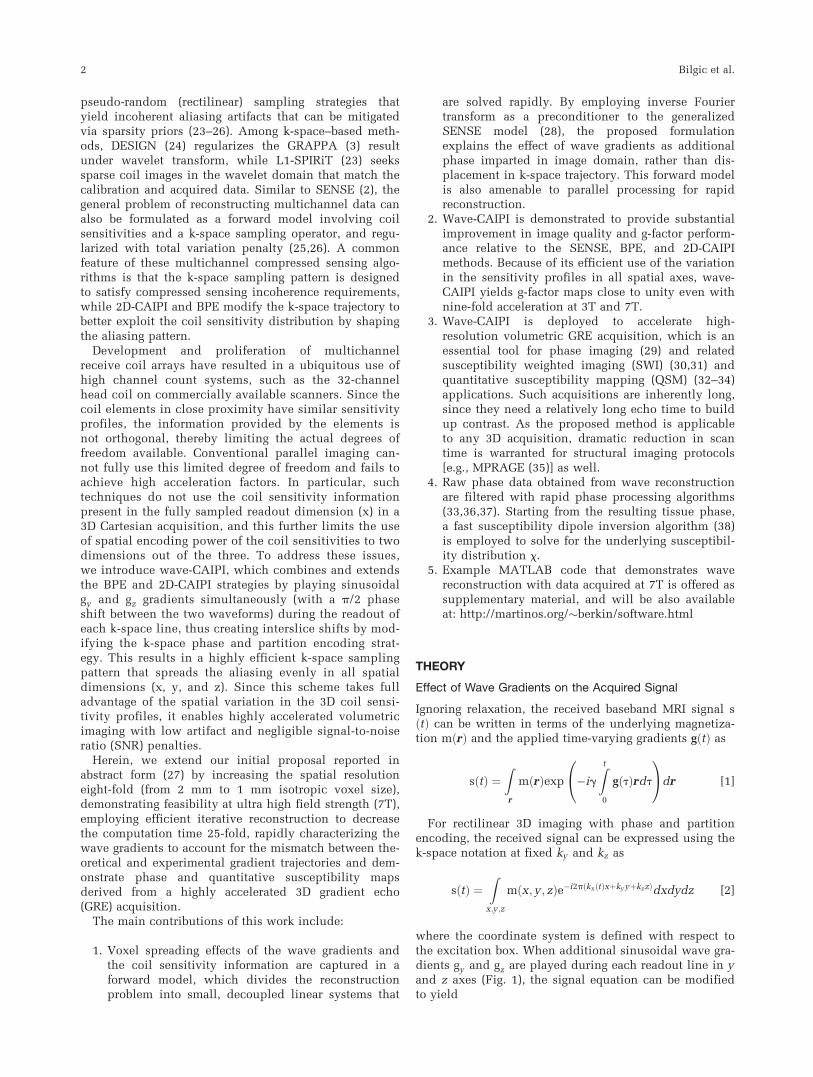
Since the underlying image is convolved with a spa-tially varying PSF, the amount of voxel spreading is afunction of ðy; zÞ coordinates. Note that the wave gra-dients gy and gz do not cause voxel spreading in the yand z directions, but the only spreading effect is alongthe x (readout) axis. This effect is demonstrated in Figure2, where the wave gradients along the y and z axes com-bined with interslice shifts give rise to spreading in allspatial directions.
Forward Model for Parallel Imaging with Wave-CAIPI
In the presence of R-fold acceleration in phase and partitionencoding, rows of image readout lines from R spatial posi-tions collapse on each other, which can be unfolded usingknowledge of the spatial encoding provided by coil sensitiv-ity profiles. This approach can be extended to the aliasinginduced by the wave sampling pattern. For simplicity, inthis example, we consider two-fold accelerated wave-CAIPIin phase encoding (ky ) direction only. In this case, twoimage locations that are half of a field of view (FOV) apartwill collapse on each other. We denote the measured signalat these locations succinctly as wave ½y1� and wave ½y2� anddrop the x and z indices. These rows of image readout linesare related to the underlying magnetization m y1½ � and m y2½ �via the convolution operations wave ½y1� ¼ F�1Psf y1½ �Fmy1½ � and wave ½y2� ¼ F�1Psf y2½ �Fm y2½ �. The forward modelthat relates the acquired data to the unknown magnetizationis then
F�1Psf y1½ �F F�1Psf y2½ �F� � m y1½ �
m y2½ �
" #¼ wave [9]
where wave ¼ wave y1½ � þwave ½y2� is the collapsedwave image due to undersampling. With the additional
FIG. 1. Gradient waveforms and k-space trajectory for wave-CAIPIimaging. Sinusoidal gy and gz gradients with a p/2 phase shiftbetween the waveforms incur a corkscrew trajectory in k-space.
The corkscrews are also staggered due to the 2D-CAIPI samplingstrategy to create interslice shifts. [Color figure can be viewed in
the online issue, which is available at wileyonlinelibrary.com.]
Wave-CAIPI for Highly Accelerated 3D Imaging 3
encoding information from n receive coil channels withsensitivity profiles Ci, this system becomes
F�1Psf y1½ �FC1 y1½ � F�1Psf y2½ �FC1 y2½ �
� �
F�1Psf y1½ �FCn y1½ � F�1Psf y2½ �FCn y2½ �
26643775 m y1½ �
m y2½ �
" #
¼
wave 1
�
wave n
26643775 [10]
Solution of Equation 10 recovers the underlying imagerows from the reduced-FOV coil images wave i. In themore general case of R-fold accelerated wave imagingwith undersampling in phase and partition encoding,the linear system in Equation 10 is modified to solve forthe set of R collapsed image rows.
Another way to view the wave acquisition is through adirect SENSE model that relates the acquired non-Cartesian k-space data to the underlying image via therelation Ei ¼ k. Here, k is the vector of k-space data, E isthe encoding matrix that includes coil sensitivities andundersampled non-Cartesian Fourier operator, and i isthe vector of the 3D image data. In this relation, theencoding matrix E is dense, and its application willrequire 3D nonuniform fast fourier transform (39). How-ever, when inverse DFT is applied to this dense matrixwithout accounting for how the wave gradients modifythe k-space trajectory, the resulting encoding becomesidentical to the PSF formulation of Equation 10. Assuch, the proposed formulation in Equation 10 employs
the inverse DFT as a preconditioner to sparsify the denseencoding, so that the 3D reconstruction problem isdecoupled into small problems that are solved independ-ently for each set of R collapsed rows.
The role of 2D-CAIPI shifting, when applied in addi-tion to the wave gradients, is to modify the rows thatwill be collapsed on each other. As such, the PSFs andcoil profiles are also shifted during reconstruction toaccount for the interslice shifts across the aliasing slices.Because a separate linear system is formed for each setof R collapsed readout lines, the reconstruction of thewhole 3D volume is highly separable and amenable toparallel processing.
The voxel spreading effect of the wave gradientsincreases the average distance across aliasing imagerows, thereby improving the variation in the coil sensi-tivity profiles of these collapsed voxels. This effect facili-tates the parallel imaging reconstruction, and is furtherexplained in Figure 3. Due to the voxel spreading effectof wave gradients in the readout direction, the data aresix-fold oversampled in kx . This extends the readoutfield of view and entirely captures the spread-out waveimages.
METHODS
Characterizing the k-Space Trajectory Traversedby Wave Gradients
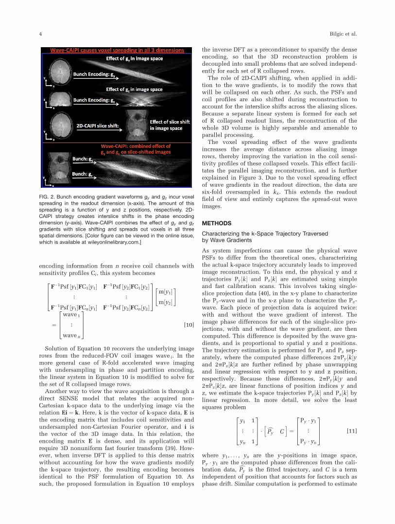
As system imperfections can cause the physical wavePSFs to differ from the theoretical ones, characterizingthe actual k-space trajectory accurately leads to improvedimage reconstruction. To this end, the physical y and ztrajectories Py ½k� and Pz½k� are estimated using simpleand fast calibration scans. This involves taking single-slice projection data (40), in the x-y plane to characterizethe Py -wave and in the x-z plane to characterize the Pz-wave. Each piece of projection data is acquired twice:with and without the wave gradient of interest. Theimage phase differences for each of the single-slice pro-jections, with and without the wave gradient, are thencomputed. This difference is deposited by the wave gra-dients, and is proportional to spatial y and z positions.The trajectory estimation is performed for Py and Pz sep-arately, where the computed phase differences 2pPy k½ �yand 2pPz k½ �z are further refined by phase unwrappingand linear regression with respect to y and z position,respectively. Because these differences, 2pPy k½ �y and2pPz k½ �z, are linear functions of position indices y andz, we estimate the k-space trajectories Py k½ � and Pz k½ � bylinear regression. In more detail, we solve the leastsquares problem
y1 1
� �
yn 1
26643775 � cPy Ch i
¼
Py � y1
�
Py � yn
26643775 [11]
where y1, . . . , yn are the y-positions in image space,Py � y1 are the computed phase differences from the cali-bration data, cPy is the fitted trajectory, and C is a termindependent of position that accounts for factors such asphase drift. Similar computation is performed to estimate
FIG. 2. Bunch encoding gradient waveforms gy and gz incur voxelspreading in the readout dimension (x-axis). The amount of this
spreading is a function of y and z positions, respectively. 2D-CAIPI strategy creates interslice shifts in the phase encoding
dimension (y-axis). Wave-CAIPI combines the effect of gy and gz
gradients with slice shifting and spreads out voxels in all threespatial dimensions. [Color figure can be viewed in the online issue,
which is available at wileyonlinelibrary.com.]
4 Bilgic et al.

cPz . The fitted trajectories cPy and cPz are combined multi-plicatively to yield the PSF estimate dPsf x; y ; z½ � ¼e�i2pðbPy k½ �yþbPz ½k�zÞ. As the PSFs depend only on the partic-ular gradient system and not the subject that is beingscanned, gradient characterization can also be performedon a phantom, independently of the in vivo acquisition.While Figure 1 depicts the gradient waveforms duringthe readout, the gx gradient is also pre-phased so thatduring the readout, the k-space is traversed from �kx;max
to þkx;max.
Iterative Parallel Imaging Reconstruction for Wave-CAIPI
The estimated PSFs are used in the generalized SENSEmodel (Eq. 10) to unfold the undersampled data acquiredwith wave gradients. Coil sensitivity profiles are com-puted by fitting 7th-order polynomials to low-resolutionhead array coil images normalized by the body coil algo-rithm (41), and up-sampled with cubic interpolation to ahigher resolution to match the high-resolution of theaccelerated acquisition.
In order to avoid forming the encoding matrices inEquation 10 explicitly, the linear system is formulated asa least-squares problem, and solved iteratively withMATLAB’S lsqr function (MathWorks Inc., Natick, Mas-sachusetts, USA). This involves simple point-wise opera-tions for multiplication with coil sensitivities and PSFs,and allows use of the fast fourier transform (FFT). Theiterative lsqr algorithm minimizes jjA �m�wjj22, where Arepresents the encoding matrix in Equation 10, while mand w stand for the unknown image rows and the wavecoil data in this equation. This formulation does notinvolve any regularization during reconstruction.
Data Acquisition and Reconstruction
Acquisition parameters for the in vivo experiments aresummarized in Table 1 and are detailed in the followingsections.
Comparing Wave-CAIPI with Conventional GREon Retrospectively Undersampled Acquisitions
Wave-CAIPI was implemented on the Siemens IDEA envi-ronment (Erlangen, Germany) for a whole-brain, 3D GREsequence. A healthy volunteer was scanned at 3T TimTrio and 7T Magnetom scanners, and fully sampledwave-CAIPI and conventional GRE data were acquired toserve as ground truth. The parameters common to bothdatasets were as follows: FOV¼ 224 � 224 � 120 mm3;voxel size¼1 � 1 � 2 mm3; 32 receive coils (42); maxi-mum wave gradient amplitude¼6 mT/m; maximum slewrate¼ 50 mT/m, using 7 sinusoidal wave cycles/readout.At 3T, scan time for fully sampled data was 5.7 min withpulse repetition time (TR)/echo time (TE)¼26/13.3 msand bandwidth¼ 70 Hz/pixel. At 7T, the scan time was 6min with TR/TE¼27/10.9 ms and bandwidth¼ 80 Hz/pixel. The fully sampled wave-CAIPI and conventionalGRE data were retrospectively undersampled in softwareenvironment to achieve a reduction factor of R¼ 3 � 3.Undersampled wave-CAIPI data were reconstructed withthe proposed parallel imaging technique, while the uni-formly undersampled normal GRE acquisition was recon-structed with the SENSE algorithm (2). Using the fullysampled data as ground truth, reconstruction artifact lev-els were quantified with the normalized root-mean-squareerror (RMSE) metric. G-factor maps were calculated toquantify the noise amplification incurred by wave-CAIPIand conventional GRE. The reciprocal maps, 1/g-factor,were also plotted to report the retained SNR.
Comparing Wave-CAIPI with 2D-CAIPI and BPEon Prospectively Undersampled Acquisitions
To assess the improvement in parallel imaging capabilityrelative to existing parallel imaging techniques,Wave-CAIPI was compared with 2D-CAIPI (16) and BPE(19–22). Two healthy subjects were scanned at 3T and 7Tusing the following parameters: FOV¼240 � 240 �
FIG. 3. R¼ two-fold acceleration causes voxels that are half of an FOV apart to collapse on each other. Since wave-CAIPI spreads outthe voxels in the x-axis, the aliasing voxels are further apart from each other. This increases the variation in coil sensitivity profiles and
improves parallel imaging. [Color figure can be viewed in the online issue, which is available at wileyonlinelibrary.com.]
Table 1Acquisition Parameters for the In Vivo Experiments
Resolution (mm3) Field Strength TR/TE (ms) Acceleration Acquisition Time Related Figure
Dataset 1 1 � 1 � 2 3T 26/13.3 3 � 3 Retrospective 38 s Fig. 4, left
Dataset 2 1 � 1 � 2 7T 27/10.9 3 � 3 Retrospective 40 s Fig.4, rightDataset 3 1 � 1 � 1 3T 40/17 3 � 3 Prospective 2.3 min Figs. 5 and 7
Dataset 4 1 � 1 � 1 7T 40/20 3 � 3 Prospective 2.3 min Figs. 6 and 8
Wave-CAIPI for Highly Accelerated 3D Imaging 5

120 mm3; 1 mm3 isotropic voxel size; TR¼ 40 ms;bandwidth¼ 70 Hz/pixel; 32 receive coils; maximum wavegradient amplitude¼ 6 mT/m; maximum slew rate¼ 50mT/m, using 7 sinusoidal wave cycles/readout. The echotimes were 17 ms at 3T and 20 ms at 7T. At each fieldstrength, R¼3 � 3 prospectively accelerated GRE datawere acquired using wave-CAIPI, 2D-CAIPI, and BPE sam-pling, where each acquisition took 2.3 min. Additionally,low-resolution (3 mm isotropic) GRE data at TR¼ 12 mswere acquired with body and head coils to estimate coilsensitivity profiles.
Processing of GRE Phase Images and QuantitativeSusceptibility Mapping
Raw phase images produced by wave-CAIPI, 2D-CAIPI,and BPE at 1 mm3 resolution and R¼ 3 � 3 accelerationwere processed by BET brain masking (43), Laplacianphase unwrapping (36) and variable kernel sophisticatedharmonic artifact reduction for phase data filtering (V-SHARP) for background phase removal (33,36,37). V-SHARP employed kernels of increasing diameters (3 to15 voxels) with a truncation threshold of 0.25 to yieldthe tissue phase f. The tissue susceptibility x is relatedto the phase f via the linear relation DFx ¼ Ff, whereF is the 3D DFT operator and D ¼ 1=3� k2
z=k2 is the sus-
ceptibility kernel in k-space (32,44). This kernel effec-tively undersamples the frequency content of thesusceptibility map on the conical surface 3k2
z ¼ k2,which makes the inversion of the relation ill-conditioned(32–34,45,46). Herein, regularized reconstruction withgradient smoothness prior is used for susceptibility map-ping. This entails the solution of the optimization prob-lem minjjDFx� Ffjj22 þ ljjMGxjj22, where M is a binarymask derived from the magnitude image that preventssmoothing across edges, and G is the spatial gradient
operator. This objective is minimized using a fastpreconditioned conjugate gradient solver (38,47) withl ¼ 0.03.
Effect of Wave Gradients within a Single Voxel
In the wideband acquisition (48), applying an additionalgz encoding gradient during the kx data readout causesundesirable voxel blurring/tilting artifact. The wave-CAIPI acquisition aims to avoid this artifact by usingsinusoidal gy and gz waveforms, which do not result inaccrual of gradient moments and avoids large intravoxelphase variations. Nonetheless, there are some intravoxelphase variations during the wave-CAIPI acquisition andpotential intravoxel blurring artifact must be character-ized. This potential blurring was characterized for the invivo acquisition setting (max wave gradient¼6 mT/m,max slew¼ 50 mT/m, 7 wave cycles/readout) to investi-gate the degree of voxel blurring incurred by wave-CAIPI. To this end, the image-space PSF acting on thevoxel was computed and integrated across the voxel toquantify the intravoxel spreading.
RESULTS
Comparing Wave-CAIPI with Conventional GRE onRetrospectively Undersampled Acquisitions
At 3T, nine-fold accelerated wave-CAIPI yielded 5.4%RMSE relative to the fully sampled data, whereas theerror was 10.7% for conventional GRE with uniformundersampling (Fig. 4, left). From g-factor analysis, gmax
for wave versus normal GRE was found to be 1.09 versus2.08 and gmean was 1.03 versus 1.42. At 7T, wave versusnormal GRE reconstruction errors were 7.0% and 12.0%,respectively (Fig. 4, right); gmax and gmean for wave ver-sus normal GRE were 1.06 versus 2.02 and 1.03 versus
FIG. 4. R¼3 � 3-fold retrospectively accelerated acquisitions. Wave-CAIPI and conventional GRE reconstructions are compared at 3T
(left) and 7T (right). Wave-CAIPI offers two-fold improvement in artifact power and maximum g-factor at both field strengths. [Color fig-ure can be viewed in the online issue, which is available at wileyonlinelibrary.com.]
6 Bilgic et al.

1.31, respectively. At both field strengths, wave-CAIPIyielded a two-fold reduction in maximum g-factor andimage artifact levels. For wave-CAIPI, the differenceimages at both 3T and 7T were mostly spatially unstruc-tured or “noise-like,” whereas for conventional acceler-ated acquisition, structured aliasing artifacts wereobserved, particularly in the high g-factor regions.
Comparing Wave-CAIPI with 2D-CAIPI and BPE onProspectively Undersampled Acquisitions
At 3T, Figure 5 depicts the corresponding k-space trajec-tories, reconstructions, and 1/g-factor maps for the threeacquisition/reconstruction methods: wave-CAIPI, 2D-CAIPI, and BPE. From g-factor analysis, maximum g-factor gmax for wave-CAIPI, 2D-CAIPI, and BPE werefound to be 1.08. At 7T, gmax for wave-CAIPI, 2D-CAIPI,and BPE were 1.05, 1.73, and 2.12 and gmean were 1.02,1.21, and 1.33 (Fig. 6). At both field strengths, wave-CAIPI yields nearly two-fold reduction in maximum g-factor relative to other methods tested.
Accelerated Phase Imaging and QuantitativeSusceptibility Mapping
Tissue phase images and susceptibility map solutionswith R¼3 � 3 fold acceleration and 1 mm3 isotropicresolution from wave-CAIPI, 2D-CAIPI, and BPE
reconstructions are presented in Figure 7 for 3Tacquisition and Figure 8 for 7T acquisition. The com-bination of Laplacian unwrapping and V-SHARP fil-tering took 14 s for phase processing, whereasregularized QSM solution was completed in 32 s forthe 3D volume.
Effect of Wave Gradients Within a Single Voxel
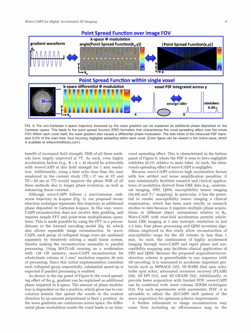
Based on wave gradient simulation inside a single voxelwith 1 mm isotropic size, the image-space PSF acting onthe voxel was found to have 0.3% side lobe amplituderelative to the main lobe (Fig. 9). As such, the intravoxelspreading effect due to wave gradients is at a negligiblelevel.
DISCUSSION
This contribution introduces wave-CAIPI, which com-bines and expands 2D-CAIPI and BPE strategies by play-ing sinusoidal gy and gz gradients during the readout ofeach phase encoding line. This results in an acquisitionstrategy that spreads the aliasing evenly in all spatialdimensions, including the fully sampled readout direc-tion. Wave-CAIPI takes full advantage of the variation inthe 3D coil sensitivity profiles and enables highly accel-erated volumetric imaging with low artifact and negligi-ble g-factor penalties.
Comparison with conventional parallel imaging revealsthat wave-CAIPI offers two-fold improvement in imageartifact level and maximum g-factor relative to normalGRE with uniform undersampling (Fig. 4). At both 3Tand 7T field strengths with nine-fold acceleration, wave-CAIPI yields close to perfect SNR retention (gmean and
FIG. 5. R¼3 � 3-fold prospectively accelerated imaging at 3T.Wave-CAIPI, 2D-CAIPI, and BPE reconstructions, 1/g-factor maps
and their respective k-space sampling patterns are demonstrated.
FIG. 6. R¼3 � 3-fold prospectively accelerated imaging at 7T.
Wave-CAIPI, 2D-CAIPI, and BPE reconstructions and 1/g-factormaps are displayed in 3 different orientations.
Wave-CAIPI for Highly Accelerated 3D Imaging 7

gmax almost unity); however, the error images relative tofully sampled data demonstrate 5%–7% RMSE. This canbe explained by the intrinsic
ffiffiffiffiRp
penalty to SNR that iscommon to all parallel imaging algorithms due to reduc-tion in the data averaging time.
Compared with the advanced parallel imaging strat-egies 2D-CAIPI and BPE, wave-CAIPI offers 1.7- and 1.8-fold improvement in maximum g-factor at 3T (Fig. 5).Residual aliasing artifacts are visible for 2D-CAIPI andBPE, while wave-CAIPI produces a high-quality, cleanimage. Reconstruction artifacts in 2D-CAIPI and BPE arealso present in the tissue phase and susceptibility maps(indicated by the arrows in Fig. 7), and noise amplifica-tion especially hampers the phase images from BPE. Incontrast, wave-CAIPI produces high-quality phase andsusceptibility maps.
At 7T, wave-CAIPI offers 1.6- and 2-fold reduction inmaximum g-factor compared with 2D-CAIPI and BPE(Fig. 6). The residual aliasing artifacts are visible for 2D-
CAIPI and BPE reconstructions, and these propagate tothe phase and susceptibility images indicated by thearrows in Figure 8.
The parallel imaging performance of all three algo-rithms is improved at 7T compared with 3T, as seen inthe g-factor maps and higher quality of the magnitudeimages (Fig. 5 vs Fig. 6). This can be explained by theclose proximity of the tight-fitting custom 7T coil (42) tothe head and the increased orthogonality across coilssensitivity profiles due to reduced wavelength at ultra-high fields. Based on these factors, the artifacts in 2D-CAIPI and BPE phase and susceptibility images appearmore subtle (enlarged detail in Fig. 8) relative to those inthe reconstructions at 3T.
As seen in 2D-CAIPI and BPE results at 3T (Fig. 5),nine-fold accelerated images have inadequate SNR dueto high g-factor penalty involved in the reconstruction.This has been mitigated by the wave-CAIPI technique,which retains close to unity g-factor penalty. With the
FIG. 7. Tissue phase and quanti-
tative susceptibility maps derivedfrom wave-CAIPI, 2D-CAIPI, andBPE at 3T and 1 mm3 isotropic
resolution are compared. Notethe artifacts indicated by the
arrows stemming from imperfectparallel imaging reconstruction.[Color figure can be viewed in
the online issue, which is avail-able at wileyonlinelibrary.com.]
FIG. 8. Tissue phase and sus-ceptibility maps from wave-CAIPI, 2D-CAIPI, and BPE
reconstructions at 7T and 1 mm3
isotropic resolution. Note theartifacts indicated by the arrows
in the susceptibility map detail.[Color figure can be viewed in
the online issue, which is avail-able at wileyonlinelibrary.com.]
8 Bilgic et al.
benefit of increased field strength, SNR of all three meth-ods have largely improved at 7T. As such, even higheracceleration factors (e.g., R¼4 � 4) should be achievablewith wave-CAIPI at this field strength for 1 mm resolu-tion. Additionally, using a later echo time than the onesemployed in the current study (TE¼ 17 ms at 3T andTE¼ 20 ms at 7T) would improve the phase SNR of allthree methods due to longer phase evolution, as well asenhancing tissue contrast.
Although wave-CAIPI follows a non-Cartesian cork-screw trajectory in k-space (Fig. 1), our proposed recon-struction technique represents this trajectory as additionalphase deposited in Cartesian k-space. In this way, wave-CAIPI reconstruction does not involve data gridding, andrequires simple FFT and point-wise multiplication opera-tions. This is made possible by using the FFT as a precon-ditioner in the forward encoding model (Eq. 8), whichalso allows separable image reconstruction. In wave-CAIPI, each group of collapsed image rows are unaliasedseparately by iteratively solving a small linear system,thereby making the reconstruction amenable to parallelprocessing. Using MATLAB running on a workstationwith 128 GB memory, wave-CAIPI reconstruction forwhole-brain volume at 1 mm3 resolution requires 30 minof processing. Since this initial implementation considerseach collapsed group sequentially, substantial speed-up isexpected if parallel processing is enabled.
As shown in the top panel of Figure 9, the voxel spread-ing effect of the gy gradient can be explained as additionalphase imparted in k-space. The amount of phase modula-tion is dependent on the y position, which gives rise to con-volution kernels that spread the voxels in the readoutdirection by an amount proportional to their y position. Asthe wave gradients are continuous across space, the differ-ential phase modulation inside the voxel leads to an intra-
voxel spreading effect. This is characterized in the bottompanel of Figure 9, where the PSF is seen to have negligiblesidelobes (0.3% relative to main lobe). As such, the intra-voxels spreading effect of wave-CAIPI is negligible.
Because wave-CAIPI achieves high acceleration factorswith low artifact and noise amplification penalties, itmay substantially facilitate research and clinical applica-tions of modalities derived from GRE data [e.g., anatomi-cal imaging, SWI, QSM, susceptibility tensor imaging(36,49) and T2* mapping]. In particular, it has the poten-tial to render susceptibility tensor imaging a clinicalexamination, which has been used strictly in researchstudies to date because it requires multiple phase acquis-itions at different object orientations relative to B0.Wave-CAIPI with nine-fold acceleration permits whole-brain GRE imaging at 1 mm isotropic resolution in just2.3 min. Fast phase processing and QSM inversion algo-rithms employed in this study allow reconstruction ofsusceptibility maps for the 3D volume in less than 1min. As such, the combination of highly acceleratedimaging through wave-CAIPI and rapid phase and sus-ceptibility mapping may facilitate clinical application ofSWI and QSM. Because the proposed acquisition/recon-struction scheme is generalizable to any sequence with3D encoding, it is warranted to accelerate important pro-tocols such as MPRAGE (35), 3D RARE (fast spin echo,turbo spin echo), attenuated inversion recovery (FLAIR)(50), 3D EPI (51), and 3D GRASE (52). Additionally, toprovide faster acquisition with limited FOV, wave-CAIPIcan be combined with inner volume ZOOM techniques(53). For such experiments with asymmetric FOV, it isadvisable to adjust the 2D-CAIPI shift pattern of thewave acquisition for optimum g-factor improvement.
A further refinement to image reconstruction maycome from including an off-resonance map in the
FIG. 9. The non-Cartesian k-space trajectory traversed by the wave gradient can be explained as additional phase deposited on theCartesian space. This leads to the point spread function (PSF) formalism that characterizes the voxel spreading effect over the whole
FOV. Within each voxel itself, the wave gradient also causes a differential phase modulation. The side lobes of the intravoxel PSF repre-sent 0.3% of the main lobe, thus incurring negligible spreading within each voxel. [Color figure can be viewed in the online issue, whichis available at wileyonlinelibrary.com.]
Wave-CAIPI for Highly Accelerated 3D Imaging 9
forward model. The voxel shift due to B0 inhomogeneityand eddy currents for wave-CAIPI is in the readoutdirection, and not more than a normal acquisition with-out the wave gradients. Accounting for the off-resonancewill further improve the image quality of wave-CAIPI bypairing the collapsed voxels with PSFs at accurate loca-tions. The actual PSFs estimation can also be improvedusing the slice-selection method proposed by Duyn et al.(40). While this is similar to current estimation scheme,it is very fast and enjoys higher SNR, which is criticalfor ultra high-resolution acquisitions.
Drawbacks and Limitations of Wave-CAIPI
Wave-CAIPI requires gradient characterization for theestimation of PSFs that are included in the forwardmodel for parallel imaging reconstruction. This requiresadditional data acquisitions, thereby increasing the totalscan time. The current PSF estimation scheme usesrapid, single-slice projections along the y and z axes.This scheme does not depend on the image contrast,since it relies on the ratio of two images with and with-out the wave gradients. As such, the shortest TR can beselected to further accelerate these calibration scans. Thetrajectory mapping depends on the particular gradientsystem being used, which is independent of the subjectbeing scanned. Therefore, the calibration data can alsobe acquired using a test phantom following the in vivoexamination.
Another limitation of wave-CAIPI is the image recon-struction computation time (30 min in MATLAB), whichis an order of magnitude longer than conventionalSENSE reconstruction. This can be mitigated by import-ing the software to the faster Cþþ platform for onlinecomputation, where parallel processing can be enabledto exploit the separable structure of wave-CAIPI recon-struction for additional speed-up.
Finally, relative sensitivity of Wave-CAIPI to motionartifacts and other factors in neuroscientific or clinicalsettings need to be evaluated.
CONCLUSION
The proposed wave-CAIPI acquisition/reconstructiontechnique involves playing sinusoidal gy and gz gradi-ent waveforms during the readout of each k-space lineand modifies the phase encoding pattern to incur inter-slice shifts across collapsing slices. This strategyspreads the aliasing in all three dimensions to allowfull use of coil sensitivity profiles and enables highlyaccelerated 3D imaging with low image artifact and neg-ligible noise amplification penalties. Compared withexisting parallel imaging techniques of SENSE, 2D-CAIPI, and BPE, wave-CAIPI demonstrates an up totwo-fold reduction in maximum g-factor at both 3T and7T. Upon nine-fold acceleration with wave-CAIPI, 3DGRE imaging achieves 1 mm isotropic voxel size in just2.3 mins with whole-brain coverage and close to perfectaccelerated SNR retention. Combined with state-of-the-art phase and susceptibility processing algorithms,wave-CAIPI may enable high-resolution phase imaging,SWI, and QSM. Its extension to increasing the resolu-tion in important 3D protocols such as MPRAGE, volu-
metric RARE, EPI, GRASE, and FLAIR may facilitatetheir clinical application.
REFERENCES
1. Sodickson D, Manning W. Simultaneous acquisition of spatial har-
monics (SMASH): fast imaging with radiofrequency coil arrays. Magn
Reson Med 1997;38:591–603.
2. Pruessmann K, Weiger M, Scheidegger MB, Boesiger P. SENSE: sensi-
tivity encoding for fast MRI. Magn Reson Med 1999;42.5:952–962.
3. Griswold M, Jakob P, Heidemann RM, Nittka M, Jellus V, Wang J,
Kiefer B, Haase A. Generalized autocalibrating partially parallel
acquisitions (GRAPPA). Magn Reson Imaging 2002;47.6:1202–1210.
4. Larkman D, Hajnal J, Herlihy AH, Coutts GA, Young IR, Ehnholm G.
Use of multicoil arrays for separation of signal from multiple slices
simultaneously excited. J Magn Reson Imaging 2001;13:313–317.
5. Moeller S, Yacoub E, Olman CA, Auerbach E, Strupp J, Harel N,
U�gurbil K. Multiband multislice GE-EPI at 7 tesla, with 16-fold accel-
eration using partial parallel imaging with application to high spatial
and temporal whole-brain fMRI. Magn Reson Med 2010;63:1144–
1153.
6. Feinberg D, Moeller S, Smith S, Auerbach E, Ramanna S, Glasser MF,
Miller K, U�gurbil K, Yacoub E. Multiplexed echo planar imaging for
sub-second whole brain FMRI and fast diffusion imaging. PLoS One
2010;5:e15710.
7. Breuer F, Blaimer M, Heidemann RM, Mueller MF, Griswold MA,
Jakob PM. Controlled aliasing in parallel imaging results in higher
acceleration (CAIPIRINHA) for multi-slice imaging. Magn Reson Med
2005;53:684–691.
8. Norris D, Koopmans PJ, Boyacio�glu R, Barth M. Power independent
of number of slices (PINS) radiofrequency pulses for low-power
simultaneous multislice excitation. Magn Reson Med 2011;66:1234–
1240.
9. Norris D, Boyacio�glu R, Schulz J, Barth M, Koopmans PJ. Application
of PINS radiofrequency pulses to reduce power deposition in RARE/
turbo spin echo imaging of the human head. Magn Reson Med 2014;
71:44–49.
10. St€ab D, Ritter C, Breuer F, Weng AM, Hahn D, K€ostler H. CAIPIRI-
NHA accelerated SSFP imaging. Magn Reson Med 2011;65:157–164.
11. Setsompop K, Gagoski B, Polimeni JR, Witzel T, Wedeen VJ, Wald
LL. Blipped-controlled aliasing in parallel imaging for simultaneous
multislice echo planar imaging with reduced g-factor penalty. Magn
Reson Med 2012;67:1210–1224.
12. Setsompop K, Cohen-Adad J, Gagoski B, Raij T, Yendiki A, Keil B,
Wedeen VJ, Wald LL. Improving diffusion MRI using simultaneous
multi-slice echo planar imaging. Neuroimage 2012;63:569–580.
13. Feinberg D, Beckett A, Chen L. Arterial spin labeling with simultane-
ous multi-slice echo planar imaging. Magn Reson Med 2013;70:1500–
1506.
14. Kim T, Shin W, Zhao T, Beall EB, Lowe MJ, Bae KT. Whole brain
perfusion measurements using arterial spin labeling with multiband
acquisition. Magn Reson Med 2013;70:1653–1661.
15. Eichner C, Jafari-Khouzani K, Cauley S, et al. Slice accelerated
gradient-echo spin-echo dynamic susceptibility contrast imaging with
blipped CAIPI for increased slice coverage. Magn Reson Med 2013.
doi: 10.1002/mrm.24960.
16. Breuer F, Blaimer M, Mueller MF, Seiberlich N, Heidemann RM,
Griswold MA, Jakob PM. Controlled aliasing in volumetric parallel
imaging (2D CAIPIRINHA). Magn Reson Med 2006;55.3:549–556.
17. Zhu K, Kerr A, Pauly J. Autocalibrating CAIPIRINHA: reformulating
CAIPIRINHA as a 3D problem. In Proceedings of the 20th Annual
Meeting of ISMRM, Melbourne, Australia, 2012. p. 518.
18. Zahneisen B, Poser BA, Ernst T, Stenger VA. Three-dimensional Fou-
rier encoding of simultaneously excited slices: generalized acquisi-
tion and reconstruction framework. Magn. Reson Med 2014;71:2071–
2081.
19. Moriguchi H, Duerk J. Bunched phase encoding (BPE): a new fast
data acquisition method in MRI. Magn Reson Med 2006;55.3:633–
648.
20. Seiberlich N, Breuer F, Ehses P, Moriguchi H, Blaimer M, Jakob PM,
Griswold MA. Using the GRAPPA operator and the generalized sam-
pling theorem to reconstruct undersampled non-Cartesian data. Magn
Reson Med 2009;61.3:705–715.
21. Moriguchi H, Sunshine J, Duerk J. Further scan time reduction of
bunched phase encoding using sensitivity encoding. In Proceedings
10 Bilgic et al.
of the 13th Annual Meeting of ISMRM, Miami Beach, Florida, USA,
2005. p. 287
22. Breuer F, Moriguchi H, Seiberlich N, Blaimer M, Jakob PM, Duerk JL,
Griswold MA. Zigzag sampling for improved parallel imaging. Magn
Reson Med 2008;60.2:474–478.
23. Lustig M, Pauly J. SPIRiT: iterative self consistent parallel imaging
reconstruction from arbitrary k space. Magn Reson Med 2010;64:457–
471.
24. Weller D, Polimeni J, Grady L, Wald LL, Adalsteinsson E, Goyal VK.
Denoising sparse images from GRAPPA using the nullspace method.
Magn Reson Med 2012;68:1176–1189.
25. Otazo R, Kim D, Axel J, Sodickson. DK. Combination of compressed
sensing and parallel imaging for highly accelerated first pass cardiac
perfusion MRI. Magn Reson Med 2010;64.3:767–776.
26. Liang D, Liu B, Wang J, Ying L. Accelerating SENSE using com-
pressed sensing. Magn Reson Med 2009;62.6:1574–1584.
27. Setsompop K, Gagoski B, Polimeni J, Wald L. Wave-CAIPIRINHA: A
Method for Reducing g-Factors in Highly Acclerated 3D Acquisitions.
In Proceedings of the 20th Annual Meeting of ISMRM, Motreal, Que-
bec, Canada, 2011. p. 478.
28. Pruessmann K, Weiger M, M., B€ornert P, Boesiger P. Advances in
sensitivity encoding with arbitrary k-space trajectories. Magn Reson
Med 2001;46:638–651.
29. Wharton S, Bowtell R. Fiber orientation-dependent white matter con-
trast in gradient echo MRI. Proc Natl Acad Sci U S A 2012;109:
18559–18564.
30. Haacke E, Xu Y, Cheng YCN, Reichenbach JR. Susceptibility
weighted imaging (SWI). Magn Reson Med 2004;52:612–618.
31. Haacke E, Tang J, Neelavalli J, Cheng Y. Susceptibility mapping as a
means to visualize veins and quantify oxygen saturation. J Magn
Reson Imaging 2010;32:663–676.
32. De Rochefort L, Liu T, Kressler B, Liu J, Spincemaille P, Lebon V,
Wu J, Wang Y. Quantitative susceptibility map reconstruction from
MR phase data using bayesian regularization: validation and applica-
tion to brain imaging. Magn Reson Med 2010;63:194–206.
33. Schweser F, Deistung A, Lehr BW, Reichenbach JR. Quantitative
imaging of intrinsic magnetic tissue properties using MRI signal
phase: an approach to in vivo brain iron metabolism? Neuroimage
2011;54:2789–807.
34. Liu T, Liu J, de Rochefort L, Spincemaille P, Khalidov I, Ledoux JR,
Wang Y. Morphology enabled dipole inversion (MEDI) from a single-
angle acquisition: comparison with COSMOS in human brain imag-
ing. Magn Reson Med 2011;66:777–783.
35. Mugler J, Brookeman J. Three-dimensional magnetization-prepared
rapid gradient-echo imaging (3D MP RAGE). Magn Reson Med 1990;
15:152–157.
36. Li W, Wu B, Avram AV, Liu C. Magnetic susceptibility anisotropy of
human brain in vivo and its molecular underpinnings. Neuroimage
2012;59:2088–2097.
37. Wu B, Li W, Guidon A, Liu C. Whole brain susceptibility mapping
using compressed sensing. Magn Reson Med 2012;67:137–147.
38. Bilgic B, Fan A, Polimeni JR, Cauley SF, Bianciardi M, Adalsteinsson
E, Wald LL, Setsompop K. Fast quantitative susceptibility mapping
with L1 regularization and automatic parameter selection. Magn
Reson Med 2013. doi: 10.1002/mrm.25029.
39. Fessler J, Sutton B. Nonuniform fast Fourier transforms using min-
max interpolation. 2003;51:560–574.
40. Duyn J, Yang Y, Frank J, van der Veen J. Simple correction method for
k-space trajectory deviations in MRI. J Magn Reson 1998;132:150–153.
41. Ying L, Sheng J. Joint image reconstruction and sensitivity estimation
in SENSE (JSENSE). Magn Reson Med 2007;57:1196–1202.
42. Keil B, Triantafyllou C, Hamm M, Wald LL. Design optimization of a
32-channel head coil at 7 T. In Proceedings of the 18th Annual Meet-
ing of ISMRM, Stockholm, Sweden, 2010. p. 1493
43. Smith S. Fast robust automated brain extraction. Hum Brain Mapp
2002;17:143–155.
44. Marques JP, Bowtell R. Application of a Fourier-based method for
rapid calculation of field inhomogeneity due to spatial variation of
magnetic susceptibility. Concepts Magn Reson Part B Magn Reson
Eng 2005;25B:65–78.
45. Liu J, Liu T, de Rochefort L, et al. Morphology enabled dipole inver-
sion for quantitative susceptibility mapping using structural consis-
tency between the magnitude image and the susceptibility map.
Neuroimage 2012;59:2560–2568.
46. Liu T, Spincemaille P, de Rochefort L, Kressler B, Wang Y. Calcula-
tion of susceptibility through multiple orientation sampling (COS-
MOS): a method for conditioning the inverse problem from measured
magnetic field map to susceptibility source image in MRI. Magn
Reson Med 2009;61:196–204.
47. Bilgic B, Chatnuntawech I, Fan AP, Setsompop K, Cauley SF, Wald
LL, Adalsteinsson E. Fast image reconstruction with L2-regulariza-
tion. J Magn Reson Imaging 2014;40:181–191.
48. Weaver J. Simultaneous multislice acquisition of MR images. Magn
Reson Med 1988;8:275–284.
49. Liu C. Susceptibility tensor imaging. Magn Reson Med 2010;63:1471–
1477.
50. Kallmes D, Hui F, Mugler J. Suppression of cerebrospinal fluid and
blood flow artifacts in FLAIR MR imaging with a single-slab three-
dimensional pulse sequence: initial experience 1. Radiology 2001;
221:251–255.
51. Poser B, Koopmans P, Witzel T, Wald L, Barth M. Three dimensional
echo-planar imaging at 7 Tesla. Neuroimage 2010;51:261–266.
52. G€unther M, Oshio K, Feinberg D. Single-shot 3D imaging techniques
improve arterial spin labeling perfusion measurements. Magn Reson
2005;54:491–498.
53. Feinberg D, Hoenninger J, Crooks L, Kaufman L, Watts JC, Arakawa.
M. Inner volume MR imaging: technical concepts and their applica-
tion. Radiology 1985;156:743–747.
Wave-CAIPI for Highly Accelerated 3D Imaging 11