viktor's notes – abscess, empyema - neurosurgery …. infection/inf5. abscess,...
TRANSCRIPT

ABSCESS, EMPYEMA Inf5 (1)
Abscess, Empyema Last updated: September 5, 2017
INTRACEREBRAL ABSCESS ....................................................................................................................... 1 ETIOPATHOPHYSIOLOGY ........................................................................................................................ 1
Predisposing conditions ......................................................................................................... 1
Sources .................................................................................................................................. 1 Etiologic agents ..................................................................................................................... 1
Pathologic Stages .................................................................................................................. 1 Location ................................................................................................................................. 2
CLINICAL FEATURES .............................................................................................................................. 3 DIAGNOSIS ............................................................................................................................................. 4
TREATMENT ........................................................................................................................................... 6
PROGNOSIS ............................................................................................................................................ 7 SPECIAL SITUATIONS ............................................................................................................................. 7
Toxoplasma gondii ................................................................................................................ 7 Aspergillosis .......................................................................................................................... 8
Candida .................................................................................................................................. 9
INTRAMEDULLARY SPINAL CORD ABSCESS ............................................................................................. 9 CLINICAL FEATURES .............................................................................................................................. 9
DIAGNOSIS ............................................................................................................................................. 9 TREATMENT ........................................................................................................................................... 9
PROGNOSIS ............................................................................................................................................ 9
SUBDURAL EMPYEMA (CRANIAL AND SPINAL) ..................................................................................... 10 ETIOPATHOPHYSIOLOGY ...................................................................................................................... 10
CLINICAL FEATURES ............................................................................................................................ 10 DIAGNOSIS ........................................................................................................................................... 10
TREATMENT ......................................................................................................................................... 10
PROGNOSIS .......................................................................................................................................... 11
CRANIAL EPIDURAL ABSCESS ................................................................................................................ 11 CLINICAL FEATURES ............................................................................................................................ 11 DIAGNOSIS ........................................................................................................................................... 11
TREATMENT ......................................................................................................................................... 11
SPINAL EPIDURAL ABSCESS .................................................................................................................... 11 ETIOPATHOPHYSIOLOGY ...................................................................................................................... 12
CLINICAL FEATURES ............................................................................................................................ 12 DIAGNOSIS ........................................................................................................................................... 12
TREATMENT ......................................................................................................................................... 13 PROGNOSIS .......................................................................................................................................... 13
INTRACEREBRAL ABSCESS
- encapsulated or free pus in brain substance.
accounts for 2% of intracranial mass lesions.
male/female ratio ≈ 2:1.
median age at presentation 30-45 years; 25% cases occur in children < 15 years!
ETIOPATHOPHYSIOLOGY
rare disease in immunocompetent individuals.
PREDISPOSING CONDITIONS
1) immunosuppression: AIDS, organ (esp. bone marrow) transplant recipients, chronic
corticosteroid therapy, neutropenia, lymphoma / leukemia.
— HIV is associated with brain abscess caused by Toxoplasma gondii, Mycobacterium
tuberculosis.
— fungi are responsible for up to 90% of cerebral abscesses among recipients of solid-
organ transplants.
2) congenital heart disease (with right-to-left shunt), pulmonary A-V fistulas - infected
venous blood bypasses pulmonary filter (gains access to cerebral arterial system).
3) IV drug abuse.
SOURCES
1) infectious focus elsewhere:
a) direct osteomyelitic spread or retrograde septic thrombophlebitis from
CONTIGUOUS CRANIAL SITE (40-50%): otitis media (pediatric or older adult
populations), sinusitis (young adults), mastoiditis, odontogenic infections, facial /
scalp infections, meningitis (rare*).
*brain abscess in child < 2 years suggests associated bacillary meningitis
b) hematogenous spread from REMOTE INFECTION SITE (30%): pulmonary
infection !!! (bronchiectasis, lung abscess), endocarditis, osteomyelitis, IV
injection with unsterile syringe / drug.
2) penetrating cranial trauma (esp. gunshot and retained bone fragments).
3) neurosurgical procedures (6-7 per 10,000 clean neurosurgical procedures).
ETIOLOGIC AGENTS
(reflect primary infective process and immune state of host).
30-60% abscesses are mixed infections!
1) streptococci (esp. Streptococcus intermedius group – S. anginosus, S. constellatus, S. milleri)
are identified in 50-70% brain abscesses.
2) anaerobic bacteria (predominantly Bacteroides species) are common in chronic otitis media or
pulmonary disease.
3) Staphylococcus aureus and Gr- rods are common after cranial penetration from surgery or
trauma.
N.B. pneumococci, meningococci, Haemophilus influenzae (major causes of bacterial
meningitis) are rarely recovered from brain abscess!
4) fungi are common in immunosuppressed:
Aspergillus fumigatus – after organ transplantation, granulocytopenia.
Candida – in chronic corticosteroid therapy, granulocytopenia, after bone marrow
transplantation, IV drug abusers.
Zygomycetes (mucormycosis) – granulocytopenia, IV drug abusers.
5) parasites are common in immunosuppressed.
intact brain parenchyma is relatively resistant to infection - in order for brain abscesses to form,
there must be pre-existing compromised area (ischemia, necrosis, hypoxia) in brain tissue.
PATHOLOGIC STAGES
ABSCESS, EMPYEMA Inf5 (2)
Abscess formation evolves through FOUR STAGES (regardless of infecting organism):
INFLAMMATION → NECROSIS → SUPPURATION → CAPSULE
1) EARLY CEREBRITIS (days 1 to 3) - perivascular infiltration of PMNs, plasma cells, and
mononuclears; marked surrounding cerebral edema.
2) LATE CEREBRITIS (days 4 to 9) - well-formed necrotic center reaches its maximum size and
is surrounded by inflammatory infiltrate of macrophages and fibroblasts; rapid new vessel
formation around developing abscess; thin capsule (fibroblasts and reticular fibers)
gradually develops; area is surrounded by cerebral edema.
3) EARLY CAPSULE FORMATION (days 10 to 13) - necrotic center decreases in size;
inflammatory infiltrate contains increasing number of fibroblasts and macrophages; mature
collagen evolves from reticulin precursors, forming capsule that is better developed on
cortical than ventricular side of lesion.
4) LATE CAPSULE FORMATION (day 14 and later) - well-formed shrinking necrotic center
surrounded by dense collagenous capsule.
depending on etiologic organism and immunologic status, there may be delayed / incomplete
encapsulation, or abscess may enlarge more quickly.
encapsulation is more complete (more mesenchymal cells forming taugher capsule) on cortical side
(than on ventricular side) - propensity of abscesses to extend and rupture into ventricular system.
encapsulation is less extensive in hematogenous abscesses.
LOCATION
frontal = temporal = parietal > cerebellar > occipital
> brainstem, intrasellar, basal ganglia, thalamus
Otogenic abscesses – temporal lobe (adults), cerebellum (children).
Sinogenic abscesses – frontal areas.
Hematogenous spread – following characteristics:
1) multiple* brain abscesses (although solitary lesions may also occur)
2) distribution of middle cerebral artery - parietal lobe predominates (highest blood flow).
3) initial location at gray matter-white matter junction.
*another cause of multiple abscesses – immunosuppression.
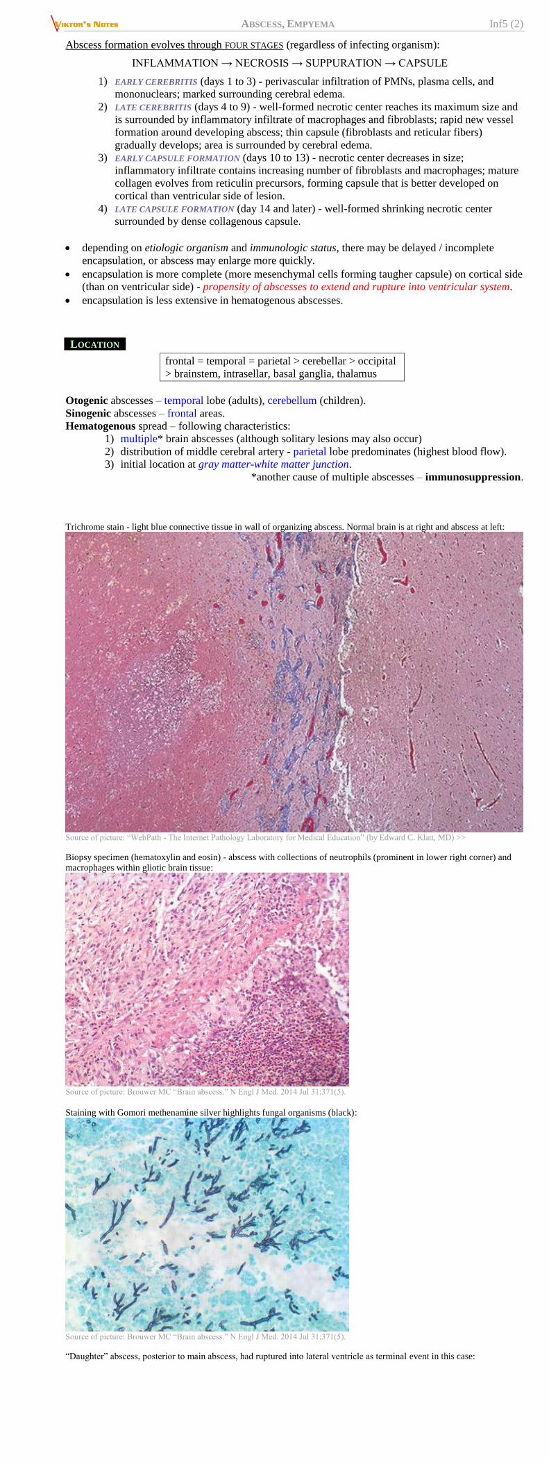
Trichrome stain - light blue connective tissue in wall of organizing abscess. Normal brain is at right and abscess at left:
Source of picture: “WebPath - The Internet Pathology Laboratory for Medical Education” (by Edward C. Klatt, MD) >>
Biopsy specimen (hematoxylin and eosin) - abscess with collections of neutrophils (prominent in lower right corner) and
macrophages within gliotic brain tissue:
Source of picture: Brouwer MC “Brain abscess.” N Engl J Med. 2014 Jul 31;371(5).
Staining with Gomori methenamine silver highlights fungal organisms (black):
Source of picture: Brouwer MC “Brain abscess.” N Engl J Med. 2014 Jul 31;371(5).
“Daughter” abscess, posterior to main abscess, had ruptured into lateral ventricle as terminal event in this case:

ABSCESS, EMPYEMA Inf5 (3)
Source of picture: “WebPath - The Internet Pathology Laboratory for Medical Education” (by Edward C. Klatt, MD) >>
Source of picture: James C.E. Underwood “General and Systematic Pathology” (1992);
Churchill Livingstone; ISBN-13: 978-0443037122 >>
CLINICAL FEATURES
- rapidly expanding infectious mass lesion (most patients have subacute course with symptoms
progressing during ≥ 2 weeks; may be indistinguishable from meningitis or encephalitis):
1) ICP↑ - prominent hemicranial or generalized headache (most common symptom! - 70-90%
patients), alterations in consciousness, vomiting, papilledema (rare finding in meningitis!).
2) focal neurological deficit (75% patients!) - seizures (focal or generalized) are particularly
prominent!

ABSCESS, EMPYEMA Inf5 (4)
3) infection – fever < 50% (i.e. may be minimal or absent!!!); nuchal rigidity is present in 25-50%
patients.
Abrupt neurologic deterioration:
a) abscess rupture into ventricular system → ventriculitis & hydrocephalus, shock & death.
b) abscess rupture into subarachnoid space → meningitis (sudden rise of CSF pressure, cell
count↑ up to 50,000/mm3, decrease in sugar content).
c) brain herniation
d) spontaneous hemorrhage
DIAGNOSIS
Lumbar puncture is contraindicated - risk of herniation!
CSF - aseptic meningeal reaction (pressure↑, 0-1000 PMNs, protein slightly↑, normal sugar)
1. Contrast-enhanced CT / MRI - low-density lesion with sharply demarcated, dense, uniform* ring
of contrast enhancement surrounded by hypodense region of edema.
*markedly irregular wall suggests tumor!
MRI is study of choice for initial detection and subsequent monitoring.
DWI has specificity 96% for differentiation from brain tumors
Cerebritis stage (MRI is superior to CT): area of hypointensity (hyperintensity on T2) with
indistinct margins and patchy contrast enhancement in periphery.
enhancing ring may appear at late cerebritis stage before true capsule has been
formed! H: DELAYED SCAN (obtained 30 min. after IV contrast) - contrast diffusion
into low-density center of abscess (vs. stage of formed true capsule - no inward
diffusion of contrast).
MRS – ↑acetate, lactate, amino acids, alanine (and ↓NAA) - highly suggestive of
cerebral abscess (but adds little value in addition to DWI)
Encapsulated stage: low T1 intensity (T2 hyperintense) lesion with diffusion restriction
surrounded by edema.
glucocorticoid use may alter appearance - only 40-60% reveal ring enhancement.
Fungal abscess in 48-year-old diabetic:
A) axial T2-MRI: central hyperintense abscess cavity with
surrounding vasogenic edema.
B) coronal post-gadolinium T1-MRI: large multiloculated abscess
cavity with enhancement of capsule and abscess wall. Note mild
mass effect + relative thinness of medial wall compared with
thicker, more irregular, lateral component.
T1-MRI with gadolinium: necrotic mass with
peripheral enhancement and surrounding edema.
Ependymal enhancement in lateral and third
ventricles (ventricular rupture, ventriculitis),
enhancement of subarachnoid space (meningitis),
mass effect with midline shift:
Diffusion restriction:

ABSCESS, EMPYEMA Inf5 (5)
Source of picture: Brouwer MC “Brain abscess.” N Engl J Med. 2014 Jul 31;371(5).
MRI of small brain abscess:
Source of picture: “WebPath - The Internet Pathology Laboratory for Medical Education” (by Edward C. Klatt, MD) >>
CT of brain abscess:
Source of picture: “WebPath - The Internet Pathology Laboratory for Medical Education” (by Edward C. Klatt, MD) >>
Abscess in right-to-left cardiac shunt:
A) CT - marked mass effect in left frontal lobe; ringlike isodense areas surrounded by low-density edema.
B) same CT after IV contrast - enhancement of periphery of multiloculated abscess cavity.
Brain abscess (MRI):
Source of picture: “WebPath - The Internet Pathology Laboratory for Medical Education” (by Edward C. Klatt, MD) >>

ABSCESS, EMPYEMA Inf5 (6)
Brain abscess (MRI):
Source of picture: “WebPath - The Internet Pathology Laboratory for Medical Education” (by Edward C. Klatt, MD) >>
2. EEG - focal slowing.
3. Etiological organism identification:
a) procedure of choice - CT- / MRI-guided stereotactic abscess aspiration (abscess is
often sterile by time of operation).
b) blood cultures (positive in ≈ 10% cases*).
c) serum should be sent for antitoxoplasma IgG (in patients with AIDS).
*when hematogenous dissemination from
remote site of infection is likely etiology
pulmonary infection is found in ≈ 10% cases (chest X-ray is mandatory for all patients!).
4. Blood – leukocytosis, ESR↑.
N.B. in significant number (≈ 40%) of patients, laboratory criteria for infection are lacking!
Distinguishing brain abscess from brain tumor:
1) C-reactive protein (CRP)↑
2) indium-111-labeled leukocyte scintigraphy (detects areas of active inflammation); false-
positive results - leukocytic infiltration into brain tumor (esp. with severe necrosis).
TREATMENT
1. Decreasing mass effect – corticosteroids (only for profound cerebral edema with impending
herniation!; may decrease penetration of antibiotics! - discontinue when edema and mass effect
improve).
2. Antimicrobial therapy: dosages → see p. Inf1 >>
antibiotics for 6-8 weeks (at least 1-2 weeks should be intravenous)
empirical therapy for AIDS patient – after results of neuroimaging (focal mass lesion without
impending herniation) and toxoplasma serology:
A) > 1 enhancing lesion OR positive toxoplasma serology = presumptive diagnosis of
TOXOPLASMA ENCEPHALITIS → start PYRIMETHAMINE (+ leucovorin) plus:
a) SULFADIAZINE – first choice
b) CLINDAMYCIN – second choice
c) ATOVAQUONE
d) AZITHROMYCIN
B) 1 enhancing lesion AND negative toxoplasma serology → brain biopsy.
N.B. rarity of toxoplasmosis in children may warrant brain biopsy
without any preceding studies.
severely ill / immunocompromised / transplant patients - MEROPENEM is first-line choice.
empirical therapy for immunocompetent patients (must cover streptococci & anaerobes):
A) PENICILLIN G 4 MMU q4h + CEFTRIAXONE 2 g q12h + METRONIDAZOLE 500 mg
q8h
B) PENICILLIN G* + METRONIDAZOLE**
*covers streptococci and anaerobes
**covers Bacteroides fragilis
C) METRONIDAZOLE + 3rd-generation cephalosporin (CEFOTAXIME, CEFTRIAXONE,
CEFTAZIDIME)* ± VANCOMYCIN OR NAFCILLIN**
*cover Enterobacteriaceae (e.g. otitic origin)
**cover Staphylococcus aureus (e.g. after cranial trauma,
neurosurgery, endocarditis)
Empiric treatment of children: CEFTRIAXONE/CEFOTAXIME + METRONIDAZOLE
D) one of penicillins + CHLORAMPHENICOL
neurosurgical patient: VANCOMYCIN + CEFEPIME + METRONIDAZOLE
FUNGAL abscesses - AMPHOTERICIN B, VORICONAZOLE.
NOCARDIA abscesses - TRIMETHOPRIM–SULFAMETHOXAZOLE or SULFADIAZINE.
Response to antibiotics is best monitored by serial CT / MRI
– abscess healing is indicated by decrease in its size.
– failure to demonstrate abscess shrinkage in 4 weeks constitutes antibiotic failure →
surgery.
– antibiotics must be continued until abscess cavity resolves completely (usually 6-8
weeks).
N.B. ring enhancement may persist for up to 9 months after cure.
3. Draining pus + taking material for culture (unless contraindicated because of suspected organism
type or patient’s clinical condition)
Even lesions with thick, well-developed ring enhancement on CT
may disappear with medical management!

ABSCESS, EMPYEMA Inf5 (7)
a) stereotactic abscess aspiration - procedure of choice; requirement - abscess > 1 cm showing
central cavity* - lower morbidity
*aspiration during cerebritis stage has unacceptable risk of hemorrhage
(esp. in children).
– leaving continuous drainage catheter is not recommended.
– if organism is known, indications for just decompression:
1) abscess close to ventricles (risk of catastrophic rupture → ventriculitits →
hydrocephalus)
2) significant mass effect (mostly if abscess > 2.5 cm)
b) complete abscess extirpation - rapid decompression; may cause damage to brain
parenchyma (→ risk of seizures); indications:
1) gas within abscess cavity
2) fungi , tbc, branching bacteria(esp. Actinomyces, Nocardia species)
3) single large (> 3 cm) & readily accessible abscess
4) abscess in posterior fossa (potential of brain stem compression)
5) retained foreign bodies (incl. bone fragments)
PROGNOSIS
CT diagnosis has been responsible for modern marked reduction in morbidity / mortality
Mortality 5-20% (if untreated ≈ 100%).
Sequelae:
1) seizure disorder (80-90% patients!) - prophylactic PHENYTOIN should be given for at
least 1 year to all patients!
2) focal motor or sensory deficits
3) behavior and learning problems
4) recurrence of abscesses
SPECIAL SITUATIONS
TOXOPLASMA GONDII
- cause of majority of focal infectious CNS lesions in AIDS patients.
Most difficult differential diagnosis is from lymphoma!
persists in CNS and eye (immunologically privileged sites) → meningoencephalitis &
chorioretinitis.
lesions typically located in:
1) cerebral cortex near gray-white junction
2) thalamus and basal ganglia
3) less often - cerebellum and brain stem; rarely - spinal cord.
Toxoplasma abscess:
Source of picture: “WebPath - The Internet Pathology Laboratory for Medical Education” (by Edward C. Klatt, MD) >>
Brain biopsy - Toxoplasma gondii cysts in microglial nodule with variety of inflammatory cell types (patient with
AIDS):
Source of picture: “WebPath - The Internet Pathology Laboratory for Medical Education” (by Edward C. Klatt, MD) >>
Clinically – acute ÷ chronic meningoencephalitis with FOCAL FEATURES (multiabscesses).
Diagnosis:
1) positive serology - antitoxoplasma IgG in serum (only indicates exposure, but not active
infection).
2) contrast neuroimaging (MRI is superior to CT) - like pyogenic abscesses or lymphoma:
multiple lesions enhance in ringed or diffuse pattern; relatively small (1–4 cm); surrounded by
edema.
– thallium SPECT (± CSF PCR for EBV) - distinguishing toxoplasmosis from
primary CNS lymphoma (focal increased uptake is seen in lymphoma) - similar
CT/MRI appearance.
– cerebral calcifications are not found in postnatally acquired infections!
3) CSF - protein↑, mononuclear pleocytosis (< 100 /mm3 ), glucose normal or ↓.
– presence of CSF antibodies may be sensitive indicator of CNS infection.
– PCR - disappointing (96–100% specificity, but only 50% sensitivity).
– Toxoplasma can be demonstrated in CSF sediment (with Wright or Giemsa stain or
organism can be cultivated).
4) DEFINITIVE DIAGNOSIS:

ABSCESS, EMPYEMA Inf5 (8)
a) 1-2 week trial of antitoxoplasma therapy (objective response must be seen
on imaging; small lesions may disappear completely in matter of weeks).
b) brain biopsy - organism detection (both free tachyzoites and encysted
bradyzoites may be found at periphery of necrotic foci).
Typical toxoplasma abscesses and response to treatment (T2- A,C,D; T1- B).
A. Multiple masses of varying sizes with propensity to involve basal ganglia and grey–white matter junction; perilesional
edema.
B. High signal on T1-MRI due to hemorrhage.
C,D. Response to toxoplasma therapy - reduced size of lesions and surrounding edema; responding lesions may show
increased intensity and surrounding low signal rim due to hemosiderin (arrow).
T2-MRI: solitary toxoplasma abscess (A) is indistinguishable from solitary primary cerebral lymphoma (B):
Enhancement in toxoplasma abscess (T2- A; T1- B): irregular rim enhancement is frequent; perilesional edema but not
mass itself is involving corpus callosum:
Treatment – for at least 6 weeks. see above >>
for mass effect – corticosteroids (discontinue as soon as possible).
Prognosis
relapses occur in 50% AIDS patients and 15-25% non-AIDS patients.
large lesions (reduce in size and have less surrounding edema) may continue to enhance for > 2
years.
primary & secondary prophylaxis in HIV-infected patients → see p. 269 >>
ASPERGILLOSIS
- immunosuppressed* patient with unremitting fever.
*Aspergillus causes 50% brain abscesses after bone marrow transplantation
ANGIOINVASIVE: multiple thrombotic infarctions / SAHs from ruptured mycotic aneurysms →
multiple brain abscesses (in major vascular territories).
radiologically similar to pyogenic abscesses.
Branching hyphae of Aspergillus invading cerebral vessel:
Source of picture: “WebPath - The Internet Pathology Laboratory for Medical Education” (by Edward C. Klatt, MD) >>
Bilateral infarction and hemorrhage (in territories of lenticulostriate perforating arteries) caused by angioinvasive
Aspergillus:

ABSCESS, EMPYEMA Inf5 (9)
chest X-ray - pulmonary infiltrates; bronchoscopy may identify infecting organism in some cases.
rapid diagnosis – Aspergillus antigen test in blood.
treatment - liposomal AMPHOTERICIN B (0.8-1.0 mg/kg/d) + FLUCYTOSINE (25 mg/kg
q6h).
New drugs for invasive aspergillosis:
VORICONAZOLE (loading 6 mg/kg IV q12h for two doses → maintenance 4 mg/kg IV
q12h).
CASPOFUNGIN (70 mg IV over 60 min single loading dose on day 1 → 50 mg/d IV).
CANDIDA
- see p. Inf1 >>
INTRAMEDULLARY SPINAL CORD ABSCESS
Only < 100 cases have been reported since 1830:
– males > females.
– PEAK INCIDENCE in 1st and 3rd decades of life.
– particular high risk factor – IV drug abuse.
– most common etiology: Staphylococcus and Streptococcus species, followed by Gr-
organisms.
solitary abscesses most likely appear in thoracic cord.
– abscesses may occur in areas of infarction (explaining septic spread to lower half of
thoracic cord).
holocord abscesses have been reported in 5 patients.
spinal cord abscesses do not destroy fiber tracts (abscess displaces fiber tracts and spreads along
axonal pathways!).
CLINICAL FEATURES
Acute cases - similar to EPIDURAL ABSCESSES (but percussion tenderness is not noted) - extremely ill
patients presenting with:
1) symptoms of infection - acute onset of back pain, fever, chills, malaise.
2) neurological symptoms – weakness ÷ paraplegia, paresthesia, bladder and bowel
incontinence.
since inflammatory process involves surrounding vasculature, spine cord infarction
may lead to irreversible paraplegia.
Chronic cases - mimic INTRAMEDULLARY TUMOR - gradually progressing neurological symptoms
predominate over those of systemic infection.
DIAGNOSIS
Neuroimaging method of choice - gadolinium-enhanced MRI:
1) mass (homogenous spinal cord enlargement on T1-MRI but high signal intensity on T2-MRI);
2) abscess margin enhances brightly with gadolinium.
CSF (can be within normal ranges!) - protein↑, pleocytosis.
Identification of infecting organism - cultures from abscess aspirate (aerobic and anaerobic bacteria,
fungi, and tuberculosis) during laminectomy.
Myelography - only widening of spinal cord.
TREATMENT
1. Antibiotics – empirically broad-spectrum antipenicillinase penicillin; minimum 4 weeks following
surgery.
2. Steroids (DEXAMETHASONE 4-10 mg q6h during entire course of treatment) – to reduce spinal
cord swelling.
3. Surgical drainage of abscess cavity - LAMINECTOMY one level above and below abscess edges:
open dura.
identify area of spinal cord involvement (swelling, hemorrhage, distended veins).
abscess aspiration for culture & stain (Gram, India ink).
myelotomy over length of abscess.
irrigate (wound and abscess cavity) with antibiotic solution.
closure in anatomical layers.
drain is optional.
PROGNOSIS
MORTALITY 10-20%.
significant percentage of patients have abscess recurrence - repeat MRIs are essential in long-
term follow-up care (enhancement of cavity will likely continue for several weeks).
ABSCESS, EMPYEMA Inf5 (10)
SUBDURAL EMPYEMA (CRANIAL AND SPINAL)
- pus collection in space between dura mater and arachnoid.
INTRACRANIAL >> SPINAL (only 50 cases reported in literature)
13-20% of localized intracranial infections.
most common in children & young adults (70% patients are in 2-3rd decade of life).
males > females (3:1).
ETIOPATHOPHYSIOLOGY
primary causes – sinusitis (esp. frontal) 50-80%, otitis media 10-20%, superficial infections of
scalp and skull, craniotomy, meningitis (very rarely!*), suppuration of subdural hematoma.
*vs. in infants subdural empyema represents infected subdural effusion
(complicating bacterial meningitis).
– in spine: hematogenous spread from distant site (most commonly), trauma, spine surgery,
dermal sinus.
pathophysiology:
a) direct spread via erosion of bone adjacent to dura mater.
b) septic thrombophlebitis of mucosal veins (e.g. of sinuses) → retrograde extension
with drainage of bacteria into regional dural veins → superior sagittal sinus →
subdural space.
brain beneath pus is molded in manner similar to that seen in subdural hematoma.
PROGRESSION
subdural space has no barriers (hence, empyema not abscess!) - empyema evolution is remarkably
rapid (along falx and over convexities).
subdural empyema may breach arachnoid (arachnoid is not very strong barrier) → meningitis.
septic thrombophlebitis extends from dural sinuses to cortical veins → cortical venous infarction of
gray and white matter drained by thrombosed vessels → brain abscess (25% patients!).
with successful treatment, thickened dura may be only residual finding.
CLINICAL FEATURES
- patient is acutely ill (entire clinical picture may evolve in as little as few hours or as long as 10 days):
1) frontal sinusitis - periorbital edema and erythema, local pain and tenderness, etc.
2) fever, chills
3) severe headache (often localized initially to side of infection), nuchal rigidity (70-80%).
4) MASS EFFECT - progressive disturbance of consciousness, increase in infant head size with
bulging fontanel.
5) FOCAL NEUROLOGICAL DEFICITS (80-90% patients; caused by cortical vein thrombophlebitis):
seizures (30-60% patients), hemiparesis, aphasia.
Spinal subdural empyema - fever with rapidly progressive spinal cord compression.
backache is not as characteristic as in spinal epidural abscess.
tenderness along spine is often absent (vs. spinal epidural abscess).
DIAGNOSIS
marked peripheral leukocytosis.
MRI (procedure of choice) – hypodense crescent adjacent to inner border of skull or adjoining falx
with mildly (markedly on T2-MRI) increased signal intensity compared with CSF;
N.B. empyema is denser than CSF; vs. benign subdural effusion - isointense (on
MRI T1- and T2) with CSF!
– contrast enhancement of empyema margin (fine, intense line).
– underlying parenchymal edema.
– mass effect.
– empyema extent is limited by attachments of dura (way to distinguish epidural from
subdural suppurative process).
cerebral arteriography* (formerly was used routinely) should be employed on emergent basis
when MRI is unavailable and subdural empyema is strongly suspected despite normal CT -
subdural avascular mass.
*myelography for spinal empyema.
subdural tap may be diagnostic in infants.
lumbar puncture should be avoided; CSF is as in cerebral abscess (aseptic meningeal reaction):
– clear and colorless;
– neutrophilic pleocytosis may be absent;
– protein 75-150 mg/dl; sugar content is normal.
– bacteria are not found (CSF is sterile!)
TREATMENT
- surgical emergency!
anticonvulsants should be administered prophylactically.
1. Intravenous antibiotic therapy (same as that for brain abscess) – against organisms typically
isolated from chronic sinusitis / otitis:
aerobic streptococci (30-50%)
anaerobic and microaerophilic streptococci (15-25%)
staphylococci (12-25%; majority of cases of spinal subdural empyema)
aerobic Gr- bacilli (3-10%)
Empiric therapy: dosages → see p. Inf1 >>
1) PENICILLIN G OR 3rd-generation cephalosporin (CEFTRIAXONE or
CEFOTAXIME)
2) METRONIDAZOLE
3) NAFCILLIN OR VANCOMYCIN
IV for at least 3 weeks after surgical drainage → PO to complete 6-week course.
2. Management of increased ICP; use of steroids (tapered rapidly after surgery) is common but
remains controversial.
3. Immediate surgical drainage:
A. CRANIAL – via craniotomy (esp. for posterior fossa subdural empyemas) or multiple burr
holes.
drains are left in subdural space for several days.
postoperatively, repeat CT / MRI scans – reoperation (drainage of loculated pockets) is
typically necessary.
B. SPINAL – laminectomy → dural incision → drainage.

ABSCESS, EMPYEMA Inf5 (11)
although extensive antibiotic irrigation of subdural space at time of surgery (BACITRACIN +
VANCOMYCIN or GENTAMICIN) is common, there are no data on benefits of this
practice.
PROGNOSIS
mortality 10-40% (almost fatal if untreated).
in 8-46% patients chronic epilepsy results; disabling hemiparesis or aphasia (5-25% survivors).
CRANIAL EPIDURAL ABSCESS
- suppurative infection in epidural space (between dura and bone).
Epidural abscesses: SPINAL >> CRANIAL (9:1)
etiology & pathogenesis ≈ subdural empyema.
– almost always associated with overlying infection in cranial bones (e.g. penetration from
chronic sinusitis or mastoiditis; most common cause is craniotomy complicated by wound
infection).
– hematogenous spread to epidural space from remote site of infection is extremely rare (vs.
extremely common cause of spinal epidural abscess!).
rare in young children; median - 6th decade.
MORBIDITY & MORTALITY are low.
remains well localized due to tight adherence of dura to overlying calvarium; but abscess often
crosses dura along emissary veins → subdural empyema, meningitis, etc.
abscesses rarely dissect beyond base of skull.
CLINICAL FEATURES
- slowly growing mass (does not produce sudden major neurologic deficits unless complicated by deep
extension*) - insidious clinical presentation:
*subdural empyema, meningitis, intraparenchymal abscess
1) unrelenting hemicranial headache or persistent fever (patient may otherwise be
asymptomatic!!!).
2) without treatment, INTRACRANIAL HYPERTENSION and FOCAL NEUROLOGIC SIGNS ultimately
develop (when infection extends into subdural space).
DIAGNOSIS
MRI - superficial, circumscribed lenticular-shaped lesion of diminished density*, with inner rim of
contrast enhancement** (thicker and more irregular than with subdural empyema).
*but higher signal intensity (on T1- and T2) than CSF
**inflamed dura
MRI is free from bony artifacts adjacent to inner table of skull.
MRI readily differentiates abscesses from sterile effusions or chronic extraaxial hematomas.
Lumbar puncture is certainly to be discouraged until after imaging has established that significant
mass effect is not present.
modest aseptic CSF reaction.
Hyponatremia is found in 30% cases.
TREATMENT
Antibiotic therapy → see SUBDURAL EMPYEMA >>
Surgical drainage - depending on extent of lesion and involvement of overlying bone:
a) burr holes
b) craniotomy
c) craniectomy (debridement of infected bone)
dural grafting may be necessary (if dura has been breached by infection).
communications between sinus cavities and epidural space may require later surgical closure.
SPINAL EPIDURAL ABSCESS
- any infectious phlegmon involving epidural space, even without demonstrable contained pus (true
abscess).
Spinal epidural space:
TRUE space – posteriorly; AP width greatest where spinal cord is smallest (T4-8 and L3-S2).
midthoracic region (T4-8) – largest amount of epidural fatty tissue – most
common location for spinal epidural abscess!
POTENTIAL space – anteriorly (because dura is adherent to posterior surface of vertebral bodies).
anterior abscesses usually occur at cervical levels.
Can occur at all ages (60% patients are 20-50 years of age).

ABSCESS, EMPYEMA Inf5 (12)
ETIOPATHOPHYSIOLOGY
a) most common (2/3) ETIOLOGY (vs. cranial epidural abscess) - HEMATOGENOUS SPREAD from
remote site.
– immunosuppression (most commonly AIDS or diabetes mellitus) is predisposing
condition in 50% cases.
– small hematoma (mild blunt trauma) provides locus minoris resistentiae - may allow
for hematogenous seeding of infection.
– hematogenous spread to vertebral body / disc (osteomyelitis / discitis) may also occur →
subsequent extension into spinal epidural space.
b) DIRECT EXTENSION (1/3) from vertebral osteomyelitis is esp. common in IV drug abusers.
– other sources of direct extension - decubitus ulcers, infected abdominal wounds, psoas
abscesses, perinephric and retropharyngeal abscesses, iatrogenic complication of lumbar
puncture.
– tuberculous epidural abscess is usually associated with vertebral osteomyelitis.
Spinal epidural abscess extends ≈ 3 spinal segments (may extend for as long as 13 segments).
Spinal cord lesion:
gross appearance of spinal cord is usually normal.
direct compression of neural tissue & inflammatory thrombosis in intraspinal vessels →
infarction → MYELOMALACIA.
microscopically - scattered areas of softening, vacuolization of cord, areas of necrosis
(disappearance of cells, loss of myelin, axonal swelling).
CLINICAL FEATURES
1. Fever.
2. Back pain & tenderness (on percussion & movement) at affected spinal level.
erythema and swelling in area of back pain.
stiff neck and headache are common.
3. Progressive compression of spinal cord (appears within hours ÷ weeks of initial symptoms):
radicular pain ↓
paresis & loss of sensation below lesion level + bowel & bladder dysfunction
↓
complete paraplegia & loss of all sensory modalities below level of lesion
(i.e. transection syndrome)
DIAGNOSIS
MRI (procedure of choice) – abscess is isointense to CSF (hyperintense on T2).
contrast enhancement of lesion occurs.
Staphylococcus aureus spondylitis with epidural abscess in IV drug abuser:
A: AP radiograph - large, paravertebral soft-tissue mass and "extra" rib pair (arrowheads) resulting from discitis
and marked bony destruction of adjacent T7 and T8 vertebrae (appearance mimics single normal vertebra).
B: Midsagittal postgadolinium T1-MRI - diffuse vertebral enhancement and obliteration of intervening disc space
except for two residual intervertebral fluid collections (again mimicking single diffusely involved vertebral body);
large posterior epidural phlegmon and abscess (arrowheads) as well as large anterior vertebral soft-tissue mass.
C: Parasagittal MRI - involvement of two adjacent posterior elements (confirming that process represents
pronounced discitis/osteomyelitis of two levels).
Lumbar puncture (demonstrates subarachnoid block) should not be performed:
1) risk for infection spread to subarachnoid space.
2) risk for herniation from decompression below area of obstruction
if meningitis is suspected - high cervical tap is often safest approach.
Normal plain CT alone does not exclude diagnosis (if MRI is unavailable → use CT myelography).
Myelography (not necessary if abscess is diagnosed by MRI/CT) - extradural block (complete in 80%
patients)
performed by cervical puncture.
needle is advanced slowly and suction applied with syringe as epidural space is approached -
if abscess has extended to level of puncture, pus is withdrawn → terminate procedure (use pus
for culture).
X-rays show osteomyelitic findings in 1/3 cases.

ABSCESS, EMPYEMA Inf5 (13)
TREATMENT
Immediate surgery: laminectomy - decompression - drainage of epidural space.
granulation tissue is commonly found in association with epidural abscesses and may require
excision during course of decompression.
Antibiotics: 4-6 weeks IV → 2-3 months oral.
empiric antibiotics should cover:
1) S. aureus (etiological agent in majority of cases!!!) – NAFCILLIN.
2) M. tuberculosis (ISONIAZID, RIFAMPIN, ETHAMBUTOL, PYRAZINAMIDE + STREPTOMYCIN
or RIFABUTIN or CLOFAZIMINE).
3) most authorities would provide additional Gr- coverage (3rd-generation cephalosporin,
quinolone, or aminoglycoside).
PROGNOSIS
- relates inversely to amount of neurologic dysfunction at time of diagnosis:
only pain → recovery without deficit.
some weakness → 50% will achieve complete resolution.
paralysis < 36 hours' duration → < 50% will show some return of motor function.
in tuberculous epidural abscess motor recovery has been reported even after paralysis lasting
for weeks.
BIBLIOGRAPHY for ch. “Infections of Nervous System” → follow this LINK >>
Viktor’s Notes℠ for the Neurosurgery Resident
Please visit website at www.NeurosurgeryResident.net