vijaya lakshmi kanchustambham - web.stanford.edu · quantum control of molecular collisions near 1...
TRANSCRIPT
1
ZARELAB GUIDE March, 2018
Research Topic Lab Member(s) Page Quantum control of molecular collisions near 1 kelvin William Edward
Perreault Nandini Mukherjee
2
Photon catalysis of gas- and condensed-phase reactions Jana Meiser Kallie Hilsabeck
3
Abiotic production of sugar phosphates and ribonucleosides in aqueous microdroplets
Jae Kyoo Lee Jooyoun Kang
4
Microdroplet chemistry for the synthesis of gold nanostructures Jae Kyoo Lee 5
Microdroplet reactions Xin Yan Yin-Hung Lai
6
Mechanistic study of reaction acceleration and heterogeneous catalytic oil upgrading in microdroplets
Yin-Hung Lai Maria T. Dulay
7
Non-invasive diagnosis by mass spectrometry and machine learning Zhenpeng Zhou Vishnu Shankar
8
Artificial intelligence enabled innovations and discoveries in chemistry Zhenpeng Zhou 9
Investigations in human sweat & breath for new biomarker discovery Vishnu Shankar Zhenpeng Zhou
10
Electrically controlled drug release from drug-loaded polymer nanocomposite films
Christian Chamberlayne Ephraim Neumann
11
Surgical margin evaluation of clear cell renal cell carcinoma (ccRCC) using desorption electrospray ionization mass spectrometry imaging (DESI-MSI) Biochemical reactions in microdroplets from a theta tip capillary
Vijaya Lakshmi Kanchustambham
12
Mass spectrometry imaging for biomedical applications Katy Margulis 13
Design of nanoparticles for biomedical applications Katy Margulis 14
In situ measurement of ligand-receptor interactions at the cell surface by ambient ionization mass spectrometry
Sarah E. Noll 15
Monitoring of neurochemical release from PC-12 cells using ambient ionization mass spectrometry
Dan Gao Sarah E. Noll Maria T. Dulay
16
Discovery of potent inhibitors against GPAT (glycerol-3-phosphate acyltransferase) for treatment of cancer and other diseases
Feng Jin 17
In-situ ambient ionization mass spectrometry for mechanistic studies of organometallic catalysis
Katherine Walker 18
Biosensors for Detection of Pathogenic Microorganisms Maria T. Dulay 19
Composite silane-based polymers as substrate material and electrospray emitter for ambient ionization mass spectrometry
Maria T. Dulay 20

2
Quantum control of molecular collisions near 1 kelvin William Edward Perreault and Nandini Mukherjee
Measurement of vector correlations in molecular scattering is an indispensable tool for mapping out interaction
potentials. We study molecular collisions such as the rotationally inelastic scattering of deuterium hydride (HD) (v = 1, j = 2) by molecular deuterium (D2) to form HD (v = 1, j = 1),1 where v and j are the vibrational and rotational quantum numbers, respectively. HD (v = 1, j = 2) is prepared using Stark-induced adiabatic Raman passage (SARP), an optical adiabatic passage technique developed in our lab which uses two partially overlapping single-mode, nanosecond pulses to coherently transfer nearly upwards of 90% of the population in a molecular ground state to an addressable excited state.2 By changing the polarization of the laser light used to perform SARP, the orientation of the excited molecule can be controlled.3 This allows us to prepare excited HD (v = 1, j = 2) with its bond axis aligned preferentially parallel or perpendicular to the lab-fixed molecular beam axis (see Fig. 1). By coexpanding pairs of gases in a single supersonic beam, we achieve collision temperatures near 1 kelvin, restricting scattering to a few lowest partial waves, such as (l=0) s and (l=1) p. This allows us to use partial wave analysis based on the conservation of angular momentum to obtain information about the scattering potential. In our recent work on the rotationally inelastic scattering of HD (v = 1, j = 2) by D2 to form HD (v = 1, j = 1),1 the scattering angular distributions showed a dramatic stereodynamic preference (~3:1) for perpendicular versus parallel alignment as can be seen in Fig. 2. The four-vector correlation measured between the initial and final velocities and the initial and final rotational angular momentum vectors of HD provides insight into the strong anisotropic forces present in the collision process.
1. W. E. Perreault, N. Mukherjee, and R. N. Zare, Science 358, 356–359 (2017). 2. W. E. Perreault, N. Mukherjee, and R. N. Zare J. Chem. Phys. 145, 154203 (2016). 3. N. Mukherjee, W. Dong, and R. N. Zare, J. Chem. Phys. 140, 074201 (2014).
Figure 1. The quantum state of HD is prepared using SARP optical fields polarized (a) parallel, in H-SARP, and (b) perpendicular, in V-SARP, with respect to the molecular beam axis. For H-SARP, the m-state refers to the quantization Z-axis parallel to the relative velocity of HD and D2. For V-SARP, the m′ state refers to the quantization Z′ axis perpendicular to the relative velocity.
Figure 2. Collisions of state-prepared HD (v = 1, j = 2) with unprepared D2 forming HD (v = 1, j = 1), where the HD molecular bond axis was aligned (a) parallel or (b) perpendicular to the relative velocity between HD and D2. The purple dashed curve gives the time-of-flight distribution of unscattered state-prepared HD (v = 1, j = 2). In both (a) and (b), the black solid curve shows fitting using partial wave analysis. See Ref. [1] for more details.

3
Photon catalysis of gas- and condensed-phase reactions
Jana Meiser, Kallie Hilsabeck
The outcomes of a collision between two molecular species are solely determined by directions and energetics of the system - i.e., the translational, vibrational, and rotational energies of the reagents, and the interacting forces among them. In previous years, research in the Zare lab has focused on bimolecular collisions (i.e. H+H2), which have provided great model systems for investigating reactions on a single-collision scale.1-3 In addition to understanding these reactions, chemists seek to develop the skills to control their outcomes. For example, radiation has long been used to control chemical reactions through the resonant excitation of vibrations and excited electronic states. Much less well studied is the use of nonresonant radiation, supplied by an infrared laser pulse, which through its electric field can influence the course of a collision. This can occur by aligning the reactants parallel to the electric field or by inducing a Stark shift directly on the potential energy surfaces (Figure 1) or both.
Figure 1. Visual representation of second-order Stark shift on potential energy curves.
In both alignment and Stark shift, the photons do interact with the molecular system through its polarizability but are not created or destroyed. It is this process of photon catalysis that we are currently investigating in our subgroup. Our study differs from what has been done previously4-5 in that nanosecond infrared pulses, much longer than the timescale of the reaction, are used to supply the strong electric field. This presents an opportunity to determine the more general effect a nonresonant electric field has on modifying total reaction yields and is also more conducive to the extension of the effect to bimolecular reactions.
In the first of two major objectives, the effect of a nonresonant laser field on photodissociation of diatomics (e.g. deuterium iodide) and “complex” molecules (e.g. phenol), is being explored and further extended to bimolecular inelastic and reactive collisions. To fulfill the second objective, a nonresonant infrared laser field will be applied to condensed-phase systems that are transparent to the incident radiation. 1. Gao, H.; Sneha, M.; Bouakline, F.; Althorpe, S. C.; Zare, R. N., Differential Cross Sections for the H+ D2→ HD (v′= 3, j′= 4–10)+ D Reaction above the Conical Intersection. The Journal of Physical Chemistry A 2015, 119 (50), 12036-12042. 2. Jambrina, P. G.; Herráez-Aguilar, D.; Aoiz, F. J.; Sneha, M.; Jankunas, J.; Zare, R. N., Quantum interference between H+ D2 quasiclassical reaction mechanisms. Nat. Chem. 2015, 7 (8), 661-667. 3. Sneha, M.; Gao, H.; Zare, R. N.; Jambrina, P.; Menéndez, M.; Aoiz, F., Multiple scattering mechanisms causing interference effects in the differential cross sections of H+ D2→ HD (v′= 4, j′)+ D at 3.26 eV collision energy. The Journal of chemical physics 2016, 145 (2), 024308. 4. Sussman, B. J.; Townsend, D.; Ivanov, M. Y.; Stolow, A., Dynamic stark control of photochemical processes. Science 2006, 314 (5797), 278-281. 5. Corrales, M.; González-Vázquez, J.; Balerdi, G.; Solá, I.; De Nalda, R.; Bañares, L., Control of ultrafast molecular photodissociation by laser-field-induced potentials. Nat. Chem. 2014, 6 (9), 785-790.
4
Abiotic production of sugar phosphates and ribonucleosides in aqueous microdroplets Jae Kyoo Lee, Jooyoun Kang
Phosphorylation is an essential chemical reaction for life. This reaction generates fundamental cell components,
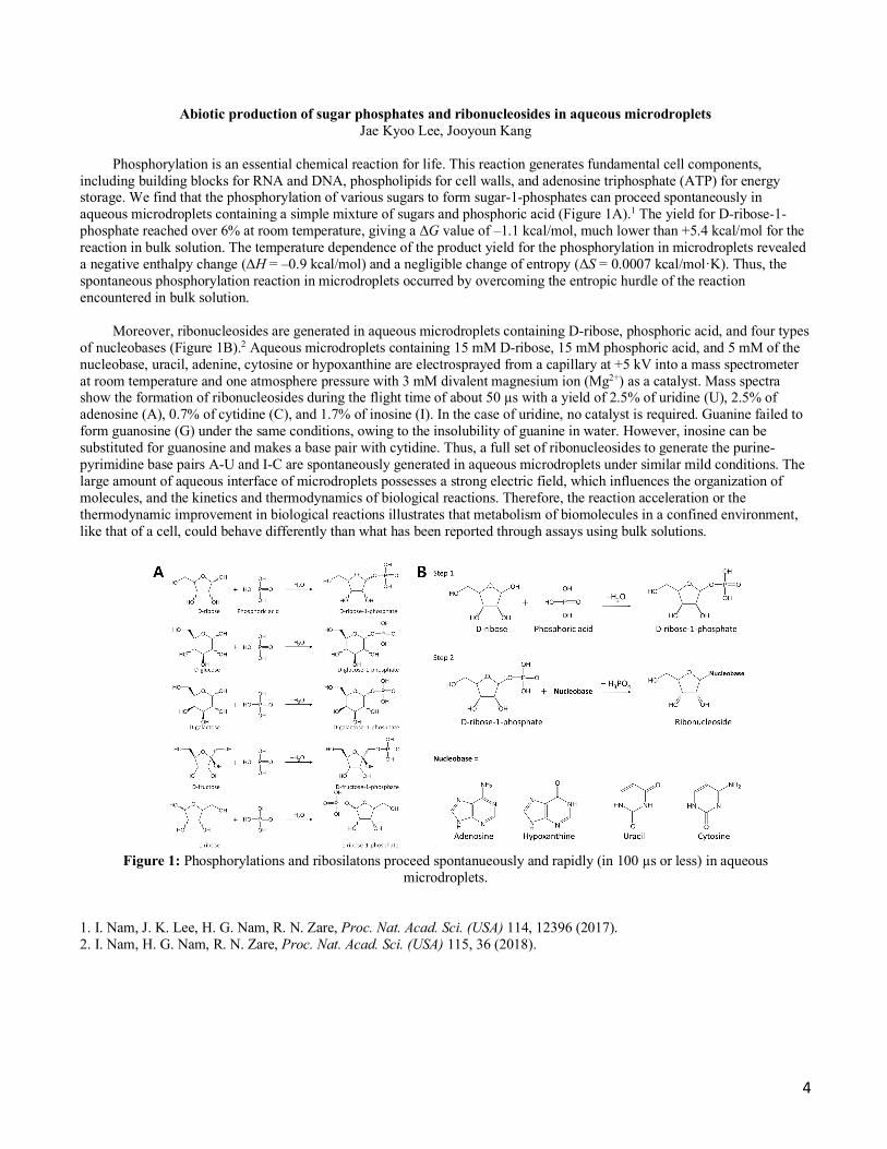
including building blocks for RNA and DNA, phospholipids for cell walls, and adenosine triphosphate (ATP) for energy storage. We find that the phosphorylation of various sugars to form sugar-1-phosphates can proceed spontaneously in aqueous microdroplets containing a simple mixture of sugars and phosphoric acid (Figure 1A).1 The yield for D-ribose-1-phosphate reached over 6% at room temperature, giving a ΔG value of –1.1 kcal/mol, much lower than +5.4 kcal/mol for the reaction in bulk solution. The temperature dependence of the product yield for the phosphorylation in microdroplets revealed a negative enthalpy change (ΔH = –0.9 kcal/mol) and a negligible change of entropy (ΔS = 0.0007 kcal/mol·K). Thus, the spontaneous phosphorylation reaction in microdroplets occurred by overcoming the entropic hurdle of the reaction encountered in bulk solution.
Moreover, ribonucleosides are generated in aqueous microdroplets containing D-ribose, phosphoric acid, and four types
of nucleobases (Figure 1B).2 Aqueous microdroplets containing 15 mM D-ribose, 15 mM phosphoric acid, and 5 mM of the nucleobase, uracil, adenine, cytosine or hypoxanthine are electrosprayed from a capillary at +5 kV into a mass spectrometer at room temperature and one atmosphere pressure with 3 mM divalent magnesium ion (Mg2+) as a catalyst. Mass spectra show the formation of ribonucleosides during the flight time of about 50 µs with a yield of 2.5% of uridine (U), 2.5% of adenosine (A), 0.7% of cytidine (C), and 1.7% of inosine (I). In the case of uridine, no catalyst is required. Guanine failed to form guanosine (G) under the same conditions, owing to the insolubility of guanine in water. However, inosine can be substituted for guanosine and makes a base pair with cytidine. Thus, a full set of ribonucleosides to generate the purine-pyrimidine base pairs A-U and I-C are spontaneously generated in aqueous microdroplets under similar mild conditions. The large amount of aqueous interface of microdroplets possesses a strong electric field, which influences the organization of molecules, and the kinetics and thermodynamics of biological reactions. Therefore, the reaction acceleration or the thermodynamic improvement in biological reactions illustrates that metabolism of biomolecules in a confined environment, like that of a cell, could behave differently than what has been reported through assays using bulk solutions.
Figure 1: Phosphorylations and ribosilatons proceed spontanueously and rapidly (in 100 µs or less) in aqueous
microdroplets. 1. I. Nam, J. K. Lee, H. G. Nam, R. N. Zare, Proc. Nat. Acad. Sci. (USA) 114, 12396 (2017). 2. I. Nam, H. G. Nam, R. N. Zare, Proc. Nat. Acad. Sci. (USA) 115, 36 (2018).
5
Microdroplet chemistry for the synthesis of gold nanostructures Jae Kyoo Lee
Chemical reactions in confined environments behave differently than the same ones in bulk solution. We have found
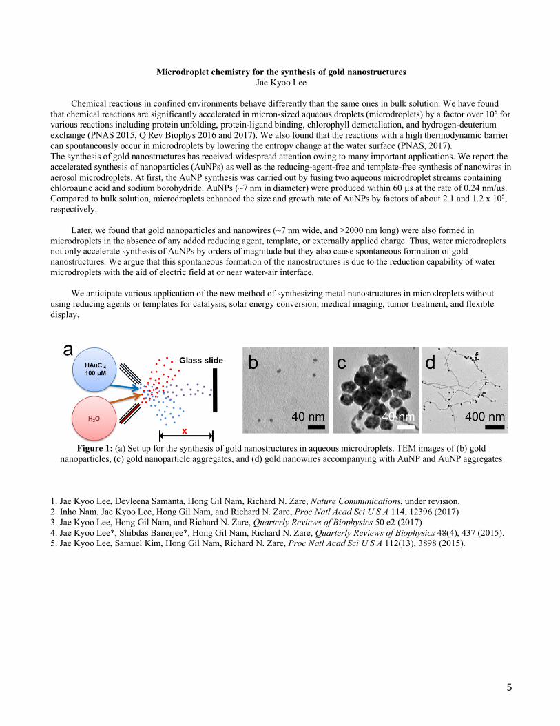
that chemical reactions are significantly accelerated in micron-sized aqueous droplets (microdroplets) by a factor over 105 for various reactions including protein unfolding, protein-ligand binding, chlorophyll demetallation, and hydrogen-deuterium exchange (PNAS 2015, Q Rev Biophys 2016 and 2017). We also found that the reactions with a high thermodynamic barrier can spontaneously occur in microdroplets by lowering the entropy change at the water surface (PNAS, 2017). The synthesis of gold nanostructures has received widespread attention owing to many important applications. We report the accelerated synthesis of nanoparticles (AuNPs) as well as the reducing-agent-free and template-free synthesis of nanowires in aerosol microdroplets. At first, the AuNP synthesis was carried out by fusing two aqueous microdroplet streams containing chloroauric acid and sodium borohydride. AuNPs (~7 nm in diameter) were produced within 60 µs at the rate of 0.24 nm/µs. Compared to bulk solution, microdroplets enhanced the size and growth rate of AuNPs by factors of about 2.1 and 1.2 x 10⁵, respectively.
Later, we found that gold nanoparticles and nanowires (~7 nm wide, and >2000 nm long) were also formed in microdroplets in the absence of any added reducing agent, template, or externally applied charge. Thus, water microdroplets not only accelerate synthesis of AuNPs by orders of magnitude but they also cause spontaneous formation of gold nanostructures. We argue that this spontaneous formation of the nanostructures is due to the reduction capability of water microdroplets with the aid of electric field at or near water-air interface.
We anticipate various application of the new method of synthesizing metal nanostructures in microdroplets without
using reducing agents or templates for catalysis, solar energy conversion, medical imaging, tumor treatment, and flexible display.
Figure 1: (a) Set up for the synthesis of gold nanostructures in aqueous microdroplets. TEM images of (b) gold
nanoparticles, (c) gold nanoparticle aggregates, and (d) gold nanowires accompanying with AuNP and AuNP aggregates 1. Jae Kyoo Lee, Devleena Samanta, Hong Gil Nam, Richard N. Zare, Nature Communications, under revision. 2. Inho Nam, Jae Kyoo Lee, Hong Gil Nam, and Richard N. Zare, Proc Natl Acad Sci U S A 114, 12396 (2017) 3. Jae Kyoo Lee, Hong Gil Nam, and Richard N. Zare, Quarterly Reviews of Biophysics 50 e2 (2017) 4. Jae Kyoo Lee*, Shibdas Banerjee*, Hong Gil Nam, Richard N. Zare, Quarterly Reviews of Biophysics 48(4), 437 (2015). 5. Jae Kyoo Lee, Samuel Kim, Hong Gil Nam, Richard N. Zare, Proc Natl Acad Sci U S A 112(13), 3898 (2015).
6
Microdroplet reactions Xin Yan, Yin-hung Lai
A recent finding worthy of attention is that many reactions can be dramatically accelerated in microdroplets compared
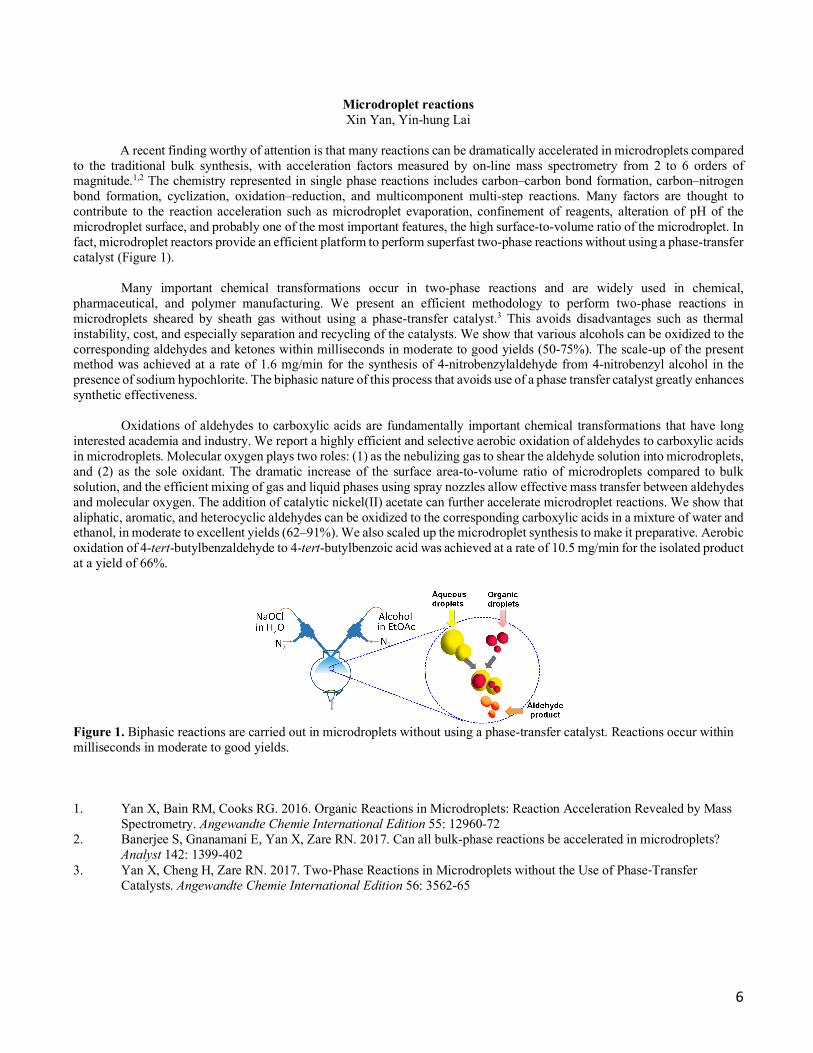
to the traditional bulk synthesis, with acceleration factors measured by on-line mass spectrometry from 2 to 6 orders of magnitude.1,2 The chemistry represented in single phase reactions includes carbon–carbon bond formation, carbon–nitrogen bond formation, cyclization, oxidation–reduction, and multicomponent multi-step reactions. Many factors are thought to contribute to the reaction acceleration such as microdroplet evaporation, confinement of reagents, alteration of pH of the microdroplet surface, and probably one of the most important features, the high surface-to-volume ratio of the microdroplet. In fact, microdroplet reactors provide an efficient platform to perform superfast two-phase reactions without using a phase-transfer catalyst (Figure 1).
Many important chemical transformations occur in two-phase reactions and are widely used in chemical, pharmaceutical, and polymer manufacturing. We present an efficient methodology to perform two-phase reactions in microdroplets sheared by sheath gas without using a phase-transfer catalyst.3 This avoids disadvantages such as thermal instability, cost, and especially separation and recycling of the catalysts. We show that various alcohols can be oxidized to the corresponding aldehydes and ketones within milliseconds in moderate to good yields (50-75%). The scale-up of the present method was achieved at a rate of 1.6 mg/min for the synthesis of 4-nitrobenzylaldehyde from 4-nitrobenzyl alcohol in the presence of sodium hypochlorite. The biphasic nature of this process that avoids use of a phase transfer catalyst greatly enhances synthetic effectiveness.
Oxidations of aldehydes to carboxylic acids are fundamentally important chemical transformations that have long interested academia and industry. We report a highly efficient and selective aerobic oxidation of aldehydes to carboxylic acids in microdroplets. Molecular oxygen plays two roles: (1) as the nebulizing gas to shear the aldehyde solution into microdroplets, and (2) as the sole oxidant. The dramatic increase of the surface area-to-volume ratio of microdroplets compared to bulk solution, and the efficient mixing of gas and liquid phases using spray nozzles allow effective mass transfer between aldehydes and molecular oxygen. The addition of catalytic nickel(II) acetate can further accelerate microdroplet reactions. We show that aliphatic, aromatic, and heterocyclic aldehydes can be oxidized to the corresponding carboxylic acids in a mixture of water and ethanol, in moderate to excellent yields (62–91%). We also scaled up the microdroplet synthesis to make it preparative. Aerobic oxidation of 4-tert-butylbenzaldehyde to 4-tert-butylbenzoic acid was achieved at a rate of 10.5 mg/min for the isolated product at a yield of 66%.
Figure 1. Biphasic reactions are carried out in microdroplets without using a phase-transfer catalyst. Reactions occur within milliseconds in moderate to good yields. 1. Yan X, Bain RM, Cooks RG. 2016. Organic Reactions in Microdroplets: Reaction Acceleration Revealed by Mass
Spectrometry. Angewandte Chemie International Edition 55: 12960-72 2. Banerjee S, Gnanamani E, Yan X, Zare RN. 2017. Can all bulk-phase reactions be accelerated in microdroplets?
Analyst 142: 1399-402 3. Yan X, Cheng H, Zare RN. 2017. Two-Phase Reactions in Microdroplets without the Use of Phase-Transfer
Catalysts. Angewandte Chemie International Edition 56: 3562-65

7
Mechanistic study of reaction acceleration and heterogeneous catalytic oil upgrading in microdroplets Yin-Hung Lai, Maria T. Dulay
Although it has been reported that microdroplets, typically generated by electrospray, provide unique environments for
accelerating a variety of chemical reactions, the mechanisms underlying this phenomenon are not fully understood. Reaction kinetics in microdroplets are often found to be significantly faster than the corresponding bulk-phase kinetics, suggesting the important role of interfacial chemistry. The rate acceleration in a model system, the nucleophilic opening of limonene oxide with morpholine, has been investigated.[1] By utilizing micro-particle imaging velocimetry (micro-PIV) to measure sizes and velocities of microdroplets,[2] significant factors contributing the rate acceleration including the surface area to volume ratio and the reaction time have been evaluated. Decreasing the size of microdroplets and increasing the reaction time dramatically increase conversion to product. It is our hope that a fundamental understanding of the mechanisms of droplet acceleration will help in future preparative-scale chemical synthesis.
Caused by the continuous depletion of conventional oil reserves, to meet growing demand for fuels, development of
alternative fuels by oil upgrading, that is, to break down asphaltene molecules into smaller fragments, becomes essential. We report a novel ambient method that expedites the degradation rate of asphaltenes by spraying sample microdroplets onto a charged paper surface containing catalytic TiO2 nanoparticles.[3] In our method, an additional supply of hydrogen source is not needed. Unlike conventional photocatalysis, degradation that takes place at the interface between oil droplets and the charged surface is electric field induced and accomplished on the sub-millisecond time scale. Moreover, the fragmentation yield is further increased by recycling the process, that is, by retreating the oil sample that has contacted the solid support. We have shown a proof-of-concept process demonstrating the effectiveness of this technique for model compounds (rubrene and hematoporphyrin) as well as undiluted/diluted crude oil samples. Recently, we have found a way to degrade compact asphaltenes and are optimizing the capability of the method.
Workflow for the heterogeneous catalytic degradation of asphaltenes (A) a spray source is used to accommodate sample solution (organic solution, in brown color), which sprays sample microdroplets onto the aqueous wet surface of the filter paper containing anatase TiO2 nanoparticles. The spray source of sample solution is electrically grounded. High voltage is applied to the wet paper coated with TiO2 nanoparticles; (B) Extracted sample is deposited on a glass platter and analyzed using laboratory-built laser desorption laser ionization mass spectrometry (L2MS).
[1] Y.-H. Lai, S. Sathyamoorthi, R. M. Bain, R. N. Zare, J. Am. Soc. Mass Spectrom. 2018. [2] E. T. Jansson, Y.-H. Lai, J. G. Santiago, R. N. Zare, J. Am. Chem. Soc. 2017, 139, 6851-6854. [3] Y.-H. Lai, Z. Zhou, B. Chanbasha, R. N. Zare, Energy Fuels 2017, 31, 12685–12690.

8
Non-invasive diagnosis by mass spectrometry and machine learning Zhenpeng Zhou, Vishnu Shankar
We are currently concerned with the non-invasive diagnosis of metabolic conditions from breath and sweat, using
desorption electrospray ionization (DESI) mass spectrometry combined with artificial intelligence (AI) techniques.
Mass spectrometry is a simple, fast, and cost-effective method to analyze various chemical species by ionizing a sample and sorting the ions based on mass-to-charge (m/z) ratios. DESI is a recently developed method, where a spray of charged droplets impacts a solid sample on a substrate and creates a thin liquid film. An additional splash of the incoming droplets creates microdroplets, which are then drawn into the mass spectrometer.
DESI offers several advantages, compared to other chemical methods, which make it a well-suited for analyzing sweat and breath for diagnostic purposes. For example, DESI allows samples to be examined with minimal preparation, which offers much potential for rapid and routine real-time analysis. Additionally, as AI methods need large amounts of data for training, testing, and validation, the ability to obtain a rich spectrum of molecular species makes DESI appealing for AI methods of analysis. In addition to the complementarity of AI and DESI, AI, specifically machine learning and statistical pattern recognition, is an appropriate technique for medical diagnostics because it can offer improved sensitivity and specificity, as well as objectivity compared to human clinical practice.
As a proof of concept, we applied DESI on latent fingerprints to obtain not only spatial patterns but also chemical maps. Samples with similar lipid compositions as those of the fingerprints were collected by swiping a glass slide across the forehead of consenting adults. A machine learning model called gradient boosting decision tree (GBDT) was applied to the samples that allowed us to distinguish between different genders, ethnicities, and ages (within 10 years). The results from 194 samples showed accuracies of 89.2%, 82.4%, and 84.3%, respectively. Specific chemical species that were determined by the feature selection of GBDT were identified by tandem mass spectrometry. The machine learning model trained on the sample data was applied to overlaid latent fingerprints from different individuals, giving accurate gender and ethnicity information from those fingerprints. The results suggest that DESI-MSI imaging of fingerprints with GDBT analysis might offer a significant advance in forensic science. (Figure 1).
Figure 1. the classification result of each pixel in the image by the pretrained model. The pixels predicted to be belong to a Chinese male are shown in blue, while the pixels predicted to be from an Indian female are shown in red. In both cases, the predictions were correct.

9
Artificial intelligence enabled innovations and discoveries in chemistry Zhenpeng Zhou
The idea of machine learning and artificial intelligence has produced surprising results in the field of theoretical and
computational chemistry. We proposed to apply the decision-making framework to problems in chemistry, for example optimizing chemical reactions, or de novo drug design.
In a recent work, we employed deep reinforcement learning to optimize chemical reactions. Our model iteratively records the results of a chemical reaction and chooses new experimental conditions to improve the reaction outcome. This model outperformed a state-of-the-art blackbox optimization algorithm by using 71% fewer steps on both simulations and real reactions. Furthermore, we introduced an efficient exploration strategy by drawing the reaction conditions from certain probability distributions, which resulted in an improvement on regret from 0.062 to 0.039 compared with a deterministic policy. Combining the efficient exploration policy with accelerated microdroplet reactions, optimal reaction conditions were determined in 30 min for the four reactions considered, and a better understanding of the factors that control microdroplet reactions was reached. Moreover, our model showed a better performance after training on reactions with similar or even dissimilar underlying mechanisms, which demonstrates its learning ability. (Figure 1)
Figure 1. Visualization of Our Model Unrolled over Three Time Steps
10
Investigations in human sweat & breath for new biomarker discovery Vishnu Shankar, Zhenpeng Zhou
Non-invasive methods, compared to traditional medical diagnostics, are pain-free, cheaper, and more efficient, which is
useful for early-detection, treatment, and illuminating important patient-level biochemical data. For example, investigations of human breath [1] revealed decreased levels of isoprene concentration for patients with chronic heart failure, which led to new connections between repair mechanisms in cholesterol biosynthesis and heart failure. In the same way, it has been suggested that human sweat [2], consisting of fatty acids, glycerides, and other lipid-based compounds, can aid novel insights in the metabolism for patients, which is measurably altered under disease states. Therefore, the objective of this study is to investigate human sweat and breath to find novel disease biomarkers.
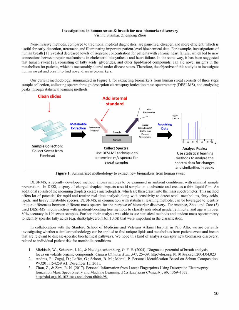
Our current methodology, summarized in Figure 1, for extracting biomarkers from human sweat consists of three steps sample collection, collecting spectra through desorption electrospray ionization mass spectrometry (DESI-MS), and analyzing peaks through statistical learning methods.
Figure 1. Summarized methodology to extract new biomarkers from human sweat
DESI-MS, a recently developed method, allows samples to be examined in ambient conditions, with minimal sample
preparation. In DESI, a spray of charged droplets impacts a solid sample on a substrate and creates a thin liquid film. An additional splash of the incoming droplets creates microdroplets, which are then drawn into the mass spectrometer. This method offers lot of potential for rapid and routine real-time analysis along with sensitivity to detect small metabolites, fatty-acids, lipids, and heavy metabolite species. DESI-MS, in conjunction with statistical learning methods, can be leveraged to identify unique differences between different mass spectra for the purpose of biomarker discovery. For instance, Zhou and Zare (3) used DESI-MS in conjunction with gradient-boosting tree methods to classify individual gender, ethnicity, and age with over 80% accuracy in 194 sweat samples. Further, their analysis was able to use statistical methods and tandem mass-spectrometry to identify specific fatty acids (e.g. dialkylglycerol(16:1|10:0)) that were important in the classification.
In collaboration with the Stanford School of Medicine and Veterans Affairs Hospital in Palo Alto, we are currently investigating whether a similar methodology can be applied to find unique lipids and metabolites from patient sweat and breath that are relevant to disease-specific biochemical pathways. We hope this kind of analysis can spur new biomarker discovery, related to individual patient risk for metabolic conditions.
1. Miekisch, W., Schubert, J. K., & Noeldge-schomburg, G. F. E. (2004). Diagnostic potential of breath analysis — focus on volatile organic compounds. Clinica Chimica Acta, 347, 25–39. http://doi.org/10.1016/j.cccn.2004.04.023
2. Andres, P.; Zugaj, D.; Laffet, G.; Schoot, B. M.; Martel, P. Personal Identification Based on Sebum Composition. WO2011154259 A1, December 15, 2011.
3. Zhou, Z., & Zare, R. N. (2017). Personal Information from Latent Fingerprints Using Desorption Electrospray Ionization Mass Spectrometry and Machine Learning. ACS Analytical Chemistry, 89, 1369–1372. http://doi.org/10.1021/acs.analchem.6b04498.
Sample'Collec+on:'Collect'Sweat'from'
Forehead'
Metabolite'Extrac+on'
Surface(
Electrospray((Pressure)(
Sample(
Ionized(Microdroplets/Analyte(Ions((Pressure,(
Electrosta0cs)(
Mass((Spectrometer(
Collect'Spectra:'Use'DESI6MS'technique'to'determine'm/z'spectra'for'
sweat'samples'
Acquiring'Data'
Analyze'Peaks:''Use'sta?s?cal'learning'methods'to'analyze'the'spectra'data'for'changes'and'similari?es'in'peaks'
Clean'slides' Add'internal'standard'
11
Electrically controlled drug release from drug-loaded polymer nanocomposite films Christian Chamberlayne, Ephraim Neumann
Medical implants, capable of releasing quantitative amounts of drugs on demand, open many possibilities in medical device applications. Toward this goal, we are developing drug-loaded polymers that release drugs upon electrical stimulation. The polymer acts both as a drug reservoir and a release mechanism for medical implants. The advantage of using electric signals is that they are easily generated and controlled, and do not require the use of specialized equipment.
Two issues facing the field of electrically stimulated drug release are: 1) loading sufficient quantities of drug, and 2)
releasing said drug with low voltages. We have demonstrated drug loadings above 40% by weight as well as drug release at low voltages (less than 1.5V). Our system is also quite versatile, allowing electrically stimulated drug release of a variety of drugs, spanning from small molecules up to small polypeptides like insulin.
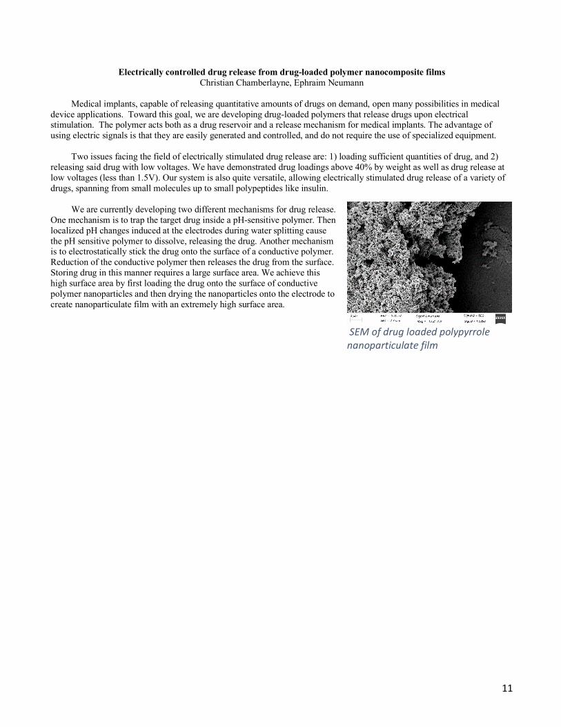
We are currently developing two different mechanisms for drug release.
One mechanism is to trap the target drug inside a pH-sensitive polymer. Then localized pH changes induced at the electrodes during water splitting cause the pH sensitive polymer to dissolve, releasing the drug. Another mechanism is to electrostatically stick the drug onto the surface of a conductive polymer. Reduction of the conductive polymer then releases the drug from the surface. Storing drug in this manner requires a large surface area. We achieve this high surface area by first loading the drug onto the surface of conductive polymer nanoparticles and then drying the nanoparticles onto the electrode to create nanoparticulate film with an extremely high surface area.
SEM of drug loaded polypyrrole nanoparticulate film

12
Surgical margin evaluation of clear cell renal cell carcinoma (ccRCC) using desorption electrospray ionization mass spectrometry imaging (DESI-MSI) Vijaya Lakshmi Kanchustambham
One of the great challenges in tumor-progression-free survival after surgical resection of solid tumors is to distinguish
the delicate boundary between positive and negative surgical margins. Conventional histopathologic evaluation of surgical margins using intraoperative frozen sections (IFS) is time-consuming, expensive, and susceptible to artefacts. The demand for fast and accurate method for cancer detection and diagnosis led to the development of both ex-vivo and in-vivo molecular imaging techniques. DESI-MSI is an ambient ionization mass spectrometry imaging technique which offers unique potential for the chemical map of clinical biomarkers.1 Using DESI-MSI, we image both normal and cancer kidney tissue sections and identify the metabolites and lipid signatures that are unique to the cancer abnormal metabolism. The classification and validation of potential biomarkers is performed by the statistical method least absolute shrinkage and selector operator (LASSO) of the mass spectrometry imaging data from paired surgical margins of patients.
Figure 1. Chemical map (m/z = 331.250) of metabolic signatures in ccRCC positive surgical margins probed by DESI-MSI
Biochemical reactions in microdroplets from a theta tip capillary
Many enzymatic reactions catalyze reactants to products while populating short-lived intermediates. The development of time-resolved mass spectrometry offers high sensitivity and selectivity for studying the dynamic processes associated with the progress of the reaction in time. However, the rapid mixing of solutions during electrospray process is needed to probe the steady-state kinetics in faster processes. Here, we use droplet chemistry as a novel approach to create microdroplets from a theta glass capillary and follow the reaction after rapid mixing of enzyme, substrate, and cofactors that likely occurs in a few microseconds or less. In addition, we would like to compare the kinetics of enzyme-catalyzed reactions in microdroplets from that in bulk solution.2 The experimental investigation of enzymatic reactions in microdroplets will provide key insights to the reaction mechanism on the surface of water droplets.
Scheme 1. Cartoon representing theta tip capillary for nanoESI experiments.
1. Banerjee, S.; Zare, R. N.; Tibshirani, R. J.; Kunder, C. A.; Nolley, R.; Fan, R.; Brooks, J. D.; Sonn, G. A. Proceedings of the National Academy of Sciences 2017, 114, 3334.
2. Bain, R. M.; Sathyamoorthi, S.; Zare, R. N. Angewandte Chemie International Edition 2017, 56, 15083.

13
Mass spectrometry imaging for biomedical applications Katy Margulis
We employ desorption electrospray ionization mass spectrometry imaging (DESI-MSI) to study a variety of
pathophysiological processes in living tissues, to distinguish between tissues of various pathologies, and to guide drug discovery.
In DESI-MSI, tissue sections are scanned directly without any sample preparation and with minimal in-process damage,
mapping their chemical composition in two-dimensional manner. To desorb and ionize the target molecules, a beam of charged droplets is directed to the tissue surface, extracting compounds into secondary droplets that are subsequently analyzed by the mass spectrometer. A program-controlled moving stage is used to scan the entire surface of the sample, while the mass spectra are recorded as a function of the x,y-position on the tissue. A two-dimensional distribution image with relative signal intensity can be generated for any specific m/z value. Hence, a single scan enables acquiring the richest chemical information per pixel, obviating the need for a specific molecular targeting, and allowing for a spatial co-localization of different molecules. Atmospheric pressure ionization conditions, minimal requirements for sample preparation, non-destructive scanning process and high sensitivity make this imaging technique exceptionally valuable for detecting chemical changes in tissue caused by pathological processes or pharmacological intervention. Basal cell carcinoma diagnostics. We established the capability DESI-MSI to distinguish between micrometer-sized tumor aggregates of basal cell carcinoma (BCC), a common skin cancer, and normal human skin. We analyzed 86 human specimens collected during Mohs micrographic surgery for BCC to cross-examine spatial distributions of numerous lipids and metabolites in BCC aggregates versus adjacent skin (Figure 1). Statistical analysis using the least absolute shrinkage and selection operation (Lasso) was employed to categorize each 200-µm diameter picture element (pixel) of investigated skin tissue map as BCC or normal. Lasso yielded an overall 94.1% diagnostic accuracy pixel by pixel of the skin map. We suggest that DESI-MSI/Lasso analysis is not limited to the diagnosis of BCC but should be applicable to a wide range of microscopic tumors. Studying pathogenesis of oncogene-driven tumors and identifying new pharmacological targets. We study initiation, progression and regression of tumors driven by various oncogenes in conditional transgenic mouse models. In these models we can intentionally activate and deactivate the oncogene in a specific organ to induce and regress the tumors. By DESI-MSI we monitor lipids and metabolites altered in each stage of tumor development and regression, which gives us insights on tumor pathogenesis and allows us to identify novel targets for pharmacological intervention (Figure 2).
Figure 1. (A) Skin lesion suspected as BCC is removed during Mohs surgery and sectioned; (B) Excised skin sections are imaged by DESI-MS to detect microscopic tumors.
Figure 2. (A) Kidney tumor (renal cell carcinoma) is initiated and subsequently regressed by activation and deactivation of MYC oncogene in conditional transgenic mice; (B) Kidney sections are imaged by DESI-MS to detect metabolic changes during tumor initiation, progression and regression; (C) Novel therapeutic targets are identified based on the information obtained by DESI-MSI, and pharmacological intervention is tested in mice models.
14
Design of nanoparticles for biomedical applications Katy Margulis
We design organic nanoparticles for efficient delivery of a wide spectrum of therapeutic agents. One specific interest is
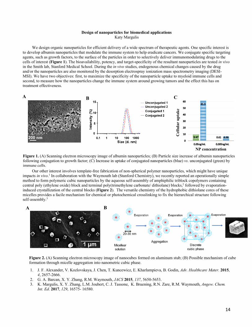
to develop albumin nanoparticles that modulate the immune system to help eradicate cancers. We conjugate specific targeting agents, such as growth factors, to the surface of the particles in order to selectively deliver immunomodulating drugs to the cells of interest (Figure 1). The bioavailability, potency, and target-specificity of the resultant nanoparticles are tested in vivo in the Smith lab, Stanford Medical School. During the in vivo studies, endogenous chemical changes caused by the drug and/or the nanoparticles are also monitored by the desorption electrospray ionization mass spectrometry imaging (DESI-MSI). We have two objectives: first, to maximize the specificity of the nanoparticle uptake to myeloid immune cells and second, to measure how the nanoparticles change the immune system around growing tumors and the effect this has on treatment effectiveness.
Our other interest involves template-free fabrication of non-spherical polymer nanoparticles, which might have unique impacts in vivo.1 In collaboration with the Waymouth lab (Stanford Chemistry), we recently reported an operationally simple method to form polymeric cubic nanoparticles by the aqueous self-assembly of amphiphilic triblock copolymers containing central poly (ethylene oxide) block and terminal poly(trimethylene carbonate/ dithiolane) blocks,2 followed by evaporation-induced crystallization of the central blocks (Figure 2). The versatile chemistry of the hydrophobic dithiolane cores of these micelles provides a facile mechanism for chemical or photochemical crosslinking to fix the hierarchical structure following self-assembly.3
1. J. F. Alexander, V. Kozlovskaya, J. Chen, T. Kuncewicz, E. Kharlampieva, B. Godin, Adv. Healthcare Mater. 2015, 4, 2657-2666.
2. G. A. Barcan, X. Y. Zhang, R.M. Waymouth, JACS 2015, 137, 5650-5653. 3. K. Margulis, X. Y. Zhang, L.M. Joubert, C. J. Tassone, K. Bruening, R.N. Zare, R.M. Waymouth, Angew. Chem.
Int. Ed. 2017, 129, 16575- 16580.
A B C
Figure 1. (A) Scanning electron microscopy image of albumin nanoparticles; (B) Particle size increase of albumin nanoparticles following conjugation to growth factor; (C) Increase in uptake of conjugated nanoparticles (blue) vs. unconjugated (green) by immune cells.
NP concentration
Cel
lula
r up
take
A B
Figure 2. (A) Scanning electron microscopy image of nanocubes formed on aluminum stub; (B) Possible mechanism of cube formation through micelle aggregation into nanometric cubic phase.
15
In situ measurement of ligand-receptor interactions at the cell surface by ambient ionization mass spectrometry Sarah E. Noll
Studies have shown links between G-protein coupled receptors (GPCRs) and a variety of psychiatric disorders, including
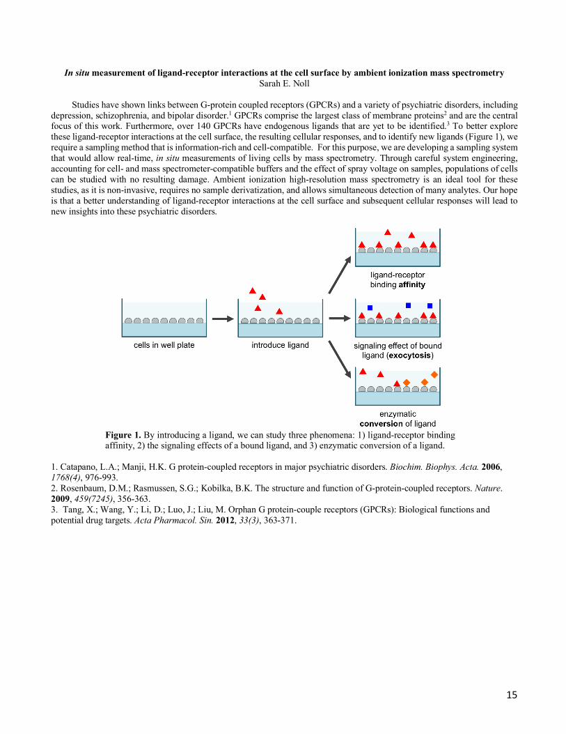
depression, schizophrenia, and bipolar disorder.1 GPCRs comprise the largest class of membrane proteins2 and are the central focus of this work. Furthermore, over 140 GPCRs have endogenous ligands that are yet to be identified.3 To better explore these ligand-receptor interactions at the cell surface, the resulting cellular responses, and to identify new ligands (Figure 1), we require a sampling method that is information-rich and cell-compatible. For this purpose, we are developing a sampling system that would allow real-time, in situ measurements of living cells by mass spectrometry. Through careful system engineering, accounting for cell- and mass spectrometer-compatible buffers and the effect of spray voltage on samples, populations of cells can be studied with no resulting damage. Ambient ionization high-resolution mass spectrometry is an ideal tool for these studies, as it is non-invasive, requires no sample derivatization, and allows simultaneous detection of many analytes. Our hope is that a better understanding of ligand-receptor interactions at the cell surface and subsequent cellular responses will lead to new insights into these psychiatric disorders.
Figure 1. By introducing a ligand, we can study three phenomena: 1) ligand-receptor binding affinity, 2) the signaling effects of a bound ligand, and 3) enzymatic conversion of a ligand.
1. Catapano, L.A.; Manji, H.K. G protein-coupled receptors in major psychiatric disorders. Biochim. Biophys. Acta. 2006, 1768(4), 976-993. 2. Rosenbaum, D.M.; Rasmussen, S.G.; Kobilka, B.K. The structure and function of G-protein-coupled receptors. Nature. 2009, 459(7245), 356-363. 3. Tang, X.; Wang, Y.; Li, D.; Luo, J.; Liu, M. Orphan G protein-couple receptors (GPCRs): Biological functions and potential drug targets. Acta Pharmacol. Sin. 2012, 33(3), 363-371.
16
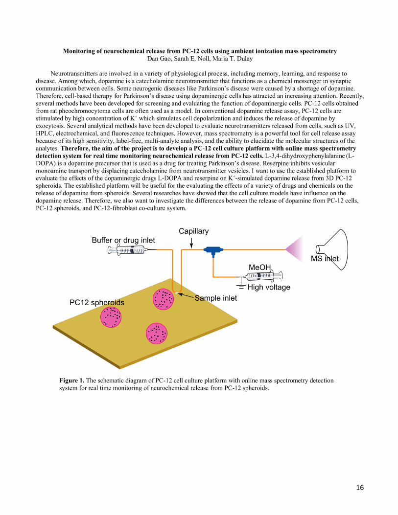
Monitoring of neurochemical release from PC-12 cells using ambient ionization mass spectrometry Dan Gao, Sarah E. Noll, Maria T. Dulay
Neurotransmitters are involved in a variety of physiological process, including memory, learning, and response to
disease. Among which, dopamine is a catecholamine neurotransmitter that functions as a chemical messenger in synaptic communication between cells. Some neurogenic diseases like Parkinson’s disease were caused by a shortage of dopamine. Therefore, cell-based therapy for Parkinson’s disease using dopaminergic cells has attracted an increasing attention. Recently, several methods have been developed for screening and evaluating the function of dopaminergic cells. PC-12 cells obtained from rat pheochromocytoma cells are often used as a model. In conventional dopamine release assay, PC-12 cells are stimulated by high concentration of K+ which simulates cell depolarization and induces the release of dopamine by exocytosis. Several analytical methods have been developed to evaluate neurotransmitters released from cells, such as UV, HPLC, electrochemical, and fluorescence techniques. However, mass spectrometry is a powerful tool for cell release assay because of its high sensitivity, label-free, multi-analyte analysis, and the ability to elucidate the molecular structures of the analytes. Therefore, the aim of the project is to develop a PC-12 cell culture platform with online mass spectrometry detection system for real time monitoring neurochemical release from PC-12 cells. L-3,4-dihydroxyphenylalanine (L-DOPA) is a dopamine precursor that is used as a drug for treating Parkinson’s disease. Reserpine inhibits vesicular monoamine transport by displacing catecholamine from neurotransmitter vesicles. I want to use the established platform to evaluate the effects of the dopaminergic drugs L-DOPA and reserpine on K+-simulated dopamine release from 3D PC-12 spheroids. The established platform will be useful for the evaluating the effects of a variety of drugs and chemicals on the release of dopamine from spheroids. Several researches have showed that the cell culture models have influence on the dopamine release. Therefore, we also want to investigate the differences between the release of dopamine from PC-12 cells, PC-12 spheroids, and PC-12-fibroblast co-culture system.
Figure 1. The schematic diagram of PC-12 cell culture platform with online mass spectrometry detection system for real time monitoring of neurochemical release from PC-12 spheroids.
Capillary
MeOHMS inlet
PC12 spheroids
Buffer or drug inlet
Sample inletHigh voltage

17
Discovery of potent inhibitors against GPAT (Glycerol 3-Phosphate Acyltransferase) for treatment of cancer and other diseases
Feng Jin
Obesity is a common social healthy focus, and about 66% American adults suffer this disease, which leads to a series of subsequent diseases, including diabetes, hypertension, cardiovascular diseases, non-alcoholic fatty liver disease, even certain types of cancer. Glycerol-3-phosphate acyltrans-ferase-1 (mtGPAT) catalyzes the following key step in the synthesis of animal fat and plays a central role in the biological process of obesity.
Scheme 1. Biological synthesis of TAG
Increased triacylglycerol(TAG) synthesis is associate with obesity and many other health problems, thus GPAT has been supported as a good target for anti-obesity treatment. However, to date, no GAPT inhibitor has been approved for clinical trials maybe due to moderate or weak inhibitory activity against GPAT. And FSG67, a GPAT inhibitor, was initiated as an anti-obesity reagent. In our precious study, we discovered FSG67 has good in vivo anticancer activity, but still with some biological shortcomings, for example, an in vitro µM anti-GPAT capacity. So further we design a series of new and potent GPAT inhibitors, of which some representative compounds may be appropriate for further clinical development.
a) b)
Scheme 2. a) Substrate-based design strategy; b) Mimic compounds, are capable of competing with A, and block the synthesis of TAG.
lysophosphatidic acid
OH
O
O P OHO
O-
O
Na+glycerol-3-phosphate
HOOH
PO
O OHOH + long-chain acyl-CoAs GPAT 1
the rate-limiting step of glycerolipid biosynthesis
phosphatidic acid
PO
OHO
HO OHOH
esterified
OHOH
HO
glyceroltriacylglycerol
OO
O
O
O
O
Z
Y
X FSG67 is X = o-COOHZ= Ph, Y = NHSO2R
18
In-situ ambient ionization mass spectrometry for mechanistic studies of organometallic catalysis Katherine Walker
High-resolution electrospray ionization mass spectrometry (ESI-MS) is an powerful technique for identifying
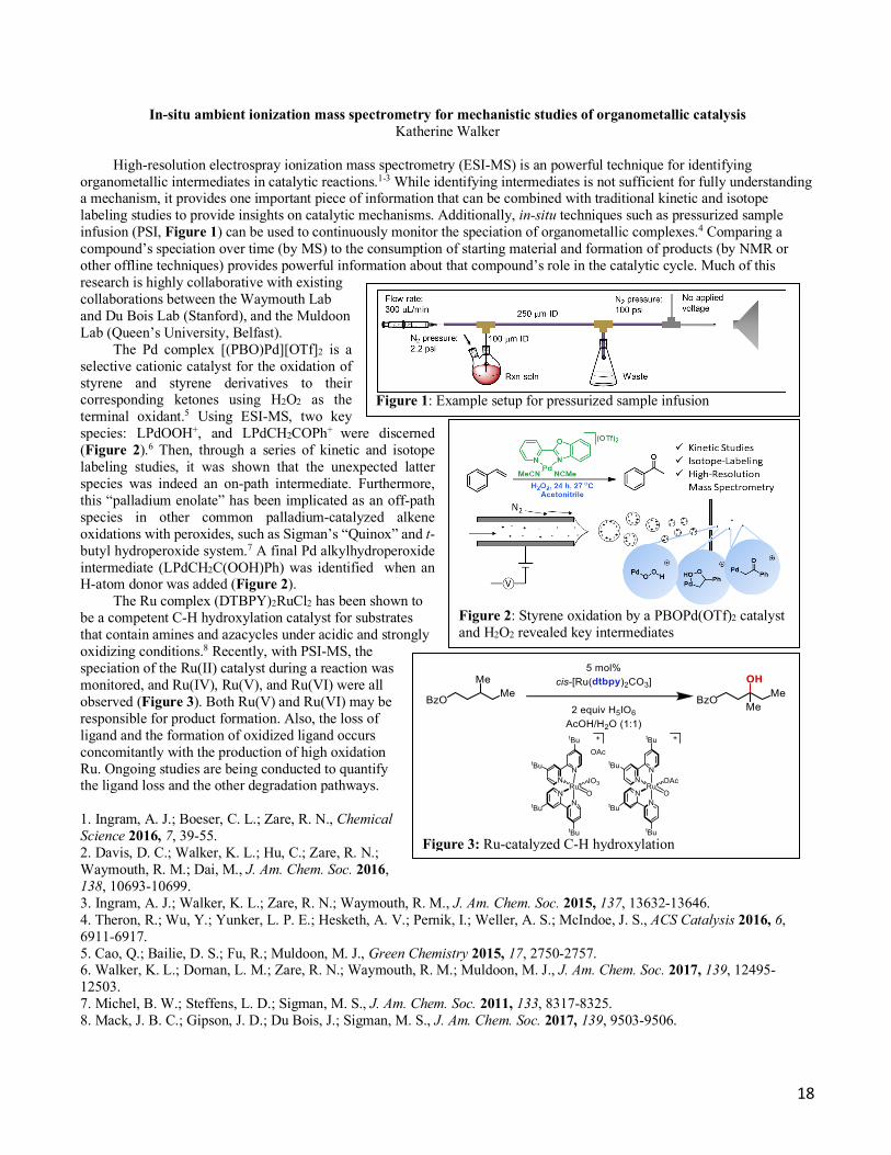
organometallic intermediates in catalytic reactions.1-3 While identifying intermediates is not sufficient for fully understanding a mechanism, it provides one important piece of information that can be combined with traditional kinetic and isotope labeling studies to provide insights on catalytic mechanisms. Additionally, in-situ techniques such as pressurized sample infusion (PSI, Figure 1) can be used to continuously monitor the speciation of organometallic complexes.4 Comparing a compound’s speciation over time (by MS) to the consumption of starting material and formation of products (by NMR or other offline techniques) provides powerful information about that compound’s role in the catalytic cycle. Much of this research is highly collaborative with existing collaborations between the Waymouth Lab and Du Bois Lab (Stanford), and the Muldoon Lab (Queen’s University, Belfast).
The Pd complex [(PBO)Pd][OTf]2 is a selective cationic catalyst for the oxidation of styrene and styrene derivatives to their corresponding ketones using H2O2 as the terminal oxidant.5 Using ESI-MS, two key species: LPdOOH+, and LPdCH2COPh+ were discerned (Figure 2).6 Then, through a series of kinetic and isotope labeling studies, it was shown that the unexpected latter species was indeed an on-path intermediate. Furthermore, this “palladium enolate” has been implicated as an off-path species in other common palladium-catalyzed alkene oxidations with peroxides, such as Sigman’s “Quinox” and t-butyl hydroperoxide system.7 A final Pd alkylhydroperoxide intermediate (LPdCH2C(OOH)Ph) was identified when an H-atom donor was added (Figure 2).
The Ru complex (DTBPY)2RuCl2 has been shown to be a competent C-H hydroxylation catalyst for substrates that contain amines and azacycles under acidic and strongly oxidizing conditions.8 Recently, with PSI-MS, the speciation of the Ru(II) catalyst during a reaction was monitored, and Ru(IV), Ru(V), and Ru(VI) were all observed (Figure 3). Both Ru(V) and Ru(VI) may be responsible for product formation. Also, the loss of ligand and the formation of oxidized ligand occurs concomitantly with the production of high oxidation Ru. Ongoing studies are being conducted to quantify the ligand loss and the other degradation pathways. 1. Ingram, A. J.; Boeser, C. L.; Zare, R. N., Chemical Science 2016, 7, 39-55. 2. Davis, D. C.; Walker, K. L.; Hu, C.; Zare, R. N.; Waymouth, R. M.; Dai, M., J. Am. Chem. Soc. 2016, 138, 10693-10699. 3. Ingram, A. J.; Walker, K. L.; Zare, R. N.; Waymouth, R. M., J. Am. Chem. Soc. 2015, 137, 13632-13646. 4. Theron, R.; Wu, Y.; Yunker, L. P. E.; Hesketh, A. V.; Pernik, I.; Weller, A. S.; McIndoe, J. S., ACS Catalysis 2016, 6, 6911-6917. 5. Cao, Q.; Bailie, D. S.; Fu, R.; Muldoon, M. J., Green Chemistry 2015, 17, 2750-2757. 6. Walker, K. L.; Dornan, L. M.; Zare, R. N.; Waymouth, R. M.; Muldoon, M. J., J. Am. Chem. Soc. 2017, 139, 12495-12503. 7. Michel, B. W.; Steffens, L. D.; Sigman, M. S., J. Am. Chem. Soc. 2011, 133, 8317-8325. 8. Mack, J. B. C.; Gipson, J. D.; Du Bois, J.; Sigman, M. S., J. Am. Chem. Soc. 2017, 139, 9503-9506.
Figure 1: Example setup for pressurized sample infusion
Figure 2: Styrene oxidation by a PBOPd(OTf)2 catalyst and H2O2 revealed key intermediates
Figure 3: Ru-catalyzed C-H hydroxylation

19
Biosensors for Detection of Pathogenic Microorganisms Maria T. Dulay
The ubiquity of pathogenic microorganisms, such as bacteria, viruses and parasites, in food, water, and blood makes it
necessary to have effective testing methods for such microorganisms. Conventional microbiological testing methods are time consuming in that they involve a morphological evaluation of the microorganism as well as tests for their viability and often an amplification step, and these methods lack the high sensitivity needed to detect low concentrations where even a single pathogenic organism in complex biological environments that may include other nonpathogenic organisms can be an infectious dose. To address the need for rapid and sensitive detection of microorganisms, our lab has been working on the development and application of cell-imprinted polymers for microorganism capture and detection. This research area began with imprinting of PDMS (polydimethylsiloxane), a viscoelastic polymer, with a variety of microorganisms, including bacteria and viruses.
The focus of this project is to develop cell imprints on polymers, including silane-based ones, whose properties can be
modified by changing a variety of reaction parameters, which is an advantage over the use of PDMS. Briefly, a cell-imprinted polymer is prepared by first creating a target bacteria template by adsorption of the bacteria onto an inert surface. Polymer reaction solution is poured over the template and the resulting polymer forms around the fixed bacteria, creating an imprint of the bacteria on the polymer, which is peeled from the template and used to capture the target bacteria from a suspension. This approach is just one of the ways that we imprint bacteria into these OSX polymers. The unique shape and chemical fingerprint of the targeted bacteria are captured in the polymer during imprinting. Capture of the targeted bacteria is achieved without laminar flow at room temperature or 30°C. Different detection schemes are coupled to the imprinted polymer biosensor, including optical methods.
Preliminary results using E. coli-GFP-imprinted sol-gel polymers
as shown in Figure 1 demonstrate the potential of these imprinted OSX polymers for the capture of target bacteria within 30 minutes at 30° C by gravitational settling (no laminar flow) in the imprinted area of the polymer. Furthermore, differentiation between the targeted chemically inactivated bacterium E. coli, and non-targeted S. typhimurium and native E. coli is demonstrated with high selectivity compared to imprinted PDMS. By designing cell-imprinted OSX polymers as biosensors for microorganisms such as bacteria, we hope to have a rapid, sensitive, and inexpensive device for the detection of pathogenic microorganisms in different environments. The use of these devices as point-of-care biosensors is one application that we are pursuing. Figure 2 show SEM images of E. coli-GFP-imprinted OSX polymers and a template.
A B
Figure 1. Fluorescent images of captured (A) inactivated E. coli-GFP (OD600 0.38 solution) and (B) inactivated S. typhimurium-GFP (OD600 0.38 solution) on an inactivated E. coli-GFP-imprinted OSX polymer.
A B C
Figure 2. SEM images of (A) imprinted OSX-1 polymer with heat-fixed E. coli-GFP (OD600 1.0 suspension) on polystyrene substrate, (B) glutaraldehyde-inactivated E. coli-GFP on OSX-2 polymer prepared with E. coli-GFP (OD600 0.44 suspension) on Thermanox plastic, and (C) bright-field image of glutaraldehyde-inactivated E. coli-GFP template at 40X magnification.

20
Composite silane-based polymers as substrate material and electrospray emitter for ambient ionization mass spectrometry
Maria T. Dulay
Two of the advantages of using these organic silane-based polymers, called organosiloxane (OSX) polymers, in the analysis of biological mixtures, such as blood or serum, that significantly simplifies analysis are (1) the low reliance on sample preparation protocols and (2) their use with analytical techniques such as mass spectrometry under ambient conditions (i.e., avoids the use of vacuum). The ability to directly analyze a complex mixture without the need for sample workup allows for high sample throughput analysis.
Sol-gel chemistry is used to synthesize OSX polymers of high mechanical
strength, flexibility, variable porosity, and polarity. Bonding or grafting of various chemical entities, such as enzymes, to the surface of these polymers is possible through the active chemical functional groups on the polymer surface. Because these OSX polymers are amenable to various formats, such as planar sheets, bulk porous materials and coatings for various surfaces and materials, they are potentially useful in areas ranging from medicine to drug and environmental monitoring. As such, utilizing the unique properties of these OSX polymers has been a key goal in its development as a platform for rapid detection of analytes, such as drugs and proteins in biological fluids.
For one platform, porous and nonporous OSX polymers are being developed as
deposition and spray surfaces in ambient ionization mass spectrometry (MS, Figure 1) for the analysis of a variety of compounds, including drugs, in complex biological media, such as urine, blood, and serum as shown in Figure 2.1 There is very little to no sample preparation required. Blood spotted onto the polymer is directly analyzed by ambient ionization mass spectrometry with good signal sensitivity.
Other platforms that involve preconcentration and chromatographic separation using these OSX polymers are being investigated. Furthermore, by incorporating other materials, such as enzymes and conductive compound into the OSX polymers we can extend their utility as materials for bioanalysis. We have recently, modified the surface of OSX polymers with the enzyme, trypsin, for an in-situ digestion device coupled to desorption electrospray ionization MS (DESI-MS) for the direct analysis of digestion products with little sample preparation prior to the digestion reaction. Currently, we are developing silane-based polymers with higher conductivities by incorporating conductive materials like carbon nanotubes (CNTs) within the network of the polymer. With these OSX polymer-CNT blends, we hope to one day electrically drive these polymers with a conventional battery as the external voltage source. In summary, this project involves both synthetic as well as analytical endeavors toward the realization of composite organic polymer platforms for rapid analysis of a variety of analytes.
1. Dulay, M.T.; Zare, R.N., Rapid Commun. Mass Spectrom. 2017, 31, 1651-1658.
Scanning electron microscope image of a porous OSX polymer.
Figure 1. OSX as a spray material for ambient ionization MS.
Figure 2. MS of 7 narcotic drugs and their deuterated analogs in urine detected by polymer spray. The drugs include morphine (6) and cocaine (7).