versatility of reverse sural artery flap (rsaf) in
TRANSCRIPT

~ 583 ~
International Journal of Orthopaedics Sciences 2021; 7(2): 583-590
E-ISSN: 2395-1958
P-ISSN: 2706-6630
IJOS 2021; 7(2): 583-590
© 2021 IJOS
www.orthopaper.com
Received: 11-02-2021
Accepted: 15-03-2021
Dr. Nilesh Shende
Assistant Professor, Plastic,
Reconstructive and Burns
Surgery, Department, Seth GS
Medical College & Kem Hospital,
Parel, Mumbai, India
Dr. Ujwal Chirde
Assistant Professor, Plastic,
Reconstructive and Burns
Surgery, Department, Seth GS
Medical College & Kem Hospital,
Parel, Mumbai, India
Dr. Vinita Puri
Professor & Head of
Department, Plastic,
Reconstructive and Burns
Surgery Department, Seth GS
Medical College & Kem Hospital,
Parel, Mumbai, India
Dr. Aniruddh Gupta
Assistant Professor, Plastic,
Reconstructive and Burns
Surgery Department, Seth GS
Medical College & Kem Hospital,
Parel, Mumbai, India
Corresponding Author:
Dr. Ujwal Chirde
Assistant Professor, Plastic,
Reconstructive and Burns
Surgery, Department, Seth GS
Medical College & Kem Hospital,
Parel, Mumbai, India
Versatility of Reverse Sural Artery Flap (RSAF) in
management of lower limb defects: Our experience
Dr. Nilesh Shende, Dr. Ujwal Chirde, Dr. Vinita Puri and Dr. Aniruddh
Gupta
DOI: https://doi.org/10.22271/ortho.2021.v7.i2h.2679
Abstract Background: Wounds around the lower 1/3rd of leg and foot are difficult to manage. Treatment options
like skin grafting, healing by secondary intention can result in an unstable scar. Free tissue transfers and
Regional flaps has its own disadvantages. To circumvent these difficulties a fasciocutaneous flap
harvested from sural angiosome was proposed. In this study, we have made an attempt to find out
different scenarios in which this flap can be used as a management option and ways to minimize the
complications associated with this flap.
Methods: A retrospective observational study was conducted over 2 years. 30 patients were studied.
Patient details was obtained from the case record, operative notes and postoperative follow-up.
Results: Patients between the age of 10 years to 65 years were studied. RSA flap with adipofascial
pedicle and fasciocutaneous pedicle was done in 21 and 9 patients respectively. Delaying of flap was
done in 9 patients, with most common indication as distal defect. Common site of defect which were
reconstructed was on the Tendoachilles region, Heel region. Various complications were loss of graft
used to cover adipofascial pedicle, Venous congestion, hematoma formation and partial skin necrosis of
the flap. None of the patients complained of any effect on routine activity during postoperative follow up.
Conclusion: It is a versatile flap and different scenario can be managed with customization in plan. With
proper selection of patients, it can be judiciously used to manage different leg defects.
Keywords: Reverse sural artery, venous congestion, adipofascial, fasciocutaneous
Introduction
Reverse Sural artery flap is a distally based fasciocutaneous flap [1, 2]. It is based on peroneal
artery perforators and anastomosis between the peroneal artery septocutaneous perforator and
sural arterial network [3, 4, 5]. It does not involve sacrificing any major vessel of limb.
Fasciocutaneous Sural flap described by Ponten in 1981, underwent various modifications
over a period of time [6]. Donski and Fogdestram later came up with a distally based
fasciocutaneous flap from the sural angiosome for repair of soft tissue defect of foot and distal
1/3rd of leg [6, 7]. In 1992, Masquelet et al. introduced the concept of Sural neurocutaneous
island flap. This flap included an adipofascial pedicle and sural nerve, it was referred to as
“Reversed Sural artery flap” [1, 2, 8].
Wounds around the lower 1/3rd of leg and foot are difficult to manage because of poor
circulation. Often, these wounds are associated with exposed bones, tendons and implants [8, 9].
Simple treatment options like skin grafting, healing by secondary intention is difficult as it can
result in an unstable scar. Free tissue transfer is an ideal option for most of these defects, but it
has disadvantages like prolonged operative time, need of an expertise in microsurgery and
distant donor site morbidity [9, 10]. Regional flaps based on Peroneal artery, Posterior tibial
artery or Anterior tibial artery results into sacrifice of a major blood vessel of the lower limb [9]. To circumvent these difficulties a fasciocutaneous flap harvested from sural angiosome was
proposed [9].
In this study we have made an attempt to find out different scenarios in which this flap can be
used as a management option and look at the ways to minimize the complications associated
with this flap.
~ 584 ~
International Journal of Orthopaedics Sciences www.orthopaper.com Aims and Objectives
1. To study the use of Reverse sural artery flap (RSAF) in
our practice.
2. To study post-operative complications of RSAF.
3. To study various means to minimize complications
associated with RSA flap.
Materials and Methods
The institutional ethical committee clearance was obtained
(IEC NO- EC/OA-110/2018). This was a retrospective
observational study which was conducted for 2 years. A total
of 30 patients, reconstructed with the reverse sural artery flap
for defect of foot and lower leg, were studied. Patient details
regarding age, sex, comorbidities, etiology of the defect,
location of the defect, size of defect, radiological evidence of
associated fracture, type of flap done, complications and
details of post-operative follow up (i.e., outcome) was
obtained from the case record, operative notes and their
follow-up in outpatient department. Details obtained were
entered into a predesigned case evaluation form. Based on this
a MS excel worksheet was prepared. This data was then used
to draw the results.
Planning and Surgical technique
The detailed history of patients was taken including age,
personal history (smoking, tobacco chewing), comorbidities
(Diabetes Mellitus, Peripheral Vascular Disease,
Hypertension). Preoperative anesthetic checkup was done.
Depending upon above details and location of the defect
decision was taken regarding delaying of the flap. Delay of
flap was done for the patients with comorbidities and distal
defect [11, 12].
Marking of the axis of flap
A horizontal line is drawn between lateral and medial
malleolus and the midpoint of the line is then marked.
Posterior aspect of leg is divided into three parts, with most
proximal line along popliteal skin crease. Midpoint of this line
is marked. This point is joined with the midpoint of
intermalleolar line which marks the vertical axis of the leg.
With the help of hand held doppler perforators of Peroneal
artery are marked. Distal most perforator is usually 5-7cms
proximal to intermalleolar line and lateral to vertical axis of
the flap [11].
The most important source of arterial supply to distally based
sural flap is from septocutaneous perforators of Peroneal
artery, which are three to six in number. The most distal
perforator is located 4-7cm above the lateral malleolus
between the tendon of peroneus longus and Tendoachilles [6, 7,
13, 14]. These perforators communicate with superficial sural
artery. Sural flap also gets additional blood supply from the
perforator of posterior tibial artery. The, inclusion of Sural
nerve and short saphenous vein provides additional arterial
supply along with septocutaneous perforators of Peroneal and
posterior tibial artery.
Venous drainage of the flap depends mainly on the small
collateral veins which accompany the short saphenous vein
and have anastomotic connections with the short saphenous
vein. This, allows the blood to bypass the valves of short
saphenous vein and flow in retrograde fashion [6, 7, 15].
Surgery was done under regional or general anesthesia
whichever was suitable. With patient in prone position, a
pneumatic tourniquet was applied to the thigh. Thorough
debridement of the wound was done and wash given.
Template of the size of defect was made. Length of the
carrying segment is then determined from the most prominent
perforator marked and then the template is transferred and
marked on the posterior aspect of leg. Thus, the flap planning
was done in reverse. This, helped to avoid traction, kinking of
the carrying segment and to assess the reach of the flap. A
tongue (fig 1) was added to the skin paddle for comfortable
inset.
Fig 1: Marking of adipofascial pedicle
Fig 2: Marking of fasciocutaneous pedicle
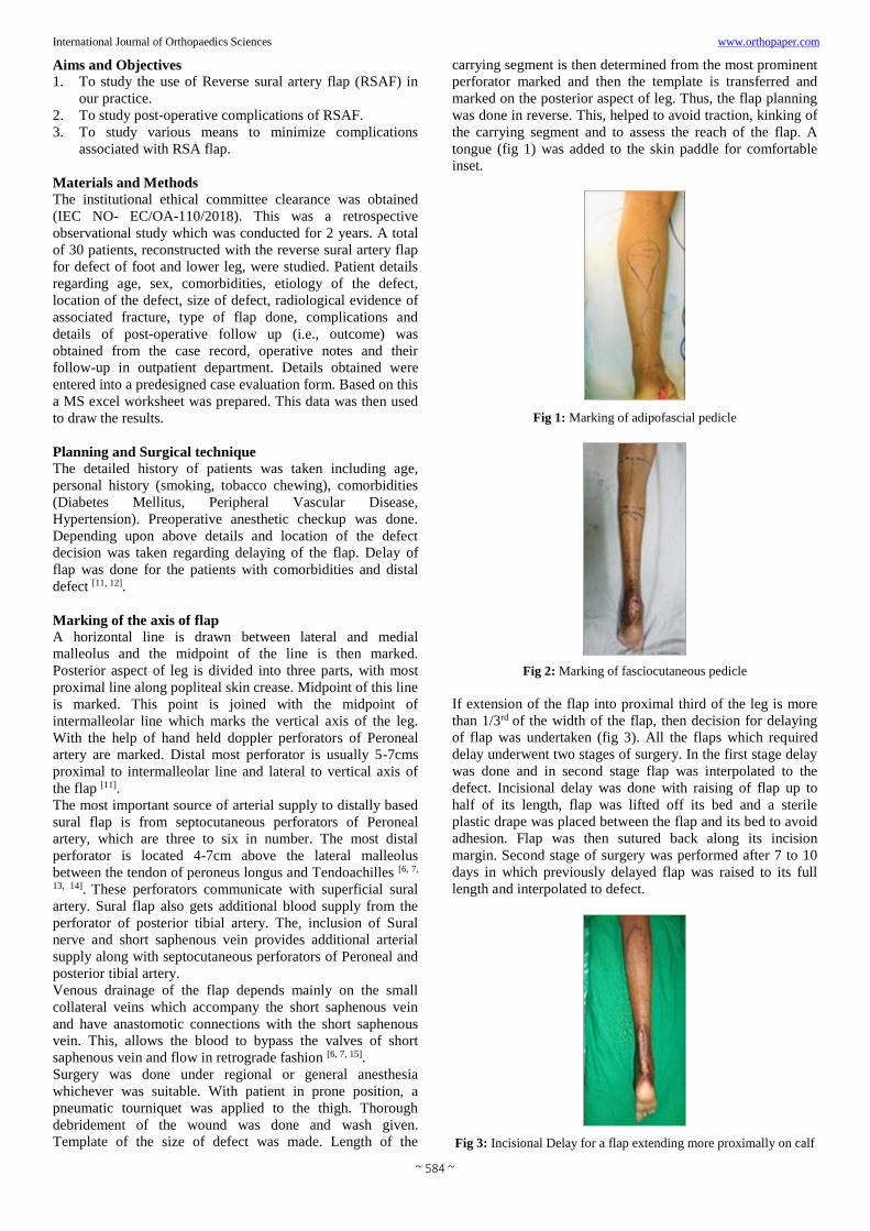
If extension of the flap into proximal third of the leg is more
than 1/3rd of the width of the flap, then decision for delaying
of flap was undertaken (fig 3). All the flaps which required
delay underwent two stages of surgery. In the first stage delay
was done and in second stage flap was interpolated to the
defect. Incisional delay was done with raising of flap up to
half of its length, flap was lifted off its bed and a sterile
plastic drape was placed between the flap and its bed to avoid
adhesion. Flap was then sutured back along its incision
margin. Second stage of surgery was performed after 7 to 10
days in which previously delayed flap was raised to its full
length and interpolated to defect.
Fig 3: Incisional Delay for a flap extending more proximally on calf

~ 585 ~
International Journal of Orthopaedics Sciences www.orthopaper.com For adipofascial pedicle the first incision was taken along the
length of the flap, which is along the previously marked
vertical axis of the leg (fig.1). Sural nerve and Short
Saphenous vein were identified. Skin flap with minimal
adipose tissue underneath was raised on both sides.
Adipofascial pedicle of the width of 3.5-4cms was designed.
Incision was then taken along the leading edge of the skin
island. Skin, superficial fascia and deep fascia was incised in
step ladder manner to include extra fascia, about 1cm, beyond
the skin incision. Sural nerve and short saphenous vein were
then divided and ligated along with the fascia. Skin island
along with adipofascial pedicle was raised, with gentle
handling, up to just short of the most prominent peroneal
artery perforator marked previously. Flap was then
interpolated to defect and insetting was done.
Fig 4: Skin island with triangular tail and adipofascial pedicle
Interpolation can be done in three ways (fig 5 & 6): a)
exteriorizing the pedicle and wrapping the pedicle with split
skin graft; b) incising the skin bridge between the defect and
the base of the flap, insetting triangular tongue into it; c)
tunneling the flap. The first method requires second surgery
for division and insetting of pedicle while the latter two are
single stage procedures.
Donor area is closed primarily as much as possible and the
remaining donor area is covered with split skin graft.
Corrugated drain is placed beneath the flap.
Fig 5: Exteriorized pedicle in defect around ankle
Fig 6: Incised skin bridge with triangular tongue inset
Fasciocutaneous pedicle was done in patients with
comorbidities (fig 7). In these cases, the donor site raw area is
larger as compared to adipofascial pedicle. Donor area is
reduced and covered with split skin graft.
Fig 7: RSA flap with fasciocutaneous pedicle
Cross leg RSA was done in patients with middle 1/3rd and
upper 1/3rd leg defect for which other treatment options were
not suitable.
Fig 8: Cross RSA flap for defect of middle third of opposite limb

~ 586 ~
International Journal of Orthopaedics Sciences www.orthopaper.com
Fig 9: Post op follow up shows well settled flap
Dressing was done by using Gamgee (cotton) rolls and Plaster
of Paris slab was applied on posterior aspect of leg. To avoid
pressure on pedicle and flap we used glove balloons for
molding the plaster slab (fig 10). Post operatively limb
elevation was given to decrease the oedema.
Fig 10: Glove balloons used for molding of plaster slab. A proximal
empty space shows a molded plaster after removal of glove balloon
Results It was a retrospective observational study of cases operated
over a duration of 2 years. A total of 30 patients between the
age of 10 years to 65 years were studied. 25 patients were
male and 5 were female. Flap was harvested from ipsilateral
side in 27 patients and from opposite leg as cross leg RSA
flap in 3 patients. RSA flap with adipofascial pedicle and
fasciocutaneous pedicle was done in 21 and 9 patients
respectively. Most common etiology for which this procedure
was done was post traumatic leg defect (26 patients). Other
etiologies were post infective leg defect (2 patients), one
patient each of non-healing ulcer and malignancy
(melanoma).
Bar Diagram 1: Etiology
Decision regarding delaying of flap was taken in 9 patients.
Most common indication for delay was distal defect
(Sole/Heel). Three patients had compromised vascularity of
leg due absent anterior tibial artery, two patients had
Peripheral vascular disease and other two had Diabetes
mellites’ patient each had history of tobacco chewing and
Hypertension. Most common site of defect which was
reconstructed with the flap was on the Tendoachilles region (7
patients) followed by Heel region (6 patients). Defect on
lower 1/3rd leg and lateral malleolus was reconstructed in 4
patients each. The cross-leg flap done for middle third and
upper third defect of opposite leg.
Fig 11: Location of defect Fig 12: Location of defect

~ 587 ~
International Journal of Orthopaedics Sciences www.orthopaper.com
Fig 13: Location of defect Fig 14: Location of defect
Operating time of surgery ranged from minimum of 90
minutes to maximum of 180 minutes with mean operating
time of 124 minutes. We have reconstructed wound size
ranging from maximum of 20X10cms to minimum size of
5X3cms. We have raised flap ranging from 9- 16cms in
length and 5-20cms in width. For all flaps, the pivot point was
at least 5cms proximal to intermalleolar line.
Out of 21 patients in which Island flap with adipofascial
paddle was done; all, but one, were less than 50years of age.
Out of this one patient had Tobacco addiction and one had
absent ATA. The patient who was more than 50 years of age,
developed complication of necrosis of the lateral skin flaps,
which were raised while harvesting adipofascial pedicle.
Out of 9 patients in which Fasciocutaneous pedicle was done,
six patients were less than 50years of age; of which five
patients had comorbidity (absent major vessel, Peripheral
vascular disease) (Chart 2) and other three patients who were
more than 50 years of age, one had Hypertension and other
one had Diabetes mellitus.
Bar diagram 2: Patient factors
All the flaps survived except for one which underwent partial
necrosis, done in patient who underwent Syme’s amputation
for post traumatic leg defect. necrosed skin of the flap was
debrided and grafted. Most common complications were; loss
of graft used to cover adipofascial pedicle along with skin
tongue, in cases were insetting of skin tongue alone would
cause compression of pedicle (2 patients) and necrosis of skin
flap on calf raised while harvesting adipofascial pedicle (2
patients). In both cases raw area formed was later grafted.
Venous congestion; hematoma formation and Partial skin
necrosis, up to 5% of the flap, which healed spontaneously,
were other complications which occurred in one patient each,
but all these flaps were salvaged. None of the patients
complained of any effect on routine activity during
postoperative follow up.

~ 588 ~
International Journal of Orthopaedics Sciences www.orthopaper.com
Fig 15: Necrosis of skin in adipofascial pedicle Fig 16: Partial necrosis of flap
Bar diagram 3: Complications
Note – All flaps were salvaged
Flowchart: Summary of all flaps co-morbidity and Complications

~ 589 ~
International Journal of Orthopaedics Sciences www.orthopaper.com Discussion The soft tissue defect around the foot, ankle and lower leg are
difficult to reconstruct due notoriously poor wound healing
secondary to poor blood supply [9]. Reconstruction with free
tissue transfer is an ideal option. However, it has limitations
such as long operating time, surgeon’s microsurgical expertise
and donor site morbidity [9, 10]. Introduction of distally based
fasciocutaneous flap and its latter modification by Masquelet
et al. [2] into neurocutaneous island flap offers a viable
alternative to conservative options like foot offloading,
repeated debridement or amputation of limb. And also, such
procedure has shorter operating time, learning curve; harvest
is straight forward and it preserves major vessels of limb [16].
Trauma was the most common cause of leg defect in our
study, which was similar to many other studies [8, 9]. Other
indications for which we used this flap was post infective
defect, osteomyelitis, chronic non healing ulcer, amputation
stump reconstruction (Syme’s amputation) [8, 9, 17].
In literature of this flap continues to be associated with
complications like flap necrosis, venous congestion and other
complications [6, 7, 8]. In our study one patient each had partial
flap necrosis and venous congestion, which is less (6%)
compared to studies which had reported a rate of 5-36% of
major ischemic event, like partial or total flap necrosis, with
reverse sural artery flap [18, 19, 20].
It has been mentioned in many studies that venous congestion
and not the inadequate blood supply is a major cause of flap
necrosis [9]. In our patients to minimize these complications
we took measures mentioned in literature [5, 6, 8, 9, 11, 15] and also
by using some of our custom modifications. Various measures
taken by us to improve venous drainage includes; 1)
incorporating saphenous vein in flap (which helps to
incorporate noncontinuous perforators), 2) exteriorize the
pedicle (to avoid compression of the pedicle; thus improving
the venous drainage), 3) wide pedicle with extra fascial cuff
beyond the distal skin margin of the flap (it helps to
incorporate the additional arterial anastomotic network along
the length of pedicle and thus further improving the
vascularity of the flap), 4) gentle handling of flap (thus
reduces trauma and post-operative inflammation in the flap),
5) use of glove balloons to mold the plaster slab to avoid
pressure, 6) post-operative limb elevation of about 30 degrees
to improve the venous drainage.
We have successfully used the flap for distal defect of sole,
midfoot region (1 case) and dorsum of foot (2 cases). In all
these patients we preferred delaying of flap. It has been
shown that a delay can significantly increase blood
circulation, where the distal portion of the flap is random
pattern [8, 11], Some of the important intraoperative
considerations we included in all of our procedures were: a)
Loupe magnification for dissection; b) Meticulous dissection
to avoid shearing of vascular pedicle and Sural nerve from the
flap; c) Inclusion of fascia beyond the distal margin by
making incision in step ladder manner; d) Lastly, use of glove
balloon for molding of plaster dressing to avoid pressure over
vascular pedicle.
As seen in literature, some believe that reverse sural artery
flap is not suitable for reconstruction of weight bearing area
of foot as it is associated with numbness [17]. In our study,
total nine patients underwent reconstruction of weight bearing
area (i.e., heel, sole), none of the patient had numbness
significant enough to interfere with routine activities. Also, to
further increase the success rate we designed adipofascial
pedicle; in young patients, those without comorbidities and
addiction; to increase reach of the flap and decreases donor
site morbidity. Other precautions that we took to decrease the
donor site complication in this set of patients were, raising the
skin flap with adequate adipose tissue carpet to preserve the
subdermal plexus [6]. Inspite, of which two patients had skin
flap necrosis on calf; of which one had tobacco addiction and
other patent was more than 50years of age. Hence a
fasciocutaneous pedicle is a better option in >50 years
patients and those with co-morbidities.
With all these precautions and modifications reverse sural
artery flap can be very effectively used for reconstruction of
distal limb defect when more complex options like free tissue
transfer is not possible.
Conclusion
The Reverse sural artery flap can be used as a very effective
alternative to free flap for various distal leg and foot defects.
It is a versatile flap and different scenario can be managed
with customization in plan; For example, in patients with
vascular disease and older age, delaying of flap and
fasciocutaneous flap are preferred method. On the other hand,
in young patients it can be used with an adipofascial pedicle,
as a single stage procedure. We can use this flap in patients
with compromised vascularity, single vessel limb where free
flap and cross leg flap is difficult. Thus, with proper selection
of patients, RSA can be judiciously used to manage different
leg defects.
References
1. Jolly G, Zgoni T. Soft Tissue Reconstruction of the Foot
with a Reverse Flow Sural Artery Neurofasciocutaneous
Flap Ostomy Wound Manage 2004;50(6):44-49.
2. Masquelet AC, Romana MC, Wolf G. Skin island flaps
supplied by the vascular axis of the sensitive superficial
nerves: anatomic study and clinical experience in the leg.
Plast Reconstr Surg 1992;89(06):1115-1121
3. Dhua S, Manashree S, Tilak BG. The Clinical Outcome
of Perforator Based Sural Artery and Propeller Flaps in
Reconstruction of Soft Tissue of Extremities. World J
Plast Surg 2019;8(1):3-11. doi: 10.29252/wjps.8.1.3.
4. Bhatt YC, Singh S, Doshi P, Vaghani SG. Reverse
Peroneal Artery Flap for Large Heel and Sole Defects: A
Reliable Coverage. World J Plast Surg 2017;6(2):212-
219.
5. Motamed S, Massod Yavari, Reza Rafiee, Hamid Reza
Hallaj Mofrad, Feaz Niazi Shahraki. Distally based sural
artery flap without sural nerve. Acta Medica
Iranica 2010;48(2):127-129.
6. Follmar Keithe, Baccarani Alessio, Baumeister Steffenp,
Levin Lscott, Erdmann Detlev. The Distally Based Sural
Flap. Plastic and Reconstructive Surgery.
2007;119(6):138e-148e
7. Ciofu RN, Zamfirescu DG. Reverse Sural flap for ankle
and heel soft tissues reconstruction Journal of Medicine
and Life 2017;10(1):94-98
8. Hassanpour S, Mohammadkhah N. Extraction of reverse
sural artery flap from the proximal third of the leg
Archives of Iranian Medicine 2008,11(2).
9. Bindesh et al. Reverse Sural Fascio Cutaneous Flap for
Soft Tissue Coverage around Foot and Ankle
International Journal of Science and Research (IJSR)
2015,4(2).
10. Sugg et al. The Reverse Superficial Sural Artery Flap
Revisited for Complex Lower Extremity and Foot
Reconstruction Plast Reconstr Surg Glob Open
2015;3:e519; doi: 10.1097/GOX.0000000000000500;

~ 590 ~
International Journal of Orthopaedics Sciences www.orthopaper.com Published online 22 September 2015.)
www.PRSGlobalOpen.com
11. Ramesha KT, Prakash Kumar MN, Shankarappa M.
Extended Reverse Sural Artery Flap’s Safety, Success
and Efficacy - A Prospective Study Journal of Clinical
and Diagnostic Research. 2014;8(5):NC08-NC11
12. Parrett BM, Pribaz JJ, Matros E, Przylecki W, Sampson
CE, Orgill DP. Risk analysis for the reverse sural
fasciocutaneous flap in distal leg reconstruction. Plast
Reconstr Surg 2009;123(05):1499-1504
13. Syed Kamran Ahmed, Boris Kwok Keung Fung. The
versatile reverse flow sural artery neurocutaneous flap: A
case series and review of literature Journal of
Orthopaedic Surgery and Research 2008;3:15
14. Kumar Satish K, Sravanan R, Ahamed Aasik. Analysis of
The Reverse Suralartery Flapas An Option for
Reconstruction of Defect of Leg and Foot. Indian Journal
of Applied Research 2019,9(4).
15. Bista N et al. The reverse sural fasciocutaneous flap for
the coverage of soft tissue defect of lower extremities
(distal 1/3 leg and foot) Nepal Med Coll J 2013;15(1):56-
61
16. Fraccalvieri M, Verna G, Dolcet M, Fava R, Rivarossa A,
Robotti E et al. The distally based superficial sural flap:
our experience in reconstructing the lower leg and foot.
Ann Plast Surg 2000;45:132-139.
17. Larrañaga et al. Reconstruction of Hind and Mid-Foot
Defects. The Surgery Journal 2017,3(3).
18. Rajacic N, Darweesh M, Jayakrishnan K, Gang RK, Jojic
S. The distally based superficial sural flap fo
reconstruction of the lower leg and foot. Br J Plast Surg
1996;49:383-389.
19. Yilmaz M, Karatas O, Barutcu A. The distally based
superficial sural artery island flap: clinical experiences
and modifications. Plast Reconstr Surg 1998;102:2358-
2367.
20. Touam C, Rostoucher P, Bhatia A, Oberlin C.
Comparative study of two series of distally based
fasciocutaneous flaps for coverage of the lower one
fourth of the leg, the ankle, and the foot. Plast Reconstr
Surg 2001;107:383-392.