vaccine title: leishmaniae-induced visceral leishmaniasis...
TRANSCRIPT

DRAFT FOR PEER REVIEW 11/17/06
Vsci519 F06 Vaccine Proposal Group 3 1
Vaccine title: Leishmaniae-induced Visceral Leishmaniasis Protection via a Subunit
Vaccine
Pathogen: Leishmania spp.
Vaccine type: Subunit vaccine with cytokine and lipopolysaccharide/attenuated
Salmonella adjuvants
Subject population: BALB/c mice (susceptible strain) vs. C57Bl/6 mice (resistant strain;
control)
Signatures of investigators

DRAFT FOR PEER REVIEW 11/17/06
Vsci519 F06 Vaccine Proposal Group 3 2
Literature Review Leishmaniasis is the second largest parasitic killer after malaria infecting 12 million people worldwide with over a half million deaths a year (Selvapandivan et al., 2005). This disease is most prevalent in the Indian subcontinent and southwest Asia, but is widely distributed through the tropics (Garg and Dube, 2006). Leishmaniasis is clinically classified in three categories: cutaneous (CL), mucocutaneous (MCL) and visceral (VL). VL, also known as kala-azar or black fever due to the skin hyperpigmentation that eventually occurs, is believed to be the most devastating of these infections. A small percentage of infected individuals may show symptoms only of the skin response termed post kala-azar dermal leishmaniasis without a visceral manifestation, and others are asymptomatic and remain immune to symptomatic disease. Malnutrition and/or stress increase the possibility of conversion from an asymptomatic to an active disease state. VL is often fatal: with no treatment, death can occur within weeks for the most acute infections (S. Saha et al., 2006). With this in mind, the focus of this proposal will be on the development of a vaccine against visceral leismaniasis. The Leishmania parasite that causes VL invades the spleen, liver, and bone marrow of the reticuloendothelial system. VL-afflicted individuals initially present with a persistent cutaneous lesion that heals. Initial symptoms may also include irregularly-occurring fever, malaise, and headaches which become more frequent, followed by cough, diarrhea, dizziness, vomiting, bleeding disorders, splenomegaly, anemia, leukopenia and/or hypergammaglobinemia. A series of illnesses including oral and respiratory problems follow years later, which leads to inflammation and mutilation of the nose, mouth, oropharynx and trachea. As the disease persists, it becomes more difficult to treat, and in many cases eventually leads to death as a result of either malnutrition or respiratory compromise. (S. Saha et al., 2006) Though Leishmania infection can occur via blood/blood product transfusion (American Association of Blood Bankers, 2003), leishmaniasis is most often caused by the intracellular protozoan parasite, Leishmania spp. which is transmitted through the bite of the female sand fly (Phlebotomus spp.). VL infection involves many leishmania species, including the complex L. donavani, L. infantum and L. tropica of the old world (cases occurring mostly in Bangladesh, northeastern India, Nepal and Sudan); new world (countries including Brazil) species include L. chagasi and L. .amazonesis (Malla and Mahajan, 2006). All leishmania species undergo a digenetic life cycle (fig. 1) that involves an extracellular developmental stage in the sand fly vector and an intracellular developmental stage in mammals (Roberts, 2006). The gut of the sand fly carries extracellular motile, flagellated Lieshmania promastigotes. In a period of 4-25 days, parasites multiply, move toward the sand fly’s pharynx, and eventually block the fly’s esophagus. A feeding sand fly then infects its host by regurgitating leishmaniae into the dermis of the skin (Rogers et al., 2006). At this stage, the parasites undergo further changes losing flagella and becoming multiplying non-motile amastigotes. Phagocytosis of the parasites by macrophages then follows, and the amastigotes multiply in phagolysosomal vacuoles. When infected macrophages rupture, the infection spreads to other macrophages systemically amplifying the infection. In addition to the human reservoir, canines can also be infected; this has implications for disease spread between canines and humans, and for the development of drug-resistant strains as both hosts are treated.

DRAFT FOR PEER REVIEW 11/17/06
Vsci519 F06 Vaccine Proposal Group 3 3
Treatments for VL have changed significantly in recent years. In Italy, for example, pentavalent antimonal (Sb) complexes were widely used until the mid-90s. Though historically effective, side effects included pancreatitis and cardiac abnormalities, and treatment failures have increased; the failures may be attributable, in part, to the evolution of resistant strains in treated canine hosts (Gradoni et al., 2003). In recent years, however, treatment has shifted to liposomal amphoetericin B (L-AmB) and the oral form of this drug, miltefosine (Murray, 2006). In Gradoni and colleagues’ study of 593 patients (2003), L-AmB treatment had minimal side effects and fewer treatment failures, but Garg and Dube (2006) describe side effects including varied toxicity challenges and problems with glucose metabolism. The varied forms of amphoetericin are 30 to 50-fold more expensive than Sb-based drugs, making this option less useful in developing countries (Gradoni et al., 2003). Leishmania has been declared endemic in some areas and the World Health Organization (WHO) has categorized it as a category 1 (emerging and uncontrolled) disease. Though initially localized primarily to sub-Mediterranean regions, disease spread is an issue. Vector spread, differential attempts to eradicate sand flies, and the development of drug-resistant Leismania strains in drug-treated canines exacerbate the challenge. Human immigration from areas of greater prevalence and emigration into high-risk areas (e.g., by soldiers deployed to Iraq and Afghanistan) demonstrate that this is a disease not limited to developing countries. As HIV infections continue to spread, the VL infection risk to immunocompromised patients does as well. The significant morbidity and mortality, lack of an effective drug treatment, and prevalence in poorer developing countries where there is “insufficient access to or impetus for developing affordable new drugs” (Murray, et al., 2006) confirm the need for control of the vector. More importantly, these factors establish a great need for research in the area of potential vaccines. It is well established that spontaneous or drug-induced recovery from cutaneous leishmaniasis or visceral leishmaniasis leads to solid immunity against future infections. These concepts provide firm rational for vaccine development.

DRAFT FOR PEER REVIEW 11/17/06
Vsci519 F06 Vaccine Proposal Group 3 4
Immunology: There are two critical responses necessary for immune suppression of Leishmania infection: activation of macrophages (i.e., enabling them to kill intracellular amastigotes) and induction of antigen-specific gamma interferon-producing CD4+ T helper (Th1) lymphocytes. Studies have demonstrated that peripheral blood mononuclear cells (PBMC) from VL patients do not proliferate or produce gamma interferon (IFN-γ) when exposed to specific antigen in vitro (Ghalib et al., 1993). Observations in humans and in sensitive BALB/c mice compared to the resistant C57Bl/6 murine strain have demonstrated that modulation of effector cells and cytokines leads to immune evasion and disease progression (Garg and Dube, 2006). A major challenge for an effective immune response to Leishmania infection is that the pathogen is harbored in a variety of immune system cells including macrophages (MP), the antigen-presenting dendritic cells (DC), and PBMCs. The infection begins when the sand fly deposits both saliva and motile promastigotes into the dermis. Sand fly salivary proteins stimulate vasodilation, inhibit coagulation, produce immunomodulatory effects (Sacks & Kamhawi, 2001) and recruit neutrophils (PMNs) and macrophages (Zer et al., 2001). Secretion of PMN-attracting factor by Leishmania and interleukin 8 (IL-8, a PMN-attracting chemokine) by the PMNs themselves, as well as a documented uptake and destruction of Leishmania, demonstrate that parasite phagocytosis by PMNs is a critical early mechanism of pathogen control (Teixeria, 2006). Another critical cell in the immune response to VL is the MP, which plays four important roles in a VL response. Macrophages are the primary host for leishmania, serve as antigen presenters, are the source of other critical cytokines (e.g., TNFα and IL-1) that modulate the T-cell response, and, if stimulated by a Th1 cell, kill intracellular parasites via phagolysosomic digestion and via oxidative challenges to the pathogen. Additional monocytes are attracted initially by cytokines in the sand fly saliva and later by PMN-secreted cytokines such as macrophage inflammatory protein (MIP) (fig. 2). CCL2, a critical cytokine for MPs, induces H2O2-dependent and nitric oxide-dependent intra- and extra-cellular microbicidal activity. CCL2 also attracts natural killer lymphocytes (NK), DCs and more MPs (Ritter & Moll, 2000); decreases (atypical) responses to other LPS-bearing pathogens (Bogdon & Rollinghoff, 1998); prevents apoptosis of infected cells (Moore & Matlashewski, 1994); and prevents intracellular pathogen digestion. Lipophosphoglycan (LPG), a pathogen surface molecule that facilitates MP binding, also contributes to the MP’s inability to successfully digest intracellular Lieshmania by inhibiting phagosome-endosome fusion (Desjardins & Descoteaux, 1997). LPG also alters monocyte migration by decreasing adhesion molecule expression, inhibits induction/release of IL-1β (Hatzigeorgiou et al., 1996) and CCL2 (Bhattacharyya et al., 2002), and scavenges O2
- (Chan et al., 1989) which gives the amastigote time to resist lysosomal digestion. Thus, in the active disease state, though macrophages are recruited, they end up serving the pathogen as a host vs. killing it, and LPG plays a critical role in the lack of ability of MP to destroy the pathogen. In opposition to the response by the MP, the Th1 response is the dominant cell-mediated response (CMI) in a successful immune response to Leishmania infection; suppression of Th1 coupled with elevation of Th2 serve as the “hallmarks of active disease” (S. Saha, 2006). IL-12

DRAFT FOR PEER REVIEW 11/17/06
Vsci519 F06 Vaccine Proposal Group 3 5
induces INF-γ production and thus supports the effective Th1 response. In contrast, IL-4 stimulation of the antibody-producing Th2 response and IL-10-mediated suppression of the Th1 response are correlated with active disease (S. Saha, 2006). The critical IL-12 and IL-10 balance is affected by the level of activity of CD40 signaling: weak CD40 stimulation increases IL-10 expression via an ERK-1/2 dependent pathway, while strong stimulation upregulates MAPK-dependent IL-12 production (Mathur, 2004). It has also been shown that exposure to anti-IL-10 antibody of T cells harvested from VL patients restores all cytokine responses. More importantly, antigen-specific T cells co-producing IL-10 and IFN-γ have been shown to expand in response to L. donavani infection (Ghalib et al., 1993). These observations support the important role for IL-10 in the suppression of critical T-cell responses that are, in part, apparently responsible for the conversion to an active disease state. In addition to the involvement of IL-10, evidence points to a classical Th2 response in the progression to an active disease state. Elevated levels of the Th2 cytokine IL-4 have been reported after there is a failure of treatment, while elevated levels of IL-13 are associated with successful treatment. In active disease, high antibody titers are found; IgG, IgM, and IgE subclasses all increase but their role is unclear (S. Saha, 2006). As noted in the introduction, hypergammaglobulinemia is a symptom of disease. Saha (2006) argues that in normal disease progression, IgG may not only “fail to protect but also actually contribute to disease progression.” Other non-antigen specific cells and processes play a role in the immune response to this pathogen. Natural killer (NK) lymphocyte action appears to have a supportive but non-essential role in the immune response. Resistance to L. major, for example, can occur if the NK cell is the only lymphoctye missing from the response (Satoskar et al., 1999). However, NK cells cause cytolysis of infected cells and secrete INF-γ, a critical cytokine for the effective Th1-stimulated response. Mast cells are recruited early in the response; their numbers increase in early infection in the BALB/c (sensitive) mouse but not in the resistant C57Bl/6 strain indicating that they play a role in prevention of active disease (B. Saha, 2004). T-cell-secreted IL-3 induces proliferation of mast cell committed progenitors – particularly in the spleen and bone marrow – that are critical areas for the disease (B. Saha, 2004). Direct lysis via the alternative complement pathway is effective on a form of the promastigote that is less infective; however, the highly-infective metacyclic promastigotes phosphorylate membrane attack components (MAC) and thus prevent lysis of infected cells. As Lieshmania exhibits so many effective strategies for evading the immune system, and because treatment for this potentially fatal disease can be expensive or unavailable in developing countries, several attempts have been made to produce a vaccine. Attempts at vaccine design include the use of recombinant protein antigens, approaches involving DNA vaccines, and development of vaccines that include Th1-inducing adjuvants. Using live parasites to induce lesions and to prevent re-infection has been tried as well, but the risks associated with this method outweighed the benefits; not only is delivering fresh cultures of a live vaccine impractical, but there is also an increased chance for localized disease, as well as dissemination in the context of HIV. Another approach has been to use promastigotes deactivated by sonification or some other means, thus exposing the immune system to the entire repertoire of antigens. This approach has also resulted in mostly unfavorable results. It seems the immune

DRAFT FOR PEER REVIEW 11/17/06
Vsci519 F06 Vaccine Proposal Group 3 6
system is overwhelmed in some way and many effector cells are lost. With the establishment of animal models, it is highly anticipated that all factors causing hindrance to further vaccine development will soon be eliminated in order to move towards routine clinical trials. (Nakhasi et al., 2005 and Handman, 2001).

DRAFT FOR PEER REVIEW 11/17/06
Vsci519 F06 Vaccine Proposal Group 3 7
Vaccine Type and Administration Route : Developing an effective vaccine against a complex organism such as Leishmania – particularly given the varied methods this protozoan uses to evade the immune system – is difficult. Vaccines containing single antigens – even when coupled with potential adjuvants – have not been successful in preventing visceral leishmaniasis. Thus, this vaccine will include a variety of potential antigens including Leishmania surface proteins and saliva from the sand fly vector, an adjuvant, a blocking antibody against a cytokine that prevents an effective immune response, and cytokines that have been shown to support pathogen destruction. The combination of critical molecules in this subunit vaccine will support the multi-faceted immune response that is necessary for a successful innate and adaptive immune response to this complex parasitic pathogen. The first subunits in the vaccine are Leishmania molecules that will serve as antigens that will stimulate an antibody-mediated response. LPG and the 63-kDa surface metalloproteinase (gp63) are found on the surface of promastigotes of all Leishmania species, and are involved in several mechanisms of pathogenesis. First, complement-activated lysis is hindered by these molecules. LPG releases the membrane attack complex (C5b-C9). Gp63 converts C3b to aC3bi type molecule; C3bi then binds to the type 3 complement receptor (CR3) on host macrophages, facilitating phagocytosis (Bogdan, 1998). LPG and gp63 are each directly involved in uptake by macrophages by interacting with other receptors. This is important because Leishmania lack means of penetrating host cells for infection (Zambrano-Villa et al., 2002). Once inside the macrophage, LPG inhibits the phagosome-endosome fusion by interacting with the host protein kinase C even across the lipid bilayer (Bogdan, 1998). Gp63 protects against cytolysis, and prevents the respiratory burst until the protozoa matures to the amastigote stage (Bogdan, 1998). The pathogen in its amastigote form is better suited to cope with acidic conditions and the respiratory burst by the expression of catalase and superoxide dismutase enzymes (Zambrano-Villa et al., 2002). Both LPG and gp63 have a history of use in vaccine trials: Pinheiro and colleagues (2006) demonstrated that intra-nasal vaccination with LPG provides protection against CL in the murine model and several gp63 peptides have been successfully tested in animal models (Handman, 2001). Thus, because of their prevalence on all Leishmania, their multiple virulent attributes, and prior demonstrations of effectiveness, LPG and gp63 are prime targets for neutralizing antibody and T cell-mediated immunity provided by an effective vaccine. To further enhance the prevantative humoral response, the vaccine will also include saliva from the sand fly vector. Norsworthy and colleagues ( 2004 ) have demonstrated that sand fly saliva that is injected along with the promastigote pathogen when it feeds aids Leishmania in establishing a prolonged infection. In their study, mice were co-injected with L. amazonensis and vector saliva or L. amazonensis alone in the ear tissue. Those that received the saliva developed larger lesions, had a higher concentration of IL-10 within the ear and draining lymph nodes, and had a suppressed NO production over controls. IL-10 levels did not remain high, but a statistical difference was found in a small, early time frame when T cell differentiation takes place. (The significance of this effect will be further understood when cytokines included in the vaccine are addressed.) The saliva was not found to produce these effects alone, but is rather utilized by the Leishmania in some unknown mechanism. Developing an antibody response to the components of the vector saliva will aid the host in preventing infection in the natural setting.

DRAFT FOR PEER REVIEW 11/17/06
Vsci519 F06 Vaccine Proposal Group 3 8
The subunit vaccine will also include as adjuvants lipopolysaccharide (LPS) and an attenuated Salmonella bacterium (BCG). LPS will be added to stimulate antigen uptake and presentation for the activation of armed effector cells of the adaptive immune response. LPS is a common adjuvant, and was shown by Norsworthy et. al (2004) to increase nitric oxide production. However, LPS, like other effective adjuvants, generally causes a strong inflammatory response; though this is a hallmark for adjuvanticity, it may not be appropriate for use in human subjects. An alternative (or supplementary) adjuvant is BCG, an attenuated Salmonella bacterium, that has been successfully and safely used in anti-Leishmania therapy in humans (Handman, 2001). Perhaps the most important component of the proposed vaccine is forced cytokine direction. Because Leishmania is an intracellular pathogen that infects MP of the innate immune system, the primary focus must be to activate effector T cells. The type of CD4+ T cell response is of particular importance. A Th2-driven response leads to B cell activation and antibody production; though antibody may help prevent future infections by way of neutralization and complement fixation, it is insufficient for the complete removal of infection once it is established. Th1 cells however, activate host macrophages and lead to lasting immunity. In any immune response, an initial dominance of one type of CD4 T cells occurs. Once this ratio has been established it is relatively fixed, as each class of effector cell secretes cytokines preventing the development of the other. For this reason, the early stages of infection are vital to determining the eventual success of the immune response. It is during this time frame that the observed increase in IL-10, an anti-Th1 cytokine, occurs in the presence of sand fly saliva. Some species of Leishmania promote a Th2 response in order to establish lifelong infection. L. major promastigotes have been found to actively suppress transcription of the gene coding for IL-12 (Carrera et al., 1996), which is needed for Th1 development. These effects can be reversed by neutralizing undesired cytokines and providing those that promote effective immunity. Anti IL-10 antibody has been shown to enhance parasite killing in conjunction with IFN-γ (Vieth et al., 1004). Neutralization of another Th2 promoter, TGFα, has also shown similar positive results (Bogdan, 1998). This vaccine includes anti IL-10 neutralizing antibody to help prevent the negative role of the sand fly saliva during the vaccine trial itself. To further promote Th1 activation, the vaccine also includes IFN-γ and IL-12. By including cytokine influence, we hope not to completely eliminate a Th2 response, but rather to push the ratio more towards a Th1 response, especially during the first few days following injection. The combination of LPG, gp63, vector saliva, LPS and BCG, anti-IL-10 antibody, IFN-γ, and IL-12 will be given to both susceptible BALB/c and resistant C57BL/6 (control) mice by intradermal injection. Recently, it has become more typical to introduce vaccines in a manner similar to normal pathogen infection. In the case of leishmania infection, dermal inoculation more effectively mimics the sand fly bite than oral delivery or intravenous/intramuscular injection, and the intradermal infection model has been shown to be an effective strategy for murine VL vaccinations (Ahmed et al., 2003). Vaccine or saline injections (negative control) will be given to a group of VL-susceptible mice as well as a number of VL resistant mice that will serve as additional controls. BALB/c mice clones are used in many experiments with Leishmania; they are susceptible to infection due to a predominately Th2 response including high levels of IL-4 production and low IFNγ (Sacks and Noben-Trauth, 2002) and have a disease

DRAFT FOR PEER REVIEW 11/17/06
Vsci519 F06 Vaccine Proposal Group 3 9
course paralleling human VL (Garg and Dube, 2006). Other mice strains such as C57BL/6 and C3H/HeJ naturally have a Th1 response and are self-healing (i.e., they are disease-resistant). We expect that both mice strains will develop neutralizing antibody against proteins within the saliva of the sand fly, as well as LPG and gp63. More importantly, we hope to have a predominant Th1 response specific to LPG and gp63 peptides, as well as cytotoxic CD8 T cells. Three weeks after vaccination we will provide a challenge of live L. major transmitted by the natural vector to subsets of both sensitive and resistant strains of vaccinated mice as well as un-vaccinated mice of the same strains (see Table 1). If the vaccine is effective and safe, we will not see disease symptoms or other side effects in either of the two pathogen-challenged vaccinated strains or in the resistant mice (C57BL/6 – positive control), but we will see VL symptoms in the negative controls (unvaccinated BALB/c) mice.
DRAFT FOR PEER REVIEW 11/17/06
Vsci519 F06 Vaccine Proposal Group 3 10
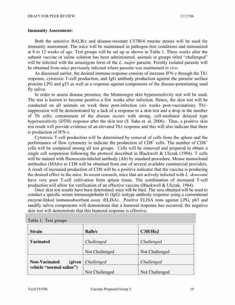
Immunity Assessment: Both the sensitive BALB/c and disease-resistant C57Bl/6 murine strains will be used for immunity assessment. The mice will be maintained in pathogen-free conditions and immunized at 8 to 12 weeks of age. Test groups will be set up as shown in Table 1. Three weeks after the subunit vaccine or saline solution has been administered, animals in groups titled “challenged” will be infected with the amastigote form of the L. major parasite. Freshly isolated parasite will be obtained from mice previously infected where parasite was maintained in vivo. As discussed earlier, the desired immune response consists of increase IFN-γ through the Th1 response, cytotoxic T-cell production, and IgG antibody production against the parasite surface proteins LPG and g53 as well as a response against components of the disease-potentiating sand fly saliva. In order to assess disease presence, the Montenegro skin hypersensitivity test will be used. The test is known to become positive a few weeks after infection. Hence, the skin test will be conducted on all animals on week three post-infection (six weeks post-vaccination). Th1-suppression will be demonstrated by a lack of a response to a skin test and a drop in the number of Th cells; containment of the disease occurs with strong, cell-mediated delayed type hypersensitivity (DTH) response after the skin test (S. Saha et. al, 2006). Thus, a positive skin test result will provide evidence of an elevated Th1 response and this will also indicate that there is production of IFN-γ.
Cytotoxic T-cell production will be determined by removal of cells from the spleen and the performance of flow cytometry to indicate the production of CD8+ cells. The number of CD8+ cells will be compared among all test groups. Cells will be removed and prepared to obtain a single cell suspension following the protocol described in Blackwell & Ulczak (1984). T cells will be stained with fluorescein-labeled antibody (Ab) by standard procedure. Mouse monoclonal antibodies (MAbs) to CD8 will be obtained from one of several available commercial providers. A result of increased production of CD8 will be a positive indicator that the vaccine is producing the desired effect in the mice. In recent research, mice that are actively infected with L. donovani have very poor T-cell cultivation from spleen tissue. The combination of increased T-cell production will allow for verification of an effective vaccine (Blackwell & Ulczak, 1984).
Once skin test results have been determined, mice will be bled. The sera obtained will be used to conduct a specific serum immunoglobulin G (IgG) isotype antibody response using a conventional enzyme-linked immunoabsorbant assay (ELISA). Positive ELISA tests against LPG, p63 and sandfly saliva components will demonstrate that a humoral response has occurred; the negative skin test will demonstrate that this humoral response is effective.
Table 1. Test groups
Strain Balb/c C3H/HeJ
Challenged Challenged Vacinated
Not Challenged Not Challenged
Challenged Challenged Non-Vacinated (given vehicle “normal saline”)
Not Challenged Not Challenged

DRAFT FOR PEER REVIEW 11/17/06
Vsci519 F06 Vaccine Proposal Group 3 11
Fig. 1: Leishmania Life Cycle (from Handman, 2001)

DRAFT FOR PEER REVIEW 11/17/06
Vsci519 F06 Vaccine Proposal Group 3 12
Fig. 2: Critical Cytokines in Immune Response to Leishmania (from Teixeira, et al., 2006)

DRAFT FOR PEER REVIEW 11/17/06
Vsci519 F06 Vaccine Proposal Group 3 13
Information Sources: Ahmen, S. M. Colmenares, L. Soong, K. Goldsmith-Pestana, L. Munstermann, R. Molina, and
D. McMahon-Pratt. Intradermal infection model for pathogenesis and vaccine studies of murine visceral leishmaniasis. Infection and Immunity, 71: 401-410, 2003.
American Association of Blood Bankers, Deferral for risk of leishmaniasis exposure. AABB Bulletin 2003: 3-14. 2003
Bhattacharyya, S. et al. Chemokine-induced leishmanicidal activity in murine macrophages via the generation of nitric oxide. Journal of Infectious Disease. 185: 1704-1708, 2002
Blackwell, Jennifer M., Ulczak, Orysia M. Immunoregulation of genetically controlled aquired responses to Leishmania donovani infection in mice: demonstration and characterization of suppressor T cells in noncure mice. Infection and Immunity. April: 97-102, 1984
Bogdan, C and Rollinghoff, M. The immune response to Leishmania: mechanisms of parasite control and evasion. International Journal for Parasitology, 28: 121-134, 1998
Carrera, L. et al. Leishmania promastigotes selectively inhibit interleukin-12 induction in bone marrow-derived macrophages from susceptible and resistant mice. Journal of Exploratory Medicine. 183: 515-526, 1996
Chan, J., Fumiwara, T., & Brennan, P. Microbial glycolipids: possible virulence factors that scavenge oxygen radicals. Proceedings of the National Academy of Sciences. 86: 2453-2457, 1989
Desjardins, M. & Descoteaux. A. Inhibition of phagolysosomal biogenesis by the Leishmania lipophosphoglycan. Journal of Experimental Medicine, 185: 2061-2068, 1997
Garg, R. and Dube, A. Animal models for vaccine studies for visceral leishmaniasis. Indian Journal of Medical Research, 123: 439-454, 2006
Ghalib W, Piuvezam M, Skeiky Y. , Siddig M, Hashim F, el-Hassan M., Russo D, and Reed S Interleukin 10 production correlates with pathology in human Leishmania donovani infections. Journal of Clinical investigations 92(1): 324–329, 1993
Gradoni, L., Gramiccia, & Scalone, A. Visceral lieshmaniasis treatment, Italy. Emerging Infectious Diseases, 9: 1617-1620, 2003 primary
Handman, E. Leishmaniasis: current status of vaccine development. Clinical Microbiology Reviews. 14: 229-243, 2001
Hatzigeorgiou, DE, Geng, J. and Zhu, B. Lipophosphoglycan from Leishmania suppresses agonist-induced interleukin-1� gene expression in human monocytes via a unique promoter sequence. Proceedings of the National Academy of Sciences USA. 93:14708-14712, 1996
Islam, M.A., Itoh, M., Shamsuzzamann S.M., Mirza, R., Matin, F., Ahmed,I., Shamsuzzaman Choudhury, A.K.M., Akram Hossain, M., Zui, X., Begam, N., Furuya, M., Leafasia, J.L., Hashiguchi, Y., Kimura,E. Diagnosis of Visceral Leishmaniasis by Enzyme linked Imunosorbent Assay Using Urine Samples. Clinical and Diagnosis Laboratory Immunology, July: 789-794, 2002
Kurstak, E. 1985. Progress in enzyme immunoassays: production of reagents, experimental design and interpretation. Bulletin of the World Health Organization 63:793-811.
Malla N. and Mahajan R. Pathophysiology of visceral leishmaniasis – some recent concepts. Indian Journal of Medical Research 267-274, 2006
Mary,C., Auriault, V., Faugere, B., Dessein, A. Control on Leishmania infantum infection is associated with CD8+ and gamma interferon- and interleukin-5-producing CD4+ antigen-specific T cells. Infection and Immunity, Nov.: 5559-5566, 1999

DRAFT FOR PEER REVIEW 11/17/06
Vsci519 F06 Vaccine Proposal Group 3 14
Mathur, R., Awasthi, A., Wadhone, P., Ramanamurthy, B., Saha, B. Reciprocal CD40 signals through p38 MAPK and ERK-1/2 induce counteracting immune response. Nature Medicine, 10: 540-544 (2004)
Moore, KJ & Matlashewski, G. Intracellular infection by Leishmania donavani inhibits macrophage apopotosis. Journal of Immunology. 152: 2930-2937, 1994
Murray, H., Berman, J., Davies, D., & Saravia, N. Advances in leishmaniasis. Lancet. 367:112- . 2006
Pinheiro, R, E. Pinot, H, Guedes, O. Filho, K de Mello, E. Saraiva, S. de Mendonca and B. Rossi-Bergmann. Protection against cutaneous leishmaniasis by intranasal vaccination with lipophosphoglycan. Vaccine (in press), 2006
Ritter, U. and Moll, H. Monocyte chemotactic protein-1 stimulates the killing of Leishmania major by human monocytes, acts synergistically with IFN-γ and is antagonized by IL-4. European Journal of Immunology. 30: 3111-3120, 2000
Roberts M., Current understandings on the immunology of leishmaniasis and recent developments in prevention and treatment. British Medical Bulletin, 75 and 76:115-130, 2006
Rogers R., Sizova O., M. Ferguson, Nikolaev A., Bates P. Synthetic Glycovaccine Protects against the bite of Leishmania-Infected Sand Flies. The Journal of Infectious Diseases, 194:512-8, 2006 PrimaryNorsworthy, N., Sun, J., Elnaiem, D., Lanzaro, G., and Soong, L. Sand Fly saliva enhances Lieshmania amazonesis infection by modulating interleuking-10 production. Infection and Immunity. 72: 1240-1247, 2004
Sacks, D and Kamhawi, S. Molecular aspects of parasite-vector and vector-host interactions in leishmaniasis. Annual Review of Microbiology. 55: 453-483, 2001.
Sacks, D., and Noben-Trauth, N. The immunology of susceptibility and resistance to Leishmania major in mice. Nature Review of Immunology. 2: 845-858, 2002
Saha, S., Mondal, S., Banerjee, A., Ghose, J., Bhowmick., S. & Ali, N. Immune responses in kala-azar, Indian Journal of Medical Research, 123: 245-266, 2006
Saha, B., Tonkal, A., Croft, S., Roy, S. , Mast cells at the host-pathogen interface: host-protection verus immune evasion in leismaniasis. Clinical Exploratory Immunology, 137: 19-23, 2004
Satoskar, A.R. et al. Mice lacking NK cells develop an efficient Th1 response and control cutaneous Leishmania major infection. Journal of Immunology 162: 6747-6754, 1999
Selvapandiyan, A., Duncan, R., Debrabant, A., Lee, N., Sreenivas, G., Salotra, P. & Nakhashi, H. Genetically modified live attenuated parasites as vaccines for leismaniasis. Indian Journal of Medical Research 123:455-466, 2006
Teixeira, M., Teixeira, C., Andrade, B., Barral-Netto, M., Barral., A. Chemikines in host-parasite interactions in leishmaniasis. Trends in Parasitology 22: 32-40, 2006 Review
Vieth, M., Will, A., Schroppel, K., Rollinghoff, M., and Gessner, A. Interleukin-10 inhibits antimicrobial activity against Leishmania major in murine macrophages. Scandanavian Journal of Immunology. 40: 403-409, 1994
Zambrano-Villa, S., Rosales-Borjas, D., Carrero, J.C., and Ortiz-Ortiz, L. How protozoan parasites evade the immune response. Trends in Parasitology 18: 272-278, 2002
Zer, R. et al. Effect of sand fly saliva on Leishmania uptake by murine macrophages. International Journal of Parasitology. 31: 810-814, 2001