theasthmacenter.org urticaria & angioedema1).pdf · urticaria & angioedema hives (or...
TRANSCRIPT

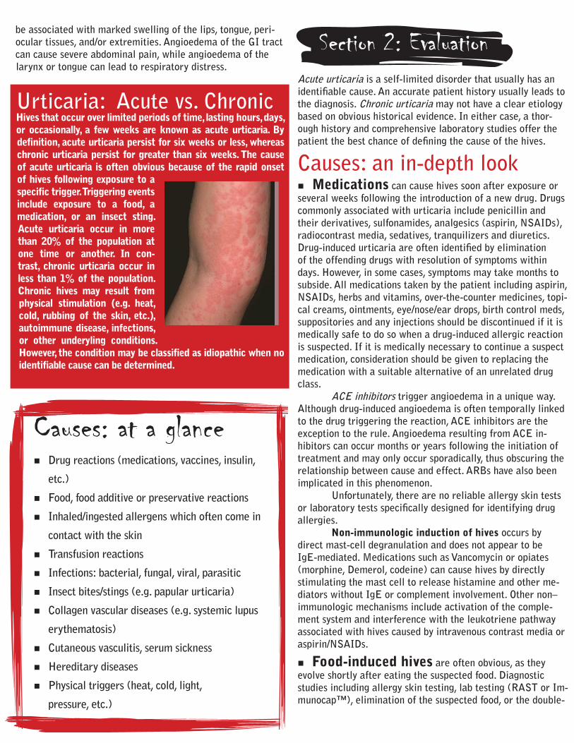
Urticaria & AngioedemaHives (or urticaria) are itchy, erythematous (reddish), often elevated skin lesions that blanch with pressure. Urticaria usually result from the release of mast-cell mediators (e.g. hista-mine, etc.) within the upper layers of the skin, causing blood ves-sel dilation, inflammation and local extravasation (or leakage) of intravascular fluid from capillaries into the skin. Hives come in all sizes and shapes, from tiny 2-3 mm to large irregularly shaped hives covering most of the body. Other erythematous, itchy rashes are often erroneously attributed to a diagnosis of hives. Such rashes include erythema multiforme, insect bites, cutaneous lymphoma, dermatitis herpetiformis, atopic dermatitis, porphyria, contact dermatitis, and dozens of other skin conditions. Individual inflammatory skin lesions persisting for days and resulting in lasting discolor-ation or hyper-pigmentation of the skin are probably not hives, but a result of some other inflammatory dis-order (e.g. vasculitis). Skin biopsy of such lesions may be helpful in reaching the correct diagnosis.
Physicians who specialize in urticaria manage-ment (e.g. allergists and dermatologists) often find it a difficult and complex disorder to diagnose and treat. Fortunately, even if an underlying cause cannot be identified, the majority of patients with hives can be successfully managed with a combination of medications and other preventive measures. Acute hives (detailed in the next section) are self-limited and resolve in days or weeks. In contrast, chronic hives recur over weeks, months and even years, but they can usually be con-
trolled with continuous use of medication. They often resolve following many months or
years of treatment.
Angioedema often results from a similar release of mediators, involving the
skin’s deeper cutaneous and sub-cutaneous tissues. In this case, more edema (fluid
accumulation and swelling) is present than that seen with
hives. Angioedema tends to
theasthmacenter.org
AdvisorAhe sthmaenter
TCEducation and Research Fund
Section 1: Definition
a test tube of warm water (44 degrees Fahrenheit) to the arm for 4-5 minutes to check for the evolution of hives.
Cholinergic or systemic heat urticaria Cholinergic urticaria consists of tiny wheals (2-3 mm)
surrounded by a large border of erythema. Triggers include exercise, anxiety, sweating and hot showers. Typically, hives begin over the upper body and spread across the skin sur-face. An intradermal injection of methacholine reproduces these tiny hives and is diagnostic of this disorder. Running or bicycling for 15 minutes in an 85-degree room may be the best test. The treatment of choice for cholinergic ur-ticaria is hydroxyzine 100-200mg/24hrs in divided doses. Cetirazine has also proven effective.
Pressure urticaria Pressure urticaria is characterized by a delayed swell-
ing that occurs 4-6 hours after pressure has been applied. There is a great deal of local swelling, but no wheal and flare at the site. However, a biopsy reveals perivascular mononuclear cell infiltrates and dermal edema commonly seen in urticaria. A sling placed over the forearm or shoulder with a 10- or 20-pound weight for 10-20 minutes is an accurate test for this condition. The delayed swelling may present as burning or pain rather than itching. Pressure urticaria often affects the soles of the feet after standing for long periods of time, especially on hard, irregular surfaces. Treatment consists of low-dose corticosteroids (15-25 mg of prednisone every other day), since antihistamines and other common urticaria medications have proven ineffec-tive.
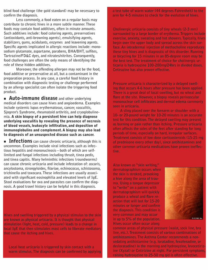
Dermatographism Also known as “skin writing,”
dermatographism occurs when the skin is stroked, provoking a hive along the area of trau-ma. Using a tongue depressor to “write” on a patient with dermatographism will quickly produce a wheal and flare re-action that will last for 15-20 minutes or longer and confirm the diagnosis. This condition is very common and may occur in up to 5% of the population. Hives occur often occur along common areas of physical pressure (waist, sock line, bra line, etc.). Treatment consists of various combinations of antihistamines. The Asthma Center recommends a non-sedating antihistamine (e.g. loratadine, fexofenadine, or desloratadine) in the morning and hydroxyzine, levocetiriz-ine (Xyzal) or Cetirizine at night. For persistent symptoms, raising hydroxyzine to 25-50 mg qid is often effective.
blind food challenge (the gold standard) may be necessary to confirm the diagnosis. Less commonly, a food eaten on a regular basis may contribute to chronic hives in a more subtle manner. These foods may contain food additives, often in minute amounts. Such additives include: food coloring agents, preservatives (antioxidants, anti-browning agents), emulsifying agents, stabilizers, gums, acidulants, enzymes and leavening agents. Specific agents implicated in allergic reactions include: mono-sodium glutamate, aspartame, parabens, BHA/BHT, sulfites, Tartrazine/FD&C dyes, and nitrates/nitrites. Consequently, food challenges are often the only means of identifying the role of these hidden additives. Moreover, the offending allergen may not be the food, food additive or preservative at all, but a contaminant in the preparation process. In any case, a careful food history in combination with diagnostic testing or challenge performed by an allergy specialist can often isolate the triggering food product.
n Auto-immune disease and other underlying medical disorders can cause hives and angioedema. Examples include systemic lupus erythematosus, cancer, vasculitis, Sjogren’s Syndrome, rheumatoid arthritis, and cryoglobuline-mia. A skin biopsy of a persistent hive can help diagnose underlying vasculitis by revealing the presence of necrosis of blood vessels, leukocyte infiltration, and deposition of immunoglobulins and complement. A biopsy may also lead to diagnosis of an unsuspected disease such as cancer.
n Infections can cause chronic urticaria, although this is uncommon. Examples include viral infections such as infec-tious hepatitis and mononucleosis—both of which are self-limited and fungal infections including thrush, tinea pedis, and tinea capitis. Many helminthic infections (roundworms) can cause chronic urticaria and include infestation of: ascaris, ancylostoma, strongyloides, filariae, echinococus, schistosoma, trichinella and toxocara. These infections are usually associ-ated with significant eosinophilia and elevated levels of IgE. Stool evaluations for ova and parasites can confirm the diag-nosis. A good travel history can be helpful in this diagnosis.
n Physical urticaria and angioedema
Hives and swelling triggered by a physical stimulus to the skin are known as physical urticaria. It is thought that physical stimulation (light, heat, cold, pressure) leads to a change in local IgE that then stimulates mast cells to liberate mediators that cause the itching and hives.
Local heat urticaria Local heat urticaria is triggered by skin contact with a
warm stimulus. The diagnosis can be confirmed by applying
be associated with marked swelling of the lips, tongue, peri-ocular tissues, and/or extremities. Angioedema of the GI tract can cause severe abdominal pain, while angioedema of the larynx or tongue can lead to respiratory distress.
Urticaria: Acute vs. ChronicHives that occur over limited periods of time, lasting hours, days, or occasionally, a few weeks are known as acute urticaria. By definition, acute urticaria persist for six weeks or less, whereas chronic urticaria persist for greater than six weeks. The cause of acute urticaria is often obvious because of the rapid onset of hives following exposure to a specific trigger. Triggering events include exposure to a food, a medication, or an insect sting. Acute urticaria occur in more than 20% of the population at one time or another. In con-trast, chronic urticaria occur in less than 1% of the population. Chronic hives may result from physical stimulation (e.g. heat, cold, rubbing of the skin, etc.), autoimmune disease, infections, or other underyling conditions. However, the condition may be classified as idiopathic when no identifiable cause can be determined.
Acute urticaria is a self-limited disorder that usually has an identifiable cause. An accurate patient history usually leads to the diagnosis. Chronic urticaria may not have a clear etiology based on obvious historical evidence. In either case, a thor-ough history and comprehensive laboratory studies offer the patient the best chance of defining the cause of the hives.
Causes: an in-depth lookn Medications can cause hives soon after exposure or several weeks following the introduction of a new drug. Drugs commonly associated with urticaria include penicillin and their derivatives, sulfonamides, analgesics (aspirin, NSAIDs), radiocontrast media, sedatives, tranquilizers and diuretics. Drug-induced urticaria are often identified by elimination of the offending drugs with resolution of symptoms within days. However, in some cases, symptoms may take months to subside. All medications taken by the patient including aspirin, NSAIDs, herbs and vitamins, over-the-counter medicines, topi-cal creams, ointments, eye/nose/ear drops, birth control meds, suppositories and any injections should be discontinued if it is medically safe to do so when a drug-induced allergic reaction is suspected. If it is medically necessary to continue a suspect medication, consideration should be given to replacing the medication with a suitable alternative of an unrelated drug class. ACE inhibitors trigger angioedema in a unique way. Although drug-induced angioedema is often temporally linked to the drug triggering the reaction, ACE inhibitors are the exception to the rule. Angioedema resulting from ACE in-hibitors can occur months or years following the initiation of treatment and may only occur sporadically, thus obscuring the relationship between cause and effect. ARBs have also been implicated in this phenomenon. Unfortunately, there are no reliable allergy skin tests or laboratory tests specifically designed for identifying drug allergies. Non-immunologic induction of hives occurs by direct mast-cell degranulation and does not appear to be IgE-mediated. Medications such as Vancomycin or opiates (morphine, Demerol, codeine) can cause hives by directly stimulating the mast cell to release histamine and other me-diators without IgE or complement involvement. Other non–immunologic mechanisms include activation of the comple-ment system and interference with the leukotriene pathway associated with hives caused by intravenous contrast media or aspirin/NSAIDs.
n Food-induced hives are often obvious, as they evolve shortly after eating the suspected food. Diagnostic studies including allergy skin testing, lab testing (RAST or Im-munocap™), elimination of the suspected food, or the double-
Causes: at a glance n Drug reactions (medications, vaccines, insulin,
etc.)
n Food, food additive or preservative reactions
n Inhaled/ingested allergens which often come in
contact with the skin
n Transfusion reactions
n Infections: bacterial, fungal, viral, parasitic
n Insect bites/stings (e.g. papular urticaria)
n Collagen vascular diseases (e.g. systemic lupus
erythematosis)
n Cutaneous vasculitis, serum sickness
n Hereditary diseases
n Physical triggers (heat, cold, light,
pressure, etc.)
Section 2: Evaluation
a test tube of warm water (44 degrees Fahrenheit) to the arm for 4-5 minutes to check for the evolution of hives.
Cholinergic or systemic heat urticaria Cholinergic urticaria consists of tiny wheals (2-3 mm)
surrounded by a large border of erythema. Triggers include exercise, anxiety, sweating and hot showers. Typically, hives begin over the upper body and spread across the skin sur-face. An intradermal injection of methacholine reproduces these tiny hives and is diagnostic of this disorder. Running or bicycling for 15 minutes in an 85-degree room may be the best test. The treatment of choice for cholinergic ur-ticaria is hydroxyzine 100-200mg/24hrs in divided doses. Cetirazine has also proven effective.
Pressure urticaria Pressure urticaria is characterized by a delayed swell-
ing that occurs 4-6 hours after pressure has been applied. There is a great deal of local swelling, but no wheal and flare at the site. However, a biopsy reveals perivascular mononuclear cell infiltrates and dermal edema commonly seen in urticaria. A sling placed over the forearm or shoulder with a 10- or 20-pound weight for 10-20 minutes is an accurate test for this condition. The delayed swelling may present as burning or pain rather than itching. Pressure urticaria often affects the soles of the feet after standing for long periods of time, especially on hard, irregular surfaces. Treatment consists of low-dose corticosteroids (15-25 mg of prednisone every other day), since antihistamines and other common urticaria medications have proven ineffec-tive.
Dermatographism Also known as “skin writing,”
dermatographism occurs when the skin is stroked, provoking a hive along the area of trau-ma. Using a tongue depressor to “write” on a patient with dermatographism will quickly produce a wheal and flare re-action that will last for 15-20 minutes or longer and confirm the diagnosis. This condition is very common and may occur in up to 5% of the population. Hives occur often occur along common areas of physical pressure (waist, sock line, bra line, etc.). Treatment consists of various combinations of antihistamines. The Asthma Center recommends a non-sedating antihistamine (e.g. loratadine, fexofenadine, or desloratadine) in the morning and hydroxyzine, levocetiriz-ine (Xyzal) or Cetirizine at night. For persistent symptoms, raising hydroxyzine to 25-50 mg qid is often effective.
blind food challenge (the gold standard) may be necessary to confirm the diagnosis. Less commonly, a food eaten on a regular basis may contribute to chronic hives in a more subtle manner. These foods may contain food additives, often in minute amounts. Such additives include: food coloring agents, preservatives (antioxidants, anti-browning agents), emulsifying agents, stabilizers, gums, acidulants, enzymes and leavening agents. Specific agents implicated in allergic reactions include: mono-sodium glutamate, aspartame, parabens, BHA/BHT, sulfites, Tartrazine/FD&C dyes, and nitrates/nitrites. Consequently, food challenges are often the only means of identifying the role of these hidden additives. Moreover, the offending allergen may not be the food, food additive or preservative at all, but a contaminant in the preparation process. In any case, a careful food history in combination with diagnostic testing or challenge performed by an allergy specialist can often isolate the triggering food product.
n Auto-immune disease and other underlying medical disorders can cause hives and angioedema. Examples include systemic lupus erythematosus, cancer, vasculitis, Sjogren’s Syndrome, rheumatoid arthritis, and cryoglobuline-mia. A skin biopsy of a persistent hive can help diagnose underlying vasculitis by revealing the presence of necrosis of blood vessels, leukocyte infiltration, and deposition of immunoglobulins and complement. A biopsy may also lead to diagnosis of an unsuspected disease such as cancer.
n Infections can cause chronic urticaria, although this is uncommon. Examples include viral infections such as infec-tious hepatitis and mononucleosis—both of which are self-limited and fungal infections including thrush, tinea pedis, and tinea capitis. Many helminthic infections (roundworms) can cause chronic urticaria and include infestation of: ascaris, ancylostoma, strongyloides, filariae, echinococus, schistosoma, trichinella and toxocara. These infections are usually associ-ated with significant eosinophilia and elevated levels of IgE. Stool evaluations for ova and parasites can confirm the diag-nosis. A good travel history can be helpful in this diagnosis.
n Physical urticaria and angioedema
Hives and swelling triggered by a physical stimulus to the skin are known as physical urticaria. It is thought that physical stimulation (light, heat, cold, pressure) leads to a change in local IgE that then stimulates mast cells to liberate mediators that cause the itching and hives.
Local heat urticaria Local heat urticaria is triggered by skin contact with a
warm stimulus. The diagnosis can be confirmed by applying
be associated with marked swelling of the lips, tongue, peri-ocular tissues, and/or extremities. Angioedema of the GI tract can cause severe abdominal pain, while angioedema of the larynx or tongue can lead to respiratory distress.
Urticaria: Acute vs. ChronicHives that occur over limited periods of time, lasting hours, days, or occasionally, a few weeks are known as acute urticaria. By definition, acute urticaria persist for six weeks or less, whereas chronic urticaria persist for greater than six weeks. The cause of acute urticaria is often obvious because of the rapid onset of hives following exposure to a specific trigger. Triggering events include exposure to a food, a medication, or an insect sting. Acute urticaria occur in more than 20% of the population at one time or another. In con-trast, chronic urticaria occur in less than 1% of the population. Chronic hives may result from physical stimulation (e.g. heat, cold, rubbing of the skin, etc.), autoimmune disease, infections, or other underyling conditions. However, the condition may be classified as idiopathic when no identifiable cause can be determined.
Acute urticaria is a self-limited disorder that usually has an identifiable cause. An accurate patient history usually leads to the diagnosis. Chronic urticaria may not have a clear etiology based on obvious historical evidence. In either case, a thor-ough history and comprehensive laboratory studies offer the patient the best chance of defining the cause of the hives.
Causes: an in-depth lookn Medications can cause hives soon after exposure or several weeks following the introduction of a new drug. Drugs commonly associated with urticaria include penicillin and their derivatives, sulfonamides, analgesics (aspirin, NSAIDs), radiocontrast media, sedatives, tranquilizers and diuretics. Drug-induced urticaria are often identified by elimination of the offending drugs with resolution of symptoms within days. However, in some cases, symptoms may take months to subside. All medications taken by the patient including aspirin, NSAIDs, herbs and vitamins, over-the-counter medicines, topi-cal creams, ointments, eye/nose/ear drops, birth control meds, suppositories and any injections should be discontinued if it is medically safe to do so when a drug-induced allergic reaction is suspected. If it is medically necessary to continue a suspect medication, consideration should be given to replacing the medication with a suitable alternative of an unrelated drug class. ACE inhibitors trigger angioedema in a unique way. Although drug-induced angioedema is often temporally linked to the drug triggering the reaction, ACE inhibitors are the exception to the rule. Angioedema resulting from ACE in-hibitors can occur months or years following the initiation of treatment and may only occur sporadically, thus obscuring the relationship between cause and effect. ARBs have also been implicated in this phenomenon. Unfortunately, there are no reliable allergy skin tests or laboratory tests specifically designed for identifying drug allergies. Non-immunologic induction of hives occurs by direct mast-cell degranulation and does not appear to be IgE-mediated. Medications such as Vancomycin or opiates (morphine, Demerol, codeine) can cause hives by directly stimulating the mast cell to release histamine and other me-diators without IgE or complement involvement. Other non–immunologic mechanisms include activation of the comple-ment system and interference with the leukotriene pathway associated with hives caused by intravenous contrast media or aspirin/NSAIDs.
n Food-induced hives are often obvious, as they evolve shortly after eating the suspected food. Diagnostic studies including allergy skin testing, lab testing (RAST or Im-munocap™), elimination of the suspected food, or the double-
Causes: at a glance n Drug reactions (medications, vaccines, insulin,
etc.)
n Food, food additive or preservative reactions
n Inhaled/ingested allergens which often come in
contact with the skin
n Transfusion reactions
n Infections: bacterial, fungal, viral, parasitic
n Insect bites/stings (e.g. papular urticaria)
n Collagen vascular diseases (e.g. systemic lupus
erythematosis)
n Cutaneous vasculitis, serum sickness
n Hereditary diseases
n Physical triggers (heat, cold, light,
pressure, etc.)
Section 2: Evaluation
Solar or actinic urticaria Hives and pruritus develop following brief exposure to
light is known as solar urticaria. Hives result from expo-sure to certain wavelengths of light and typically are lim-ited to sun-exposed areas. Symptoms often resolve quickly. However, in rare instances, generalized light exposure can lead to anaphylaxis. There are different types of solar urticaria, which are classified according to the wavelength of light that induces the eruption. A diagnosis is made by testing patients with specific wavelengths of light. Effec-tive therapy varies depending on the type of solar urticar-ia. Sunblock containing zinc oxide, titanium oxide, or both are particularly effective in avoidance treatment.
Aquagenic urticaria involves an outbreak of hives following contact with water that is not temperature-de-pendent. The diagnosis is confirmed by placing a compress with distilled or tap water against the skin for a few min-utes (the water should be tepid). Other forms of physical urticaria must be ruled out before making the diagnosis.
Cold urticaria is characterized by the rapid onset of pruritus, erythema and swelling following exposure to a cold stimulus. The swelling is local to the site of exposure. Initially, symptoms get worse as the skin is warmed. Swim-ming in cold water can lead to anaphylaxis and drowning. Therefore, individuals with cold urticaria should always swim with a buddy, and an Epi-pen™ should be kept read-ily available. Many cases of cold urticaria are IgE-mediat-ed, but cold urticaria may be associated with other dis-eases including: cryoglobulinemia, cold agglutinin disease, cryofibrinogenemia, and paroxysmal cold hemoglobinuria. The treatment of choice is with the antihistamine cypro-heptadine (Periactin) for cold urticaria.
Exercise-induced hives may be a variant of vibratory or heat-induced hives. Usually symptoms evolve in conjunc-tion with prolonged walks or exercising. Symptoms may be limited to intense itching or may evolve into general-ized hives, which ultimately subside following cessation of exercise. It is often best treated by taking an antihistamine 1-2 hours before exercising. A severe form of this phe-nomenon is that of exercise-induced anaphylaxis in which the patient becomes hypotensive and breaks out in hives. This latter form of exercise-induced hives is preventable in some patients by avoiding meals for 4 hours prior to exercise, which indicates that it may be a food-dependent reaction. Pre-treatment with an antihistamine is recom-mended, along with maknig sure an Epi-pen™ is on hand.
n Cutaneous vasculitis Biopsy of hives which last more than 24 hours may reveal
a necrotizing vasculitis involving small vessel infiltrates. Individual hives resolving within 24 horus without inflammation are rarely a sign of vasculitis. Immunofluorescent studies reveal the presence of immunoglobulins and complement. Systemic vasculitis with hives often has elevated sedimentation rates, arthralgia, fever, etc. Cutaneous vasculitis is often associated with low complement levels (hypocomplementemic vasculitis), in which the vasculitis is limited to the skin without other systemic involvement.
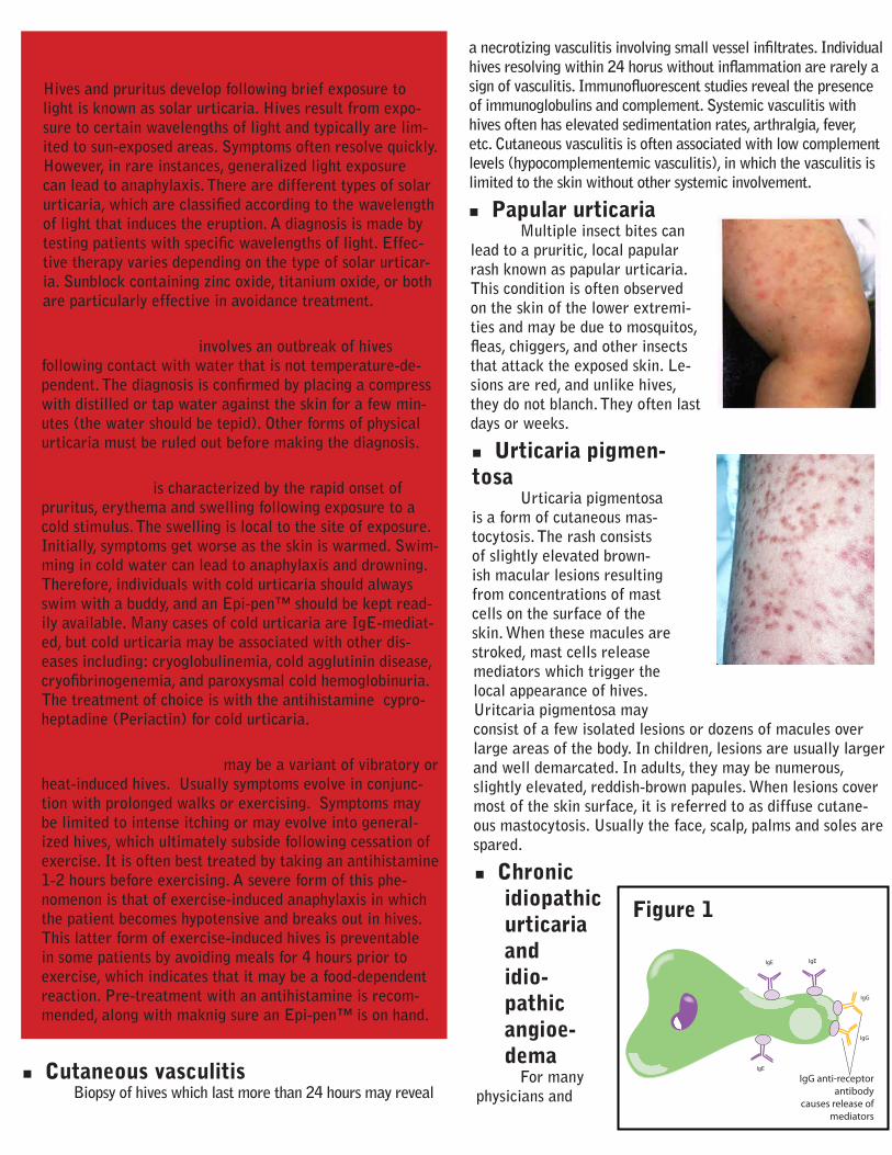
n Papular urticaria Multiple insect bites can lead to a pruritic, local papular rash known as papular urticaria. This condition is often observed on the skin of the lower extremi-ties and may be due to mosquitos, fleas, chiggers, and other insects that attack the exposed skin. Le-sions are red, and unlike hives, they do not blanch. They often last days or weeks.
n Urticaria pigmen-tosa Urticaria pigmentosa is a form of cutaneous mas-tocytosis. The rash consists of slightly elevated brown-ish macular lesions resulting from concentrations of mast cells on the surface of the skin. When these macules are stroked, mast cells release mediators which trigger the local appearance of hives. Uritcaria pigmentosa may consist of a few isolated lesions or dozens of macules over large areas of the body. In children, lesions are usually larger and well demarcated. In adults, they may be numerous, slightly elevated, reddish-brown papules. When lesions cover most of the skin surface, it is referred to as diffuse cutane-ous mastocytosis. Usually the face, scalp, palms and soles are spared.
n Chronic idiopathic urticaria and idio-pathic angioe-dema
For many physicians and
Figure 1
IgE
IgE
IgE
IgG
IgG
IgG anti-receptor antibody
causes release of mediators
patients, the management of chronic urticaria is frustrating because of the lack of an identifiable cause, and the variable response to medical therapy. Less than 25% of individuals with chronic urticaria have an identifiable cause of their rash. The remaining 75% are referred to as chronic idiopathic urticaria or hives “without a known cause.” If only a small number of chronic urticaria sufferers have an identifiable cause, why work up patients so thoroughly? There are two reasons: 1. In the event that a cause can be iden-tified, the trigger can either be eliminated or managed more ef-fectively with a focused, therapeutic approach. 2. It is medically necessary to rule out an underying medical condition. Patients with chronic idiopathic urticaria usually have normal blood work and no signs of underlying systemic disease. Some studies have shown that up to 40% of such patients ap-pear to have an autoimmune cutaneous disorder. Skin biopsy reveals a non–necrotizing perivascular, mononuclear cell in-filtrate. Frequently, an increase in mast cells and basophils is also noted. Further, the skin biopsy reveals an increase of CD4+ cells, neutrophils, and eosinophils, similar to that seen in late-phase allergic reactions. In the New England Journal of Medicine, Hide, Francis and Grattan, et al. reported the presence of anti-IgE recep-tor antibodies in 30-40% of patients with idiopathic urticaria. These patients suffer from an autoimmune disease in which auto-antibodies are directed at the IgE receptors on mast cells causing release of mediators which induce hives. (See Fig. 1)
Hereditary forms of urticaria and angioedemaFamilial cold urticaria This is a form of periodic fever, a rare form of cold intol-erance inherited as an autosomal dominant trait. Cold exposure causes a burning rash, fever, chills, arthralgia, myalgia, head-ache, and leukocytosis.
Hereditary vibratory angioedema This reaction consists of pruritus and swelling minutes after a vibratory stimulus. Treatment consists of avoidance of vibratory stimulation and use of antihistamines.
Hereditary angioedema (HAE) and acquired C1-INH deficiency Hereditary angioedema is due to an autosomal domi-nant disorder resulting in a decrease in the quantity and/or func-tion of C1-esterase inhibitor concentrate (C1-INH). It affects any part of the body, including the tongue, larynx, extremities and intestines. Hives and itching are always absent. Episodes often last days. GI symptoms often mimic an acute abdomen or abdominal obstruction. Biopsy shows no cellular infiltrate and the edema results from the release of vasoactive factors such as kinins. Low or absent C1-INH levels lead to complement consumption with decreases in C3 and C4. In fact, patients often have a low C4 level even if symptoms are not active. A de-creased quantitative C1-INH level is diagnostic, however 15%
of these patients may have normal quantitative C1-INH levels that are defective, requiring a functional assay for C1-INH to confirm the diagnosis. Treatment of hereditary angioedema is unique. It doesn’t respond to epinephrine, steroids, antihistamines or leukotriene antagonists, usually effective in treating al-lergic angioedema. Instead, the stimulation of the hepatic production of C1-INH by the use of anabolic steroids such as Stanazole is used to prevent attacks. Less com-monly, fresh frozen plasma concentrate infusions are used to treat acute symptoms. The good news is that C1-INH concentrate infusion had been available in Europe for decades and is now approved for use in the United States. An infusion of C1-INH concentrate will quickly resolve active symptoms of HAE by restoring the proper balance of inhibition of this system. When upper airway angioedema does not respond to medical treatment, a tracheotomy is the procedure of choice to restore a patient's airway. Finally, low C1-INH levels and resulting angioe-dema may be the result of an underlying autoimmune disease or cancer in which the underlying disease inac-tivates C1-INH and effectively creates the same clinical symptoms as seen in hereditary angioedema.
Angioedema, or deep swelling of the soft tis-sue, may be a result of drug allergy, food al-lergy, bee sting allergy, complement disorders, or autoimmune disease as well as other uncommon or rare disease processes (see table). In contrast to urticaria, angioedema is often not pruritic (itchy), but rather, can be painful and tender. Allergic triggers of angioedema are typically IgE-mediated and are often easily identified based on the demonstrated temporal relationship between exposure and development of symptoms. A family history of angioedema might point to a unique disorder known as CI-INH deficiency, or hereditary angioedema (HAE). In this disorder, there is a deficiency in the quantity and/or function of the enzyme C1-INH. This deficiency results in episodes of angioedema which can involve the skin, the airway, and/or the GI tract. Episodes of angioedema related to this disorder can be severe and life-threatening and respond poorly to conventional therapy. Unique to this form of angioedema is the absence of urti-caria or itching.
Section 3: Angioedema in-depth

Solar or actinic urticaria Hives and pruritus develop following brief exposure to
light is known as solar urticaria. Hives result from expo-sure to certain wavelengths of light and typically are lim-ited to sun-exposed areas. Symptoms often resolve quickly. However, in rare instances, generalized light exposure can lead to anaphylaxis. There are different types of solar urticaria, which are classified according to the wavelength of light that induces the eruption. A diagnosis is made by testing patients with specific wavelengths of light. Effec-tive therapy varies depending on the type of solar urticar-ia. Sunblock containing zinc oxide, titanium oxide, or both are particularly effective in avoidance treatment.
Aquagenic urticaria involves an outbreak of hives following contact with water that is not temperature-de-pendent. The diagnosis is confirmed by placing a compress with distilled or tap water against the skin for a few min-utes (the water should be tepid). Other forms of physical urticaria must be ruled out before making the diagnosis.
Cold urticaria is characterized by the rapid onset of pruritus, erythema and swelling following exposure to a cold stimulus. The swelling is local to the site of exposure. Initially, symptoms get worse as the skin is warmed. Swim-ming in cold water can lead to anaphylaxis and drowning. Therefore, individuals with cold urticaria should always swim with a buddy, and an Epi-pen™ should be kept read-ily available. Many cases of cold urticaria are IgE-mediat-ed, but cold urticaria may be associated with other dis-eases including: cryoglobulinemia, cold agglutinin disease, cryofibrinogenemia, and paroxysmal cold hemoglobinuria. The treatment of choice is with the antihistamine cypro-heptadine (Periactin) for cold urticaria.
Exercise-induced hives may be a variant of vibratory or heat-induced hives. Usually symptoms evolve in conjunc-tion with prolonged walks or exercising. Symptoms may be limited to intense itching or may evolve into general-ized hives, which ultimately subside following cessation of exercise. It is often best treated by taking an antihistamine 1-2 hours before exercising. A severe form of this phe-nomenon is that of exercise-induced anaphylaxis in which the patient becomes hypotensive and breaks out in hives. This latter form of exercise-induced hives is preventable in some patients by avoiding meals for 4 hours prior to exercise, which indicates that it may be a food-dependent reaction. Pre-treatment with an antihistamine is recom-mended, along with maknig sure an Epi-pen™ is on hand.
n Cutaneous vasculitis Biopsy of hives which last more than 24 hours may reveal
a necrotizing vasculitis involving small vessel infiltrates. Individual hives resolving within 24 horus without inflammation are rarely a sign of vasculitis. Immunofluorescent studies reveal the presence of immunoglobulins and complement. Systemic vasculitis with hives often has elevated sedimentation rates, arthralgia, fever, etc. Cutaneous vasculitis is often associated with low complement levels (hypocomplementemic vasculitis), in which the vasculitis is limited to the skin without other systemic involvement.
n Papular urticaria Multiple insect bites can lead to a pruritic, local papular rash known as papular urticaria. This condition is often observed on the skin of the lower extremi-ties and may be due to mosquitos, fleas, chiggers, and other insects that attack the exposed skin. Le-sions are red, and unlike hives, they do not blanch. They often last days or weeks.
n Urticaria pigmen-tosa Urticaria pigmentosa is a form of cutaneous mas-tocytosis. The rash consists of slightly elevated brown-ish macular lesions resulting from concentrations of mast cells on the surface of the skin. When these macules are stroked, mast cells release mediators which trigger the local appearance of hives. Uritcaria pigmentosa may consist of a few isolated lesions or dozens of macules over large areas of the body. In children, lesions are usually larger and well demarcated. In adults, they may be numerous, slightly elevated, reddish-brown papules. When lesions cover most of the skin surface, it is referred to as diffuse cutane-ous mastocytosis. Usually the face, scalp, palms and soles are spared.
n Chronic idiopathic urticaria and idio-pathic angioe-dema
For many physicians and
Figure 1
IgE
IgE
IgE
IgG
IgG
IgG anti-receptor antibody
causes release of mediators
patients, the management of chronic urticaria is frustrating because of the lack of an identifiable cause, and the variable response to medical therapy. Less than 25% of individuals with chronic urticaria have an identifiable cause of their rash. The remaining 75% are referred to as chronic idiopathic urticaria or hives “without a known cause.” If only a small number of chronic urticaria sufferers have an identifiable cause, why work up patients so thoroughly? There are two reasons: 1. In the event that a cause can be iden-tified, the trigger can either be eliminated or managed more ef-fectively with a focused, therapeutic approach. 2. It is medically necessary to rule out an underying medical condition. Patients with chronic idiopathic urticaria usually have normal blood work and no signs of underlying systemic disease. Some studies have shown that up to 40% of such patients ap-pear to have an autoimmune cutaneous disorder. Skin biopsy reveals a non–necrotizing perivascular, mononuclear cell in-filtrate. Frequently, an increase in mast cells and basophils is also noted. Further, the skin biopsy reveals an increase of CD4+ cells, neutrophils, and eosinophils, similar to that seen in late-phase allergic reactions. In the New England Journal of Medicine, Hide, Francis and Grattan, et al. reported the presence of anti-IgE recep-tor antibodies in 30-40% of patients with idiopathic urticaria. These patients suffer from an autoimmune disease in which auto-antibodies are directed at the IgE receptors on mast cells causing release of mediators which induce hives. (See Fig. 1)
Hereditary forms of urticaria and angioedemaFamilial cold urticaria This is a form of periodic fever, a rare form of cold intol-erance inherited as an autosomal dominant trait. Cold exposure causes a burning rash, fever, chills, arthralgia, myalgia, head-ache, and leukocytosis.
Hereditary vibratory angioedema This reaction consists of pruritus and swelling minutes after a vibratory stimulus. Treatment consists of avoidance of vibratory stimulation and use of antihistamines.
Hereditary angioedema (HAE) and acquired C1-INH deficiency Hereditary angioedema is due to an autosomal domi-nant disorder resulting in a decrease in the quantity and/or func-tion of C1-esterase inhibitor concentrate (C1-INH). It affects any part of the body, including the tongue, larynx, extremities and intestines. Hives and itching are always absent. Episodes often last days. GI symptoms often mimic an acute abdomen or abdominal obstruction. Biopsy shows no cellular infiltrate and the edema results from the release of vasoactive factors such as kinins. Low or absent C1-INH levels lead to complement consumption with decreases in C3 and C4. In fact, patients often have a low C4 level even if symptoms are not active. A de-creased quantitative C1-INH level is diagnostic, however 15%
of these patients may have normal quantitative C1-INH levels that are defective, requiring a functional assay for C1-INH to confirm the diagnosis. Treatment of hereditary angioedema is unique. It doesn’t respond to epinephrine, steroids, antihistamines or leukotriene antagonists, usually effective in treating al-lergic angioedema. Instead, the stimulation of the hepatic production of C1-INH by the use of anabolic steroids such as Stanazole is used to prevent attacks. Less com-monly, fresh frozen plasma concentrate infusions are used to treat acute symptoms. The good news is that C1-INH concentrate infusion had been available in Europe for decades and is now approved for use in the United States. An infusion of C1-INH concentrate will quickly resolve active symptoms of HAE by restoring the proper balance of inhibition of this system. When upper airway angioedema does not respond to medical treatment, a tracheotomy is the procedure of choice to restore a patient's airway. Finally, low C1-INH levels and resulting angioe-dema may be the result of an underlying autoimmune disease or cancer in which the underlying disease inac-tivates C1-INH and effectively creates the same clinical symptoms as seen in hereditary angioedema.
Angioedema, or deep swelling of the soft tis-sue, may be a result of drug allergy, food al-lergy, bee sting allergy, complement disorders, or autoimmune disease as well as other uncommon or rare disease processes (see table). In contrast to urticaria, angioedema is often not pruritic (itchy), but rather, can be painful and tender. Allergic triggers of angioedema are typically IgE-mediated and are often easily identified based on the demonstrated temporal relationship between exposure and development of symptoms. A family history of angioedema might point to a unique disorder known as CI-INH deficiency, or hereditary angioedema (HAE). In this disorder, there is a deficiency in the quantity and/or function of the enzyme C1-INH. This deficiency results in episodes of angioedema which can involve the skin, the airway, and/or the GI tract. Episodes of angioedema related to this disorder can be severe and life-threatening and respond poorly to conventional therapy. Unique to this form of angioedema is the absence of urti-caria or itching.
Section 3: Angioedema in-depth

Managing angioedemaAllergic angioedema resulting from mast cell mediator release and inflammation often responds well to corticoster-oids. However it may take hours or days to respond to this therapy. In emergency management of angioedema (e.g. airway obstruction), an injection of epinephrine (.3-.5 cc) is often effective within minutes and can be life saving. HAE and acquired C1-INH deficiency-related an-gioedema is often difficult to treat with poor a response to epinephrine and other corticosteroids. Effective treatment involves administration of medication which promote C1 INH production and/or administration of C1 INH through infusion of fresh, frozen plasma. An infusion of C1-INH concentrate will quickly
resolve active symptoms of HAE by restoring the proper balance of inhibition of this system. When upper airway angioedema of any type does not respond to treatment and the airway is significantly obstructed, then a tracheotomy is the procedure of choice. In summary, chronic urticaria and angioedema are varied in their mechanisms and presentations and will often re-quire comprehensive investigations in order to identify a cause. Fortunately, even those with chronic conditions without a clear cause often respond to carefully planned maintenance programs and emergency backup, which will ultimately lead to a better quality of life. n
Food and drug reactions Eliminate suspected foods and medications, allergy tests, challenge with suspected foods, elimination diets, eliminate artificial flavor, color, preservatives, additives, contaminants with antibiotics and/or hormones, and natural salicylates from diet
Contact or inhaled allergens Skin tests, radioallergosorbent test, immunocap, in vitro and in vivo challenges
Malignancy with angioedema CH50, C1q, C4, C1 INH determinations
Cold urticaria Ice cube test
Solar (Actinic) urticaria Exposure to defined wavelengths of light, labs tests for red cell protoporphyrin, fecal protoporphyrin, and coproporphyrin
Dermographism Stroking with narrow object (e.g. tongue blade)
Pressure urticaria Application of pressure for defined time and inten-sity
Vibratory urticaria Vibration with laboratory vortex for 4 minutes
Aquagenic urticaria Challenge with tap water at various temperatures
Urticaria pigmentosa Skin biopsy, test for dermographism
Hereditary angioedema C4, C2, C1 INH; quantitative and functional
Familial cold urticaria Challenge by cold exposure, measurement of tem-perature, white blood cell count
Idiopathic/autoimmune Skin biopsy, immunofluorescence, (negative), autologous skin test, antithyroglobulin and antimi-crosomal antibodies, etc.
Approaches to the management of chronic urticaria
Differential diagnosis of angioedeman Cellulitis n Lymphedeman SLE and other collagen vascular disordersn Rosenthal-Melkersson syndromen Obstruction of the vena cavan Cancern Hypoalbuminemia
Laboratory work-up for chronic idiopathic urticaria (no cause) and angioedeman CBC with differential
n Erythrocyte sedimentation rate (ESR)
n Anti-nuclear antibody (ANA)
n Urinalysis
n Liver function tests (LFT)
n Tests for mononucleosis and hepatitis
n ANCA (for patients with vasculitis)
n Thyroid profile and thyroid autoantibodies
n Complement profile (C3, C4, CH 50)
n C1 INH levels (quantitative and functional) for angioe-dema
n Tryptase level
n Cryoglobulins (for cold-induced urticaria)
n Stool for ova and parasites
n Skin biopsy, immunofluorescence
Managing chronic urticaria1. H1 antihistamines. Histamine released from mast cells is the primary chemical mediator involved in the de-velopment of urticaria. Antihistamines competitively bind histamine receptor sites in the skin, thus suppressing the development of hives and the related pruritis (itching). Non-sedating antihistamines such as loratadine (Claritin), desloratidine (Clarinex) and fexofenodine (Al-legra) are best administered in the morning, in order to avoid
drowsiness and impairment of function as seen with first-generation antihistamines. Zyrtec (Cetirazine) and Xyzal (Levocetirizine) are minimally sedating and appear to be well-tolerated by most patients. Some patients are fortunate and their urticaria can be controlled with a single, daily dose of the non-sedating or minimally sedating antihista-mines, however, many patients with chronic urticaria require multiple antihistamines, often at higher than conventional doses. The non-sedating/minimally sedating newer H1 antihistamines are frequently administered in combination with older generation H1 antihistamines such as Benadryl (diphenhydramine) and Atarax (hydroxyzine), which are often administered at night. Doses of hydroxyzine or diphenhydramine between 25 and 50 mg up to four times per day may be required in order to provide adequate control of symptoms. Physicians and patients must be mindful of the possible sedating and anticholinergic side effects of these older agents and take the necessary precautions.
2. H2 antihistamines (e.g. cimetidine) benefit a num-ber of patients with hives when taken in conjunction with H1 antihistamines, resulting in better overall control.
3. Leukotriene receptor antagonists such as Singulair (montelukast) have proven effective in some pa-tients with chronic urticaria and angioedema, apparently by blocking the leukotriene pathway, which plays a role in mast cell-induced inflammation.
4. Oral corticosteroids. (e.g. prednisone, meth-ylprednisolone, Medrol) are powerful anti-inflammatory medications which are highly effective in the management of urticaria. Most patients will experience significant relief of their hives with an oral corticosteroid. Initially, a tapering course of prednisone (30-40 mg/day) will prove effective in controlling severe cases, but as the dose is tapered, hives may have a tendency to recur. Some patients will require low-dose daily treatment (5-10 mg daily) while others can benefit from alternate-day treat-ment in which a low dose is given every other morning. Alternate-day treatment with corticosteroids allows control of hives without the potential for steroid dependency associated with long-term use of daily steroids. Extreme caution must be exercised with the administration of short courses or chronic use of oral corticosteroids.
5. Alternative treatments are available, however, their efficacy is highly variable, and they are not approved for general use. Some of the alternative treatments pre-sented in medical literature include: intravenous gam-maglobulin, methotrexate, Plaquenil (hydroxychloroquin), cyclosporine, (omalizumab, Xolair) and UV light therapy.
Section 4: Treatment