ubiquitylation, neddylation and the dna damage response
TRANSCRIPT

on April 7, 2018http://rsob.royalsocietypublishing.org/Downloaded from
rsob.royalsocietypublishing.org
ReviewCite this article: Brown JS, Jackson SP. 2015
Ubiquitylation, neddylation and the DNA
damage response. Open Biol. 5: 150018.
http://dx.doi.org/10.1098/rsob.150018
Received: 28 January 2015
Accepted: 9 March 2015
Subject Area:cellular biology/molecular biology
Keywords:DNA damage response, double-strand break
repair, ubiquitin, NEDD8, MLN4924
Author for correspondence:Stephen P. Jackson
e-mail: [email protected]
& 2015 The Authors. Published by the Royal Society under the terms of the Creative Commons AttributionLicense http://creativecommons.org/licenses/by/4.0/, which permits unrestricted use, provided the originalauthor and source are credited.
Ubiquitylation, neddylation and the DNAdamage response
Jessica S. Brown and Stephen P. Jackson
The Wellcome Trust and Cancer Research UK Gurdon Institute, University of Cambridge, Cambridge CB2 1QN, UK
1. SummaryFailure of accurate DNA damage sensing and repair mechanisms manifests as a
variety of human diseases, including neurodegenerative disorders, immuno-
deficiency, infertility and cancer. The accuracy and efficiency of DNA damage
detection and repair, collectively termed the DNA damage response (DDR),
requires the recruitment and subsequent post-translational modification (PTM)
of a complex network of proteins. Ubiquitin and the ubiquitin-like protein (UBL)
SUMO have established roles in regulating the cellular response to DNA double-
strand breaks (DSBs). A role for other UBLs, such as NEDD8, is also now emerging.
This article provides an overview of the DDR, discusses our current understanding
of the process and function of PTM by ubiquitin and NEDD8, and reviews the lit-
erature surrounding the role of ubiquitylation and neddylation in DNA repair
processes, focusing particularly on DNA DSB repair.
2. IntroductionOrganisms have developed elaborate cellular pathways that encompass the
sensing, signalling and repair of damaged DNA, collectively termed the DNA
damage response (DDR), in order to protect themselves from the long-term
adverse effects of DNA damage [1,2]. The cascade of events that takes place fol-
lowing an insult to DNA involves the recruitment and localization of DNA
damage sensor and mediator proteins into visible sub-nuclear foci [3]. Signalling
pathways link these, often chromatin-associated complexes, with transducer/
effector proteins that move throughout the nucleus, serving to amplify the
DNA damage signal and to coordinate global as well as more localized cellular
changes. The cellular response to DNA damage largely depends on the nature
of the insult and subsequent type of damage, with DNA double-strand breaks
(DSBs) being the most cytotoxic. The whole DDR process is tightly controlled
by reversible protein post-translational modifications (PTMs), including phos-
phorylation, poly(ADP-ribosyl)ation, ubiquitylation, sumoylation, methylation,
acetylation and others, which regulate protein stability, localization and activity
without the need for changes in de novo protein synthesis.
DNA damage comes in many different forms, which may arise in isolation,
or occur as a complex mixture depending on the nature of the insult. In
addition, spontaneously arising DNA lesions contribute to mutagenesis and
ageing [4]. DNA damage can result from endogenous sources, such as reactive
oxygen species or other by-products of cellular metabolism, DNA mismatches
during replication or as a result of abortive topoisomerase activity. DNA DSBs
can also arise through programmed cellular events, such as during chromoso-
mal crossover and recombination in meiosis or through V(D)J and class-switch
recombination in developing lymphocytes to generate immune receptor and
antibody diversity [5–7]. Alternatively, exogenous sources of DNA damage
include ionizing radiation (IR), ultraviolet light (UV) and environmental
carcinogens, including those derived from tobacco smoke.
Clinical syndromes arising due to hereditary defects in DDR proteins are
typified by immunodeficiency, infertility, neurodegeneration, cancer predisposi-
tion and, in some cases, accelerated ageing, highlighting some of the physiological

rsob.royalsocietypublishing.orgOpen
Biol.5:150018
2
on April 7, 2018http://rsob.royalsocietypublishing.org/Downloaded from
processes that rely on functional DNA repair pathways [1,2].
Genomic instability in particular is a hallmark of cancer, and
many tumours are deficient in one or more DNA repair path-
ways. This, along with the inherent replication stress in many
tumours, provides a therapeutic window for cytotoxic che-
motherapeutics that act through the generation of DNA
damage and has also led to the clinical development of small
molecule inhibitors of key DDR enzymes [8–10].
2.1. Sensing a DNA double-strand breakA DSB is detected very quickly by various DSB ‘sensor’
proteins that subsequently direct signalling and repair via
one of two predominant DSB repair pathways in human
cells: homologous recombination (HR) or non-homologous
end-joining (NHEJ). One of these DSB sensors is the Ku
protein, a heterodimer formed by two structurally related
polypeptides of 70 and 83 kDa (Ku70 and Ku80, respectively)
[11,12]. Ku is a highly abundant DNA-binding protein,
capable of binding free DNA ends, and is essential for
repair by NHEJ [13,14]. DNA binding of Ku occurs rapidly
following a DSB and is independent of DNA sequence
[15–17]. Ku is able to self-associate and the binding of two
Ku molecules to either side of the DSB enables bridging of
Ku and stabilization of the DNA ends, while maintaining
access to the DNA ends by ligation enzymes [18–20]. In
addition, Ku serves to recruit all other core components of
the NHEJ complex, including DNA-PKcs [17,21,22],
XRCC4/LIG4 [23,24], XLF [25] and the recently identified
PAXX protein [26,27], to enable DNA end-ligation/repair.
Another DSB sensor is the (MRN) protein complex com-
prising MRE11 (meiotic recombination 11), RAD50 and NBS1
(Nijmegen breakage syndrome 1) [28–31]. MRE11 has intrinsic
DNA-binding activity [32], as well as endo- and exonuclease
activity [33,34]. It is important for the short-range stabiliza-
tion of DNA ends and, together with its binding partner CtIP
(also known as RBBP8; retinoblastoma binding protein 8),
promotes initiation of DNA end resection to promote HR
[35,36]. The MRE11–RAD50 components of MRN also par-
tially unwind DNA ends and are believed to play a role in
the long-range tethering of DNA molecules, whereas NBS1
contributes to recruitment and activation of ATM (ataxia-
telangiectasia mutated) kinase, which mediates downstream
signalling events [37–40].
The poly(ADP-ribose) polymerase proteins PARP1
and PARP2 also recognize both single- and double-stranded
DNA lesions, with such binding triggering their enzymatic
activities to synthesize poly (ADP)-ribose (PAR) chains
attached to PARP1/2 themselves as well as other proteins in
the vicinity of DNA breaks [41–43]. The best-described DDR
function for PARP is in single-strand break (SSB) repair,
where PAR chains promote recruitment of DNA repair factors
such as XRCC1 (X-ray repair cross-complementing protein 1)
and LIG3 (DNA ligase 3) [44,45]. PARP1 also promotes DSB
repair by alternative NHEJ [46]. It is not yet clear how a particu-
lar DSB might promote the recruitment of one over another
DNA damage sensing molecule and, indeed, studies have
shown that both MRN and Ku co-localize at some DSBs, at
least initially [16]. DSB repair pathway choice at various
levels of the signalling cascade is an area of intense current
research, with recent work highlighting how it is at least
in part controlled by cell cycle status [47] and chromatin
structure [48].
2.2. Signalling events following a double-strand breakEven a single DSB can evoke a complex cellular response
that occurs not only in the vicinity of the break but also
globally throughout the cell to coordinate the most appro-
priate outcomes. DNA DSB signalling events are largely
coordinated by the apical phosphatidylinositol 3-kinase-
related kinases (PIKKs): ATM, ATR (ataxia-telangiectasia
and Rad3-related protein) and DNA-PKcs. These kinases
preferentially phosphorylate serine or threonine residues
followed by a glutamine residue (S/TQ) [49]. Hundreds of
potential substrates have been identified for ATM and ATR
[50], although the physiological relevance of many of these is
still not known. Interestingly, DNA-PKcs itself is the only
physiologically relevant DNA-PK substrate identified to date
[51,52]. As described above, ATM is predominantly activa-
ted following DSB formation by its interaction with NBS1
[37–39], although ATM activation can be potentiated by
other factors, particularly in the context of a damaged or dis-
rupted chromatin state [53]. ATR, however, is activated by
RPA (replication protein A)-bound single-stranded DNA
(ssDNA), which can arise either as a result of replication
stress (following uncoupling of the replication helicase and
polymerase) or following DNA end resection, as associated
with HR-mediated DSB repair [54]. ATR is recruited to RPA-
ssDNA by its obligate partner ATRIP (ATR-interacting
protein), where it is activated by TOPBP1 (topoisomerase bind-
ing partner 1) [55–57]. Germline mutations in ATM or ATR
result in ataxia-telangiectasia [58,59] and Seckel syndrome,
respectively [60,61]. The catalytic activity of DNA-PK is
brought about via Ku-mediated DNA binding [21] and pro-
motes NHEJ [62–64]. Germline mutations in DNA-PKcs
result in severe combined immunodeficiency syndromes [65].
A critical early step in the cellular response to DNA DSBs is
phosphorylation of histone H2AX on serine 139 (known as
gH2AX) [66], largely by ATM in response to IR although func-
tional redundancy exists with ATR and DNA-PK [67]. MDC1
(mediator of DNA damage checkpoint protein 1) directly
binds gH2AX through its carboxyl-terminal BRCT repeats
and potentiates the gH2AX signal, by both promoting its
phosphorylation and curtailing its dephosphorylation [68].
Spreading of gH2AX to over a megabase from the site of the
initial lesion [66] is required to effectively sustain the DNA
damage signal sufficiently to recruit and retain mediator pro-
teins such as 53BP1 at IR-induced foci. MDC1 also serves as
a docking site for recruitment of other DDR proteins on the
damaged chromatin and is the cornerstone molecule for cross-
talk between phosphorylation and ubiquitylation signalling
cascades in the DDR (figure 1).
As well as coordinating the local recruitment of mediator
proteins to DNA DSBs, the PIKKs also phosphorylate effector
molecules that regulate more global cellular responses, includ-
ing transcription, apoptosis, senescence and delayed cell cycle
progression. CHK1 (checkpoint kinase 1) and CHK2 (check-
point kinase 2) are two well-characterized substrates of ATR
and ATM, respectively, that function throughout the nucleus
[69]. CHK1 is important for activation and maintenance of
the G2/M checkpoint, whereas CHK2 is considered to work
mainly, but not exclusively, in the G1/S checkpoint [70].
CDC25A is a principal substrate of CHK1, phosphorylation
of which leads to SCFbTRCP-mediated degradation and
activation of the intra-S and G2/M checkpoints [71]. A critical
substrate of CHK2 is p53, phosphorylation of which promotes

proteinX
RNF8
MDC1
HERC2
RNF168UBC13
Me Me
BARD1 BRCA1
P P
P
P
P P P PMRN
RAP80
UbUbUbUb
Ub
Ub
UbUb
Ub
53BP1
ATM
DSB
Figure 1. Simplified illustration of the major protein players involved in ubiquitin signalling following DSB induction. See text for details. Horizontal lines representDNA. P, phosphorylation; Ub, ubiquitylation; Me, methylation. Protein X denotes unknown protein.
rsob.royalsocietypublishing.orgOpen
Biol.5:150018
3
on April 7, 2018http://rsob.royalsocietypublishing.org/Downloaded from
stabilization and subsequent apoptosis when the number of
DNA lesions exceeds the repair capacity of the cell [72,73].
A third checkpoint kinase, MAPKAP-K2 (MK2), has also
been characterized, which is the effector kinase of the p38
SAPK pathway [74]. In response to DSBs, the p38MAPK/
MK2 pathway is activated downstream of ATM/ATR and
functions independently of CHK1 activation to maintain G2/
M and intra-S phase arrest [75]. Major substrates of MK2
following DNA damage include CDC25 family members as
well as protein complexes important for RNA biology [74–76].
Together, the signalling pathways described above are critical
for cell survival following DNA damage as they regulate the
kinetics of cell cycle progression, allowing sufficient time for
DNA repair.
2.3. DNA repair pathwaysAs mentioned previously, in human cells, the predominant DSB
repair pathways are classical NHEJ and HR. NHEJ is initiated
by the binding of Ku to DNA ends, which subsequently recruits
DNA-PKcs, end-processing and ligation factors to the break. It
is a relatively rapid repair pathway that occurs in all cell cycle
stages. In the absence of classical NHEJ proteins, DNA DSBs
can still be ligated, albeit more slowly, by an alternative NHEJ
pathway (alt-NHEJ; also known as MMEJ—microhomology
mediated end-joining) [77]. While it is generally assumed
that alt-NHEJ only plays a detectable role in DSB repair when
classical NHEJ is compromised, it appears to function more pre-
dominantly in the repair of certain types of breaks, such as those
generated during immunoglobulin gene class-switch recombi-
nation [78]. Alt-NHEJ is thought to contribute to the excessive
genomic deletions and chromosomal translocations seen in
various tumours. PARP1, XRCC1, LIG3, LIG1 and CtIP have
all been shown to play a role in alt-NHEJ; however, the exact
mechanism of repair has not been fully defined [79].
In contrast to NHEJ, DSB repair by HR requires significant
DNA end-processing and is initiated following 30 –50 DNA
end resection coordinated by CtIP and the MRN complex [2].
The resulting ssDNA is stabilized through RPA (replication
protein A) coating, leading to ATR activation. BRCA2 (breast
cancer type 2 susceptibility protein), probably with the help
of BRCA1 (breast cancer type 1 susceptibility protein) and
PALB2 (partner and localizer of BRCA2), promotes the loading
of RAD51 onto RPA-coated ssDNA, which then enables strand
invasion of a homologous DNA sequence in a sister chromatid
and formation of a complex DNA arrangement that is resolved
by mechanisms that may or may not result in crossover of the
two DNA molecules [80]. HR is restricted to S/G2 phases of
the cell cycle and, compared with NHEJ, is a relatively slow
process, sometimes taking hours to complete. Hereditary
defects in genes for HR factors such as BRCA1 and BRCA2
lead to cancer predisposition syndromes and tumours that
are reliant on alternative repair pathways for survival. This
observation led to the idea of using the synthetic lethality
concept to treat such cancers and the clinical development of
PARP inhibitors for the treatment of BRCA1- and BRCA2-
deficient tumours [81–83]. Inhibitors of PARP activity with
molecules such as olaparib/LynparzaTM (AstraZeneca) inhibit
SSB repair and also cause PARP to become trapped on DNA
repair intermediates, resulting in DNA replication associated
DSBs [84]. PARP inhibitors therefore generate one-ended
DNA DSBs in cells that are normally repaired by HR processes;
consequently, PARP inhibitors selectively kill HR deficient
tumours [81,82].
Besides DNA DSBs, DNA damage can also consist of DNA
SSBs, damaged DNA bases, DNA mismatches, as well as inter-
and intrastrand DNA cross-links, which engage with specialized
cellular pathways for their repair (table 1).
3. Post-translational modification withubiquitin
3.1. Ubiquitin and ubiquitin conjugation to substratesUbiquitin is a highly evolutionarily conserved, small (76 amino
acid residue) protein, originally identified through its ability to
mediate ATP-dependent protein degradation in reticulocyte

Table 1. Brief description of DNA repair pathways in human cells. See text for details on repair by NR and NHEJ.
DNA repair pathways
mismatch repair (MMR) DNA mismatches can arise during normal DNA replication and are repaired through MMR pathways involving the
collective actions of a nuclease, polymerase and ligase [85]. Hereditary defects in MMR genes, such as occur in
Lynch syndrome (also known as HNPCC, hereditary non-polyposis colorectal cancer) result in tumours with high
levels of microsatellite instability
SSB repair SSBs are recognized by PARP, which synthesizes PAR chains in the vicinity of the DNA break and promotes recruitment
of DNA repair factors such as XRCC1 and LIG3 [86]. SSBs can occur as a result of IR or treatment with various
chemical agents, and also arise as intermediates during BER and NER (see below)
base excision repair (BER) involves the recognition, excision and replacement of damaged bases in cells, using enzymes that overlap with those
required for SSB repair [87]
nucleotide excision repair
(NER)
NER removes helix-distorting lesions from DNA, in particular the UV-induced photo lesions CPD (cyclobutane
pyrimidine dimers) and 6-4PP ( pyrimidine 6-4 pyrimidone photoproducts). Xeroderma pigmentosum (XP) is the
archetypal human NER-deficiency syndrome, causing extreme sensitivity to UV light and very high incidences of
skin malignancies. NER involves removal of a short oligonucleotide that includes the damaged lesion and
subsequent restoration of the DNA sequence using the undamaged DNA as a template. Two sub-pathways of NER,
global genome NER (GG-NER) and transcription coupled NER (TC-NER) use different mechanisms to recognize DNA
lesions and promote either repair of DNA lesions throughout the genome or lesions encountered during active
transcription, respectively [88]
trans-lesion synthesis (TLS) TLS is a DNA damage bypass mechanism that protects against DSB break generation following replication fork stalling.
It employs specialized DNA polymerases, principally from the Y-family, to replicate past the damaged DNA template
and is inherently error-prone [89]
DNA interstrand cross-link
(ICL) repair
ICLs can arise following exposure to a range of environmental mutagens, but are particularly abundant in cells
following exposure to alkylating or platinum-based chemotherapeutics [90]. Fanconi anaemia is a rare genetic
disorder causing aplastic anaemia, developmental defects and cancer predisposition, which is characterized by
hypersensitivity to DNA interstrand cross-linking agents. It is caused by autosomal recessive mutations in one of 15
known genes that are required for ICL repair. The core Fanconi anaemia complex is made up of eight proteins
(FANCA, B, C, E, F, G, L and M) required for the detection and repair of ICLs. ICLs can stall progression of the
replication fork, causing replication fork collapse and the generation of a DNA DSB requiring coordination between
translesion synthesis and homologous recombination mechanisms for repair [89]
rsob.royalsocietypublishing.orgOpen
Biol.5:150018
4
on April 7, 2018http://rsob.royalsocietypublishing.org/Downloaded from
extracts [91–93]. Four genes encode ubiquitin in the human
genome (UBC, UBB, UBA52 and UBA80), which are first
transcribed either fused to ribosomal proteins (UBA52,UBA80), or as linear poly-ubiquitin chains that require proces-
sing to ubiquitin monomers (UBC, UBB) [94–96]. Full-length
ubiquitin is a precursor peptide, requiring cleavage to expose
a carboxyl-terminal di-glycine motif. Ubiquitin is then cova-
lently conjugated via its carboxyl-terminus to target proteins,
generally to the 1-amino group on a substrate lysine. This
conjugation involves a three-step enzymatic process, first
described in the 1980s [97,98], using an E1- (activating), E2-
(conjugating) and E3- (ligase) enzyme (subsequently referred
to as E1, E2 and E3, respectively; see figure 2 for further
description of the enzymatic cascade).
A pyramid of enzymatic complexity exists to enable con-
jugation of ubiquitin to a plethora of substrates and the
subsequent regulation of a wide range of biological processes.
In humans, there are eight known E1s, two of which are
specific for ubiquitin (UBA1 and UBA6) [100], 35 active E2s
[101] and there are predicted to be more than 1000 E3s
[102]. E3s can be divided into three major families: RING
(really interesting new gene), HECT (homology to E6AP car-
boxyl-terminus) and RBR (ring between ring) [102,103]. The
RING E3s bind simultaneously to the ubiquitin-charged E2
and substrate (either directly, or through E3-binding part-
ners), facilitating the transfer of ubiquitin to the substrate,
without the E3 binding ubiquitin directly. The RING
domain of such E3s contains seven highly conserved cysteine
residues and a highly conserved histidine residue that coordi-
nate binding to two central Zn2þ ions, the structure of which
is essential for E2 binding [104]. The term RING E3 ligase is
rather a misnomer therefore, as RING E3s contain no catalytic
activity per se, although they do catalyse the transfer of ubi-
quitin from the E2 to substrate by positioning the ubiquitin
moiety into a favourable position for conjugation [105]. In
the case of HECT and RBR E3s, however, ubiquitin is trans-
ferred from the E2 to an active site cysteine in the E3 and
then to the substrate. U-box E3s are a smaller family of E3
enzymes, originally called E4s, as they were shown to fine-
tune pre-formed ubiquitin chains [106]. They contain a U-box
motif that has a similar three-dimensional structure to the
RING domain but lacks the conserved Zn2þ binding residues;
U-box containing proteins have subsequently been shown to
have independent E3 ligase activity [107].
Unlike sumoylation and perhaps also neddylation, there is
no target consensus motif for ubiquitin conjugation of

ATP
GG
HECT/RBR
RING
GG~AMP + PPI
E1
E2E1
sub
sub
E2
E2
E2
Ub
Ub
Ub
Ub
Ub
DUB
Ub
Ub
Cys
Cys
Cys
Lys
subLys
~GG
~GG
GG
~
GGGG
Figure 2. Illustration of ubiquitylation cascade. Ubiquitin is produced as a precursor polypeptide and cleaved to reveal a carboxyl-terminal GG- motif. In an ATP-dependentreaction, an E1 enzyme transforms this motif into a ubiquitin-adenylate intermediate, which reacts with a Cys in the catalytic domain of the E1 to form an E1�Ub,thioester linkage. At least for UBA1 (the best-characterized ubiquitin E1), a second ubiquitin molecule is adenylated and remains non-covalently linked to the E1 adenyla-tion active site. Double loading of the E1 with ubiquitin is believed to potentiate transfer of ubiquitin from the E1 to the E2 [99]. The ubiquitin-charged E1 is recognized byan E2 conjugating enzyme and ubiquitin is transferred to the catalytic cysteine of the E2 via a thioester linkage. Ubiquitin is subsequently conjugated to a substrate lysine,through E2 recognition of a substrate/E3 ligase complex. E1 and E3 binding sites to the E2 overlap, ensuring progression of the ubiquitylation cascade. RING E3s facilitatetransfer of ubiquitin from the E2 to substrate without binding ubiquitin directly. Alternatively, ubiquitin is transferred to an active site cysteine in HECT/RBR E3s beforeforming an isopeptide linkage with the substrate lysine. Multiple cycles of substrate binding to ubiquitin-charged E2s lead to ubiquitin chain formation. Ubiquitylation canbe reversed by de-ubiquitylating enzymes (DUBs).
rsob.royalsocietypublishing.orgOpen
Biol.5:150018
5
on April 7, 2018http://rsob.royalsocietypublishing.org/Downloaded from
substrates. In the majority of cases (especially for polyubiquity-
lation with K48 and K11 chains), ubiquitylation is not
restricted to a particular substrate lysine, although examples
of lysine-specific ubiquitylation do exist, for example K164
mono-ubiquitylation on PCNA (proliferating cell nuclear
antigen). Depending on the E3 and substrate involved, lysine
specificity can be determined by the E3, interacting partners
of the E3 and/or the substrate itself [103].
Like all PTMs, ubiquitylation is dynamic and can be
reversed by deubiquitylating enzymes (DUBs). There are five
families of DUBs, comprising a total of 79 genes in the human
genome (UCHs, ubiquitin carboxyl-terminal hydrolases;
USPs, ubiquitin-specific proteases; OTUs, ovarian tumour
proteases; MJDs, Machado-Josephin domain proteases; and
JAMMs, JAB1/MPN/MOV34 metalloenzymes, also known as
MPNþ). Broadly speaking, the function of DUBs can be divided
into three groups: processing of linear ubiquitin chains to gener-
ate free ubiquitin, editing of ubiquitin chains and reversal of
ubiquitylation on substrates [108,109]. DUBs are generally
highly specific for ubiquitin versus other UBLs, peptide versus
isopeptide linkages, linkage chain type and different substrates.
In addition, some DUBs have a preference for distal rather than
proximal ubiquitin molecules, some preferentially cleave mono-
ubiquitin and others have specially evolved for the recycling of
ubiquitin following proteasomal degradation [108,109]. The
balance between ubiquitylating and deubiquitylating activity
is essential for regulating a wide range of cellular processes,
encompassing all those described for ubiquitylation below.
3.2. Function of ubiquitylation and the ‘ubiquitin code’Some substrates are modified by a single ubiquitin moiety
on a single residue, termed mono-ubiquitylation, while others
are multi-mono-ubiquitylated. Mono-ubiquitylation has been
shown to regulate lysosomal degradation of proteins [110]
and also mediates protein–protein interactions, such as regu-
lating the recruitment of translesion synthesis polymerases
following mono-ubiquitylation of PCNA after DNA damage
[111]. Ubiquitin, however, contains seven lysine residues (K6,
K11, K27, K29, K33, K48, K63), enabling the formation of ubi-
quitin chains and thus the poly-ubiquitylation of substrates.
In addition, linear ubiquitin chains (also known as M1 linkage)
can be formed using the amino-terminal methionine of ubiqui-
tin [112]. The linkage specificity is largely determined by the
pairing of specific E2s and E3s [113]. K48 poly-ubiquitylation
was the first described, and labels proteins for proteasome-
mediated degradation [114]. With the exception of K63-linked
chains, all other chains accumulate following proteasomal inhi-
bition in yeast [115], suggesting at least some role in directing
proteasomal degradation for all non-K63-linked chains. Like
mono-ubiquitylation, mediating non-proteasomal protein–
protein interactions is an important additional function for
poly-ubiquitylation, particularly K63 chains, and a number
of different ubiquitin-binding domains (UBDs) have been
characterized on a wide range of proteins [116,117]. The non-
degradative K63 chains are particularly important in regulating
the DDR and are discussed in more detail below. Other
described functions for poly-ubiquitylation include the regu-
lation of protein activity and localization [118,119]. Although
mixed and branched ubiquitin chains are readily formed
in vitro, the prevalence and physiological relevance of such
linkages in vivo is still not fully clear [118]. Given the heterogen-
eity of the ubiquitin code, it is perhaps not surprising that
ubiquitylation regulates a wide range of biological processes,
including cell cycle progression, transcription, apoptosis and
inflammation, as well as DNA damage signalling and repair.

rsob.royalsocietypublishing.orgOpen
Biol.5:150018
6
on April 7, 2018http://rsob.royalsocietypublishing.org/Downloaded from
As such, its function or dysfunction is associated with various
diseases, especially neurodegenerative disorders, viral diseases
and cancer, and targeting ubiquitin-system components is
currently an active area for drug development.
3.3. Ubiquitin-like proteinsSince the discovery of ubiquitin, a number of UBLs have been
described, based on their structural similarity and sequence
homology to ubiquitin. As discussed in more detail elsewhere,
excluding ubiquitin itself, nine UBL families have been defined:
NEDD8 (neural precursor cell expressed developmentally
downregulated protein 8), SUMO1–3 (small ubiquitin-related
modifier-1, -2 or -3), ISG15 (interferon-stimulated gene 15),
URM1 (ubiquitin-related modifier 1), UFM1 (ubiquitin-fold
modifier 1), FAT1 (HLA-F adjacent transcript 10), FAU (Finkel-
Biskis-Reilly murine sarcoma virus ubiquitously expressed,
also known as FUB1 or MNSF-b), ATG12 (autophagy-related
protein 12), ATG8 (autophagy-related protein 8) and paralogues
MAP1LC3 (microtubule-associated protein 1 light chain 3)-A, -B
and -C and GABARAPL (gamma-aminobutyric acid receptor-
associated protein-like)-1, -2 and -3 [100,120,121]. Like ubiquitin,
the UBLs rely on an E1–E2–E3 enzymatic cascade to mediate
their conjugation to substrates, although the enzymes used and
downstream effects of conjugation are specific to each UBL.
Along with SUMO, NEDD8 is one of the best-characterized
UBLs to date, and the specifics of the NEDD8 conjugation
pathway along with published functions of neddylation in the
DDR are discussed further in following sections.
4. Role of ubiquitylation in DNA double-strand break repair
A role for ubiquitylation in the DDR first came through the dis-
covery that the post-replication repair protein yeast Rad6 is
actually a ubiquitin E2 [122] which, in association with the
E3 Rad18, mono-ubiquitylates the replication factor PCNA
on lysine 164 in response to DNA damage [123]. Subsequently,
mono-ubiquitylation of PCNA by human RAD18 was shown
to regulate the switch from the use of replicative polymerases
to damage-tolerant polymerases at the replication fork
[124,125]. Notably, in vertebrate cells RAD18-independent ubi-
quitylation of PCNA K164 also occurs, suggesting some
differences between the regulation of post-replication repair
in yeast and higher eukaryotes [126]. Mono-, as well as poly-
ubiquitylation of PCNA is therefore crucial for coordinating
DNA damage tolerance events in both yeast and human cells
[89]. Ubiquitylation has since been shown to regulate almost
all DNA repair pathways and, in particular, is integral to the
early signalling events following DNA DSBs (figure 1).
4.1. Double-strand break signalling by RNF8 andRNF168
DNA damage sites are enriched with K63 ubiquitin chains
[127,128], and RNF8 provides a critical link between phos-
phorylation and ubiquitylation events in the DDR (figure 1).
Three independent studies in 2007 identified the RING ubi-
quitin E3 ligase RNF8 as a predominant E3 regulating
ubiquitylation at DSB sites [129–131]. These studies demon-
strated that RNF8 binds ATM-phosphorylated MDC1 via its
FHA (forkhead-associated) domain, mediates K63-linked ubi-
quitylation at DNA damage sites with the E2 UBE2N (also
known as UBC13) and is required for the recruitment of both
53BP1 and the RAP80/BRCA1 complex to DSB sites. In
addition to binding phosphorylated MDC1, the FHA domain
of mammalian RNF8 was also shown to interact with ATM-
phosphorylated HERC2 (also a RING E3 ligase) following
DNA damage to form an MDC1–RNF8–HERC2 complex
[132]. Although it is not yet known whether the E3 ligase
activity of HERC2 is required for its function in the DDR, in
human cells, HERC2 has been reported to stabilize the inter-
action between RNF8 and UBE2N and also maintains the
level of RNF8, to promote RNF8-mediated ubiquitylation and
subsequent downstream events at DNA damage sites [132].
Notably, however, knockout of the gene for HERC2 in the
chicken DT40 cell line does not cause DNA damage hypersen-
sitivity or defects in ubiquitin accumulation at DNA damage
sites [133], suggesting that at least some functions for this
very large protein are not conserved throughout vertebrates.
The ubiquitylation signal required for DDR signalling is
not sufficiently maintained by RNF8 alone, but is highly
dependent on the activity of a second RING E3 ubiquitin
ligase, RNF168. RNF168 MIUs (motif interacting with ubiqui-
tin domains) and UMI (UIM and MIU-related ubiquitin-
binding domain) bind as-yet undefined, RNF8-ubiquitylated
substrates flanking DSB sites [127,128,134]. RNF168 then pro-
motes ubiquitylation of histone H2A K13/15, thereby
promoting assembly of 53BP1 and BRCA1/RAP80 complexes
at sites of DNA damage [135]. Recent work has shed light on
how a hierarchy of ubiquitin E3 recruitment is achieved in the
DDR and has demonstrated that regions outside ubiquitin-
binding domains, named LR motifs, as well as properties of
the substrates themselves determine recruitment and substrate
specificity of the E3 [136,137]. Current models propose that
RNF8 ubiquitylates an as-yet unidentified non-nucleosomal
substrate that serves as a docking site for RNF168 recruitment
and subsequent ubiquitylation of lysines 13/15 on histone
H2A(X), enabling recruitment of 53BP1 and BRCA1 complexes
to DNA damage sites [135].
Functionally, bi-allelic heterozygous nonsense mutations
in the gene encoding RNF168 cause RIDDLE syndrome
(radiosensitivity, immunodeficiency, dysmorphic features and
learning difficulties) [127,138] and depletion of either RNF8 or
RNF168 in cells causes hypersensitivity to DSB inducing
agents. Other studies have also demonstrated a role for RNF8/
168 in immunoglobulin class-switch recombination [139], telo-
mere end-protection [140,141] and transcriptional repression at
DNA damage sites [142]. Interestingly, the DDR is suppressed
during mitosis at the level of RNF8 function [143,144], highlight-
ing the importance of RNF8-mediated ubiquitylation in driving
DSB repair and associated signalling events.
4.2. Ubiquitylation impacts on 53BP1 and BRCA1functions
The recruitment of 53BP1 and BRCA1 to DSB sites is essen-
tially triggered by the same signal: RNF8/168-mediated
ubiquitylation (figure 1). However, 53BP1 and BRCA1 have
seemingly opposing functions, with 53BP1 promoting NHEJ
[145] and BRCA1 promoting HR [146]. Until recently, despite
being dependent on histone H2A K13/15 ubiquitylation, the
recruitment of 53BP1 to DNA damage sites appeared to be

rsob.royalsocietypublishing.orgOpen
Biol.5:150018
7
on April 7, 2018http://rsob.royalsocietypublishing.org/Downloaded from
through its TUDOR domain binding of histone H4K20me2
[147]. The demonstration that 53BP1 also contains a ubiquity-
lation-dependent recruitment (UDR) motif, required for its
interaction with histone H2A ubiquitylated on K15 (H2A
K15ub), has now explained the RNF168 dependency of
53BP1 recruitment to sites flanking DNA DSBs [148], with
the current model proposing that 53BP1 binds nucleosomes
bivalently, through interacting with both H4K20me2 and
H2A K15ub at DNA damage sites [148].
The seminal studies on RNF8 demonstrated that the main-
tenance of BRCA1 at DSB sites depends on RAP80 binding of
the BRCA1-A complex, via its ubiquitin-binding modules
(UIMs), to K63-linked ubiquitin chains on chromatin
[129–131]. Early recruitment of BRCA1 to damaged sites
occurs independently of RAP80 however [149], and may be
explained by the direct binding of BRCA1 to DNA [150]. In
addition to its UIMs, RAP80 binding to SUMO at DNA
damage sites via a SUMO-interacting motif (SIM) is also impor-
tant for its recruitment to DSB sites [151,152]. Notably, BRCA1
contains a RING domain and forms a stable heterodimer with
the RING domain of BARD1 to function as a ubiquitin E3
ligase. While BRCA1/BARD1 has been implicated as the E3
for a range of substrates at DNA damage sites, including histone
H2A and CtIP [153], several studies suggest that the ligase
activity of BRCA1 is not critical for its tumour suppressive func-
tions and role in promoting HR, although its ability to interact
with BARD1 through the RING domain and other proteins in
the BRCA1-A complex is important [154–156]. The mechanisms
regulating the competition between 53BP1 and BRCA1 are still
not well defined, although clearly an antagonistic relationship
exists [157–159]. Further studies will help determine the
complex relationship between these two proteins as well as
the determinants for DSB pathway choice.
4.3. Other ubiquitin E3 ligases linked to the DNAdamage response
A number of other ubiquitin E3 ligases have been implicated in
the DDR. While negative regulation of ubiquitylation signal-
ling events is a well-documented function of DUBs [160],
RNF169 has been found to add an additional layer of regu-
lation to the early ubiquitylation signalling cascade initiated
by RNF8/168, by also acting as a negative regulator of the
pathway. RNF169 is closely related to RNF168, with a very
similar overall domain architecture [137]. Recent studies have
shown that RNF169 competes with 53BP1 and BRCA1/
RAP80 binding to RNF168-ubiquitylated histone H2A and
consequently affects the balance of repair between HR and
NHEJ [137,161,162]. In addition, the two HECT-E3 ligases
TRIP12 (thyroid hormone receptor interactor 12) and UBR5
(ubiquitin protein ligase E3 component amino-recognin 5)
function to limit RNF168 and ubiquitin accumulation at DSB
sites by regulating steady-state levels of RNF168 [163]. Another
ubiquitin E3, the RNF20/RNF40 heterodimer, is responsible
for histone H2B mono-ubiquitylation on transcriptionally
active chromatin and has also been shown to cause histone
H2B K120 mono-ubiquitylation following DNA damage,
which appears important for timely repair by both HR and
NHEJ [164,165]. FANCL is the RING E3 subunit of the Fanconi
anaemia complex [166], which functions with the E2 UBE2T
[167] to mono-ubiquitylate FANCD2 and FANCI following
interstrand cross-link (ICL) detection, and is therefore crucial
for ICL repair. FANCD2/FANCI mono-ubiquitylation pro-
motes retention of the core Fanconi anaemia complex on
damaged chromatin and has been shown to be an essential
step in coordinating recruitment of a number of key proteins
to ICL repair sites, including the nuclease FAN1 [168–170]
and CtIP [171]. Both PRP19 (pre-mRNA processing factor 19)
and RFWD3 (RING finger and WD repeat domain 3) have
been implicated in ATR activation following the generation
of RPA-coated ssDNA [172–176]. RFWD3 is a RING ubiquitin
E3 that has been shown to promote p53 stability following
DNA damage [177]. RFWD3 also physically interacts with
RPA and appears important for DNA DSB repair [175,176].
PRP19 is a U-box ubiquitin E3, which in a complex with
CDC5L, PRL1 and SPF27 has an established role in pre-
mRNA splicing [178]. Recent studies have shown that PRP19
also interacts with RPA and suggest that PRP19 is important
for the ubiquitylation of RPA following DNA damage, which
promotes ATRIP accumulation and subsequent ATR activation
at damaged sites [173,174]. How the functions of PRP19 and
RFWD3 are coordinated in respect to DNA damage is not
currently known.
STUBLs (SUMO-targeted ubiquitin ligases) are SIM
(SUMO-interacting motif )-containing ubiquitin E3 ligases
that bind and ubiquitylate sumoylated proteins. They play
important roles in the dissociation of highly sumoylated com-
plexes at DSB sites and link sumoylation to proteasomal
degradation. The first described STUBL was Slx8-Rfp in
Schizosaccharomyces pombe (Slx5–Slx8 in Saccharomycescerevisiae), the lack of which was shown to cause an accumu-
lation of sumoylated proteins and yeast DNA damage
hypersensitivity [179]. A similar level of DNA damage sensi-
tivity is seen in human cells deficient in the Slx5–Slx8
homologue RNF4, which is important for DSB repair by
both HR and NHEJ as well as for the release/turnover of sev-
eral key DSB repair proteins from DNA damage sites,
including MDC1, 53BP1, RPA, RAD51, FANCI and FANCD
[180–182]. RNF4 accumulation at DNA DSB sites is depen-
dent on its tandem SIM domains, and its recruitment most
probably occurs in response to a collection of sumoylated
proteins at DNA damage sites, where it functions to ubiqui-
tylate and target proteins for removal via the proteasome
and/or the p97/VCP segregase [181,182]. In fact, it has
recently been demonstrated that the ubiquitin E3 ligase
activity of RNF4 depends on RNF4 binding to SUMO2/3
polymeric chains and subsequent RNF4 dimerization [183].
In addition to its role in promoting the turnover of proteins,
RNF4 might also be important for the formation of hybrid
SUMO/ubiquitin chains at DNA damage sites, which med-
iate the recruitment of proteins containing both SIMs and
UBDs, such as RAP80 [151]. A second human STUBL with
a role in the DDR has now also been described in the litera-
ture—RNF111 (also known as Arkadia)—although in this
case, the function of ubiquitylation is not to target the sub-
strate for proteasomal degradation; rather, RNF111
preferentially ubiquitylates sumoylated XPC following UV
damage to promote its recruitment to damaged DNA [184].
4.4. Links between the ubiquitin – proteasome systemand VCP/p97 in the DNA damage response
The proteasome is a more than 2.5 megadalton protease that
functions to degrade ubiquitylated proteins [185], and

rsob.royalsocietypublishing.orgOpen
Biol.5:150018
8
on April 7, 2018http://rsob.royalsocietypublishing.org/Downloaded from
consists of two major components: the 28-subunit core par-
ticle (also known as the 20S subunit) and the regulatory
particle (also known as the 19S subunit). Substrate entry
into the central chamber of the barrel-shaped 20S subunit,
where the proteolytic active sites reside, is regulated by the
19S subunit, which recognizes ubiquitin tagged proteins. Pro-
teasomal subunits can be detected at DNA DSB sites
[181,186] and a functional proteasome is important for
DNA DSB repair, being required for the turnover of DNA
damage checkpoint proteins such as CDC25A [187] as well
as the turnover of DNA repair proteins such as MDC1,
BRCA1 and RPA [181,188,189].
VCP (valosin-containing protein; also known as p97 or
Cdc48 in budding yeast) is a hexameric AAA (ATPases-associ-
ated with various cellular activities)-ATPase, that uses ATP
hydrolysis to structurally unfold or remodel proteins [190].
VCP associates with a number of different ubiquitin-binding
domain-containing cofactors that regulate the activity and
functions of VCP and of which UFD1 and NPL4 are the most
studied. The segregase/chaperone functions of VCP are the
best described, where VCP-UFD1-NPL4 binds misfolded
and/or polyubiquitylated proteins, extracts them from protein
complexes, cellular surfaces or chromatin and delivers them to
the proteasome for degradation. By its association with ubiqui-
tin chain-editing proteins and DUBs, VCP can also modify
ubiquitin chains to facilitate protein turnover. A growing
amount of evidence demonstrates that chromatin extraction
of DNA repair proteins by VCP is a general mechanism for
removing ubiquitylated proteins from DNA damage sites
[191]. In global genome nucleotide excision repair (GG-NER),
there is strong evidence to suggest that VCP is important for
the removal of CRL4DDB1-ubiquitylated DDB2 and XPC from
DNA damage sites [192], as well as for the extraction of chro-
matin/PCNA-associated CDT1 following DNA damage
[193]. VCP is also important for DSB repair and is recruited
to DNA DSB sites, where it most probably functions to facilitate
the clearance of K48-linked ubiquitylated proteins [191,194].
DVC1 (also known as SPRTN and C1orf124) has recently
been shown to be a VCP adaptor protein, required for the
recruitment of VCP to stalled replication forks [195,196].
DVC1 depletion in cells enhances UV-induced mutagenesis,
and it has been proposed that VCP might facilitate extraction
of translesion synthesis polymerases during post-replication
repair [195,196]. In vivo, DVC1 deficiency causes genomic
instability and progerioid phenotypes [197,198]. In addition,
patients with biallelic germline mutations in DVC1 develop
early onset hepatocellular carcinoma [198].
Interestingly, UFD1 and NPL4 also bind SUMO through
their SIMs [199], and in budding yeast have been shown to
extract sumoylated Rad52 from chromatin, thereby restricting
Rad51-dependent HR [200]. The function of VCP may there-
fore extend beyond the ubiquitylation system and most
probably plays critical but as-yet relatively undefined roles
in DSB repair.
4.5. Deubiquitylating enzymes and double-strand breakrepair
Various DUBs have now been directly implicated in DNA
DSB repair, and the role of DUBs in the DDR has been
more extensively reviewed elsewhere [160]. OTUB1 promotes
p53 stabilization and also functions as a negative regulator of
RNF168-dependent ubiquitylation at sites of DNA damage
[201,202]. OTUB1 inhibits the RNF168 E2 enzyme UBE2N
(and probably also other E2s) independently of its de-
ubiquitylating activity [203,204]. USP44 is recruited to sites
of DNA damage and has been shown to antagonize
RNF168 recruitment to such sites [205]. It is also important
for the regulation of the mitotic spindle checkpoint
[206,207]. USP1 functions in DNA ICL repair by regulating
FANCD2 mono-ubiquitylation [208], and in its absence cells
are hypersensitive to DNA cross-linking agents because of
constitutive chromatin association of FANCD2 [209]. USP28
is recruited to DNA DSBs in a 53BP1-dependent manner
and was reported to be important for the stability of NBS1,
CHK2, 53BP1, CLSPN and TOPBP1 after DNA damage
[210]. It has been subsequently demonstrated, however, that
USP28 depletion has little effect on DNA repair, and there-
fore USP28 is unlikely to play a major role in regulating
DNA DSB responses [211]. POH1 (also known as PSMD14)
is a proteasome associated DUB which has been shown to
negatively regulate K63 ubiquitin and 53BP1 accumulation
at DNA DSBs as well as to facilitate HR through promoting
RAD51 loading [186]. BAP1 (BRCA1-associated protein 1; also
known as UCHL2) is encoded by a tumour suppressor gene,
which is frequently mutated in a range of cancers [212].
Depletion of BAP1 results in hypersensitivity to DNA dama-
ging agents, and it has been proposed that BAP1 is important
for the recruitment of HR effector proteins and the polycomb
deubiquitylase complex to sites of DNA damage [213,214]. In
addition to the above, a recently published siRNA screen has
systematically characterized all known human DUBs against a
panel of DDR assays, thereby highlighting likely DSB repair
functions for various DUBs [215]. One of the top hits from this
screen, UCHL5, was subsequently shown to promote DNA
resection and HR through stabilization of NFRKB (nuclear
factor related to kappa-B-binding protein), a component of the
INO80 chromatin-remodelling complex [215].
5. Post-translational modification withNEDD8
In 1992, 10 genes were identified that showed developmental
downregulation of their expression in the mouse brain and
were named NEDD1–10 (neural precursor cell expressed,
developmentally downregulated 1–10) [216]. NEDD8 was
later shown to be a UBL, and of the UBLs that is the most
highly related to ubiquitin at the sequence and secondary struc-
ture levels (figure 3a) [217,218]. NEDD8 is conjugated to target
proteins by an enzymatic neddylation cascade that is extremely
well conserved from yeast to human, and is an essential path-
way in all organisms studied, with the curious and currently
unexplained exception of Saccharomyces cerevisiae [219,220].
Like ubiquitin, NEDD8 is first synthesized as a precursor
(from a single NEDD8 gene however) that is processed at the
conserved carboxyl-terminal Gly76 residue by the hydrolase
activity of deneddylating enzymes, exposing a di-glycine
motif that is required for NEDD8 covalent attachment to
target substrates. Both NEDP1 (also known as DEN1 or
SENP8) and UCHL3 have been described as NEDD8 proces-
sing enzymes [221,222]. NEDD8 is conjugated to a substrate
lysine (predominantly on cullin proteins) in a three-step enzy-
matic process analogous to ubiquitylation, using E1, E2 and E3
enzymes specific for neddylation (figure 3b). The NEDD8 E1,

NEDD8 (H. sapiens) 60606060
76
6
63
E1
N8
E2 E3
E3E2E1N8
N8
CSNATP
AMP + PPI
MLN4924
DCUN1D1–5
NAE1-UBA3 CUL 1-4CUL 5
UBE2M/RBX1UBE2F/RBX2
sub
N8
sub
11 27 29 33 48
767676
Ubl1 (S. pombe)Rub1 (S. cerevisiae)
Ubiquitin (H. sapiens)
NEDD8 (H. sapiens)Ubl1 (S. pombe)
Rub1 (S. cerevisiae)Ubiquitin (H. sapiens)
(b)
(a)
Figure 3. NEDD8 sequence homology and the neddylation cascade. (a) Sequence alignment of human NEDD8, ubiquitin and the NEDD8 homologues in Sacchar-omyces pombe (ubl1) and Saccharomyces cerevisiae (rub1) using CLUSTALW2 (http://www.ebi.ac.uk/Tools/msa/clustalw2/). Lysines critical for ubiquitin chain formationare outlined in blue. Residue 72 (Arg in ubiquitin and Ala in NEDD8) critical for E1 specificity is outlined in red. Colours of amino acids relate to their physiochemicalproperties: red, small (small þ hydrophobic, including aromatic Y); blue, acidic; magenta, basic; green, hydroxyl þ sulfhydryl þ amine þ G. Asterisk (*) denotesfully conserved residue, the symbols :/. denote conservation between groups of strongly/weakly similar properties. (b) Representation of the major neddylationpathway components. NEDD8 (N8) is conjugated in an ATP-dependent cascade involving an E1 (NAE1-UBA3), E2 (UBE2M or F) and E3 (RBX1 or -2) to cullinsubstrates (sub). The enzymatic pathway is analogous to ubiquitylation (see figure 2 for details). DCUN1D1 – 5 are cofactors for the NEDD8 E3s. Neddylation isreversed by the CSN complex. MLN4924 inhibits UBA3. See text for details.
rsob.royalsocietypublishing.orgOpen
Biol.5:150018
9
on April 7, 2018http://rsob.royalsocietypublishing.org/Downloaded from
NAE (NEDD8 activating enzyme), is composed of the NAE1-
UBA3 heterodimer, with NAE1 and UBA3 being homologues
of the amino- and carboxyl-terminal regions, respectively, of
the ubiquitin E1 UBE1. Given the extent of homology between
both NEDD8 and ubiquitin, as well as between NAE and
UBE1, an interesting question is how specificity between the
UBL and E1 is achieved. Site-directed mutagenesis studies
identified Arg72 in ubiquitin (Ala72 in NEDD8) as being critical
for E1 specificity [223], as Arg190 on UBA3 repels Arg72 on
ubiquitin [224]. A NEDD8 Ala72Arg mutant and ubiquitin
Arg72Ala mutant is capable of binding the ubiquitin and
NEDD8 E1, respectively [218,225,226], highlighting the impor-
tance of this residue. Specificity between the respective
ubiquitin and NEDD8 pathway enzymes is maintained through
a unique interaction between an amino-terminal sequence on
UBE2M and a binding groove of UBA3 [227]. Despite these
features, however, NEDD8 activation by the ubiquitin E1
UBE1 is possible and has been demonstrated in circumstances
when the NEDD8:ubiquitin balance in cells is perturbed—
either through overexpression of NEDD8 or following
chemical or physiological depletion of ubiquitin [228,229].
There are two known NEDD8 E2s: UBE2M and UBE2F
[219,230–232], which preferentially partner with the E3 RBX1
(also known as ROC1) or RBX2 (also known as ROC2 or
RNF7), respectively [232]. RBX1 and -2 are RING E3s which sim-
ultaneously bind their substrate and NEDD8�UBE2M/F to
catalyse NEDD8 transfer [233]. Interestingly, RBX1 and RBX2
also function as the ubiquitin E3s for cullin-RING complexes
[234,235]. An additional group of proteins—Dcn1 (defective in
cullin neddylation 1) in budding yeast and DCUN1D1–5
(defective in cullin neddylation 1, domain-containing 1–5,
also known as DCNL1–5) in humans—have been described
that act as cofactors for RBX1 and RBX2, potentiating their ned-
dylation activity [233,236–240]. The DCUN1D proteins contain
a UBD and a conserved, unique PONY (potentiating neddyla-
tion) domain that binds amino-terminally acetylated UBE2M/
F and the cullin substrate [238,240]. DCUN1D1–5 do not
appear to affect E2/E3 substrate specificity and are each able
to bind all of the cullins via their PONY domains. However,
they potentiate the neddylation activity of different E3-cullin
complexes to varying degrees [240]. Currently, substrate speci-
ficity of NEDD8 is only loosely defined at the level of the E2,
with UBE2M/RBX1 and UBE2F/RBX2 preferentially neddylat-
ing CUL1–4 or CUL5, respectively [232]. Why five human
orthologues of DCN1 exist remains unknown, and subcellular
localization or varying expression levels of the DCUN1D pro-
teins may prove to be important for spatio-temporal
regulation of cullin-RING ligase (CRL) activity [239–241].
SR = substrate receptor example SRs include:SKP2, bTRIP
example SRs include:CDT2, DDB2, CSA
CRL CRL1/SCF CRL4
Ub Ub UbRBX1 RBX1
UBE2M UBE2M
adaptorSR F-box DCAF
cullin CUL1
SKP1 DDB1
CUL4
E3
E2
N8 N8 N8
N8 N8 N8
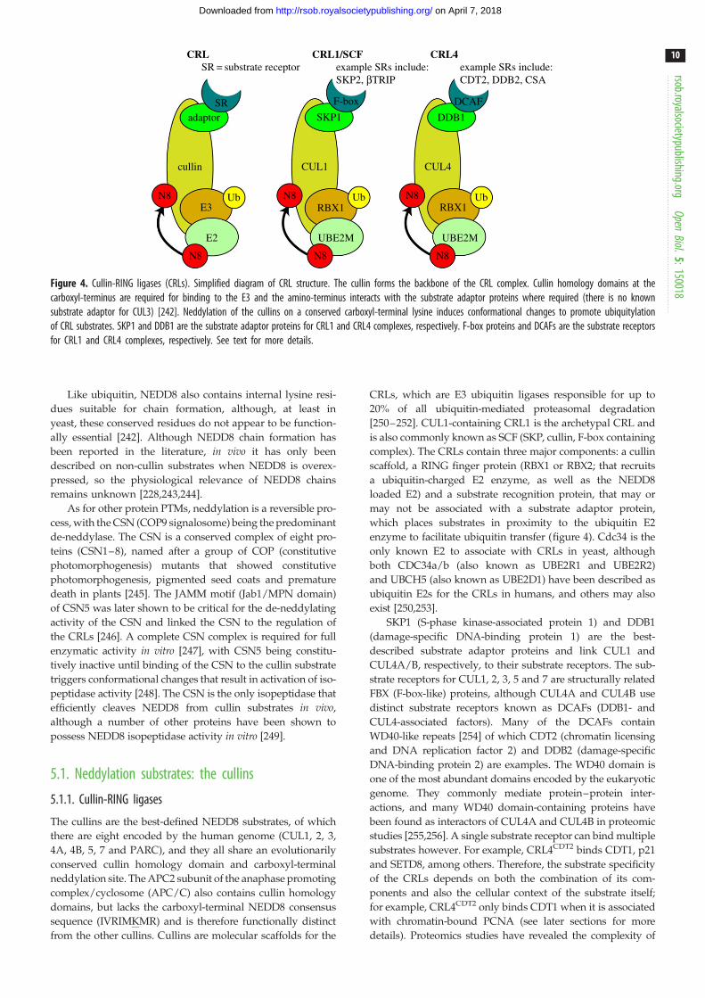
Figure 4. Cullin-RING ligases (CRLs). Simplified diagram of CRL structure. The cullin forms the backbone of the CRL complex. Cullin homology domains at thecarboxyl-terminus are required for binding to the E3 and the amino-terminus interacts with the substrate adaptor proteins where required (there is no knownsubstrate adaptor for CUL3) [242]. Neddylation of the cullins on a conserved carboxyl-terminal lysine induces conformational changes to promote ubiquitylationof CRL substrates. SKP1 and DDB1 are the substrate adaptor proteins for CRL1 and CRL4 complexes, respectively. F-box proteins and DCAFs are the substrate receptorsfor CRL1 and CRL4 complexes, respectively. See text for more details.
rsob.royalsocietypublishing.orgOpen
Biol.5:150018
10
on April 7, 2018http://rsob.royalsocietypublishing.org/Downloaded from
Like ubiquitin, NEDD8 also contains internal lysine resi-
dues suitable for chain formation, although, at least in
yeast, these conserved residues do not appear to be function-
ally essential [242]. Although NEDD8 chain formation has
been reported in the literature, in vivo it has only been
described on non-cullin substrates when NEDD8 is overex-
pressed, so the physiological relevance of NEDD8 chains
remains unknown [228,243,244].
As for other protein PTMs, neddylation is a reversible pro-
cess, with the CSN (COP9 signalosome) being the predominant
de-neddylase. The CSN is a conserved complex of eight pro-
teins (CSN1–8), named after a group of COP (constitutive
photomorphogenesis) mutants that showed constitutive
photomorphogenesis, pigmented seed coats and premature
death in plants [245]. The JAMM motif (Jab1/MPN domain)
of CSN5 was later shown to be critical for the de-neddylating
activity of the CSN and linked the CSN to the regulation of
the CRLs [246]. A complete CSN complex is required for full
enzymatic activity in vitro [247], with CSN5 being constitu-
tively inactive until binding of the CSN to the cullin substrate
triggers conformational changes that result in activation of iso-
peptidase activity [248]. The CSN is the only isopeptidase that
efficiently cleaves NEDD8 from cullin substrates in vivo,
although a number of other proteins have been shown to
possess NEDD8 isopeptidase activity in vitro [249].
5.1. Neddylation substrates: the cullins
5.1.1. Cullin-RING ligases
The cullins are the best-defined NEDD8 substrates, of which
there are eight encoded by the human genome (CUL1, 2, 3,
4A, 4B, 5, 7 and PARC), and they all share an evolutionarily
conserved cullin homology domain and carboxyl-terminal
neddylation site. The APC2 subunit of the anaphase promoting
complex/cyclosome (APC/C) also contains cullin homology
domains, but lacks the carboxyl-terminal NEDD8 consensus
sequence (IVRIMKMR) and is therefore functionally distinct
from the other cullins. Cullins are molecular scaffolds for the
CRLs, which are E3 ubiquitin ligases responsible for up to
20% of all ubiquitin-mediated proteasomal degradation
[250–252]. CUL1-containing CRL1 is the archetypal CRL and
is also commonly known as SCF (SKP, cullin, F-box containing
complex). The CRLs contain three major components: a cullin
scaffold, a RING finger protein (RBX1 or RBX2; that recruits
a ubiquitin-charged E2 enzyme, as well as the NEDD8
loaded E2) and a substrate recognition protein, that may or
may not be associated with a substrate adaptor protein,
which places substrates in proximity to the ubiquitin E2
enzyme to facilitate ubiquitin transfer (figure 4). Cdc34 is the
only known E2 to associate with CRLs in yeast, although
both CDC34a/b (also known as UBE2R1 and UBE2R2)
and UBCH5 (also known as UBE2D1) have been described as
ubiquitin E2s for the CRLs in humans, and others may also
exist [250,253].
SKP1 (S-phase kinase-associated protein 1) and DDB1
(damage-specific DNA-binding protein 1) are the best-
described substrate adaptor proteins and link CUL1 and
CUL4A/B, respectively, to their substrate receptors. The sub-
strate receptors for CUL1, 2, 3, 5 and 7 are structurally related
FBX (F-box-like) proteins, although CUL4A and CUL4B use
distinct substrate receptors known as DCAFs (DDB1- and
CUL4-associated factors). Many of the DCAFs contain
WD40-like repeats [254] of which CDT2 (chromatin licensing
and DNA replication factor 2) and DDB2 (damage-specific
DNA-binding protein 2) are examples. The WD40 domain is
one of the most abundant domains encoded by the eukaryotic
genome. They commonly mediate protein–protein inter-
actions, and many WD40 domain-containing proteins have
been found as interactors of CUL4A and CUL4B in proteomic
studies [255,256]. A single substrate receptor can bind multiple
substrates however. For example, CRL4CDT2 binds CDT1, p21
and SETD8, among others. Therefore, the substrate specificity
of the CRLs depends on both the combination of its com-
ponents and also the cellular context of the substrate itself;
for example, CRL4CDT2 only binds CDT1 when it is associated
with chromatin-bound PCNA (see later sections for more
details). Proteomics studies have revealed the complexity of

rsob.royalsocietypublishing.orgOpen
Biol.5:150018
11
on April 7, 2018http://rsob.royalsocietypublishing.org/Downloaded from
the CRL network [255] and diversity of CRL substrates
[257,258], culminating in more than 200 unique CRL complexes
that play key roles in cell cycle regulation, embryogenesis,
DNA replication and repair [251].
5.1.2. Regulation of cullin-RING ligase activity
CRL activity towards its substrate is tightly controlled, and
regulation is multi-factorial. Neddylation of the cullins, on a
conserved carboxyl-terminal lysine, serves to increase the ubi-
quitylating activity of the CRLs by triggering conformational
changes within the cullin and by preventing association with
the CRL inhibitor CAND1 (cullin-associated NEDD8-
dissociated 1) [259]. CAND1 binds to de-neddylated cullins,
promotes dissociation of the cullin from the substrate receptor
and inhibits CRL activity in vitro [260,261]. As well as showing
inhibitory CRL effects, however, genetic studies demonstrated
that CAND1 also promotes CRL activity, with this functional
paradox being explained by the importance of CAND1
for maintaining an available pool of substrate receptors and
therefore promoting the dynamics of CRL formation and
dissociation [262–264].
CSN regulation of CRL activity is also more complex than
initially thought. CSN association with the CRL maintains
the cullin in a de-neddylated state. This, along with its associ-
ation with the de-ubiquitylating enzyme USP15 [265],
inhibits the ubiquitylation of CRL substrates and protects
substrate receptor proteins from auto-degradation by the
CRL. In some cases, the CSN may also physically block sub-
strate access. Taken together, CSN dissociation from the CRL
has therefore become a recognized mechanism for promoting
substrate ubiquitylation [266–269].
Independently of cullin association with the negative
regulators CSN and CAND1, cullin neddylation and thus
activation also appears to be dependent on the formation of a
complete CRL unit, including cullin binding to adaptor proteins
and substrate receptor subunits, possibly through inducing a
conformational change or dimerization of the CRL, which pro-
motes neddylation [270]. PTMs of the substrate binding motif or
‘degron’ also regulate CRL interaction with its target and sub-
sequent activation in some cases [271]. Further structural and
biochemical studies will better determine the mechanisms of
CRL activation. Clearly, however, spatio-temporal regulation
of CRL activity is critical and is achieved through a fine balance
of promoting and inhibitory mechanisms.
5.2. The neddylation inhibitor MLN4924Many CRL components are either overexpressed or mutated in
human cancers [272,273], making CRL inhibition an attractive
anti-cancer strategy. In addition, success of the proteasome
inhibitor bortezomib (Velcadew; Takeda Oncology) for the
treatment of multiple myeloma and mantle cell lymphoma
has paved the way for novel therapeutics that inhibit
ubiquitin-mediated protein degradation pathways.
MLN4924 (Takeda Oncology) is an irreversible inhibitor of
UBA3 that potently and specifically inhibits UBA3 activation of
NEDD8 in cells, and subsequently inhibits global NEDD8 con-
jugation and CRL activity [252]. MLN4924 is an AMP analogue
which is catalysed by UBA3 to form a covalent UBA3-
MLN4924 adduct that can no longer be used by the NEDD8
machinery [274]. Therefore, treatment of cells with MLN4924
rapidly leads to the accumulation of CRL substrates, including
CDT1, p27 and NRF2, among others [252]. The predominant
cellular phenotype, in a range of cell lines tested, following
prolonged (8 h) MLN4924 treatment is S-phase arrest and
DNA re-replication, thought to be largely due to CRL4CDT2
inhibition [275], although CRL1 is also likely to contribute.
CRL4CDT2 inhibition causes stabilization of CDT1 and SETD8
(also known as SET8 or PR-SET7), both of which contribute
to the re-replication observed. CDT1 is required for loading
of the DNA replication helicase complex (MCM2–7) onto chro-
matin in the G1 phase of the cell cycle, and CRL4CDT2/
SCFSKP2-mediated degradation of CDT1 during S-phase pre-
vents DNA re-replication [276]. SETD8 catalyses histone
monomethylation on Lys 20 of histone H4 (H4K20me1), and
PCNA-dependent degradation of SETD8 by CRL4CDT2
during S-phase inhibits DNA re-replication (although the
mechanism by which SETD8 promotes loading of the replica-
tion complex onto chromatin remains unknown) [277,278].
DNA re-replication is a recognized cause of DNA damage
[279] and prolonged MLN4924 treatment causes replication-
dependent activation of ATM- and ATR-mediated DDR
signalling, DSB formation as measured by gH2AX formation
and eventual apoptosis or cellular senescence [279–281].
In addition to re-replication itself being a cause of DNA
damage, recently it has been shown that CRL-dependent ubi-
quitylation is required for dissociation of the DNA replisome
in Saccharomyces cerevisiae and Xenopus [282,283]. It will be
interesting to determine how failure to remove the MCM com-
plex on completion of DNA replication might contribute
to DSB generation following treatment with MLN4924.
Regardless of mechanism, cell survival in yeast mutants
that undergo DNA re-replication or in human cells follow-
ing MLN4924 treatment is reduced in DSB repair deficient
backgrounds [284,285], clearly suggesting that cell death fol-
lowing MLN4924 treatment is at least in part due to the
generation of DNA DSBs.
Pre-clinical data suggest that inhibiting neddylation might
be an effective anti-cancer strategy for solid tumours and hae-
matological malignancies [286]. MLN4924 is well tolerated and
has shown anti-tumour activity in phase I oncology trials invol-
ving haematological and solid tumours [287] (see also http://
meetinglibrary.asco.org). Phase I combination trials are
on-going (http://www.cancer.gov/clinicaltrials).
5.3. Non-cullin neddylation substratesA number of non-cullin substrates for NEDD8 have been
described in the literature, including p53 [288], the ribosomal
protein L11 [289] and histone H4 [290]. In addition, proteomics
studies have identified neddylation targets both within
and outside the known CRL network [243,244]. However,
these studies have largely relied on the overexpression of
tagged-NEDD8 constructs as their bait, and as conjugation
of NEDD8 to target proteins using the ubiquitin machinery
can occur in these circumstances [229], it is difficult to be
convinced that proteins other than cullins are true NEDD8
substrates in vivo. It is also worth noting that, while MLN4924
specifically inhibits neddylation, it also leads to potent
inhibition of CRL ubiquitylating activity. Consequently,
demonstrating that neddylation of a substrate is MLN4924
dependent does not discriminate between conjugation being
mediated by the NEDD8 or ubiquitin machineries. Further-
more, histones are heavily ubiquitylated by CRLs and are
often identified as neddylation substrates when NEDD8

rsob.roy
12
on April 7, 2018http://rsob.royalsocietypublishing.org/Downloaded from
is overexpressed. A recent excellent review by leading labora-
tories in the neddylation field has suggested criteria for
identifying physiological neddylation substrates and, to date,
only the cullins fulfil all these criteria [291].
alsocietypublishing.orgOpen
Biol.5:150018
6. Role of neddylation in the DNA damageresponse
6.1. CUL4A and CUL4BOf all the cullins, CUL4A and CUL4B are the most strongly
linked to the DDR. Higher eukaryotes such as Homo sapiens,
Mus musculus and Xenopus tropicalis have both types of
CUL4, which share approximately 85% sequence identity
and are derived from a common CUL4 ancestor found in Cae-norhabditis elegans, Drosophila melanogaster and S. pombe. At a
cellular level, a large amount of functional redundancy exists
between CUL4A and CUL4B. Some non-overlapping func-
tions of the two proteins have been revealed by mouse
genetic studies however, because although Cul4A2/2 mice
are viable with no overt developmental defects [292], presum-
ably due to functional compensation by Cul4B, Cul4A2/2 male
mice are sterile, which is thought to be due to a failure to
resolve recombination crossover intermediates during meiosis
[293]. In addition, mutations in CUL4B are linked to an
X-linked mental retardation syndrome in man [294] and
Cul4B2/2 mice display embryonic lethality due to develop-
mental failure of extra-embryonic tissues [295]. Beyond these
limited examples, CUL4A and CUL4B appear to be function-
ally redundant with one another, and the roles described
below for CRL4 in the DDR cannot be linked to either one of
the proteins in isolation.
6.2. CRL4 and nucleotide excision repairBoth GG-NER and TC-NER (table 1) use mechanisms invol-
ving CRL4 to recognize DNA lesions and promote repair
[88]. DDB2 is the DNA damage sensor protein for GG-NER
and is the substrate receptor of the CUL4–DDB1 CRL complex
(CRL4DDB2), which in the nuclear fraction of unperturbed cells
is closely associated with the CSN [296]. In the current model,
upon binding to UV-damaged sites, CRL4DDB2 dissociates
from the CSN, leading to activation of the CRL and ubiquityla-
tion of XPC and DDB2 [266,296]. Interestingly, the inhibitory
effects of the CSN on CRL4DDB2 appear to be independent of
CSN5 de-neddylase activity [266]. Dimerization of CRL4DDB2
upon binding to DNA may also be important for facilitating
ubiquitylation of its substrates [297]. Ubiquitylation of DDB2
reduces its affinity for DNA and causes its proteasome-
mediated degradation, whereas ubiquitylation of XPC potenti-
ates its association with the DNA, enabling repair [298]. In
addition, CRL4DDB2 ubiquitylates histone H3 and H4 in the
vicinity of the DNA lesions, facilitating access of the NER
repair machinery [299]. Mutations in the DNA-binding
region of DDB2 give rise to the archetypal NER syndrome
xeroderma pigmentosum [300,301].
CSA (Cockayne syndrome type A protein) is another sub-
strate receptor for CUL4–DDB1, and CRL4CSA is recruited to
TC-NER sites where it is believed to ubiquitylate the SWI/
SNF ATPase CSB (Cockayne syndrome type B protein) and is
important for repair and ensuing transcriptional re-start
[266,302]. Although CSA and DDB2 are quite distinct at the
sequence level, they are structurally very similar in their
complex formation with DDB1, and like CRL4DDB2, CRL4CSA
activity appears to be regulated by its association/dissociation
with the CSN [266].
6.3. Cullin-RING ligase regulation of DNA damage cellcycle checkpoint responses
CRLs play prominent roles in cell cycle regulation [276] and are
also important for cell cycle checkpoint responses following
DNA damage. As described above, CDT1 degradation by
CRL4CDT2/SCFSKP2 during normal S-phase is essential in pre-
venting DNA re-replication. In addition, following DNA
damage, CRL4CDT2 promotes CDT1 degradation in a chroma-
tin-bound PCNA-dependent manner. CDT1 contains a ‘PIP
degron’, which is made up of a canonical PCNA binding
motif (the PIP box; through which CDT1 binds PCNA) and a
basic patch of four amino acids downstream (Bþ4), which
mediates the interaction between CDT1 and CRL4CDT2.
CRL4CDT2 interacting with both PCNA and CDT1 via the PIP
degron is a common mechanism by which CRL4CDT2 interacts
with its substrates [303], although how this interaction is regu-
lated to occur only in the context of chromatin-bound PCNA is
not known [304].
CDC25A is a phosphatase required for activation of
cyclin-dependent kinases, enabling cell cycle progression.
Following DNA damage, CDC25A is phosphorylated in a
predominantly CHEK1/CHEK2-dependent manner, leading
to its ubiquitylation by SCFbTRCP, degradation and subse-
quent cell cycle arrest [305]. Similarly, a phospho-degron
signal on CLASPIN (a protein required for ATR-mediated
CHEK1 activation following stalled replication forks and
DNA end resection during HR) triggers its ubiquitylation by
SCFbTRCP and subsequent degradation, enabling checkpoint
recovery [306,307].
6.4. Neddylation and double-strand break repairIn vitro studies using Xenopus laevis egg extracts have shown
that, upon binding DNA, Ku80 is polyubiquitylated with K48
chains by the cullin 1 containing complex SCFFBXL12 and
removed from the DNA [308,309]. Truncation mutants of
Ku80 that are proficient in DNA binding, but not DNA
repair, are efficiently ubiquitylated, suggesting that in this
system DNA binding rather than the completion of repair is
the signal for Ku ubiquitylation and release [309]. The mechan-
ism by which Ku is extracted from the DNA remains unclear,
however. Postow et al. [309] showed that Ku removal is inde-
pendent of the proteasome and hypothesized that VCP
unfolds Ku, thereby promoting its removal from DNA [310].
Ku forms a highly stable ring-like structure that threads onto
DNA ends [311], and failure to remove Ku from repaired
DNA will likely interfere with cellular processes such as DNA
replication and transcription [312,313], highlighting the need
for an active mechanism to release DNA bound Ku [310].
It has not yet been determined whether a CRL-dependent
mechanism of Ku release is conserved in human cells.
In human cells, inhibition of NEDD8 conjugation has been
shown to hypersensitize cells to DNA damaging agents such as
mitomycin C, cisplatin and IR [314–321]. Synergy between
MLN4924 and DNA cross-linking agents also clearly exists
across a range of cell lines [314,316–319]. The mechanisms

rsob.royalsocietypublishing.orgOpen
Biol.5:150018
13
on April 7, 2018http://rsob.royalsocietypublishing.org/Downloaded from
behind this synergy are not fully elucidated and have not been
consistent between studies [316] but may include inhibitory
effects on assembly of the Fanconi complex [317], promotion
of reactive oxygen species (ROS) generation [318] and
increased replication fork collapse due to BRCA2/RAD51
stabilization [314]. Given the wide-ranging cellular effects of
neddylation inhibition, it is likely that the synergy observed
is multi-factorial and potentially cell-type specific. Further
studies will refine our understanding of the mechanisms
involved.
Recent studies have linked neddylation with DSB repair
more directly and NEDD8 has been shown to localize
to DNA damage sites [290]. In this study by Ma et al., it was
concluded that RNF111 functions as the NEDD8 E3
ligase responsible for NEDD8 accumulation at sites of DNA
damage. More specifically, the authors concluded that
RNF111-mediated neddylation of histone H4 is essential for
RNF168 recruitment to DSB sites [290]. RNF111, also known
as Arkadia, is a ubiquitin E3 ligase with an established role
in TGF-b signalling [322], and more recently has been shown
to function as a STUBL in PML degradation and in NER
[184,323]. Whether the effects of RNF111 depletion on DSB
repair reported by Ma et al. are due to the ubiquitylation
rather than the suggested neddylation activity of RNF111 has
not been established. In addition, as recently published, like
most ubiquitin E3s, the RING structure of RNF111 does not
favour interaction with the NEDD8 E2 UBE2M [291,324], and
as a consequence, it would be interesting to determine the
NEDD8 E2 responsible for the proposed neddylation function
of RNF111.
Other studies linking neddylation and DSB repair include
a published report by Wu et al. [325] that the CUL1 substrate
receptor SKP2 is important for NBS1 K63 ubiquitylation and
subsequent ATM activation. Interestingly, it has also been
demonstrated that NBS1 ubiquitylation by RNF8 is important
for efficient NBS1 recruitment to DNA damage sites [326],
and both pathways appear to be important for repair by
HR. More recently, it has been proposed that RNF111-
mediated neddylation regulates the extent of DNA end
resection and neddylation inhibition increases rates of HR
[327]. RNF168 has also been reported to function as a
NEDD8 E3 ligase in response to DNA damage, where it has
been reported to neddylate histone H2A and subsequently
negatively regulate the ubiquitylation of H2A and the down-
stream recruitment of BRCA1 [328]. As discussed above, the
existence of non-cullin neddylation substrates is a contentious
issue in the field [291], and further studies are required to sub-
stantiate H2A as a neddylation substrate and RNF168 as a
NEDD8 E3 ligase.
7. SummaryThe importance of PTMs in the DDR has been appreciated
for many years, and the role of ubiquitin and the UBLs
in DSB repair has been an area of active research over the
last decade. A major unanswered question in the field is
the identity of the RNF8 substrate responsible for RNF168
recruitment. In addition, it is likely that the full spectrum of
ubiquitin-system proteins important for the DDR has not
yet been fully established. Indeed, it seems likely that further
work in this area will uncover new mechanistic insights
into DDR processes as well as provide promising new
drug targets.
Given the complexity of the crosstalk between SUMO and
ubiquitin systems during cellular responses to DNA DSBs
[329], it would probably not be surprising if similar levels
of crosstalk exist between the neddylation and ubiquitylation
systems. As the predominant function of neddylation in the
literature to date is to regulate the ubiquitylating activity of
the CRLs, we feel that it will be particularly interesting to
determine how CRL activity is regulated in the context of
responses to DNA damage. While MLN4924 is an extremely
useful tool for investigating the cellular consequences of
neddylation inhibition, prolonged treatment with MLN4924
has wide-ranging cellular effects and generates high levels
of DNA DSBs [252]. Potent inhibition of neddylation is
achieved within minutes in cells treated with MLN4924, so
short treatment times should be used wherever possible
and appropriate experimentally. Likewise, given the potential
utilization of NEDD8 by the ubiquitin machinery, caution
should be exercised when interpreting results using overex-
pressed NEDD8 systems, and reagents should be validated
as being specific for NEDD8. Inhibitors specific for indivi-
dual CRL complexes are not available, but would be
extremely helpful for determining the fine-tuning of the
complex CRL network.
Studying the mechanisms by which ubiquitin and
the UBLs regulate DSB repair and associated processes is a
technical challenge, not least because of the functional redun-
dancy in these systems. A better understanding of how these
PTMs interconnect, as well as the development of more small
molecule inhibitors such as MLN4924 will enable the cellular
functions of these and connected enzymatic pathways to be
determined through approaches that until now have not
been feasible. In particular, further advances in proteomics
techniques will better define the substrates of various
PTMs, and evolving gene-targeting and cell engineering
approaches [330,331] will complement the use of inhibitors
in determining the functions of PTM pathway members.
Being frequently mutated or overexpressed in human can-
cers, the ubiquitin and UBL pathways offer a spectrum of
attractive drug targets, and with the recent approval of
PARP inhibitor olaparib/LynparzaTM (AstraZeneca) by the
European Medicines Agency and the US Food and Drug
Administration for the treatment of BRCA-mutated, high-
grade serous epithelial ovarian cancer, the precedent is now
set for DDR inhibitors in the clinic.
Acknowledgements. We thank Natasha Lukashchuk and Yaron Galantyfor useful discussions and also Kate Dry for critical reading ofthe manuscript.
Funding statement. J.S.B. is funded by the Wellcome Trust ClinicalFellowship (grant no. 094794/Z/10/Z). Research in the Jackson lab-oratory is funded by Cancer Research UK programme grant C6/A11224, the European Research Council and the European Commu-nity Seventh Framework Programme grant agreement no. HEALTH-F2-2010-259893 (DDResponse). Core funding is provided by CRUK(C6946/A14492) and the Wellcome Trust (WT092096). S.P.J. receiveshis salary from the University of Cambridge UK, supplementedby CRUK.
Author contributions. Both J.S.B. and S.P.J. contributed to drafting andrevising the manuscript. Both authors gave final approval beforesubmission.
Conflict of interests. J.S.B. has no competing interests. S.P.J. has no con-flicts of interest but discloses that he is a founder and shareholderof MISSION Therapeutics Ltd.

14
on April 7, 2018http://rsob.royalsocietypublishing.org/Downloaded from
References
rsob.royalsocietypublishing.orgOpen
Biol.5:150018
1. Jackson SP, Bartek J. 2009 The DNA-damageresponse in human biology and disease. Nature461, 1071 – 1078. (doi:10.1038/nature08467)
2. Ciccia A, Elledge SJ. 2010 The DNA damageresponse: making it safe to play with knives.Mol. Cell 40, 179 – 204. (doi:10.1016/j.molcel.2010.09.019)
3. Polo SE, Jackson SP. 2011 Dynamics of DNA damageresponse proteins at DNA breaks: a focus on proteinmodifications. Genes Dev. 25, 409 – 433. (doi:10.1101/gad.2021311)
4. Lindahl T. 1993 Instability and decay of the primarystructure of DNA. Nature 22, 709 – 715. (doi:10.1038/362709a0)
5. Bassing CH, Alt FW. 2004 The cellular response togeneral and programmed DNA double strandbreaks. DNA Repair 3, 781 – 796. (doi:10.1016/j.dnarep.2004.06.001)
6. Schlissel MS, Kaffer CR, Curry JD. 2006 Leukemiaand lymphoma: a cost of doing business foradaptive immunity. Genes Dev. 20, 1539 – 1544.(doi:10.1101/gad.1446506)
7. Richardson C, Horikoshi N, Pandita TK. 2004 Therole of the DNA double-strand break responsenetwork in meiosis. DNA Repair 3, 1149 – 1164.(doi:10.1016/j.dnarep.2004.05.007)
8. Basu B, Yap TA, Molife LR, de Bono JS. 2012Targeting the DNA damage response in oncology:past, present and future perspectives. Curr. Opin.Oncol. 24, 316 – 324. (doi:10.1097/CCO.0b013e32835280c6)
9. Curtin NJ. 2012 DNA repair dysregulation fromcancer driver to therapeutic target. Nat. Rev. Cancer12, 801 – 817. (doi:10.1038/nrc3399)
10. Furgason JM, Bahassi EM. 2013 Targeting DNArepair mechanisms in cancer. Pharmacol. Ther. 137,298 – 308. (doi:10.1016/j.pharmthera.2012.10.009)
11. Griffith AJ, Blier PR, Mimori T, Hardin JA. 1992 Kupolypeptides synthesized in vitro assemble intocomplexes which recognize ends of double-strandedDNA. J. Biol. Chem. 267, 331 – 338.
12. Mimori T, Hardin JA, Steitz JA. 1986 Characterizationof the DNA-binding protein antigen Ku recognizedby autoantibodies from patients with rheumaticdisorders. J. Biol. Chem. 261, 2274 – 2278.
13. Smider V, Rathmell WK, Lieber MR, Chu G. 1994Restoration of X-ray resistance and V(D)Jrecombination in mutant cells by Ku cDNA. Science266, 288 – 291. (doi:10.1126/science.7939667)
14. Taccioli GE et al. 1994 Ku80: product of the XRCC5gene and its role in DNA repair and V(D)Jrecombination. Science 265, 1442 – 1445. (doi:10.1126/science.8073286)
15. Mari P-O et al. 2006 Dynamic assembly of end-joining complexes requires interaction betweenKu70/80 and XRCC4. Proc. Natl Acad. Sci. USA 103,18 597 – 18 602. (doi:10.1073/pnas.0609061103)
16. Britton S, Coates J, Jackson SP. 2013 A new methodfor high-resolution imaging of Ku foci to deciphermechanisms of DNA double-strand break repair.
J. Cell Biol. 202, 579 – 595. (doi:10.1083/jcb.201303073)
17. Uematsu N et al. 2007 Autophosphorylation ofDNA-PKCS regulates its dynamics at DNA double-strand breaks. J. Cell Biol. 177, 219 – 229. (doi:10.1083/jcb.200608077)
18. Ramsden DA, Gellert M. 1998 Ku protein stimulatesDNA end joining by mammalian DNA ligases: adirect role for Ku in repair of DNA double-strandbreaks. EMBO J. 17, 609 – 614. (doi:10.1093/emboj/17.2.609)
19. Cary RB, Peterson SR, Wang J, Bear DG, BradburyEM, Chen DJ. 1997 DNA looping by Ku and theDNA-dependent protein kinase. Proc. Natl Acad. Sci.USA 94, 4267 – 4272. (doi:10.1073/pnas.94.9.4267)
20. De Vries E, van Driel W, Bergsma WG, Arnberg AC,van der Vliet PC. 1989 HeLa nuclear proteinrecognizing DNA termini and translocating on DNAforming a regular DNA-multimeric protein complex.J. Mol. Biol. 208, 65 – 78. (doi:10.1016/0022-2836(89)90088-0)
21. Gottlieb T, Jackson S. 1993 The DNA-dependentprotein kinase: requirement for DNA ends andassociation with Ku antigen. Cell 72, 131 – 142.(doi:10.1016/0092-8674(93)90057-W)
22. Smith GC, Jackson SP. 1999 The DNA-dependentprotein kinase. Genes Dev. 13, 916 – 934. (doi:10.1101/gad.13.8.916)
23. Costantini S, Woodbine L, Andreoli L, Jeggo PA,Vindigni A. 2007 Interaction of the Ku heterodimerwith the DNA ligase IV/Xrcc4 complex and itsregulation by DNA-PK. DNA Repair 6, 712 – 722.(doi:10.1016/j.dnarep.2006.12.007)
24. Hsu HL, Yannone SM, Chen DJ. 2002 Defininginteractions between DNA-PK and ligase IV/XRCC4.DNA Repair 1, 225 – 235. (doi:10.1016/S1568-7864(01)00018-0)
25. Yano K, Morotomi-Yano K, Wang S-Y, Uematsu N,Lee K-J, Asaithamby A, Weterings E, Chen DJ. 2008Ku recruits XLF to DNA double-strand breaks. EMBORep. 9, 91 – 96. (doi:10.1038/sj.embor.7401137)
26. Ochi T et al. 2015 PAXX, a paralog of XRCC4 andXLF, interacts with Ku to promote DNA double-strand break repair. Science 347, 185 – 188. (doi:10.1126/science.1261971)
27. Xing M et al. 2015 Interactome analysis identifies anew paralogue of XRCC4 in non-homologous endjoining DNA repair pathway. Nat. Commun. 6, 6233.(doi:10.1038/ncomms7233)
28. Dolganov GM, Maser RS, Novikov A, Tosto L, ChongS, Bressan DA, Petrini JH. 1996 Human Rad50 isphysically associated with human Mre11:identification of a conserved multiprotein compleximplicated in recombinational DNA repair. Mol. Cell.Biol. 16, 4832 – 4841.
29. Ogawa H, Johzuka K, Nakagawa T, Leem SH,Hagihara AH. 1995 Functions of the yeast meioticrecombination genes, MRE11 and MRE2. Adv.Biophys. 31, 67 – 76. (doi:10.1016/0065-227X(95)99383-Z)
30. Trujillo KM, Yuan SS, Lee EY, Sung P. 1998 Nucleaseactivities in a complex of human recombination andDNA repair factors Rad50, Mre11, and p95. J. Biol.Chem. 273, 21 447 – 21 450. (doi:10.1074/jbc.273.34.21447)
31. Usui T, Ohta T, Oshiumi H, Tomizawa J, Ogawa H,Ogawa T. 1998 Complex formation and functionalversatility of Mre11 of budding yeast inrecombination. Cell 95, 705 – 716. (doi:10.1016/S0092-8674(00)81640-2)
32. De Jager M, Dronkert ML, Modesti M, Beerens CE,Kanaar R, van Gent DC. 2001 DNA-binding andstrand-annealing activities of human Mre11:implications for its roles in DNA double-strand breakrepair pathways. Nucleic Acids Res. 29, 1317 – 1325.(doi:10.1093/nar/29.6.1317)
33. Paull TT, Gellert M. 1998 The 30 to 50 exonucleaseactivity of Mre 11 facilitates repair of DNA double-strand breaks. Mol. Cell 1, 969 – 979. (doi:10.1016/S1097-2765(00)80097-0)
34. Paull TT, Gellert M. 1999 Nbs1 potentiates ATP-driven DNA unwinding and endonuclease cleavageby the Mre11/Rad50 complex. Genes Dev. 13,1276 – 1288. (doi:10.1101/gad.13.10.1276)
35. Jazayeri A, Falck J, Lukas C, Bartek J, Smith GCM,Lukas J, Jackson SP. 2006 ATM- and cell cycle-dependent regulation of ATR in response to DNAdouble-strand breaks. Nat. Cell Biol. 8, 37 – 45.(doi:10.1038/ncb1337)
36. Sartori A, Lukas C, Coates J, Mistrik M, Fu S. 2007Human CtIP promotes DNA end resection. Nature450, 509 – 514. (doi:10.1038/nature06337)
37. Falck J, Coates J, Jackson SP. 2005 Conserved modesof recruitment of ATM, ATR and DNA-PKcs to sites ofDNA damage. Nature 434, 605 – 611. (doi:10.1038/nature03442)
38. Uziel T, Lerenthal Y, Moyal L, Andegeko Y, MittelmanL, Shiloh Y. 2003 Requirement of the MRN complexfor ATM activation by DNA damage. EMBO J. 22,5612 – 5621. (doi:10.1093/emboj/cdg541)
39. Lee J-H, Paull TT. 2005 ATM activation by DNAdouble-strand breaks through the Mre11-Rad50-Nbs1 complex. Science 308, 551 – 554. (doi:10.1126/science.1108297)
40. Lamarche BJ, Orazio NI, Weitzman MD. 2010 TheMRN complex in double-strand break repair andtelomere maintenance. FEBS Lett. 584, 3682 – 3695.(doi:10.1016/j.febslet.2010.07.029)
41. Weinfeld M, Chaudhry MA, D’Amours D, Pelletier JD,Poirier GG, Povirk LF, Lees-Miller SP. 1997Interaction of DNA-dependent protein kinase andpoly(ADP-ribose) polymerase with radiation-inducedDNA strand breaks. Radiat. Res. 148, 22 – 28.(doi:10.2307/3579534)
42. Bramson J, Prevost J, Malapetsa A, Noe AJ, PoirierGG, DesNoyers S, Alaoui-Jamali M, Panasci L. 1993Poly(ADP-ribose) polymerase can bind melphalandamaged DNA. Cancer Res. 53, 5370 – 5373.
43. D’Silva I, Pelletier JD, Lagueux J, D’Amours D,Chaudhry MA, Weinfeld M, Lees-Miller SP, Poirier

rsob.royalsocietypublishing.orgOpen
Biol.5:150018
15
on April 7, 2018http://rsob.royalsocietypublishing.org/Downloaded from
GG. 1999 Relative affinities of poly(ADP-ribose)polymerase and DNA-dependent protein kinase forDNA strand interruptions. Biochim. Biophys. Acta1430, 119 – 126. (doi:10.1016/S0167-4838(98)00278-7)
44. El-Khamisy SF, Masutani M, Suzuki H, Caldecott KW.2003 A requirement for PARP-1 for the assembly orstability of XRCC1 nuclear foci at sites of oxidativeDNA damage. Nucleic Acids Res. 31, 5526 – 5533.(doi:10.1093/nar/gkg761)
45. Mortusewicz O, Rothbauer U, Cardoso MC,Leonhardt H. 2006 Differential recruitment of DNAligase I and III to DNA repair sites. Nucleic Acids Res.34, 3523 – 3532. (doi:10.1093/nar/gkl492)
46. Wang M, Wu W, Wu W, Rosidi B, Zhang L, Wang H,Iliakis G. 2006 PARP-1 and Ku compete for repair ofDNA double strand breaks by distinct NHEJpathways. Nucleic Acids Res. 34, 6170 – 6182.(doi:10.1093/nar/gkl840)
47. Chapman JR, Taylor MRG, Boulton SJ. 2012 Playingthe end game: DNA double-strand break repairpathway choice. Mol. Cell 47, 497 – 510. (doi:10.1016/j.molcel.2012.07.029)
48. Jha DK, Pfister SX, Humphrey TC, Strahl BD. 2014SET-ting the stage for DNA repair. Nat. Struct. Mol.Biol. 21, 655 – 657. (doi:10.1038/nsmb.2866)
49. Kim S-T, Lim D-S, Canman CE, Kastan MB. 1999Substrate specificities and identification of putativesubstrates of ATM kinase family members. J. Biol.Chem. 274, 37 538 – 37 543. (doi:10.1074/jbc.274.53.37538)
50. Matsuoka S et al. 2007 ATM and ATR substrateanalysis reveals extensive protein networksresponsive to DNA damage. Science 316,1160 – 1166. (doi:10.1126/science.1140321)
51. Mahaney BL, Meek K, Lees-Miller SP. 2009 Repair ofionizing radiation-induced DNA double-strandbreaks by non-homologous end-joining. Biochem. J.417, 639 – 650. (doi:10.1042/BJ20080413)
52. Wang C, Lees-Miller SP. 2013 Detection and repairof ionizing radiation-induced DNA double strandbreaks: new developments in nonhomologous endjoining. Int. J. Radiat. Oncol. Biol. Phys. 86,440 – 449. (doi:10.1016/j.ijrobp.2013.01.011)
53. Kaidi A, Jackson SP. 2013 KAT5 tyrosinephosphorylation couples chromatin sensing to ATMsignalling. Nature 498, 70 – 74. (doi:10.1038/nature12201)
54. Costanzo V, Shechter D, Lupardus PJ, Cimprich KA,Gottesman M, Gautier J. 2003 An ATR- and Cdc7-dependent DNA damage checkpoint that inhibitsinitiation of DNA replication. Mol. Cell 11, 203 – 213.(doi:10.1016/S1097-2765(02)00799-2)
55. Zou L, Elledge SJ. 2003 Sensing DNA damagethrough ATRIP recognition of RPA – ssDNAcomplexes. Science 300, 1542 – 1548. (doi:10.1126/science.1083430)
56. Cimprich KA, Cortez D. 2008 ATR: an essentialregulator of genome integrity. Nat. Rev. Mol. CellBiol. 9, 616 – 627. (doi:10.1038/nrm2450)
57. Nam EA, Cortez D. 2011 ATR signalling: more thanmeeting at the fork. Biochem. J. 436, 527 – 536.(doi:10.1042/BJ20102162)
58. Ziv Y, Bar-Shira A, Pecker I, Russell P, Jorgensen TJ,Tsarfati I, Shiloh Y. 1997 Recombinant ATM proteincomplements the cellular A-T phenotype. Oncogene15, 159 – 167. (doi:10.1038/sj.onc.1201319)
59. Shiloh Y. 2014 ATM: expanding roles as a chiefguardian of genome stability. Exp. Cell Res. 329,154 – 161. (doi:10.1016/j.yexcr.2014.09.002)
60. O’Driscoll M, Ruiz-Perez VL, Woods CG, Jeggo PA,Goodship JA. 2003 A splicing mutation affectingexpression of ataxia-telangiectasia and Rad3-relatedprotein (ATR) results in Seckel syndrome. Nat. Genet.33, 497 – 501. (doi:10.1038/ng1129)
61. O’Driscoll M, Gennery AR, Seidel J, Concannon P,Jeggo PA. 2004 An overview of three new disordersassociated with genetic instability: LIG4 syndrome,RS-SCID and ATR-Seckel syndrome. DNA Repair 3,1227 – 1235. (doi:10.1016/j.dnarep.2004.03.025)
62. Finnie N, Gottlieb T. 1995 DNA-dependent proteinkinase activity is absent in xrs-6 cells: Implicationsfor site-specific recombination and DNA double-strand break repair. Proc. Natl Acad. Sci. USA 92,320 – 324. (doi:10.1073/pnas.92.1.320)
63. Blunt T et al. 1995 Defective DNA-dependentprotein kinase activity is linked to V(D)Jrecombination and DNA repair defects associatedwith the murine scid mutation. Cell 80, 813 – 823.(doi:10.1016/0092-8674(95)90360-7)
64. Kurimasa A, Kumano S. 1999 Requirement for thekinase activity of human DNA-dependent proteinkinase catalytic subunit in DNA strand breakrejoining. Mol. Cell. Biol. 19, 3877.
65. Van der Burg M et al. 2009 A DNA-PKcs mutation ina radiosensitive T – B – SCID patient inhibits Artemisactivation and nonhomologous end-joining. J. Clin.Invest. 119, 91 – 98. (doi:10.1172/JCI37141)
66. Rogakou E, Pilch D, Orr A, Ivanova V, Bonner W.1998 DNA double-stranded breaks induce histoneH2AX phosphorylation on serine 139. J. Biol. Chem.273, 5858 – 5868. (doi:10.1074/jbc.273.10.5858)
67. Stiff T, Driscoll MO, Rief N, Iwabuchi K, Lo M, JeggoPA. 2004 ATM and DNA-PK function redundantly tophosphorylate H2AX after exposure to ionizingradiation. Cancer Res. 64, 2390 – 2396. (doi:10.1158/0008-5472.CAN-03-3207)
68. Stucki M, Clapperton JA, Mohammad D, Yaffe MB,Smerdon SJ, Jackson SP. 2005 MDC1 directly bindsphosphorylated histone H2AX to regulate cellularresponses to DNA double-strand breaks. Cell 123,1213 – 1226. (doi:10.1016/j.cell.2005.09.038)
69. Stracker TH, Usui T, Petrini JHJ. 2009 Taking thetime to make important decisions: the checkpointeffector kinases Chk1 and Chk2 and the DNAdamage response. DNA Repair 8, 1047 – 1054.(doi:10.1016/j.dnarep.2009.04.012)
70. Bartek J, Lukas J. 2003 Chk1 and Chk2 kinases incheckpoint control and cancer. Cancer Cell 3,421 – 429. (doi:10.1016/S1535-6108(03)00110-7)
71. Reinhardt H, Yaffe M. 2009 Kinases that control thecell cycle in response to DNA damage: Chk1, Chk2,and MK2. Curr. Opin. Cell Biol. 21, 245 – 255.(doi:10.1016/j.ceb.2009.01.018)
72. Hirao A et al. 2002 Chk2 is a tumor suppressor thatregulates apoptosis in both an ataxia telangiectasia
mutated (ATM)-dependent and an ATM-independent manner. Mol. Cell. Biol. 22, 6521 –6532. (doi:10.1128/MCB.22.18.6521-6532.2002)
73. Takai H et al. 2002 Chk2-deficient mice exhibitradioresistance and defective p53-mediatedtranscription. EMBO J. 21, 5195 – 5205. (doi:10.1093/emboj/cdf506)
74. Manke IA, Nguyen A, Lim D, Stewart MQ, Elia AEH,Yaffe MB. 2005 MAPKAP kinase-2 is a cell cyclecheckpoint kinase that regulates the G2/M transitionand S phase progression in response to UVirradiation. Mol. Cell 17, 37 – 48. (doi:10.1016/j.molcel.2004.11.021)
75. Reinhardt HC, Aslanian AS, Lees JA, Yaffe MB. 2007p53-deficient cells rely on ATM- and ATR-mediatedcheckpoint signaling through the p38MAPK/MK2pathway for survival after DNA damage. Cancer Cell11, 175 – 189. (doi:10.1016/j.ccr.2006.11.024)
76. Blasius M, Wagner S. 2014 A quantitative 14-3-3interaction screen connects the nuclear exosometargeting complex to the DNA damage response. GenesDev. 28, 1977 – 1982. (doi:10.1101/gad.246272.114)
77. Deriano L, Roth DB. 2013 Modernizing thenonhomologous end-joining repertoire: alternativeand classical NHEJ share the stage. Annu. Rev.Genet. 47, 433 – 455. (doi:10.1146/annurev-genet-110711-155540)
78. Yan CT et al. 2007 IgH class switching andtranslocations use a robust non-classical end-joiningpathway. Nature 449, 478 – 482. (doi:10.1038/nature06020)
79. Frit P, Barboule N, Yuan Y, Gomez D, Calsou P. 2014Alternative end-joining pathway(s): bricolage atDNA breaks. DNA Repair 17, 81 – 97. (doi:10.1016/j.dnarep.2014.02.007)
80. Park J-Y, Zhang F, Andreassen PR. 2014 PALB2: thehub of a network of tumor suppressors involved inDNA damage responses. Biochim. Biophys. Acta1846, 263 – 275. (doi:10.1016/j.bbcan.2014.06.003)
81. Farmer H et al. 2005 Targeting the DNA repair defect inBRCA mutant cells as a therapeutic strategy. Nature434, 917 – 921. (doi:10.1038/nature03445)
82. Bryant HE et al. 2005 Specific killing of BRCA2-deficient tumours with inhibitors of poly(ADP-ribose) polymerase. Nature 434, 913 – 917. (doi:10.1038/nature03443)
83. Fong P et al. 2009 Inhibition of poly(ADP-ribose)polymerase in tumors from BRCA mutation carriers.N. Engl. J. Med. 361, 123 – 134. (doi:10.1056/NEJMoa0900212)
84. Helleday T. 2011 The underlying mechanism for thePARP and BRCA synthetic lethality: clearing up themisunderstandings. Mol. Oncol. 5, 387 – 393.(doi:10.1016/j.molonc.2011.07.001)
85. Jiricny J. 2006 The multifaceted mismatch-repairsystem. Nat. Rev. Mol. Cell Biol. 7, 335 – 346.(doi:10.1038/nrm1907)
86. Caldecott KW. 2008 Single-strand break repair andgenetic disease. Nat. Rev. Genet. 9, 619 – 631.(doi:10.1038/nrg2380)
87. Dianov GL, Hubscher U. 2013 Mammalian baseexcision repair: the forgotten archangel. Nucleic AcidsRes. 41, 3483 – 3490. (doi:10.1093/nar/gkt076)

rsob.royalsocietypublishing.orgOpen
Biol.5:150018
16
on April 7, 2018http://rsob.royalsocietypublishing.org/Downloaded from
88. Fousteri M, Mullenders LHF. 2008 Transcription-coupled nucleotide excision repair in mammaliancells: molecular mechanisms and biological effects.Cell Res. 18, 73 – 84. (doi:10.1038/cr.2008.6)
89. Sale JE. 2012 Competition, collaboration andcoordination—determining how cells bypass DNAdamage. J. Cell Sci. 125, 1633 – 1643. (doi:10.1242/jcs.094748)
90. Deans AJ, West SC. 2011 DNA interstrand crosslinkrepair and cancer. Nat. Rev. Cancer 11, 467 – 480.(doi:10.1038/nrc3088)
91. Ciehanover A, Hod Y, Hershko A. 1978 A heat-stablepolypeptide component of an ATP-dependentproteolytic system from reticulocytes. Biochem.Biophys. Res. 81, 1100 – 1105. (doi:10.1016/0006-291X(78)91249-4)
92. Hershko A, Ciechanover A. 1980 Proposed role ofATP in protein breakdown: conjugation of proteinwith multiple chains of the polypeptide of ATP-dependent proteolysis. Proc. Natl Acad. Sci. USA 77,1783 – 1786. (doi:10.1073/pnas.77.4.1783)
93. Wilkinson K, Urban M, Haas A. 1980 Ubiquitin is theATP-dependent proteolysis factor I of rabbitreticulocytes. J. Biol. Chem. 1, 7529 – 7532.
94. Ozkaynak E, Finley D. 1987 The yeast ubiquitingenes: a family of natural gene fusions. EMBO J. 6,1429 – 1439.
95. Baker RT, Board PG. 1987 The human ubiquitingene family: structure of a gene and pseudogenesfrom the Ub B subfamily. Nucleic Acids Res. 15,443 – 463. (doi:10.1093/nar/15.2.443)
96. Wiborg O, Pedersen M, Wind A. 1985 The humanubiquitin multigene family: some genes containmultiple directly repeated ubiquitin codingsequences. EMBO J. 4, 755 – 759.
97. Hershkos A, Heller H, Elias S, Ciechanover A. 1983Components of ubiquitin-protein ligase systemresolution, affinity purification. J. Biol. Chem. 258,8206 – 8214.
98. Ciechanover A, Elias S, Heller H, Hershko A. 1982‘Covalent affinity’ purification of ubiquitin-activatingenzyme. J. Biol. Chem. 257, 2537 – 2542.
99. Pickart CM, Kasperek EM, Beal R, Kim A. 1994Substrate properties of site-specific mutantubiquitin protein (G76A) reveal unexpectedmechanistic features of ubiquitin-activating enzyme(E1). J. Biol. Chem. 269, 7115 – 7123.
100. Schulman BA, Harper JW. 2009 Ubiquitin-likeprotein activation by E1 enzymes: the apex fordownstream signalling pathways. Nat. Rev. Mol. CellBiol. 10, 319 – 331. (doi:10.1038/nrm2673)
101. Van Wijk SJL, Timmers HTM. 2010 The family ofubiquitin-conjugating enzymes (E2s): decidingbetween life and death of proteins. FASEB J. 24,981 – 993. (doi:10.1096/fj.09-136259)
102. Berndsen CE, Wolberger C. 2014 New insightsinto ubiquitin E3 ligase mechanism. Nat.Struct. Mol. Biol. 21, 301 – 307. (doi:10.1038/nsmb.2780)
103. Mattiroli F, Sixma TK. 2014 Lysine-targetingspecificity in ubiquitin and ubiquitin-likemodification pathways. Nat. Struct. Mol. Biol. 21,308 – 316. (doi:10.1038/nsmb.2792)
104. Deshaies RJ, Joazeiro CAP. 2009 RING domain E3ubiquitin ligases. Annu. Rev. Biochem. 78,399 – 434. (doi:10.1146/annurev.biochem.78.101807.093809)
105. Plechanovova A, Jaffray EG, Tatham MH, NaismithJH, Hay RT. 2012 Structure of a RING E3 ligase andubiquitin-loaded E2 primed for catalysis. Nature489, 115 – 120. (doi:10.1038/nature11376)
106. Koegl M, Hoppe T, Schlenker S, Ulrich HD, MayerTU, Jentsch S. 1999 A novel ubiquitination factor,E4, is involved in multiubiquitin chain assembly.Cell 96, 635 – 644. (doi:10.1016/S0092-8674(00)80574-7)
107. Hatakeyama S, Yada M, Matsumoto M, Ishida N,Nakayama KI. 2001 U box proteins as a new familyof ubiquitin-protein ligases. J. Biol. Chem. 276,33 111 – 33 120. (doi:10.1074/jbc.M102755200)
108. Komander D, Clague MJ, Urbe S. 2009 Breaking thechains: structure and function of thedeubiquitinases. Nat. Rev. Mol. Cell Biol. 10,550 – 563. (doi:10.1038/nrm2731)
109. Turcu F, Ventii K, Wilkinson K. 2009 Regulation andcellular roles of ubiquitin-specific deubiquitinatingenzymes. Annu. Rev. Biochem. 78, 363 – 397.(doi:10.1146/annurev.biochem.78.082307.091526)
110. Mukhopadhyay D, Riezman H. 2007 Proteasome-independent functions of ubiquitin in endocytosisand signaling. Science 315, 201 – 205. (doi:10.1126/science.1127085)
111. Bienko M et al. 2005 Ubiquitin-binding domains inY-family polymerases regulate translesion synthesis.Science 310, 1821 – 1824. (doi:10.1126/science.1120615)
112. Kirisako T et al. 2006 A ubiquitin ligase complexassembles linear polyubiquitin chains. EMBO J. 25,4877 – 4887. (doi:10.1038/sj.emboj.7601360)
113. Rieser E, Cordier SM, Walczak H. 2013 Linearubiquitination: a newly discovered regulator of cellsignalling. Trends Biochem. Sci. 38, 94 – 102.(doi:10.1016/j.tibs.2012.11.007)
114. Hershko A, Ciechanover A. 1998 The ubiquitinsystem. Annu. Rev. Biochem. 67, 425 – 479. (doi:10.1146/annurev.biochem.67.1.425)
115. Xu P et al. 2009 Quantitative proteomics reveals thefunction of unconventional ubiquitin chains inproteasomal degradation. Cell 137, 133 – 145.(doi:10.1016/j.cell.2009.01.041)
116. Hurley JH, Lee S, Prag G. 2006 Ubiquitin-bindingdomains. Biochem. J. 399, 361 – 372. (doi:10.1042/BJ20061138)
117. Husnjak K, Dikic I. 2012 Ubiquitin-binding proteins:decoders of ubiquitin-mediated cellular functions.Annu. Rev. Biochem. 81, 291 – 322. (doi:10.1146/annurev-biochem-051810-094654)
118. Komander D. 2009 The emerging complexity ofprotein ubiquitination. Biochem. Soc. Trans. 37,937 – 953. (doi:10.1042/BST0370937)
119. Komander D, Rape M. 2012 The ubiquitin code.Annu. Rev. Biochem. 81, 203 – 229. (doi:10.1146/annurev-biochem-060310-170328)
120. Hochstrasser M. 2009 Origin and function ofubiquitin-like proteins. Nature 458, 422 – 429.(doi:10.1038/nature07958)
121. Van der Veen AG, Ploegh HL. 2012 Ubiquitin-likeproteins. Annu. Rev. Biochem. 81, 323 – 357.(doi:10.1146/annurev-biochem-093010-153308)
122. Jentsch S, McGrath J, Varshavsky A. 1986 The yeastDNA repair gene RAD6 encodes a ubiquitin-conjugating enzyme. Nature 329, 131 – 134.(doi:10.1038/329131a0)
123. Hoege C, Pfander B, Moldovan G-L, Pyrowolakis G,Jentsch S. 2002 RAD6-dependent DNA repair is linkedto modification of PCNA by ubiquitin and SUMO.Nature 419, 135 – 141. (doi:10.1038/nature00991)
124. Kannouche PL, Wing J, Lehmann AR. 2004Interaction of human DNA polymerase h withmonoubiquitinated PCNA. Mol. Cell 14, 491 – 500.(doi:10.1016/S1097-2765(04)00259-X)
125. Watanabe K, Tateishi S, Kawasuji M, Tsurimoto T,Inoue H, Yamaizumi M. 2004 Rad18 guides poletato replication stalling sites through physicalinteraction and PCNA monoubiquitination. EMBO J.23, 3886 – 3896. (doi:10.1038/sj.emboj.7600383)
126. Simpson LJ, Ross A-L, Szuts D, Alviani CA,Oestergaard VH, Patel KJ, Sale JE. 2006 RAD18-independent ubiquitination of proliferating-cellnuclear antigen in the avian cell line DT40. EMBORep. 7, 927 – 932. (doi:10.1038/sj.embor.7400777)
127. Stewart GS et al. 2009 The RIDDLE syndromeprotein mediates a ubiquitin-dependent signalingcascade at sites of DNA damage. Cell 136,420 – 434. (doi:10.1016/j.cell.2008.12.042)
128. Doil C et al. 2009 RNF168 binds and amplifiesubiquitin conjugates on damaged chromosomes toallow accumulation of repair proteins. Cell 136,435 – 446. (doi:10.1016/j.cell.2008.12.041)
129. Kolas NK et al. 2007 Orchestration of the DNA-damageresponse by the RNF8 ubiquitin ligase. Science 318,1637 – 1640. (doi:10.1126/science.1150034)
130. Huen MSY, Grant R, Manke I, Minn K, Yu X, YaffeMB, Chen J. 2007 RNF8 transduces the DNA-damagesignal via histone ubiquitylation and checkpointprotein assembly. Cell 131, 901 – 914. (doi:10.1016/j.cell.2007.09.041)
131. Mailand N, Bekker-Jensen S, Faustrup H, MelanderF, Bartek J, Lukas C, Lukas J. 2007 RNF8ubiquitylates histones at DNA double-strand breaksand promotes assembly of repair proteins. Cell 131,887 – 900. (doi:10.1016/j.cell.2007.09.040)
132. Bekker-Jensen S, Danielsen JR, Fugger K, Gromova I,Nerstedt A, Lukas C, Bartek J, Lukas J, Mailand N.2010 HERC2 coordinates ubiquitin-dependentassembly of DNA repair factors on damagedchromosomes. Nat. Cell Biol. 12, 80 – 86. (doi:10.1038/ncb2008)
133. Oestergaard VH, Pentzold C, Pedersen RT, Iosif S,Alpi A, Bekker-Jensen S, Mailand N, Lisby M. 2012RNF8 and RNF168 but not HERC2 are required forDNA damage-induced ubiquitylation in chickenDT40 cells. DNA Repair 11, 892 – 905. (doi:10.1016/j.dnarep.2012.08.005)
134. Pinato S, Gatti M, Scandiuzzi C, Confalonieri S,Penengo L. 2011 UMI, a novel RNF168 ubiquitinbinding domain involved in the DNA damagesignaling pathway. Mol. Cell. Biol. 31, 118 – 126.(doi:10.1128/MCB.00818-10)

rsob.royalsocietypublishing.orgOpen
Biol.5:150018
17
on April 7, 2018http://rsob.royalsocietypublishing.org/Downloaded from
135. Mattiroli F, Vissers JHA, van Dijk WJ, Ikpa P, CitterioE, Vermeulen W, Marteijn JA, Sixma TK. 2012RNF168 ubiquitinates K13 – 15 on H2A/H2AX todrive DNA damage signaling. Cell 150, 1182 – 1195.(doi:10.1016/j.cell.2012.08.005)
136. Mattiroli F, Uckelmann M, Sahtoe DD, van Dijk WJ,Sixma TK. 2014 The nucleosome acidic patch plays acritical role in RNF168-dependent ubiquitination ofhistone H2A. Nat. Commun. 5, 3291. (doi:10.1038/ncomms4291)
137. Panier S, Ichijima Y, Fradet-Turcotte A, Leung CCY,Kaustov L, Arrowsmith CH, Durocher D. 2012Tandem protein interaction modules organize theubiquitin-dependent response to DNA double-strandbreaks. Mol. Cell 47, 383 – 395. (doi:10.1016/j.molcel.2012.05.045)
138. Stewart GS et al. 2007 RIDDLE immunodeficiencysyndrome is linked to defects in 53BP1-mediatedDNA damage signaling. Proc. Natl Acad. Sci. USA104, 16 910 – 16 915. (doi:10.1073/pnas.0708408104)
139. Kracker S, Durandy A. 2011 Insights into the B cellspecific process of immunoglobulin class switchrecombination. Immunol. Lett. 138, 97 – 103.(doi:10.1016/j.imlet.2011.02.004)
140. Rai R, Li J, Zheng H, Lok G. 2011 The E3 ubiquitinligase RNF8 stabilizes TPP1 to promote telomereend protection. Nat. Struct. 18, 1400 – 1407.(doi:10.1038/nsmb.2172)
141. Peuscher MH, Jacobs JJL. 2011 DNA-damageresponse and repair activities at uncapped telomeresdepend on RNF8. Nat. Cell Biol. 13, 1139 – 1145.(doi:10.1038/ncb2326)
142. Shanbhag NM, Rafalska-Metcalf IU, Balane-BolivarC, Janicki SM, Greenberg RA. 2010 ATM-dependentchromatin changes silence transcription in cis toDNA double-strand breaks. Cell 141, 970 – 981.(doi:10.1016/j.cell.2010.04.038)
143. Giunta S, Belotserkovskaya R, Jackson SP. 2010 DNAdamage signaling in response to double-strandbreaks during mitosis. J. Cell Biol. 190, 197 – 207.(doi:10.1083/jcb.200911156)
144. Orthwein A, Fradet-Turcotte A, Noordermeer SM,Canny MD, Brun CM, Strecker J, Escribano-Diaz C,Durocher D. 2014 Mitosis inhibits DNA double-strand break repair to guard against telomerefusions. Science 344, 189 – 193. (doi:10.1126/science.1248024)
145. Noon AT, Goodarzi AA. 2011 53BP1-mediated DNAdouble strand break repair: insert bad pun here.DNA Repair 10, 1071 – 1076. (doi:10.1016/j.dnarep.2011.07.012)
146. Li M, Greenberg R. 2012 Links between genomeintegrity and BRCA1 tumor suppression. TrendsBiochem. Sci. 37, 418 – 424. (doi:10.1016/j.tibs.2012.06.007)
147. Huyen Y, Zgheib O. 2004 Methylated lysine 79 ofhistone H3 targets 53BP1 to DNA double-strandbreaks. Nature 432, 406 – 411. (doi:10.1038/nature03114)
148. Fradet-Turcotte A et al. 2013 53BP1 is a reader of theDNA-damage-induced H2A Lys 15 ubiquitin mark.Nature 499, 50 – 54. (doi:10.1038/nature12318)
149. Hu Y, Scully R, Sobhian B, Xie A, Shestakova E,Livingston DM. 2011 RAP80-directed tuning ofBRCA1 homologous recombination function ationizing radiation-induced nuclear foci. Genes Dev.25, 685 – 700. (doi:10.1101/gad.2011011)
150. Bekker-Jensen S, Lukas C, Kitagawa R, Melander F,Kastan MB, Bartek J, Lukas J. 2006 Spatialorganization of the mammalian genomesurveillance machinery in response to DNA strandbreaks. J. Cell Biol. 173, 195 – 206. (doi:10.1083/jcb.200510130)
151. Guzzo CM, Berndsen CE, Zhu J, Gupta V, Datta A,Greenberg RA, Wolberger C, Matunis MJ. 2012RNF4-dependent hybrid SUMO-ubiquitin chains aresignals for RAP80 and thereby mediate therecruitment of BRCA1 to sites of DNA damage. Sci.Signal. 5, ra88. (doi:10.1126/scisignal.2003485)
152. Hu X, Paul A, Wang B. 2012 Rap80 protein recruitmentto DNA double-strand breaks requires binding to bothsmall ubiquitin-like modifier (SUMO) and ubiquitinconjugates. J. Biol. Chem. 287, 25 510 – 25 519.(doi:10.1074/jbc.M112.374116)
153. Huen MSY, Sy SMH, Chen J. 2010 BRCA1 and itstoolbox for the maintenance of genome integrity.Nat. Rev. Mol. Cell Biol. 11, 138 – 148. (doi:10.1038/nrm2831)
154. Reid LJ, Shakya R, Modi AP, Lokshin M, Cheng J-T,Jasin M, Baer R, Ludwig T. 2008 E3 ligase activity ofBRCA1 is not essential for mammalian cell viabilityor homology-directed repair of double-strand DNAbreaks. Proc. Natl Acad. Sci. USA 105, 20 876 –20 881. (doi:10.1073/pnas.0811203106)
155. Shakya R et al. 2011 BRCA1 tumor suppressiondepends on BRCT phosphoprotein binding, but notits E3 ligase activity. Science 334, 525 – 528.(doi:10.1126/science.1209909)
156. Drost R et al. 2011 BRCA1 RING function is essentialfor tumor suppression but dispensable for therapyresistance. Cancer Cell 20, 797 – 809. (doi:10.1016/j.ccr.2011.11.014)
157. Bunting SF et al. 2010 53BP1 inhibits homologousrecombination in Brca1-deficient cells by blockingresection of DNA breaks. Cell 141, 243 – 254.(doi:10.1016/j.cell.2010.03.012)
158. Chapman JR et al. 2013 RIF1 is essential for 53BP1-dependent nonhomologous end joining andsuppression of DNA double-strand break resection.Mol. Cell 49, 858 – 871. (doi:10.1016/j.molcel.2013.01.002)
159. Escribano-Dıaz C et al. 2013 A cell cycle-dependentregulatory circuit composed of 53BP1-RIF1 andBRCA1-CtIP controls DNA repair pathway choice.Mol. Cell 49, 872 – 883. (doi:10.1016/j.molcel.2013.01.001)
160. Jacq X, Kemp M, Martin NMB, Jackson SP. 2013Deubiquitylating enzymes and DNA damageresponse pathways. Cell Biochem. Biophys. 67,25 – 43. (doi:10.1007/s12013-013-9635-3)
161. Poulsen M, Lukas C, Lukas J, Bekker-Jensen S,Mailand N. 2012 Human RNF169 is a negativeregulator of the ubiquitin-dependent response toDNA double-strand breaks. J. Cell Biol. 197,189 – 199. (doi:10.1083/jcb.201109100)
162. Chen J, Feng W, Jiang J, Deng Y, Huen MSY. 2012Ring finger protein RNF169 antagonizes theubiquitin-dependent signaling cascade at sites ofDNA damage. J. Biol. Chem. 287, 27 715 – 27 722.(doi:10.1074/jbc.M112.373530)
163. Gudjonsson T et al. 2012 TRIP12 and UBR5 suppressspreading of chromatin ubiquitylation at damagedchromosomes. Cell 150, 697 – 709. (doi:10.1016/j.cell.2012.06.039)
164. Nakamura K et al. 2011 Regulation of homologousrecombination by RNF20-dependent H2Bubiquitination. Mol. Cell 41, 515 – 528. (doi:10.1016/j.molcel.2011.02.002)
165. Moyal L et al. 2011 Requirement of ATM-dependentmonoubiquitylation of histone H2B for timely repairof DNA double-strand breaks. Mol. Cell 41,529 – 542. (doi:10.1016/j.molcel.2011.02.015)
166. Meetei AR et al. 2003 A novel ubiquitin ligase isdeficient in Fanconi anemia. Nat. Genet. 35,165 – 170. (doi:10.1038/ng1241)
167. Machida YJ, Machida Y, Chen Y, Gurtan AM, KupferGM, D’Andrea AD, Dutta A. 2006 UBE2T is the E2 inthe Fanconi anemia pathway and undergoesnegative autoregulation. Mol. Cell 23, 589 – 596.(doi:10.1016/j.molcel.2006.06.024)
168. MacKay C et al. 2010 Identification of KIAA1018/FAN1, a DNA repair nuclease recruited to DNAdamage by monoubiquitinated FANCD2. Cell 142,65 – 76. (doi:10.1016/j.cell.2010.06.021)
169. Kratz K et al. 2010 Deficiency of FANCD2-associatednuclease KIAA1018/FAN1 sensitizes cells tointerstrand crosslinking agents. Cell 142, 77 – 88.(doi:10.1016/j.cell.2010.06.022)
170. Smogorzewska A et al. 2010 A genetic screenidentifies FAN1, a Fanconi anemia-associatednuclease necessary for DNA interstrand crosslinkrepair. Mol. Cell 39, 36 – 47. (doi:10.1016/j.molcel.2010.06.023)
171. Murina O, von Aesch C, Karakus U, Ferretti LP, BolckHA, Hanggi K, Sartori AA. 2014 FANCD2 and CtIPcooperate to repair DNA interstrand crosslinks.Cell Rep. 7, 1030 – 1038. (doi:10.1016/j.celrep.2014.03.069)
172. Zhang N, Kaur R, Akhter S, Legerski RJ. 2009 Cdc5Linteracts with ATR and is required for the S-phasecell-cycle checkpoint. EMBO Rep. 10, 1029 – 1035.(doi:10.1038/embor.2009.122)
173. Marechal A et al. 2014 PRP19 transforms into asensor of RPA-ssDNA after DNA damage and drivesATR activation via a ubiquitin-mediated circuitry.Mol. Cell 53, 235 – 246. (doi:10.1016/j.molcel.2013.11.002)
174. Wan L, Huang J. 2014 The PSO4 protein complexassociates with replication protein A (RPA) andmodulates the activation of ataxia telangiectasia-mutated and RAD3-related (ATR). J. Biol. Chem.289, 6619 – 6626. (doi:10.1074/jbc.M113.543439)
175. Gong Z, Chen J. 2011 E3 ligase RFWD3 participatesin replication checkpoint control. J. Biol. Chem. 286,22 308 – 22 313. (doi:10.1074/jbc.M111.222869)
176. Liu S, Chu J, Yucer N, Leng M, Wang SY, Chen BPC,Hittelman WN, Wang Y. 2011 RING finger and WDrepeat domain 3 (RFWD3) associates with

rsob.royalsocietypublishing.orgOpen
Biol.5:150018
18
on April 7, 2018http://rsob.royalsocietypublishing.org/Downloaded from
replication protein A (RPA) and facilitates RPA-mediated DNA damage response. J. Biol. Chem.286, 22 314 – 22 322. (doi:10.1074/jbc.M111.222802)
177. Fu X et al. 2010 RFWD3-Mdm2 ubiquitin ligasecomplex positively regulates p53 stability inresponse to DNA damage. Proc. Natl Acad. Sci. USA107, 4579 – 4584. (doi:10.1073/pnas.0912094107)
178. Chan S-P, Kao D-I, Tsai W-Y, Cheng S-C. 2003 ThePrp19p-associated complex in spliceosomeactivation. Science 302, 279 – 282. (doi:10.1126/science.1086602)
179. Prudden J, Pebernard S, Raffa G, Slavin DA, PerryJJP, Tainer JA, McGowan CH, Boddy MN. 2007SUMO-targeted ubiquitin ligases in genomestability. EMBO J. 26, 4089 – 4101. (doi:10.1038/sj.emboj.7601838)
180. Yin Y, Seifert A, Chua JS, Maure J-F, Golebiowski F,Hay RT. 2012 SUMO-targeted ubiquitin E3 ligaseRNF4 is required for the response of human cells toDNA damage. Genes Dev. 26, 1196 – 1208. (doi:10.1101/gad.189274.112)
181. Galanty Y, Belotserkovskaya R, Coates J, Jackson SP.2012 RNF4, a SUMO-targeted ubiquitin E3 ligase,promotes DNA double-strand break repair.Genes Dev. 26, 1179 – 1195. (doi:10.1101/gad.188284.112)
182. Gibbs-Seymour I et al. 2015 Ubiquitin-SUMOcircuitry controls activated Fanconi anemia IDcomplex dosage in response to DNA damage.Mol. Cell 57, 150 – 164. (doi:10.1016/j.molcel.2014.12.001)
183. Rojas-Fernandez A, Plechanovova A, Hattersley N,Jaffray E, Tatham MH, Hay RT. 2014 SUMO chain-induced dimerization activates RNF4. Mol. Cell 53,880 – 892. (doi:10.1016/j.molcel.2014.02.031)
184. Poulsen SL et al. 2013 RNF111/Arkadia is a SUMO-targeted ubiquitin ligase that facilitates the DNAdamage response. J. Cell Biol. 201, 797 – 807.(doi:10.1083/jcb.201212075)
185. Finley D. 2009 Recognition and processing ofubiquitin-protein conjugates by the proteasome.Annu. Rev. Biochem. 78, 477 – 513. (doi:10.1146/annurev.biochem.78.081507.101607)
186. Butler LR et al. 2012 The proteasomal de-ubiquitinating enzyme POH1 promotes thedouble-strand DNA break response. EMBO J. 31,3918 – 3934. (doi:10.1038/emboj.2012.232)
187. Molinari M, Mercurio C, Dominguez J, Goubin F,Draetta GF. 2000 Human Cdc25 A inactivation inresponse to S phase inhibition and its role inpreventing premature mitosis. EMBO Rep. 1,71 – 79. (doi:10.1093/embo-reports/kvd018)
188. Wu W, Sato K, Koike A, Nishikawa H, Koizumi H,Venkitaraman AR, Ohta T. 2010 HERC2 is an E3ligase that targets BRCA1 for degradation. CancerRes. 70, 6384 – 6392. (doi:10.1158/0008-5472.CAN-10-1304)
189. Shi W, Ma Z, Willers H, Akhtar K, Scott SP, Zhang J,Powell S, Zhang J. 2008 Disassembly of MDC1 foci iscontrolled by ubiquitin-proteasome-dependentdegradation. J. Biol. Chem. 283, 31 608 – 31 616.(doi:10.1074/jbc.M801082200)
190. Meyer H, Bug M, Bremer S. 2012 Emergingfunctions of the VCP/p97 AAA-ATPase in theubiquitin system. Nat. Cell Biol. 14, 117 – 123.(doi:10.1038/ncb2407)
191. Dantuma NP, Acs K, Luijsterburg MS. 2014 Should Istay or should I go: VCP/p97-mediated chromatinextraction in the DNA damage response. Exp. CellRes 329, 9 – 17. (doi:10.1016/j.yexcr.2014.08.025)
192. Puumalainen M-R, Lessel D, Ruthemann P,Kaczmarek N, Bachmann K, Ramadan K, Naegeli H.2014 Chromatin retention of DNA damage sensorsDDB2 and XPC through loss of p97 segregase causesgenotoxicity. Nat. Commun. 5, 3695. (doi:10.1038/ncomms4695)
193. Raman M, Havens CG, Walter JC, Harper JW. 2011A genome-wide screen identifies p97 as anessential regulator of DNA damage-dependent CDT1destruction. Mol. Cell 44, 72 – 84. (doi:10.1016/j.molcel.2011.06.036)
194. Meerang M et al. 2011 The ubiquitin-selectivesegregase VCP/p97 orchestrates the response toDNA double-strand breaks. Nat. Cell Biol. 13,1376 – 1382. (doi:10.1038/ncb2367)
195. Davis EJ, Lachaud C, Appleton P, Macartney TJ, NathkeI, Rouse J. 2012 DVC1 (C1orf124) recruits the p97protein segregase to sites of DNA damage. Nat. Struct.Mol. Biol. 19, 1093 – 1100. (doi:10.1038/nsmb.2394)
196. Mosbech A et al. 2012 DVC1 (C1orf124) is a DNAdamage-targeting p97 adaptor that promotesubiquitin-dependent responses to replication blocks.Nat. Struct. Mol. Biol. 19, 1084 – 1092. (doi:10.1038/nsmb.2395)
197. Maskey RS, Kim MS, Baker DJ, Childs B, MalureanuLA, Jeganathan KB, Machida Y, van Deursen JM,Machida YJ. 2014 Spartan deficiency causesgenomic instability and progeroid phenotypes. Nat.Commun. 5, 5744. (doi:10.1038/ncomms6744)
198. Lessel D et al. 2014 Mutations in SPRTN cause earlyonset hepatocellular carcinoma, genomic instabilityand progeroid features. Nat. Genet. 46, 1239 – 1244.(doi:10.1038/ng.3103)
199. Nie M, Aslanian A, Prudden J, Heideker J, VashishtAA, Wohlschlegel JA, Yates JR, Boddy MN. 2012Dual recruitment of Cdc48 ( p97)-Ufd1-Npl4ubiquitin-selective segregase by small ubiquitin-likemodifier protein (SUMO) and ubiquitin in SUMO-targeted ubiquitin ligase-mediated genome stabilityfunctions. J. Biol. Chem. 287, 29 610 – 29 619.(doi:10.1074/jbc.M112.379768)
200. Bergink S, Ammon T, Kern M, Schermelleh L,Leonhardt H, Jentsch S. 2013 Role of Cdc48/p97 asa SUMO-targeted segregase curbing Rad51-Rad52interaction. Nat. Cell Biol. 15, 526 – 532. (doi:10.1038/ncb2729)
201. Sun X-X, Challagundla KB, Dai M-S. 2011 Positiveregulation of p53 stability and activity by thedeubiquitinating enzyme Otubain 1. EMBO J. 31,576 – 592. (doi:10.1038/emboj.2011.434)
202. Nakada S et al. 2010 Non-canonical inhibition ofDNA damage-dependent ubiquitination by OTUB1.Nature 466, 941 – 946. (doi:10.1038/nature09297)
203. Juang Y-C et al. 2012 OTUB1 co-opts Lys48-linkedubiquitin recognition to suppress E2 enzyme
function. Mol. Cell 45, 384 – 397. (doi:10.1016/j.molcel.2012.01.011)
204. Wiener R, Zhang X, Wang T, Wolberger C. 2012 Themechanism of OTUB1-mediated inhibition ofubiquitination. Nature 483, 618 – 622. (doi:10.1038/nature10911)
205. Mosbech A, Lukas C, Bekker-Jensen S, Mailand N.2013 The deubiquitylating enzyme USP44counteracts the DNA double-strand break responsemediated by the RNF8 and RNF168 ubiquitinligases. J. Biol. Chem. 288, 16 579 – 16 587. (doi:10.1074/jbc.M113.459917)
206. Zhang Y, Foreman O, Wigle DA, Kosari F, VasmatzisG, Salisbury JL, van Deursen J, Galardy PJ. 2012USP44 regulates centrosome positioning to preventaneuploidy and suppress tumorigenesis. J. Clin.Invest. 122, 4362 – 4374. (doi:10.1172/JCI63084)
207. Stegmeier F et al. 2007 Anaphase initiation isregulated by antagonistic ubiquitination anddeubiquitination activities. Nature 446, 876 – 881.(doi:10.1038/nature05694)
208. Nijman SMB, Huang TT, Dirac AMG, BrummelkampTR, Kerkhoven RM, D’Andrea AD, Bernards R. 2005The deubiquitinating enzyme USP1 regulates theFanconi anemia pathway. Mol. Cell 17, 331 – 339.(doi:10.1016/j.molcel.2005.01.008)
209. Oestergaard VH et al. 2007 Deubiquitination ofFANCD2 is required for DNA crosslink repair. Mol. Cell28, 798 – 809. (doi:10.1016/j.molcel.2007.09.020)
210. Zhang D, Zaugg K, Mak TW, Elledge SJ. 2006 A rolefor the deubiquitinating enzyme USP28 in control ofthe DNA-damage response. Cell 126, 529 – 542.(doi:10.1016/j.cell.2006.06.039)
211. Knobel PA, Belotserkovskaya R, Galanty Y, SchmidtCK, Jackson SP, Stracker TH. 2014 USP28 is recruitedto sites of DNA damage by the tandem BRCTdomains of 53BP1 but plays a minor role in double-strand break metabolism. Mol. Cell. Biol. 34,2062 – 2074. (doi:10.1128/MCB.00197-14)
212. Carbone M, Yang H, Pass HI, Krausz T, Testa JR,Gaudino G. 2013 BAP1 and cancer. Nat. Rev. Cancer13, 153 – 159. (doi:10.1038/nrc3459)
213. Ismail IH, Davidson R, Gagne JP, Xu ZZ, Poirier GG,Hendzel MJ. 2014 Germline mutations in BAP1impair its function in DNA double-strand breakrepair. Cancer Res. 74, 4282 – 4294. (doi:10.1158/0008-5472.CAN-13-3109)
214. Yu H et al. 2014 Tumor suppressor anddeubiquitinase BAP1 promotes DNA double-strandbreak repair. Proc. Natl Acad. Sci. USA 111,285 – 290. (doi:10.1073/pnas.1309085110)
215. Nishi R, Wijnhoven P, le Sage C, Tjeertes J, Galanty Y,Forment JV, Clague MJ, Urbe S, Jackson SP. 2014Systematic characterization of deubiquitylatingenzymes for roles in maintaining genome integrity.Nat. Cell Biol. 16, 1016 – 1026. (doi:10.1038/ncb3028)
216. Kumar S, Tomooka Y, Noda M. 1992 Identificationof a set of genes with developmentally down-regulated expression in the mouse brain. Biochem.Biophys. Res. 185, 1155 – 1161. (doi:10.1016/0006-291X(92)91747-E)
217. Kamitani T, Kito K, Nguyen HP, Yeh ETH. 1997Characterization of NEDD8, a developmentally
rsob.royalsocietypublishing.orgOpen
Biol.5:150018
19
on April 7, 2018http://rsob.royalsocietypublishing.org/Downloaded from
down-regulated ubiquitin-like protein. J. Biol.Chem. 272, 28 557 – 28 562. (doi:10.1074/jbc.272.45.28557)
218. Whitby FG, Xia G, Pickart CM, Hill CP. 1998 Crystalstructure of the human ubiquitin-like protein NEDD8and interactions with ubiquitin pathway enzymes.J. Biol. Chem. 273, 34 983 – 34 991. (doi:10.1074/jbc.273.52.34983)
219. Liakopoulos D, Doenges G, Matuschewski K, JentschS. 1998 A novel protein modification pathwayrelated to the ubiquitin system. EMBO J. 17,2208 – 2214. (doi:10.1093/emboj/17.8.2208)
220. Lammer D, Mathias N, Laplaza JM, Jiang W, Liu Y,Callis J, Goebl M, Estelle M. 1998 Modification ofyeast Cdc53p by the ubiquitin-related proteinRub1p affects function of the SCFCdc4 complex.Genes Dev. 12, 914 – 926. (doi:10.1101/gad.12.7.914)
221. Mendoza HM, Shen L-N, Botting C, Lewis A, Chen J,Ink B, Hay RT. 2003 NEDP1, a highly conservedcysteine protease that deNEDDylates cullins. J. Biol.Chem. 278, 25 637 – 25 643. (doi:10.1074/jbc.M212948200)
222. Wada H, Kito K, Caskey LS, Yeh ET, Kamitani T. 1998Cleavage of the C-terminus of NEDD8 by UCH-L3.Biochem. Biophys. Res. Commun. 251, 688 – 692.(doi:10.1006/bbrc.1998.9532)
223. Burch TJ, Haas AL. 1994 Site-directed mutagenesisof ubiquitin. Differential roles for arginine in theinteraction with ubiquitin-activating enzyme.Biochemistry 33, 7300 – 7308. (doi:10.1021/bi00189a035)
224. Souphron J, Waddell MB, Paydar A, Tokgoz-GromleyZ, Roussel MF, Schulman BA. 2008 Structuraldissection of a gating mechanism preventingmisactivation of ubiquitin by NEDD80s E1.Biochemistry 47, 8961 – 8969. (doi:10.1021/bi800604c)
225. Walden H, Podgorski MS, Huang DT, Miller DW,Howard RJ, Minor DL, Holton JM, Schulman BA.2003 The structure of the APPBP1-UBA3-NEDD8-ATPcomplex reveals the basis for selective ubiquitin-likeprotein activation by an E1. Mol. Cell 12,1427 – 1437. (doi:10.1016/S1097-2765(03)00452-0)
226. Bohnsack RN, Haas AL. 2003 Conservation inthe mechanism of Nedd8 activation by thehuman AppBp1-Uba3 heterodimer. J. Biol.Chem. 278, 26 823 – 26 830. (doi:10.1074/jbc.M303177200)
227. Huang DT, Paydar A, Zhuang M, Waddell MB,Holton JM, Schulman BA. 2005 Structural basis forrecruitment of Ubc12 by an E2 binding domain inNEDD80s E1. Mol. Cell 17, 341 – 350. (doi:10.1016/j.molcel.2004.12.020)
228. Leidecker O, Matic I, Mahata B, Pion E, Xirodimas DP.2012 The ubiquitin E1 enzyme Ube1 mediates NEDD8activation under diverse stress conditions. Cell Cycle11, 1142 – 1150. (doi:10.4161/cc.11.6.19559)
229. Hjerpe R, Thomas Y, Chen J, Zemla A, Curran S,Shpiro N, Dick LR, Kurz T. 2012 Changes in the ratioof free NEDD8 to ubiquitin triggers NEDDylation byubiquitin enzymes. Biochem. J. 441, 927 – 936.(doi:10.1042/BJ20111671)
230. Gong L, Yeh ETH. 1999 Identification of theactivating and conjugating enzymes of theNEDD8 conjugation pathway. J. Biol. Chem. 274,12 036 – 12 042. (doi:10.1074/jbc.274.17.12036)
231. Osaka F, Kawasaki H, Aida N, Saeki M, Chiba T,Kawashima S, Tanaka K, Kato S. 1998 A newNEDD8-ligating system for cullin-4A. Genes Dev. 12,2263 – 2268. (doi:10.1101/gad.12.15.2263)
232. Huang DT et al. 2009 E2-RING expansion of theNEDD8 cascade confers specificity to cullinmodification. Mol. Cell 33, 483 – 495. (doi:10.1016/j.molcel.2009.01.011)
233. Scott DC, Monda JK, Grace CRR, Duda DM, KriwackiRW, Kurz T, Schulman BA. 2010 A dual E3mechanism for Rub1 ligation to Cdc53. Mol. Cell 39,784 – 796. (doi:10.1016/j.molcel.2010.08.030)
234. Kamura T, Conrad MN, Yan Q, Conaway RC, ConawayJW. 1999 The Rbx1 subunit of SCF and VHL E3ubiquitin ligase activates Rub1 modification ofcullins Cdc53 and Cul2. Genes Dev. 13, 2928 – 2933.(doi:10.1101/gad.13.22.2928)
235. Petroski MD, Deshaies RJ. 2005 Function andregulation of cullin-RING ubiquitin ligases. Nat. Rev.Mol. Cell Biol. 6, 9 – 20. (doi:10.1038/nrm1547)
236. Kurz T, Ozlu N, Rudolf F, O’Rourke SM, Luke B,Hofmann K, Hyman AA, Bowerman B, Peter M.2005 The conserved protein DCN-1/Dcn1p isrequired for cullin neddylation in C. elegans andS. cerevisiae. Nature 435, 1257 – 1261. (doi:10.1038/nature03662)
237. Kim AY et al. 2008 SCCRO (DCUN1D1) is an essentialcomponent of the E3 complex for neddylation.J. Biol. Chem. 283, 33 211 – 33 220. (doi:10.1074/jbc.M804440200)
238. Kurz T, Chou Y-C, Willems AR, Meyer-Schaller N,Hecht M-L, Tyers M, Peter M, Sicheri F. 2008 Dcn1functions as a scaffold-type E3 ligase for cullinneddylation. Mol. Cell 29, 23 – 35. (doi:10.1016/j.molcel.2007.12.012)
239. Meyer-Schaller N et al. 2009 The human Dcn1-likeprotein DCNL3 promotes Cul3 neddylation atmembranes. Proc. Natl Acad. Sci. USA 106,12 365 – 12 370. (doi:10.1073/pnas.0812528106)
240. Monda JK, Scott DC, Miller DJ, Lydeard J, King D,Harper JW, Bennett EJ, Schulman BA. 2013Structural conservation of distinctive N-terminalacetylation-dependent interactions across a family ofmammalian NEDD8 ligation enzymes. Structure 21,42 – 53. (doi:10.1016/j.str.2012.10.013)
241. Wu K, Yan H, Fang L, Wang X, Pfleger C, Jiang X,Huang L, Pan Z-Q. 2011 Mono-ubiquitination drivesnuclear export of the human DCN1-like proteinhDCNL1. J. Biol. Chem. 286, 34 060 – 34 070.(doi:10.1074/jbc.M111.273045)
242. Girdwood D, Xirodimas DP, Gordon C. 2011 Theessential functions of NEDD8 are mediated viadistinct surface regions, and not by polyneddylationin Schizosaccharomyces pombe. PLoS ONE 6, e20089.(doi:10.1371/journal.pone.0020089)
243. Xirodimas DP, Sundqvist A, Nakamura A, Shen L,Botting C, Hay RT. 2008 Ribosomal proteins aretargets for the NEDD8 pathway. EMBO Rep. 9,280 – 286. (doi:10.1038/embor.2008.10)
244. Jones J, Wu K, Yang Y, Guerrero C. 2008 A targetedproteomic analysis of the ubiquitin-like modifierNedd8 and associated proteins. J. Proteome 7,1274 – 1287. (doi:10.1021/pr700749v)
245. Wei N, Deng XW. 1992 COP9: a new genetic locusinvolved in light-regulated development and geneexpression in arabidopsis. Plant Cell 4, 1507 – 1518.(doi:10.1105/tpc.4.12.1507)
246. Cope GA, Suh GSB, Aravind L, Schwarz SE, ZipurskySL, Koonin EV, Deshaies RJ. 2002 Role of predictedmetalloprotease motif of Jab1/Csn5 in cleavage ofNedd8 from Cul1. Science 298, 608 – 611. (doi:10.1126/science.1075901)
247. Sharon M, Mao H, Boeri Erba E, Stephens E, ZhengN, Robinson CV. 2009 Symmetrical modularity ofthe COP9 signalosome complex suggests itsmultifunctionality. Structure 17, 31 – 40. (doi:10.1016/j.str.2008.10.012)
248. Lingaraju GM, Bunker RD, Cavadini S, Hess D,Hassiepen U, Renatus M, Fischer ES, Thoma NH. 2014Crystal structure of the human COP9 signalosome.Nature 512, 161 – 165. (doi:10.1038/nature13566)
249. Rabut G, Peter M. 2008 Function and regulation ofprotein neddylation. ‘Protein modifications: beyondthe usual suspects’ review series. EMBO Rep. 9,969 – 976. (doi:10.1038/embor.2008.183)
250. Lydeard JR, Schulman BA, Harper JW. 2013 Buildingand remodelling Cullin-RING E3 ubiquitin ligases.EMBO Rep. 14, 1050 – 1061. (doi:10.1038/embor.2013.173)
251. Sarikas A, Hartmann T, Pan Z-Q. 2011 The cullinprotein family. Genome Biol. 12, 220. (doi:10.1186/gb-2011-12-4-220)
252. Soucy TA et al. 2009 An inhibitor of NEDD8-activating enzyme as a new approach to treatcancer. Nature 458, 732 – 736. (doi:10.1038/nature07884)
253. Shibata E, Abbas T, Huang X, Wohlschlegel JA,Dutta A. 2011 Selective ubiquitylation of p21 andCdt1 by UBCH8 and UBE2G ubiquitin-conjugatingenzymes via the CRL4Cdt2 ubiquitin ligase complex.Mol. Cell. Biol. 31, 3136 – 3145. (doi:10.1128/MCB.05496-11)
254. Lee J, Zhou P. 2007 DCAFs, the missing link of theCUL4-DDB1 ubiquitin ligase. Mol. Cell 26, 775 – 780.(doi:10.1016/j.molcel.2007.06.001)
255. Bennett EJ, Rush J, Gygi SP, Harper JW. 2010Dynamics of cullin-RING ubiquitin ligase networkrevealed by systematic quantitative proteomics. Cell143, 951 – 965. (doi:10.1016/j.cell.2010.11.017)
256. Xu C, Min J. 2011 Structure and function of WD40domain proteins. Protein Cell 2, 202 – 214. (doi:10.1007/s13238-011-1018-1)
257. Emanuele MJ et al. 2011 Global identification ofmodular cullin-RING ligase substrates. Cell 147,459 – 474. (doi:10.1016/j.cell.2011.09.019)
258. Larance M, Kirkwood KJ, Xirodimas DP, Lundberg E,Uhlen M, Lamond AI. 2011 Characterization ofMRFAP1 turnover and interactions downstream ofthe NEDD8 pathway. Mol. Cell. Proteomics 11,M111.014407. (doi:10.1074/mcp.M111.014407)
259. Duda DM, Borg LA, Scott DC, Hunt HW, Hammel M,Schulman BA. 2008 Structural insights into NEDD8
rsob.royalsocietypublishing.orgOpen
Biol.5:150018
20
on April 7, 2018http://rsob.royalsocietypublishing.org/Downloaded from
activation of cullin-RING ligases: conformationalcontrol of conjugation. Cell 134, 995 – 1006.(doi:10.1016/j.cell.2008.07.022)
260. Zheng J et al. 2002 CAND1 binds to unneddylatedCUL1 and regulates the formation of SCF ubiquitinE3 ligase complex. Mol. Cell 10, 1519 – 1526.(doi:10.1016/S1097-2765(02)00784-0)
261. Liu J, Furukawa M, Matsumoto T, Xiong Y, Hill C,Carolina N. 2002 NEDD8 modification of CUL1dissociates p120, an inhibitor of CUL1-SKP1 bindingand SCF ligases. Mol. Cell 10, 1511 – 1518. (doi:10.1016/S1097-2765(02)00783-9)
262. Zemla A, Thomas Y, Kedziora S, Knebel A, Wood NT,Rabut G, Kurz T. 2013 CSN- and CAND1-dependentremodelling of the budding yeast SCF complex. Nat.Commun. 4, 1641. (doi:10.1038/ncomms2628)
263. Pierce NW et al. 2013 Cand1 promotes assembly ofnew SCF complexes through dynamic exchange of Fbox proteins. Cell 153, 206 – 215. (doi:10.1016/j.cell.2013.02.024)
264. Wu S, Zhu W, Nhan T, Toth JI, Petroski MD, Wolf DA.2013 CAND1 controls in vivo dynamics of the cullin 1-RING ubiquitin ligase repertoire. Nat. Commun. 4,1642. (doi:10.1038/ncomms2636)
265. Zhou C, Wee S, Rhee E, Naumann M, Dubiel W,Wolf DA. 2003 Fission yeast COP9/signalosomesuppresses cullin activity through recruitmentof the deubiquitylating enzyme Ubp12p. Mol.Cell 11, 927 – 938. (doi:10.1016/S1097-2765(03)00136-9)
266. Fischer ES et al. 2011 The molecular basis ofCRL4DDB2/CSA ubiquitin ligase architecture,targeting, and activation. Cell 147, 1024 – 1039.(doi:10.1016/j.cell.2011.10.035)
267. Emberley ED, Mosadeghi R, Deshaies RJ. 2012Deconjugation of Nedd8 from Cul1 is directlyregulated by Skp1-F-box and substrate, and theCOP9 signalosome inhibits deneddylated SCF by anoncatalytic mechanism. J. Biol. Chem. 287,29 679 – 29 689. (doi:10.1074/jbc.M112.352484)
268. Enchev RI, Scott DC, da Fonseca PCA, Schreiber A,Monda JK, Schulman BA, Peter M, Morris EP. 2012Structural basis for a reciprocal regulation betweenSCF and CSN. Cell Rep. 2, 616 – 627. (doi:10.1016/j.celrep.2012.08.019)
269. Schmidt MW, McQuary PR, Wee S, Hofmann K, WolfDA. 2009 F-box-directed CRL complex assembly andregulation by the CSN and CAND1. Mol. Cell 35,586 – 597. (doi:10.1016/j.molcel.2009.07.024)
270. Chew E-H, Hagen T. 2007 Substrate-mediatedregulation of cullin neddylation. J. Biol. Chem. 282,17 032 – 17 040. (doi:10.1074/jbc.M701153200)
271. Duda DM, Scott DC, Calabrese MF, Zimmerman ES,Zheng N, Schulman BA. 2011 Structural regulationof cullin-RING ubiquitin ligase complexes. Curr.Opin. Struct. Biol. 21, 257 – 264. (doi:10.1016/j.sbi.2011.01.003)
272. Watson IR, Irwin MS, Ohh M. 2011 NEDD8 pathwaysin cancer, Sine Quibus Non. Cancer Cell 19,168 – 176. (doi:10.1016/j.ccr.2011.01.002)
273. Melchor L et al. 2009 Comprehensivecharacterization of the DNA amplification at 13q34in human breast cancer reveals TFDP1 and CUL4A as
likely candidate target genes. Breast Cancer Res. 11,R86. (doi:10.1186/bcr2456)
274. Brownell JE et al. 2010 Substrate-assisted inhibitionof ubiquitin-like protein-activating enzymes: theNEDD8 E1 inhibitor MLN4924 forms a NEDD8-AMPmimetic in situ. Mol. Cell 37, 102 – 111. (doi:10.1016/j.molcel.2009.12.024)
275. Lin JJ, Milhollen MA, Smith PG, Narayanan U, DuttaA. 2010 NEDD8-targeting drug MLN4924 elicits DNArereplication by stabilizing Cdt1 in S phase,triggering checkpoint activation, apoptosis, andsenescence in cancer cells. Cancer Res. 70, 10 310 –10 320. (doi:10.1158/0008-5472.CAN-10-2062)
276. Rizzardi LF, Cook JG. 2012 Flipping the switch from G1to S phase with E3 ubiquitin ligases. Genes Cancer 3,634 – 648. (doi:10.1177/1947601 912473307)
277. Abbas T, Shibata E, Park J, Jha S, Karnani N, Dutta A.2010 CRL4Cdt2 regulates cell proliferation and histonegene expression by targeting PR-Set7/Set8 fordegradation. Mol. Cell 40, 9 – 21. (doi:10.1016/j.molcel.2010.09.014)
278. Tardat M, Brustel J, Kirsh O, Lefevbre C, Callanan M,Sardet C, Julien E. 2010 The histone H4 Lys 20methyltransferase PR-Set7 regulates replicationorigins in mammalian cells. Nat. Cell Biol. 12,1086 – 1093. (doi:10.1038/ncb2113)
279. Zhu W, Dutta A. 2006 An ATR- and BRCA1-mediatedFanconi anemia pathway is required for activatingthe G2/M checkpoint and DNA damage repair uponrereplication. Mol. Cell. Biol. 26, 4601 – 4611.(doi:10.1128/MCB.02141-05)
280. Milhollen MA, Narayanan U, Soucy TA, Veiby PO,Smith PG, Amidon B. 2011 Inhibition of NEDD8-activating enzyme induces rereplication andapoptosis in human tumor cells consistent withderegulating CDT1 turnover. Cancer Res. 71,3042 – 3051. (doi:10.1158/0008-5472.CAN-10-2122)
281. Jia L, Li H. 2011 Induction of p21-dependentsenescence by an NAE inhibitor, MLN4924, as amechanism of growth suppression. Neoplasia(New York, NY) 13, 561 – 569. (doi:10.1593/neo.11420)
282. Priego Moreno S, Bailey R, Campion N, Herron S,Gambus A. 2014 Polyubiquitylation drives replisomedisassembly at the termination of DNA replication.Science 346, 477 – 481. (doi:10.1126/science.1253585)
283. Maric M, Maculins T, De Piccoli G, Labib K. 2014 Cdc48and a ubiquitin ligase drive disassembly of the CMGhelicase at the end of DNA replication. Science 346,1253596. (doi:10.1126/science.1253596)
284. Archambault V, Ikui A, Drapkin B, Cross F. 2005Disruption of mechanisms that prevent rereplicationtriggers a DNA damage response. Mol. Cell25, 6707 – 6721. (doi:10.1128/MCB.25.15.6707-6721.2005)
285. Blank JL et al. 2013 Novel DNA damage checkpointsmediating cell death induced by the NEDD8-activating enzyme inhibitor MLN4924. CancerRes. 73, 225 – 234. (doi:10.1158/0008-5472.CAN-12-1729)
286. Zhao Y, Morgan MA, Sun Y. 2014 Targetingneddylation pathways to inactivate cullin-RING
ligases for anticancer therapy. Antioxid. RedoxSignal. 21, 2383 – 2400. (doi:10.1089/ars.2013.5795)
287. Swords RT et al. In press. Pevonedistat (MLN4924),a First-in-Class NEDD8-activating enzyme inhibitor,in patients with acute myeloid leukaemia andmyelodysplastic syndromes: a phase 1 study.Br. J. Haematol. (doi:10.1111/bjh.13323)
288. Xirodimas DP, Saville MK, Bourdon J-C, Hay RT, LaneDP. 2004 Mdm2-mediated NEDD8 conjugation ofp53 inhibits its transcriptional activity. Cell 118,83 – 97. (doi:10.1016/j.cell.2004.06.016)
289. Sundqvist A, Liu G, Mirsaliotis A, Xirodimas DP.2009 Regulation of nucleolar signalling to p53through NEDDylation of L11. EMBO Rep. 10,1132 – 1139. (doi:10.1038/embor.2009.178)
290. Ma T, Chen Y, Zhang F, Yang C, Wang S, Yu X. 2013RNF111-dependent neddylation activates DNAdamage-induced ubiquitination. Mol. Cell 49,897 – 907. (doi:10.1016/j.molcel.2013.01.006)
291. Enchev RI, Schulman BA, Peter M. 2014 Proteinneddylation: beyond cullin – RING ligases. Nat.Rev. Mol. Cell Biol. 16, 30 – 44. (doi:10.1038/nrm3919)
292. Liu L et al. 2009 CUL4A abrogation augments DNAdamage response and protection against skincarcinogenesis. Mol. Cell 34, 451 – 460. (doi:10.1016/j.molcel.2009.04.020)
293. Yin Y et al. 2011 The E3 ubiquitin ligase cullin 4Aregulates meiotic progression in mousespermatogenesis. Dev. Biol. 356, 51 – 62. (doi:10.1016/j.ydbio.2011.05.661)
294. Tarpey PS et al. 2007 Mutations in CUL4B, whichencodes a ubiquitin E3 ligase subunit, cause anX-linked mental retardation syndrome associatedwith aggressive outbursts, seizures, relativemacrocephaly, central obesity, hypogonadism,pes cavus, and tremor. Am. J. Hum. Genet. 80,345 – 352. (doi:10.1086/511134)
295. Liu L, Yin Y, Li Y, Prevedel L, Lacy EH, Ma L, Zhou P.2012 Essential role of the CUL4B ubiquitin ligase inextra-embryonic tissue development during mouseembryogenesis. Cell Res. 22, 1258 – 1269. (doi:10.1038/cr.2012.48)
296. Groisman R, Polanowska J, Kuraoka I, Sawada J,Saijo M, Drapkin R, Kisselev AF, Tanaka K, NakataniY. 2003 The ubiquitin ligase activity in the DDB2and CSA complexes is differentially regulated by theCOP9 signalosome in response to DNA damage. Cell113, 357 – 367. (doi:10.1016/S0092-8674(03)00316-7)
297. Yeh JI et al. 2012 Damaged DNA induced UV-damaged DNA-binding protein (UV-DDB)dimerization and its roles in chromatinized DNArepair. Proc. Natl Acad. Sci. USA 109, E2737 – E2746.(doi:10.1073/pnas.1110067109)
298. Sugasawa K et al. 2005 UV-induced ubiquitylationof XPC protein mediated by UV-DDB-ubiquitin ligasecomplex. Cell 121, 387 – 400. (doi:10.1016/j.cell.2005.02.035)
299. Wang H, Zhai L, Xu J, Joo H-Y, Jackson S,Erdjument-Bromage H, Tempst P, Xiong Y, Zhang Y.2006 Histone H3 and H4 ubiquitylation by the

rsob.royalsocietypublishing.orgOpen
Biol.5:150018
21
on April 7, 2018http://rsob.royalsocietypublishing.org/Downloaded from
CUL4-DDB-ROC1 ubiquitin ligase facilitates cellularresponse to DNA damage. Mol. Cell 22, 383 – 394.(doi:10.1016/j.molcel.2006.03.035)
300. Tang J, Chu G. 2002 Xeroderma pigmentosumcomplementation group E and UV-damaged DNA-binding protein. DNA Repair 1, 601 – 616. (doi:10.1016/S1568-7864(02)00052-6)
301. Scrima A et al. 2008 Structural basis of UV DNA-damage recognition by the DDB1-DDB2 complex. Cell135, 1213 – 1223. (doi:10.1016/j.cell.2008.10.045)
302. Tornaletti S. 2009 DNA repair in mammalian cells:transcription-coupled DNA repair: directing youreffort where it’s most needed. Cell. Mol. Life Sci. 66,1010 – 1020. (doi:10.1007/s00018-009-8738-x)
303. Havens CG, Walter JC. 2011 Mechanism of CRL4(Cdt2),a PCNA-dependent E3 ubiquitin ligase. Genes Dev. 25,1568 – 1582. (doi:10.1101/gad.2068611)
304. Ulrich HD. 2014 Two-way communications betweenubiquitin-like modifiers and DNA. Nat. Struct. Mol.Biol. 21, 317 – 324. (doi:10.1038/nsmb.2805)
305. Sancar A, Lindsey-Boltz LA, Unsal-Kacmaz K, Linn S.2004 Molecular mechanisms of mammalian DNArepair and the DNA damage checkpoints. Annu. Rev.Biochem. 73, 39 – 85. (doi:10.1146/annurev.biochem.73.011303.073723)
306. Mailand N, Bekker-Jensen S, Bartek J, Lukas J. 2006Destruction of Claspin by SCFbTrCP restrains Chk1activation and facilitates recovery from genotoxicstress. Mol. Cell 23, 307 – 318. (doi:10.1016/j.molcel.2006.06.016)
307. Peschiaroli A, Dorrello NV, Guardavaccaro D, VenereM, Halazonetis T, Sherman NE, Pagano M. 2006SCFbetaTrCP-mediated degradation of Claspinregulates recovery from the DNA replicationcheckpoint response. Mol. Cell 23, 319 – 329.(doi:10.1016/j.molcel.2006.06.013)
308. Postow L, Funabiki H. 2013 An SCF complexcontaining Fbxl12 mediates DNA damage-inducedKu80 ubiquitylation. Cell Cycle 12, 587 – 595.(doi:10.4161/cc.23408)
309. Postow L, Ghenoiu C, Woo EM, Krutchinsky AN,Chait BT, Funabiki H. 2008 Ku80 removal from DNAthrough double strand break-induced ubiquitylation.J. Cell Biol. 182, 467 – 479. (doi:10.1083/jcb.200802146)
310. Postow L. 2012 Destroying the ring: freeingDNA from Ku with ubiquitin. FEBS Lett. 585,2876 – 2882. (doi:10.1016/j.febslet.2011.05.046)
311. Walker JR, Corpina RA, Goldberg J. 2001 Structureof the Ku heterodimer bound to DNA and itsimplications for double-strand break repair. Nature412, 607 – 614. (doi:10.1038/35088000)
312. Frit P, Li RY, Arzel D, Salles B, Calsou P. 2000 Kuentry into DNA inhibits inward DNA transactionsin vitro. J. Biol. Chem. 275, 35 684 – 35 691. (doi:10.1074/jbc.M004315200)
313. Ono M, Tucker PW, Capra JD. 1996 Ku is a generalinhibitor of DNA-protein complex formation andtranscription. Mol. Immunol. 33, 787 – 796. (doi:10.1016/0161-5890(96)00030-2)
314. Garcia K et al. 2014 Nedd8-activating enzymeinhibitor MLN4924 provides synergy with mitomycinC through interactions with ATR, BRCA1/BRCA2 andchromatin dynamics pathways. Mol. Cancer Ther. 13,1625 – 1635. (doi:10.1158/1535-7163.MCT-13-0634)
315. Guihard S, Ramolu L, Macabre C, Wasylyk B, Noel G,Abecassis J, Jung AC. 2012 The NEDD8 conjugationpathway regulates p53 transcriptional activity andhead and neck cancer cell sensitivity to ionizingradiation. Int. J. Oncol. 41, 1531 – 1540. (doi:10.3892/ijo.2012.1584)
316. Jazaeri A et al. 2013 Overcoming platinumresistance in preclinical models of ovarian cancerusing the neddylation inhibitor MLN4924. Mol.Cancer Ther 12, 1958 – 1967. (doi:10.1158/1535-7163.MCT-12-1028)
317. Kee Y, Huang M, Chang S, Moreau LA, Park E, SmithPG, D’Andrea AD. 2012 Inhibition of the Nedd8system sensitizes cells to DNA interstrand cross-linking agents. Mol. Cancer Res. 10, 369 – 377.(doi:10.1158/1541-7786.MCR-11-0497)
318. Nawrocki ST et al. 2013 Disrupting proteinNEDDylation with MLN4924 is a novel strategy totarget cisplatin resistance in ovarian cancer. Clin.Cancer Res. 19, 3577. (doi:10.1158/1078-0432.CCR-12-3212)
319. Pan W-W, Zhou J-J, Yu C, Xu Y, Guo L-J, Zhang H-Y,Zhou D, Song F-Z, Fan H-Y. 2013 Ubiquitin E3 ligaseCRL4CDT2/DCAF2 as a potential chemotherapeutictarget for ovarian surface epithelial cancer. J. Biol.Chem. 288, 29 680 – 29 691. (doi:10.1074/jbc.M113.495069)
320. Wei D, Li H, Yu J, Sebolt JT, Zhao L, Lawrence TS,Smith PG, Morgan MA, Sun Y. 2011Radiosensitization of human pancreatic cancer cellsby MLN4924, an investigational NEDD8-activating
enzyme inhibitor. Cancer Res. 72, 282 – 293.(doi:10.1158/0008-5472.CAN-11-2866)
321. Yang D, Tan M, Wang G, Sun Y. 2012 The p21-dependent radiosensitization of human breastcancer cells by MLN4924, an investigationalinhibitor of NEDD8 activating enzyme. PLoS ONE 7,e34079. (doi:10.1371/journal.pone.0034079)
322. Koinuma D, Shinozaki M, Komuro A. 2003 Arkadiaamplifies TGF-b superfamily signalling throughdegradation of Smad7. EMBO J. 22, 6458 – 6470.(doi:10.1093/emboj/cdg632)
323. Erker Y, Neyret-Kahn H, Seeler JS, Dejean A, Atfi A, LevyL. 2013 Arkadia, a novel SUMO-targeted ubiquitinligase involved in PML degradation. Mol. Cell. Biol. 33,2163 – 2177. (doi:10.1128/MCB.01019-12)
324. Scott DC, Sviderskiy VO, Monda JK, Lydeard JR, ChoSE, Harper JW, Schulman BA. 2014 Structure of a RINGE3 trapped in action reveals ligation mechanismfor the ubiquitin-like protein NEDD8. Cell 157,1671 – 1684. (doi:10.1016/j.cell.2014.04.037)
325. Wu J et al. 2012 Skp2 E3 ligase integrates ATMactivation and homologous recombination repair byubiquitinating NBS1. Mol. Cell 46, 1 – 11. (doi:10.1016/j.molcel.2012.03.022)
326. Lu C-S et al. 2012 The RING finger protein RNF8ubiquitinates Nbs1 to promote DNA double-strandbreak repair by homologous recombination. J. Biol.Chem. 287, 43 984 – 43 994. (doi:10.1074/jbc.M112.421545)
327. Jimeno S, Fernandez-Avila MJ, Cruz-Garcia A,Cepeda-Garcia C, Gomez-Cabello D, Huertas P. 2015Neddylation inhibits CtIP-mediated resection andregulates DNA double strand break repair pathwaychoice. Nucleic Acids Res 43, 987 – 999. (doi:10.10.1093/nar/gku1384)
328. Li T, Guan J, Huang Z, Hu X, Zheng X. 2014 RNF168-mediated H2A neddylation antagonizes itsubiquitination and regulates DNA damage repair.J. Cell Sci. 127, 2238 – 2248. (doi:10.1242/jcs.138891)
329. Jackson SP, Durocher D. 2013 Regulation of DNAdamage responses by ubiquitin and SUMO. Mol. Cell49, 795 – 807. (doi:10.1016/j.molcel.2013.01.017)
330. Doudna JA, Charpentier E. 2014 The new frontier ofgenome engineering with CRISPR-Cas9. Science346, 1258096. (doi:10.1126/science.1258096)
331. Hsu PD, Lander ES, Zhang F. 2014 Development andapplications of CRISPR-Cas9 for genome engineering.Cell 157, 1262– 1278. (doi:10.1016/j.cell.2014.05.010)