rnf20 links histon h2b ubiquitylation
TRANSCRIPT

Article
RNF20 Links Histone H2B
Ubiquitylation withInflammation and Inflammation-Associated CancerGraphical Abstract
Highlights
d RNF20 and H2Bub1 modulate the NF-kB response
d RNF20-deficient mice are prone to colonic inflammation and
colorectal cancer
d Reduced H2Bub1 may create a pro-tumoral
microenvironment
d Reduced H2Bub1 is associated with colitis and colorectal
cancer in humans
Tarcic et al., 2016, Cell Reports 14, 1462–1476February 16, 2016 ª2016 The Authorshttp://dx.doi.org/10.1016/j.celrep.2016.01.020
Authors
Ohad Tarcic, Ioannis S. Pateras, Tomer
Cooks, ..., Eli Pikarsky, Vassilis G.
Gorgoulis, Moshe Oren
In Brief
Ubiquitination of histone H2B (H2Bub1),
primarily by the E3 ligase RNF20, is
reduced in many advanced cancers.
Tarcic et al. report that downregulation of
RNF20 and H2Bub1 promotes chronic
colonic inflammation and inflammation-
associated colorectal cancer in mice and
humans, partly by augmenting NF-kB
activity and attenuating the antitumoral T
cell response.

Cell Reports
Article
RNF20 Links Histone H2B Ubiquitylation withInflammation and Inflammation-Associated CancerOhad Tarcic,1 Ioannis S. Pateras,2 Tomer Cooks,1 Efrat Shema,1 Julia Kanterman,3 Hadas Ashkenazi,3 Hana Boocholez,3
Ayala Hubert,4 Ron Rotkopf,5 Michal Baniyash,3 Eli Pikarsky,6,7 Vassilis G. Gorgoulis,2,8,9 and Moshe Oren1,*1Department of Molecular Cell Biology, The Weizmann Institute, Rehovot 7610001, Israel2Molecular Carcinogenesis Group, Department of Histology-Embryology, School of Medicine, University of Athens, 11527 Athens, Greece3The Lautenberg Center for General and Tumor Immunology, Israel-Canada Medical Research Institute, Faculty of Medicine, The Hebrew
University, Jerusalem 91120, Israel4Sharett Institute of Oncology, Hebrew University-Hadassah Medical Center, Jerusalem 91120, Israel5Department of Biological Services, Weizmann Institute of Science, Rehovot 7610001, Israel6Department of Immunology and Cancer Research7Department of Pathology
Hebrew University-Hadassah Medical School, Jerusalem 91120, Israel8Biomedical Research Foundation, Academy of Athens, 11527 Athens, Greece9Faculty Institute for Cancer Sciences, University of Manchester, Manchester Academic Health Science Centre, Manchester M13 9WL, UK*Correspondence: [email protected]
http://dx.doi.org/10.1016/j.celrep.2016.01.020
This is an open access article under the CC BY-NC-ND license (http://creativecommons.org/licenses/by-nc-nd/4.0/).
SUMMARY
Factors linking inflammation and cancer are of greatinterest. We now report that the chromatin-targetingE3 ubiquitin ligase RNF20/RNF40, driving histoneH2Bmonoubiquitylation (H2Bub1),modulates inflam-mation and inflammation-associated cancer in miceand humans. Downregulation of RNF20 and H2Bub1favors recruitment of p65-containing nuclear factorkB (NF-kB) dimers over repressive p50 homodimersand decreases the heterochromatin mark H3K9me3on a subset of NF-kB target genes to augment theirtranscription. Concordantly, RNF20+/� mice are pre-disposed to acute and chronic colonic inflammationand inflammation-associated colorectal cancer, withexcessivemyeloid-derived suppressor cells (MDSCs)that may quench antitumoral T cell activity. Notably,colons of human ulcerative colitis patients, as wellas colorectal tumors, reveal downregulation ofRNF20/RNF40 and H2Bub1 in both epithelium andstroma, supporting the clinical relevance of our tissueculture and mouse model findings.
INTRODUCTION
Inflammation is a crucial protective response conserved in all
multicellular organisms (Medzhitov, 2010). It entails activation
of many cell types through several signaling pathways including
nuclear factor kappa B (NF-kB) and signal transducer and acti-
vator of transcription 3 (STAT3), resulting in release of cytokines
and chemokines. Inflammation is tightly regulated and is effec-
tively turned off when no longer needed (Lawrence et al., 2001;
O’Dea and Hoffmann, 2010). Malfunction of the shutdown
1462 Cell Reports 14, 1462–1476, February 16, 2016 ª2016 The Auth
machinery might result in chronic inflammation, promoting pa-
thologies such as inflammatory bowel disease (IBD) including ul-
cerative colitis (UC) and Crohn’s disease (Majno and Joris, 2004;
Kumar and Robbins, 2007). Although proper inflammatory
response may suppress cancer (Mantovani et al., 2008), chronic
inflammation can promote tumor initiation and progression (Brig-
ati et al., 2002; Pikarsky et al., 2004; Grivennikov et al., 2010;
Schetter et al., 2010; Ben-Neriah and Karin, 2011; Hanahan
and Weinberg, 2011). Notably, IBD patients carry an increased
cancer risk (Eaden et al., 2001).
NF-kB transcription factors consist of different dimeric com-
binations of p50, p52, p65 (RelA), c-Rel, and RelB, binding
DNA as either hetero- or homodimers (DiDonato et al., 2012).
Since only p65, c-Rel, and RelB contain transcriptional activa-
tion domains (TAD), dimers comprising these subunits can acti-
vate transcription while the others repress transcription unless
they recruit specific co-activators (Heinz et al., 2013; Kogure
et al., 2013). Additionally, NF-kB regulates diverse processes
such as apoptosis, cell proliferation, differentiation, and tumor-
igenesis (DiDonato et al., 2012; Perkins, 2012).
Eukaryotic DNA is packaged in nucleosomes consisting of
four core histones (H2A, H2B, H3, and H4), which undergo
many post-translational modifications (PTM) that control chro-
matin dynamics and transcription (Campos and Reinberg,
2009). One such PTM is monoubiquitylation of mammalian
histone H2B on lysine 120 (H2Bub1), executed primarily by
the RNF20 (hBRE1)/RNF40 E3 ligase complex (Hwang et al.,
2003).
H2Bub1 resides mainly downstream to transcription start
sites (TSS), in preferential association with highly transcribed
genes (Minsky et al., 2008). It participates in RNA polymerase
II (RNA Pol II)-dependent transcription, cooperatively with the
facilitates chromatin transcription (FACT) and polymerase-
associated factor (PAF) complex (Pavri et al., 2006). H2Bub1
can also negatively regulate transcription and may contribute
ors

to heterochromatin silencing (Zhang et al., 2008). In HeLa cells,
H2Bub1 modulates transcription of specific gene sets (Shema
et al., 2008): one subgroup, enriched in proto-oncogenes, is
upregulated upon partial RNF20 depletion, while the other,
including the p53 tumor suppressor, is downregulated. Further-
more, H2Bub1 impedes recruitment of TFIIS, a factor capable
of relieving paused RNA Pol II (Shema et al., 2011). Conversely,
H2Bub1 upregulates other transcripts by favoring recruitment
of the SWI/SNF chromatin remodeling complex (Shema-Yaa-
coby et al., 2013).
Notably, RNF20 displays tumor suppressor features (Shema
et al., 2008). Moreover, RNF20 and H2Bub1 are reduced in
many cancers (Prenzel et al., 2011; Chernikova et al., 2012;
Hahn et al., 2012; Urasaki et al., 2012; Thompson et al., 2013;
Wang et al., 2013; Bedi et al., 2014). H2Bub1 levels are low in
both embryonic and adult stemcells but increase during differen-
tiation and may be a prerequisite for proper execution of the dif-
ferentiation program (Fuchs et al., 2012; Karpiuk et al., 2012).
Additionally, RNF20 andH2Bub1 are implicated in theDNAdam-
age response, replication stress, and chromosomal stability (Kari
et al., 2011;Moyal et al., 2011;Nakamura et al., 2011;Shilohet al.,
2011; Chernikova and Brown, 2012; Thompson et al., 2013).
H2Bub1 is coupled to ongoing transcription (Pavri et al., 2006;
Zhang et al., 2008). Its modulation affects the transcriptional
response to epidermal growth factor (EGF) (Shema et al.,
2008), estrogen (Prenzel et al., 2011; Bedi et al., 2014), interferon
gamma (Buro et al., 2010), and androgens (Jaaskelainen et al.,
2012). We now report that RNF20 depletion, leading to
decreased H2Bub1, augments the ability of the proinflammatory
cytokine tumor necrosis factor alpha (TNF-a) to upregulate a
panel of inflammation-associated genes. Importantly, mice with
reduced RNF20 and H2Bub1 are predisposed to chronic colonic
inflammation. Notably, colons of UC patients tend to underex-
press RNF20 and RNF40 mRNA, in conjunction with H2Bub1
downregulation in both epithelial and stromal compartments.
Moreover, RNF20-low mice succumb preferentially to inflamma-
tion-associated colorectal cancer (CRC) and their tumors exhibit
reducedH2Bub1, as observed also in humanCRC.Hence, atten-
uation of RNF20 and H2Bub1 may facilitate the emergence of
chronic inflammation and subsequent cancer.
RESULTS
RNF20 Restricts NF-kB Target Gene Transcription inResponse to TNF-aTo explore links between H2Bub1 and inflammation, we moni-
tored H2Bub1 levels in cells exposed to TNF-a, employing
non-transformed human mammary epithelial MCF10A cells.
TNF-a downregulated transiently H2Bub1, both globally (Figures
1A and S1A) and at promoters and bodies of several cytokine
genes (Figure S1B). This was not due to a decrease in RNF20
mRNA (Figure S1C) or protein (Figure 1A). As expected, RNF20
knockdown caused substantial H2Bub1 loss (Figure S1A,
siRNF20).
TNF-a transactivated many genes including IL8 and IL6, as re-
vealed by analysis of heterogeneous nuclear RNA (hnRNA) cor-
responding to primary transcripts (Figure 1B). Notably, RNF20
knockdown (Figure S1C) accelerated and prolonged this effect
Cell R
(Figure 1B), upregulating mature cytokine mRNA (Figures S1D
and S1E). In contrast, RNF20 knockdown downregulated the
non-inflammatory genes p53 and Bcl2 (Figure S1F). Hence
RNF20, presumably through H2Bub1, restricts the activation of
a set of inflammation-associated genes. Notably, knockdown
of additional H2Bub1machinery genes (RNF40 and hRAD6A) eli-
cited similar effects (Figures S1G and S1H). Augmented cytokine
gene expression was seenwith three different RNF20 small inter-
fering RNA (siRNA) oligos (Figure S1I), ruling out off-target
effects.
In non-stimulated state, promoter regions of several proinflam-
matory cytokine genes are decorated with H3K9me3 (Figures 1C
and S1J); this heterochromatin mark decreased transiently upon
TNF-a exposure, concomitantly with the transcription wave (Fig-
ure S1J). Remarkably, H3K9me3 was selectively depleted from
such promoters upon RNF20 knockdown, along with increased
binding of paused (Ser5-phosphorylated) RNA Pol II (Figure 1D);
this may underpin the accelerated transcriptional response to in-
flammatory stimuli (Figure 1B).
RNF20 Favors p50 Binding and Disfavors p65 Binding tokB SitesTranscriptional induction of cytokine genes by TNF-a is largely
mediated through NF-kB activation (Hayden and Ghosh, 2008;
Ben-Neriah and Karin, 2011). Indeed, p65 depletion practically
ablated the response (FigureS2A). The canonical NF-kB response
to TNF-a employs primarily p65/p50 dimers (Hayden and Ghosh,
2012). Interestingly, rather than compromising IL8 and CXCL1
expression, p50 knockdown upregulated those genes (Fig-
ure S2B). Hence, in this system, repressive p50 homodimers
may outweigh activating p65-containing dimers, while partial
p50 depletion alleviates repression and promotes transcription
by favoring p65-containing dimers. We therefore employed chro-
matin immunoprecipitation (ChIP)-qPCR to assess the associa-
tion of p65 and p50 with relevant kB sites. In non-treated cells,
p65 occupancy was very low, just above a control gene (p21/
CDKN1A) lacking kB sites (Figure 2A). TNF-a increased p65 occu-
pancy, which was further augmented by RNF20 depletion in IL8
and CXCL1. RNF20 knockdown did not affect TNF-a-induced
IkB degradation (Figure S2C) or p65 nuclear translocation (Fig-
ure S2D). However, it reduced basal and TNF-a-induced p50
binding, modestly in IL6 and more strongly in IL8 (Figure 2B),
and affected differentially the global recruitment of NF-kB sub-
units to chromatin upon TNF-a treatment, favoring p65 over p50
(Figure 2C).
We next performed sequential ChIP (‘‘re-ChIP’’), reacting the
chromatin first with anti-p65 and then anti-p50 antibodies. As ex-
pected, TNF-a increased the abundance of putative p65-p50
heterodimers (Figure 2D). Notably, this was further moderately
augmented by RNF20 knockdown. Thus, RNF20 depletion alters
the ratio between chromatin-associated NF-kB subunits in favor
of p65, facilitating transcriptional activation.
RNF20 Downregulation Augments the TNF-a Responsein Mouse Colonocytes, Intestinal Organoids, and InnateImmune CellsTo extend our observations to non-cancerous mouse epithelial
cells, young adult mouse colonocytes (YAMC) (Whitehead
eports 14, 1462–1476, February 16, 2016 ª2016 The Authors 1463
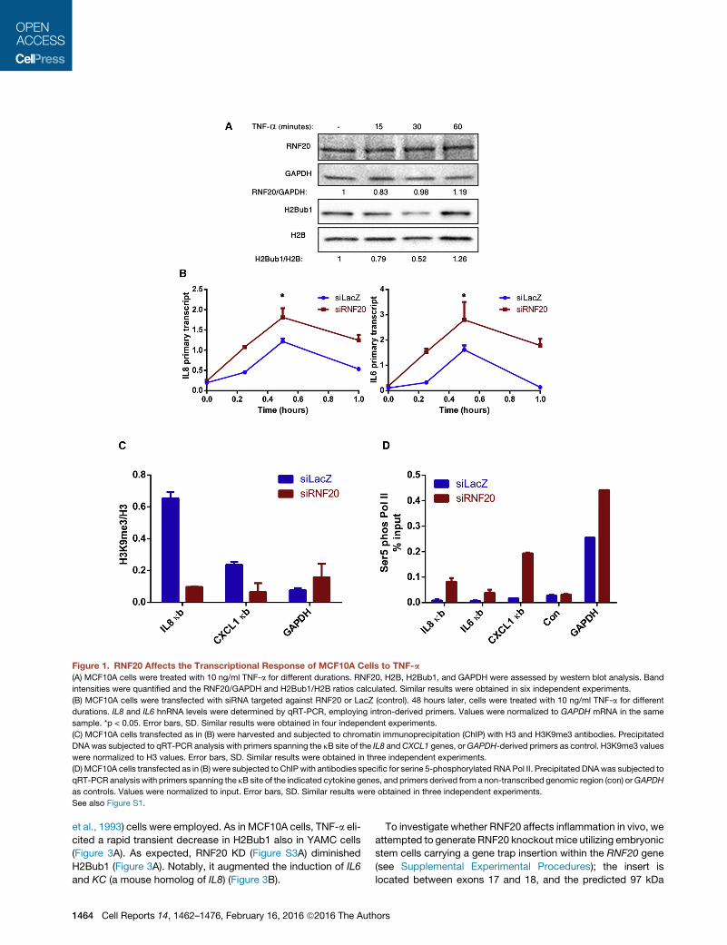
Figure 1. RNF20 Affects the Transcriptional Response of MCF10A Cells to TNF-a
(A) MCF10A cells were treated with 10 ng/ml TNF-a for different durations. RNF20, H2B, H2Bub1, and GAPDH were assessed by western blot analysis. Band
intensities were quantified and the RNF20/GAPDH and H2Bub1/H2B ratios calculated. Similar results were obtained in six independent experiments.
(B) MCF10A cells were transfected with siRNA targeted against RNF20 or LacZ (control). 48 hours later, cells were treated with 10 ng/ml TNF-a for different
durations. IL8 and IL6 hnRNA levels were determined by qRT-PCR, employing intron-derived primers. Values were normalized to GAPDH mRNA in the same
sample. *p < 0.05. Error bars, SD. Similar results were obtained in four independent experiments.
(C) MCF10A cells transfected as in (B) were harvested and subjected to chromatin immunoprecipitation (ChIP) with H3 and H3K9me3 antibodies. Precipitated
DNA was subjected to qRT-PCR analysis with primers spanning the kB site of the IL8 andCXCL1 genes, orGAPDH-derived primers as control. H3K9me3 values
were normalized to H3 values. Error bars, SD. Similar results were obtained in three independent experiments.
(D) MCF10A cells transfected as in (B) were subjected to ChIP with antibodies specific for serine 5-phosphorylated RNA Pol II. Precipitated DNAwas subjected to
qRT-PCR analysis with primers spanning the kB site of the indicated cytokine genes, and primers derived from a non-transcribed genomic region (con) orGAPDH
as controls. Values were normalized to input. Error bars, SD. Similar results were obtained in three independent experiments.
See also Figure S1.
et al., 1993) cells were employed. As in MCF10A cells, TNF-a eli-
cited a rapid transient decrease in H2Bub1 also in YAMC cells
(Figure 3A). As expected, RNF20 KD (Figure S3A) diminished
H2Bub1 (Figure 3A). Notably, it augmented the induction of IL6
and KC (a mouse homolog of IL8) (Figure 3B).
1464 Cell Reports 14, 1462–1476, February 16, 2016 ª2016 The Auth
To investigate whether RNF20 affects inflammation in vivo, we
attempted to generate RNF20 knockout mice utilizing embryonic
stem cells carrying a gene trap insertion within the RNF20 gene
(see Supplemental Experimental Procedures); the insert is
located between exons 17 and 18, and the predicted 97 kDa
ors

Figure 2. RNF20 Modulates NF-kB Chromatin Binding
(A)MCF10A cells were transfectedwith siRNA targeted against LacZ (control) or RNF20 for 48 hr and treatedwith 10 ng/ml TNF-a for 30min. Cells were subjected
to ChIP analysis with p65-specific antibodies. Precipitated DNA was subjected to qRT-PCR with primers spanning the kB sites of the IL8,CXCL1, and IL6 genes
or CDKN1A (p21)-derived primers as control. Values were normalized to input. Error bars, SD. Similar results were obtained in five independent experiments.
(B)MCF10A cells were transfected and analyzed as in (A), but employing p50-specific antibodies. Error bars, SD. Similar results were obtained in five independent
experiments.
(C) MCF10A cells were transfected and treated with TNF-a as in (A). Total chromatin was enriched as described in Supplemental Experimental Procedures, and
chromatin-associated RNF20, p50 and p65 was determined by western blot analysis. Numbers indicate relative intensities of the p50 and p65 bands after
normalization to the corresponding H2B signal; ratios in the control (siLacZ) untreated sample were set as 1.0. Relative p65/p50 ratio was calculated accordingly.
Similar results were obtained in three independent experiments.
(D) MCF10A cells were transfected and treated as in (A). Sequential chromatin immunoprecipitation (Re-ChIP) was performed as described in the Supplemental
Experimental Procedures, employing p65 antibodies followed by p50 antibodies. qRT-PCR analysis was as in (A). Error bars, SD. Similar results were obtained in
three independent experiments.
See also Figure S2.
protein should lack the C0 terminal RING domain. Such truncated
protein was undetectable (Figure S3B), suggesting its intrinsic
instability. Our attempts to obtain viable homozygous knockout
progeny failed, owing to preimplantation embryonic lethality.
Heterozygous RNF20+/� mice were viable and appeared
normal. We derived small intestinal 3D organoid cultures (Sato
et al., 2011) from wild-type (WT) and RNF20+/� mice. As ex-
pected, RNF20+/� organoids had less RNF20 mRNA and
H2Bub1 than WT organoids (Figures 3C and S3C). Importantly,
TNF-a elicited a rapid decrease in H2Bub1 (Figure 3C), and
NF-kB target gene inductionwas augmented in RNF20+/� relative
to WT organoids (Figures 3D and S3D). Likewise, peritoneal in-
flammatory leukocytes (mostly monocytes) and bone marrow-
derived macrophages from RNF20+/� mice, underexpressing
RNF20 (Figures S3E and S3F), displayed an augmented TNF-a
Cell R
response at both RNA (Figures 3E and S3F) and secreted cyto-
kine (Figure 3F) levels.
Thus, in non-cancerous epithelial cells, intestinal organoids,
and innate immune cells, reduced RNF20/H2Bub1 augments
the proinflammatory transcriptional response.
RNF20 Heterozygous Mice Are Prone to Severe ColonicInflammationDextran sodium sulfate (DSS) is extensively used to studymouse
colonic inflammation (Clapper et al., 2007). To explore the
impact of RNF20 on inflammation in vivo, mice were exposed
to 2% DSS (3 cycles, 5 days each; Figure 4A). Inflammation
was assessed by colonoscopy, using a five-parameter scoring
system (Becker et al., 2006) as described (Cooks et al., 2013).
By day 7, inflammation became significantly more severe in
eports 14, 1462–1476, February 16, 2016 ª2016 The Authors 1465

(legend on next page)
1466 Cell Reports 14, 1462–1476, February 16, 2016 ª2016 The Authors

RNF20+/� colons than inWT colons (Figure 4B). Although inflam-
mation subsided partially fromday 65 onward, it remained higher
in the heterozygotes. Moreover, by day 26 RNF20+/� mice dis-
played augmented splenomegaly (Figure 4C), and by day 65
they had shorter colons compared to WT mice (Figures 4D and
S4A); both features persisted till day 180 (Figures S4B and
S4C). Concordantly, DSS-treated RNF20+/� mice lost more
weight than WT mice (Figure S4D). Thus, RNF20+/� mice are
prone to augmented acute and chronic inflammation.
RNF20+/� mice express less RNF20 in all tissues. To assess
the contribution of the bone marrow (BM)-derived compartment,
we performed BM transplantation studies where BM of each ge-
notype was transplanted into irradiated mice of each genotype.
Reassuringly, WT mice reconstituted with WT BM were less
inflamed than RNF20+/� mice reconstituted with RNF20+/� BM
(Figure 4E). Interestingly, both WT mice reconstituted with
RNF20+/� BM and RNF20+/� mice reconstituted with WT BM
displayed intermediate inflammation, suggesting that both the
intestinal epithelial compartment and the myeloid compartment
contribute to the increased inflammation in RNF20+/� mice,
consistent with the augmented cytokine responses in both cell
types in vitro (Figures 3 and S3).
Remarkably, RNF20+/� colons had compromised barrier func-
tion, resulting in increased fluorescence in the blood of mice
force-fed dextran FITC (Figure 4F). This intestinal leakiness,
perhaps due to reduced expression of tight junction genes
(e.g., cingulin, Figure S4E), may offer an additional explanation
for the epithelial contribution to the augmented inflammation.
Interestingly, c-Myc expression was elevated in RNF20+/� co-
lons (Figure S4E).
Although average RNF20 mRNA levels in RNF20+/� colons
and lungs (Figures S4F and S4G) was approximately half that
of WT mice, it varied greatly among individual mice (Figure 4G).
H2Bub1 correlated well with RNF20 mRNA (Figure S4H).
Remarkably, regardless of genotype, RNF20 mRNA levels
(measured in the non-inflamed lungs) were strongly inversely
correlated with colonic inflammatory score (Figure 4G), as
seen also with interferon gamma (IFN-g) secretion from colon
slices at day 7, MIP2 mRNA expression at day 26, and colon
length and spleen weight at day 180 (Figures S4I–S4L). This ar-
gues strongly that the increased inflammation in RNF20+/� mice
is indeed due to the actual downregulation of RNF20 (and pre-
sumably H2Bub1).
Figure 3. Reduced RNF20 Augments the TNF-a Response in Mouse C
(A) YAMC (young adult mouse colonocytes) cells were transfected with siRNA ta
10 ng/ml TNF-a for 30 min. H2B and H2Bub1 levels were assessed by Western
(B) YAMC cells were transfected as in (A). Forty-eight hours later, cells were treat
qRT-PCR, and normalized to b-actin mRNA in the same sample. *p < 0.05. Error
(C) Small intestine organoids fromWT or RNF20+/�micewere either treated or not
by western blot analysis. The H2B andH2Bub1 panels are from two separate gels
in four independent experiments.
(D) Small intestine organoids fromWTor RNF20+/�micewere treatedwith 10 ng/m
PCR and normalized to b-actinmRNA in the same sample. *p < 0.05, **p < 0.01. E
(E) Mice were injected intraperitoneally with 4% thioglycolate. Four days later, per
with 10 ng/ml TNF-a for 30 min. Cytokine mRNA levels were determined by qRT-P
SD. Similar results were obtained in three independent experiments.
(F) Mice were injected intraperitoneally with 4% thioglycolate. Four days later, peri
for different durations. Secreted cytokines were quantified by ELISA. Similar res
See also Figure S3.
Cell R
Increased MDSC Activation in RNF20+/� MiceUnder inflammatory conditions, immune cells infiltrate the
colonic epithelium. Notably, immune patches composed mainly
of T cells, B cells, and macrophages were more abundant
in DSS-treated RNF20+/� than in WT mice (Figures S5A and
S5B). RNF20+/� mice displayed more infiltrating macrophages
(F4/80 positive, Figures S5C and S5D), including M2 macro-
phages (CD206+, Figure 4H), as well as more T cells (CD3+,
Figures S5E and S5F), at both day 26 and 65. Importantly,
RNF20-deficient mice had more myeloid-derived suppressor
cells (MDSCs), identified by IHC staining for CD11b+ and Gr1
(Figure 4I) and CD11b positivity by FACS (Figure S5G). Those
MDSCs were more active, producing excessive ROS and nitric
oxide (Figure S5H) and overexpressing Arginase 1 (Figure S5I).
Furthermore, T cell receptor z chain was downregulated in
RNF20+/� splenic T cells (Figure S5J), indicative of impaired
T cell function. CD3 epsilon was comparable in WT and
RNF20+/� mice (Figure S5J), further arguing that the reduced
T cell functionality in the latter is mediated by MDSCs. Impor-
tantly, RNF20+/� MDSCs revealed a significantly greater capac-
ity to downregulate z chain levels in co-incubated naive T cells
(Figure 4J). Hence, reduced RNF20/H2Bub1 might promote
immunosuppression via increased MDSC activation.
Elevated NF-kB Activity and DNA Damage in RNF20+/�
MiceConsistent with the enhanced NF-kB activation in cultured cells,
inflamed colons of RNF20+/� mice revealed stronger nuclear
p65 staining than WT mice, while p50 exhibited an opposite
trend (Figure 5A). Nuclear g-H2AX foci, indicative of DNA dam-
age, were more abundant in RNF20+/� colons (Figure 5B), prob-
ably reflecting the combined effect of increased DNA-damaging
ROS (Schetter et al., 2010) and compromised DNA double
strand break repair (Chernikova et al., 2010; Moyal et al.,
2011; Nakamura et al., 2011). Notably, despite the marked in-
crease in apparent DNA damage, p53 was only mildly elevated
(Figure 5B); p21, the canonical p53 target gene, was hardly
induced at all. This suggests a compromised p53 response,
probably due to p53 mRNA downregulation in RNF20+/� mice
(Figure S4E). Thus, while RNF20+/� colonocytes accumulate
more DNA damage, their ability to mount a full DNA damage
response is compromised, potentially exacerbating genomic
instability.
ells, Intestinal Organoids, and Peritoneal Inflammatory Leukocytes
rgeted against RNF20 or LacZ as control and either treated or not treated with
blot analysis. Similar results were obtained in five independent experiments.
ed with 10 ng/ml TNF-a for 1 hr. IL6 and KC mRNA levels were determined by
bars, SD. Similar results were obtained in five independent experiments.
treated with 10 ng/ml TNF-a for 30min. H2B and H2Bub1 levels were assessed
loaded with identical aliquots of the same extract. Similar results were obtained
l TNF-a for different durations. CytokinemRNA levels were determined by qRT-
rror bars, SE. The graph represents average of four independent experiments.
itoneal inflammatory leukocytes were collected and either treated or not treated
CR and normalized to b-actinmRNA in the same sample. *p < 0.05. Error bars,
toneal inflammatory leukocytes were removed and treated with 10 ng/ml TNF-a
ults were obtained in three independent experiments.
eports 14, 1462–1476, February 16, 2016 ª2016 The Authors 1467

(legend on next page)
1468 Cell Reports 14, 1462–1476, February 16, 2016 ª2016 The Authors
RNF20+/� Mice Are Predisposed to Inflammation-Associated Colorectal TumorsChronic colonic inflammation promotes colorectal cancer (Terzi�c
et al., 2010; Ullman and Itzkowitz, 2011). In the mouse, this is
greatly enhanced by combining DSS with the chemical carcin-
ogen azoxymethane (AOM) (Ward et al., 1973; Tanaka et al.,
2003). We therefore injectedmice with AOM (1 mg/g bodyweight)
7 days prior to DSS exposure (Figure 6A). Colonoscopy revealed
more polyps in RNF20+/�mice than inWTmice (Figure 6B; repre-
sentative images in Figure S6A). Moreover, while RNF20+/� ade-
nomas often progressed to carcinoma by day 180, none did in
WT mice (Figure 6C, DSS-AOM). Likewise, DSS alone elicited
carcinomas by day 180 in several RNF20+/� but no WT animals
(Figure 6C, DSS). Some RNF20+/� mice developed mucinous
carcinoma with neoplastic glands invading the submucosa and
penetrating the muscularis propria (T2), indicative of an aggres-
sive course (Figure S6B). Hence, reduced RNF20 promotes
both initiation and progression of inflammation-associated CRC
in mice. Notably, the impact of partial RNF20 depletion on tumor
progression was more pronounced than on inflammation. Thus,
beyond augmented inflammation, additional effects of RNF20
shortage might also promote cancer in this setting.
While total H2B staining was similar in tumors and non-
tumorous crypts (Figure 6D, left panels), polyps and carcinomas
in both genotypes often displayed reduced H2Bub1 in both
epithelial and stromal compartments (Figures 6D, 6E, and S6C,
arrows), implying that colorectal tumorigenesis is associated
with further H2Bub1 decrease.
Lower H2Bub1 May Promote Human Ulcerative Colitisand Colorectal CancerTo evaluate the human relevance of our findings, we analyzed a
gene expression array including data from 27 healthy people,
35 UC patients with non-inflamed colons, and 38 UC patients
with inflamed colons (Sen and Bhaumik, 2013). For patients
sampled multiple times, mean values were used. Remarkably,
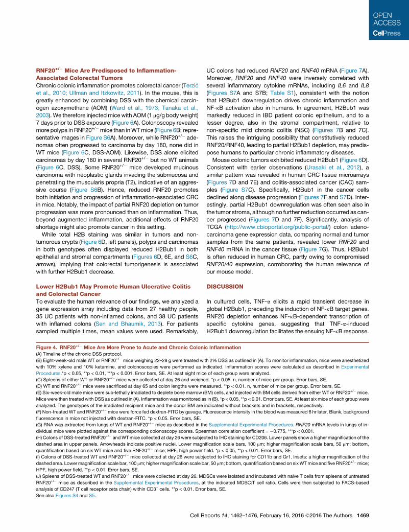
Figure 4. RNF20+/� Mice Are More Prone to Acute and Chronic Coloni
(A) Timeline of the chronic DSS protocol.
(B) Eight-week-old male WT or RNF20+/� mice weighing 22–28 g were treated wi
with 10% xylene and 10% ketamine, and colonoscopies were performed as in
Procedures.*p < 0.05, **p < 0.01, ***p < 0.001. Error bars, SE. At least eight mice
(C) Spleens of either WT or RNF20+/� mice were collected at day 26 and weighe
(D) WT and RNF20+/� mice were sacrificed at day 65 and colon lengths were me
(E) Six-week-old male mice were sub-lethally irradiated to deplete bone marrow (B
Mice were then treated with DSS as outlined in (A). Inflammation wasmonitored as
analyzed. The genotypes of the irradiated recipient mice and the donor BM are i
(F) Non-treated WT and RNF20+/� mice were force fed dextran-FITC by gavage. F
fluorescence in mice not injected with dextran-FITC. *p < 0.05. Error bars, SE.
(G) RNA was extracted from lungs of WT and RNF20+/� mice as described in th
dividual mice were plotted against the corresponding colonoscopy scores. Spea
(H) Colons of DSS-treated RNF20+/� andWTmice collected at day 26 were subjec
dashed area in upper panels. Arrowheads indicate positive nuclei. Lower magn
quantification based on six WT mice and five RNF20+/� mice; HPF, high power fi
(I) Colons of DSS-treated WT and RNF20+/� mice collected at day 26 were subje
dashed area. Lowermagnification scale bar, 100 mm; highermagnification scale ba
HPF, high power field. **p < 0.01. Error bars, SE.
(J) Spleens of DSS-treated WT and RNF20+/� mice were collected at day 26. MD
RNF20+/� mice as described in the Supplemental Experimental Procedures, at
analysis of CD247 (T cell receptor zeta chain) within CD3+ cells. **p < 0.01. Error
See also Figures S4 and S5.
Cell R
UC colons had reduced RNF20 and RNF40 mRNA (Figure 7A).
Moreover, RNF20 and RNF40 were inversely correlated with
several inflammatory cytokine mRNAs, including IL6 and IL8
(Figures S7A and S7B; Table S1), consistent with the notion
that H2Bub1 downregulation drives chronic inflammation and
NF-kB activation also in humans. In agreement, H2Bub1 was
markedly reduced in IBD patient colonic epithelium, and to a
lesser degree, also in the stromal compartment, relative to
non-specific mild chronic colitis (NSC) (Figures 7B and 7C).
This raises the intriguing possibility that constitutively reduced
RNF20/RNF40, leading to partial H2Bub1 depletion, may predis-
pose humans to particular chronic inflammatory diseases.
Mouse colonic tumors exhibited reduced H2Bub1 (Figure 6D).
Consistent with earlier observations (Urasaki et al., 2012), a
similar pattern was revealed in human CRC tissue microarrays
(Figures 7D and 7E) and colitis-associated cancer (CAC) sam-
ples (Figure S7C). Specifically, H2Bub1 in the cancer cells
declined along disease progression (Figures 7F and S7D). Inter-
estingly, partial H2Bub1 downregulation was often seen also in
the tumor stroma, although no further reduction occurred as can-
cer progressed (Figures 7D and 7F). Significantly, analysis of
TCGA (http://www.cbioportal.org/public-portal/) colon adeno-
carcinoma gene expression data, comparing normal and tumor
samples from the same patients, revealed lower RNF20 and
RNF40 mRNA in the cancer tissue (Figure 7G). Thus, H2Bub1
is often reduced in human CRC, partly owing to compromised
RNF20/40 expression, corroborating the human relevance of
our mouse model.
DISCUSSION
In cultured cells, TNF-a elicits a rapid transient decrease in
global H2Bub1, preceding the induction of NF-kB target genes.
RNF20 depletion enhances NF-kB-dependent transcription of
specific cytokine genes, suggesting that TNF-a-induced
H2Bub1 downregulation facilitates the ensuing NF-kB response.
c Inflammation
th 2% DSS as outlined in (A). To monitor inflammation, mice were anesthetized
dicated. Inflammation scores were calculated as described in Experimental
of each group were analyzed.
d. *p < 0.05. n, number of mice per group. Error bars, SE.
asured. **p < 0.01. n, number of mice per group. Error bars, SE.
M) cells, and injected with BM cells derived from either WT or RNF20+/� mice.
in (B). *p < 0.05, **p < 0.01. Error bars, SE. At least six mice of each groupwere
ndicated without brackets and in brackets, respectively.
luorescence intensity in the blood was measured 6 hr later. Blank, background
e Supplemental Experimental Procedures. RNF20 mRNA levels in lungs of in-
rman correlation coefficient = �0.775, ***p < 0.001.
ted to IHC staining for CD206. Lower panels show a higher magnification of the
ification scale bars, 100 mm; higher magnification scale bars, 50 mm; bottom,
eld. *p < 0.05, **p < 0.01. Error bars, SE.
cted to IHC staining for CD11b and Gr1. Insets: a higher magnification of the
r, 50 mm; bottom, quantification based on sixWTmice and five RNF20+/�mice;
SCs were isolated and incubated with naive T cells from spleens of untreated
the indicated MDSC:T cell ratio. Cells were then subjected to FACS-based
bars, SE.
eports 14, 1462–1476, February 16, 2016 ª2016 The Authors 1469
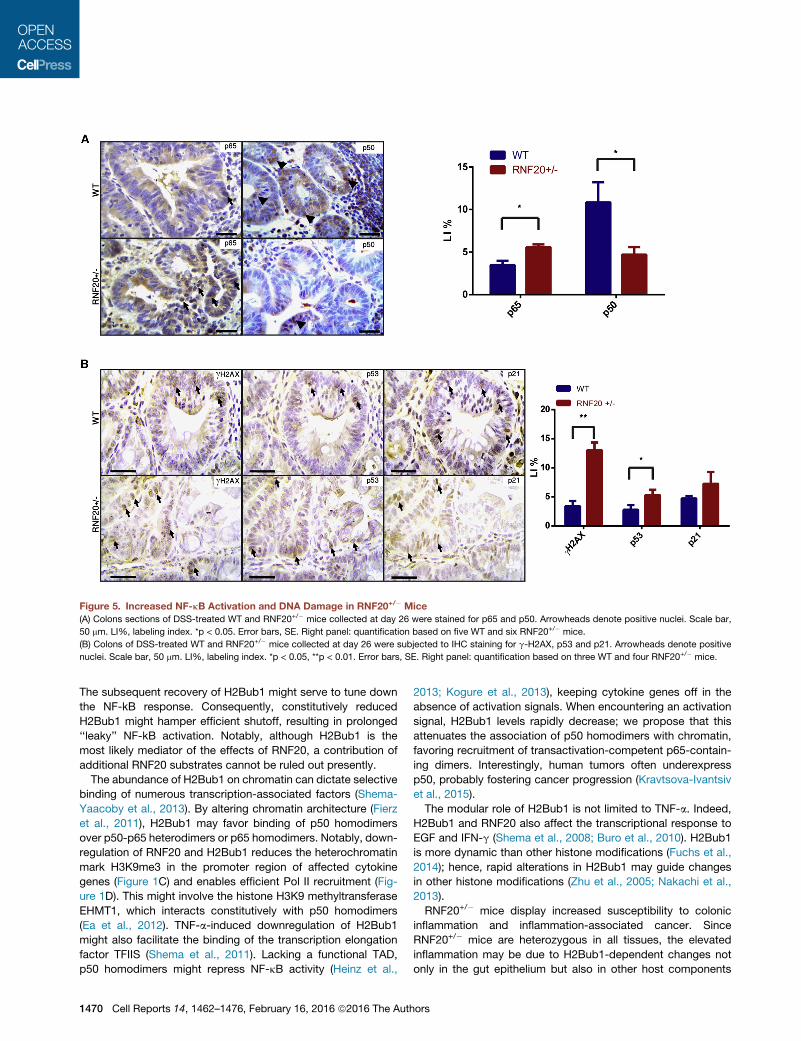
Figure 5. Increased NF-kB Activation and DNA Damage in RNF20+/� Mice
(A) Colons sections of DSS-treated WT and RNF20+/� mice collected at day 26 were stained for p65 and p50. Arrowheads denote positive nuclei. Scale bar,
50 mm. LI%, labeling index. *p < 0.05. Error bars, SE. Right panel: quantification based on five WT and six RNF20+/� mice.
(B) Colons of DSS-treated WT and RNF20+/� mice collected at day 26 were subjected to IHC staining for g-H2AX, p53 and p21. Arrowheads denote positive
nuclei. Scale bar, 50 mm. LI%, labeling index. *p < 0.05, **p < 0.01. Error bars, SE. Right panel: quantification based on three WT and four RNF20+/� mice.
The subsequent recovery of H2Bub1 might serve to tune down
the NF-kB response. Consequently, constitutively reduced
H2Bub1 might hamper efficient shutoff, resulting in prolonged
‘‘leaky’’ NF-kB activation. Notably, although H2Bub1 is the
most likely mediator of the effects of RNF20, a contribution of
additional RNF20 substrates cannot be ruled out presently.
The abundance of H2Bub1 on chromatin can dictate selective
binding of numerous transcription-associated factors (Shema-
Yaacoby et al., 2013). By altering chromatin architecture (Fierz
et al., 2011), H2Bub1 may favor binding of p50 homodimers
over p50-p65 heterodimers or p65 homodimers. Notably, down-
regulation of RNF20 and H2Bub1 reduces the heterochromatin
mark H3K9me3 in the promoter region of affected cytokine
genes (Figure 1C) and enables efficient Pol II recruitment (Fig-
ure 1D). This might involve the histone H3K9 methyltransferase
EHMT1, which interacts constitutively with p50 homodimers
(Ea et al., 2012). TNF-a-induced downregulation of H2Bub1
might also facilitate the binding of the transcription elongation
factor TFIIS (Shema et al., 2011). Lacking a functional TAD,
p50 homodimers might repress NF-kB activity (Heinz et al.,
1470 Cell Reports 14, 1462–1476, February 16, 2016 ª2016 The Auth
2013; Kogure et al., 2013), keeping cytokine genes off in the
absence of activation signals. When encountering an activation
signal, H2Bub1 levels rapidly decrease; we propose that this
attenuates the association of p50 homodimers with chromatin,
favoring recruitment of transactivation-competent p65-contain-
ing dimers. Interestingly, human tumors often underexpress
p50, probably fostering cancer progression (Kravtsova-Ivantsiv
et al., 2015).
The modular role of H2Bub1 is not limited to TNF-a. Indeed,
H2Bub1 and RNF20 also affect the transcriptional response to
EGF and IFN-g (Shema et al., 2008; Buro et al., 2010). H2Bub1
is more dynamic than other histone modifications (Fuchs et al.,
2014); hence, rapid alterations in H2Bub1 may guide changes
in other histone modifications (Zhu et al., 2005; Nakachi et al.,
2013).
RNF20+/� mice display increased susceptibility to colonic
inflammation and inflammation-associated cancer. Since
RNF20+/� mice are heterozygous in all tissues, the elevated
inflammation may be due to H2Bub1-dependent changes not
only in the gut epithelium but also in other host components
ors
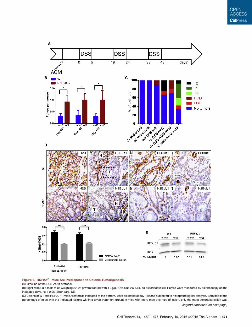
Figure 6. RNF20+/� Mice Are Predisposed to Colonic Tumorigenesis
(A) Timeline of the DSS-AOM protocol.
(B) Eight-week-old male mice weighing 22–28 g were treated with 1 mg/g AOM plus 2% DSS as described in (A). Polyps were monitored by colonoscopy on the
indicated days. *p < 0.05. Error bars, SE.
(C) Colons of WT and RNF20+/� mice, treated as indicated at the bottom, were collected at day 180 and subjected to histopathological analysis. Bars depict the
percentage of mice with the indicated lesions within a given treatment group; in mice with more than one type of lesion, only the most advanced lesion was
(legend continued on next page)
Cell Reports 14, 1462–1476, February 16, 2016 ª2016 The Authors 1471

such as immune cells, a notion supported by the enhanced
TNF-a response of RNF20+/� inflammatory leukocytes and
macrophages (Figures 3E, 3F, and S3F), as well as by the bone
marrow chimera experiments. Furthermore, we cannot exclude
RNF20/H2Bub1 effects on gut microflora.
Notably, RNF20 mRNA levels varied greatly between individ-
ual mice (Figure 4G), and inflammation inversely correlated
with RNF20 mRNA irrespective of genotype. This raises the
intriguing possibility that in humans, where inter-individual differ-
ences in H2Bub1 levels are likely broader than in inbred mice,
such inherent differences might affect susceptibility to chronic
inflammatory diseases. Indeed, human ulcerative colitis (UC) is
significantly associated with reducedRNF20/40 expression (Fig-
ure 7A), and lower RNF20/40 correlates with augmented inflam-
matory cytokine expression in human CRC (Figures S7A and
S7B; Table S1).
Remarkably, both with DSS alone and with DSS+AOM,
aggressive tumors were observed exclusively in RNF20+/� but
not WT mice (Figure 6C). Indeed, as in our mouse tumors,
many human sporadic CRC tumors exhibit reduced H2Bub1 (Ur-
asaki et al., 2012), which declines as tumors progress (Figures
7D–7F). Reduced RNF20 and RNF40mRNA in human CRC (Fig-
ure 7G) provides at least one plausible mechanistic explanation
for the frequent depletion of H2Bub1. Conceivably, compro-
mised H2Bub1 probably contributes to colorectal carcinogenesis
by more than just augmenting inflammation. H2Bub1 facilitates
DNA repair (Moyal et al., 2011; Nakamura et al., 2011; Shiloh
et al., 2011); indeed, RNF20+/� mice display elevated g-H2AX
(Figure 5B), suggesting exacerbated DNA damage. Furthermore,
RNF20+/� mice exhibit abundant M2 macrophages, providing
a tumor supportive environment. Importantly, they display
enhanced recruitment and augmented activation of MDSCs,
which suppress anti-tumoral T cell activity and thereby facilitate
tumor progression (Katoh et al., 2013). Such non-cell autono-
mous effects of reduced RNF20 might underpin the increased
tumor progression in RNF20+/� mice as well as the association
between lower H2Bub1 and advanced human cancer.
Stem cells (SCs) typically display low H2Bub1, which in-
creases markedly during SC differentiation and is required for
their differentiation (Fuchs et al., 2012; Karpiuk et al., 2012).
Hence, reduced H2Bub1 might also increase the tendency of
colonic epithelial cells to adopt stem cell-like features. Of note,
NF-kB has a crucial role in crypt stem cell expansion (Myant
et al., 2013; Schwitalla et al., 2013). Intriguingly, like RNF20,
the stem cell E3 ligase RNF43 has also been implicated in
colonic tumor suppression (Koo et al., 2012).
In sum, our study highlights non cell-autonomous functions of
H2Bub1 and implies that low H2Bub1 favors inflammation and
cancer. It remains to be determined whether, like in mice, also
in humans inter-individual variations in constitutive H2Bub1
considered. LGD, low grade dysplasia; HGD, high grade dysplasia; Tis, carcinom
muscularis propria; n, number of mice per group.
(D) Colons of DSS+AOM-treatedmice of the indicated genotype were collected at
normal tissue; T, tumor. Panels on the right show a highermagnification of the dash
25 mm for higher magnification. Lower panel: quantification based on eight WT a
(E) Colons of DSS+AOM-treated mice of the indicated genotypes were collected a
western blot analysis for H2Bub1 and H2B.
See also Figure S6.
1472 Cell Reports 14, 1462–1476, February 16, 2016 ª2016 The Auth
levels may be a risk factor for UC and CRC and perhaps addi-
tional inflammatory diseases and inflammation-associated
cancers.
EXPERIMENTAL PROCEDURES
Cell Culture and siRNA Transfections
MCF10A cells were cultured at 37�C in DMEMF-12 medium (Biological Indus-
tries) supplemented with penicillin and streptomycin as well as 10 mg/ml insu-
lin, 0.1 mg/ml cholera toxin, 0.5 mg/ml hydrocortisone, 5% heat-inactivated
horse serum, and 10 ng/ml EGF. YAMC cells were cultured at 32�C in RPMI-
1640 (Biological Industries) supplemented with penicillin and streptomycin
and 10�6 M hydrocortisone, 10�5 M a-thioglycerol, 1 mg/ml insulin, 5 units/
ml mouse INF-g, and 5% fetal calf serum. Small intestine organoids were pre-
pared as described (Sato et al., 2011).
siRNA-mediated knockdown was with Dharmafect 1 reagent according to
the manufacturer’s protocol (Dharmacon RNAi technologies, Thermo Fisher
Scientific). siRNA oligonucleotide SmartPools (siLacZ, siRNF20, si-p65, si-
p50, siRNF40, and siRad6) were from Dharmacon. Final concentration of all
siRNA oligonucleotides was 25 nM. TNF-a from R&D Systems was applied
at a final concentration of 10 ng/ml. Cells were washed twice with cold PBS,
harvested, centrifuged for 2 min at 4,000 RPM, and frozen at�80�C until lysis.
Mouse Handling
DSS treatment, colonoscopy, and analysis of mouse tissues were as
described (Cooks et al., 2013). Azoxymethane (Wako Chemicals, 011-
20171; 1 mg/g) was injected intraperitoneally (i.p.) 7 days prior to the first
DSS treatment. For bone marrow (BM) transplantation, mice were irradiated
with 950 rad and injected intravenously with 5 3 106 BM-derived cells. Mice
were then treated for 6 weeks with Ciproxin and subjected to DSS treatment.
For intestinal permeability experiments mice were force fed by gavage with
Dextran FITC (T7816, Sigma-Aldrich; 0.6 g/g). After 6 hr, mice were bled and
blood fluorescence intensity was measured. Procedures involving animals
conformed to institutional guidelines approved by the Animal Ethics Commit-
tee of the Weizmann Institute (IACUC 00800211-1).
Inflammation severity and polyps were scored by colonoscopy according to
a previously published five- parameter scoring system (Becker et al., 2006);
data was analyzed with the R statistical analysis software, employing
ANOVA. H&E slides of each mouse were analyzed by two independent pathol-
ogists, and tumors were scored according to their grade: low grade dysplasia
(LGD), high grade dysplasia (HGD), carcinoma in situ (Tis), tumor invading the
submucosa (T1), and tumor invading the muscularis propria (T2).
To obtain peritoneal inflammatory leukocytes, mice were injected i.p. with
4% thioglycolate. Four days later, leukocytes were collected frommouse peri-
toneum and plated in a tissue culture dish.
Human Ethical Issues
Primary colon biopsies for IHC studies were obtained from Hebrew University-
Hadassah Medical Center under approval by the Institutional Review Board
(HMO-0145-13), verifying that informed consent is not required.
SUPPLEMENTAL INFORMATION
Supplemental Information includes Supplemental Experimental Procedures,
seven figures, and one table and can be found with this article online at
http://dx.doi.org/10.1016/j.celrep.2016.01.020.
a in situ; T1, carcinoma invading the submucosa; T2, carcinoma invading the
day 65. Consecutive slides were stained for H2B and H2Bub1, respectively. N,
ed areas in the left H2Bub1 panels. Scale bars, 200 mm for lowermagnification,
nd ten RNF20+/� mice. ***p < 0.001. Error bars, SE.
t day 180. Polyps and adjacent normal tissue were extracted and subjected to
ors

(legend on next page)
Cell Reports 14, 1462–1476, February 16, 2016 ª2016 The Authors 1473
AUTHOR CONTRIBUTIONS
O.T. designed and performed the experiments, and wrote themanuscript. T.C.
provided advice and helped in conducting experiments. E.S. generated the
RNF20 knockout mice. I.P. and V.G. conducted histological staining and anal-
ysis. E.P. and A.H. conducted pathological analysis and provided suggestions
for further analyses. M.B., J.K., H.A., and H.B. conducted the experiments pre-
sented in Figures 4J, S5G, S5H, and S5J. R.R. conducted bio-statistical anal-
ysis of human data. M.O. supervised the work and wrote the manuscript.
ACKNOWLEDGMENTS
We thank Steven A. Johnsen for the kind gift of antibodies, Neil Perkins, Fiona
Oakley, Intidhar Galy, and Ronny Drapkin for helpful suggestions and re-
agents, and Hagai Marmor, Nir Itzkovits, and Liad Margalit for help with exper-
iments. This work was supported in part by grant 293438 (RUBICAN) from the
European Research Council, the Dr. Miriam and Sheldon G. Adelson Medical
Research Foundation, a Center of Excellence grant (1779/11) from the Israel
Science Foundation, a Center of Excellence grant from the Flight Attendant
Medical Research Institute (FAMRI), EC FP7 funding (INFLACARE, agreement
223151, and INSPiRE, agreement 284460), grant R37 CA40099 from the Na-
tional Cancer Institute, and the Lower Saxony-Israeli Association. M.O. is
incumbent of the Andre Lwoff Chair in Molecular Biology.
Received: October 1, 2015
Revised: November 24, 2015
Accepted: January 1, 2016
Published: February 4, 2016
REFERENCES
Becker, C., Fantini, M.C., and Neurath, M.F. (2006). High resolution colonos-
copy in live mice. Nat. Protoc. 1, 2900–2904.
Bedi, U., Scheel, A.H., Hennion, M., Begus-Nahrmann, Y., R€uschoff, J., and
Johnsen, S.A. (2014). SUPT6H controls estrogen receptor activity and cellular
differentiation by multiple epigenomic mechanisms. Oncogene 34, 465–473.
Ben-Neriah, Y., and Karin, M. (2011). Inflammation meets cancer, with NF-kB
as the matchmaker. Nat. Immunol. 12, 715–723.
Brigati, C., Noonan, D.M., Albini, A., and Benelli, R. (2002). Tumors and inflam-
matory infiltrates: friends or foes? Clin. Exp. Metastasis 19, 247–258.
Buro, L.J., Chipumuro, E., and Henriksen, M.A. (2010). Menin and RNF20
recruitment is associated with dynamic histone modifications that regulate
signal transducer and activator of transcription 1 (STAT1)-activated tran-
scription of the interferon regulatory factor 1 gene (IRF1). Epigenetics Chro-
matin 3, 16.
Campos, E.I., and Reinberg, D. (2009). Histones: annotating chromatin. Annu.
Rev. Genet. 43, 559–599.
Figure 7. Reduced RNF20 and RNF40 mRNA and H2Bub1 in Human Co
(A) Publishedmicroarray data (Sen andBhaumik, 2013) was analyzed, comparing
pathologies (Normal) to those of ulcerative colitis patients (UC), regardless of infl
(B) Colon samples from 25 non-specific mild chronic colitis (non-specific colitis [N
total H2B. Representative cases are shown. Insets: higher magnification of dashe
25 mm.
(C) IHC from (B) was quantified for relative H2Bub1 staining intensity in epitheliu
(D) A tissue microarray from 75 human CRC patients, classified using the tumor no
upper right indicates relative H2Bub1 intensity (H2Bub1/H2B). TNM stages: I, carc
III, carcinoma invading through the muscularis propria into the pericolorectal tiss
(E) Representative H2B and H2Bub1 staining of normal and cancerous human co
scale bar, 100 mm; higher magnification scale bar, 25 mm.
(F) Quantification of the data in (D). *p < 0.05, ***p < 0.001.
(G)RNF20 andRNF40mRNA levels were compared between colorectal cancer (C
www.cbioportal.org/public-portal/). **p < 0.01. Error bars, SE.
See also Figure S7 and Table S1.
1474 Cell Reports 14, 1462–1476, February 16, 2016 ª2016 The Auth
Chernikova, S.B., and Brown, J.M. (2012). R-loops and genomic instability in
Bre1 (RNF20/40)-deficient cells. Cell Cycle 11, 2980–2984.
Chernikova, S.B., Dorth, J.A., Razorenova, O.V., Game, J.C., and Brown, J.M.
(2010). Deficiency in Bre1 impairs homologous recombination repair and cell
cycle checkpoint response to radiation damage in mammalian cells. Radiat.
Res. 174, 558–565.
Chernikova, S.B., Razorenova, O.V., Higgins, J.P., Sishc, B.J., Nicolau, M.,
Dorth, J.A., Chernikova, D.A., Kwok, S., Brooks, J.D., Bailey, S.M., et al.
(2012). Deficiency in mammalian histone H2B ubiquitin ligase Bre1 (Rnf20/
Rnf40) leads to replication stress and chromosomal instability. Cancer Res.
72, 2111–2119.
Clapper, M.L., Cooper, H.S., and Chang, W.C. (2007). Dextran sulfate sodium-
induced colitis-associated neoplasia: a promising model for the development
of chemopreventive interventions. Acta Pharmacol. Sin. 28, 1450–1459.
Cooks, T., Pateras, I.S., Tarcic, O., Solomon, H., Schetter, A.J., Wilder, S., Loz-
ano, G., Pikarsky, E., Forshew, T., Rosenfeld, N., et al. (2013). Mutant p53
prolongs NF-kB activation and promotes chronic inflammation and inflamma-
tion-associated colorectal cancer. Cancer Cell 23, 634–646.
DiDonato, J.A., Mercurio, F., and Karin, M. (2012). NF-kB and the link between
inflammation and cancer. Immunol. Rev. 246, 379–400.
Ea, C.K., Hao, S., Yeo, K.S., and Baltimore, D. (2012). EHMT1 protein binds to
nuclear factor-kB p50 and represses gene expression. J. Biol. Chem. 287,
31207–31217.
Eaden, J.A., Abrams, K.R., and Mayberry, J.F. (2001). The risk of colorectal
cancer in ulcerative colitis: a meta-analysis. Gut 48, 526–535.
Fierz, B., Chatterjee, C., McGinty, R.K., Bar-Dagan, M., Raleigh, D.P., and
Muir, T.W. (2011). Histone H2B ubiquitylation disrupts local and higher-order
chromatin compaction. Nat. Chem. Biol. 7, 113–119.
Fuchs, G., Shema, E., Vesterman, R., Kotler, E., Wolchinsky, Z., Wilder, S., Go-
lomb, L., Pribluda, A., Zhang, F., Haj-Yahya, M., et al. (2012). RNF20 and
USP44 regulate stem cell differentiation by modulating H2B monoubiquityla-
tion. Mol. Cell 46, 662–673.
Fuchs, G., Hollander, D., Voichek, Y., Ast, G., and Oren, M. (2014). Cotran-
scriptional histone H2B monoubiquitylation is tightly coupled with RNA poly-
merase II elongation rate. Genome Res. 24, 1572–1583.
Grivennikov, S.I., Greten, F.R., and Karin, M. (2010). Immunity, inflammation,
and cancer. Cell 140, 883–899.
Hahn,M.A., Dickson, K.A., Jackson, S., Clarkson, A., Gill, A.J., andMarsh, D.J.
(2012). The tumor suppressor CDC73 interacts with the ring finger proteins
RNF20 and RNF40 and is required for the maintenance of histone 2B monou-
biquitination. Hum. Mol. Genet. 21, 559–568.
Hanahan, D., and Weinberg, R.A. (2011). Hallmarks of cancer: the next gener-
ation. Cell 144, 646–674.
Hayden, M.S., and Ghosh, S. (2008). Shared principles in NF-kappaB
signaling. Cell 132, 344–362.
lonic Pathologies
averageRNF20 andRNF40mRNA levels in colons of individuals with no colonic
ammation status. *p < 0.05, **p < 0.01. Error bars, SE.
SC]) patients and 19 ulcerative colitis (UC) patients were stained for H2Bub1 or
d area. Lower magnification scale bar, 100 mm; higher magnification scale bar,
m and stroma. ***p < 0.001. Error bars, SE.
de metastasis (TNM) system, was stained for H2Bub1 and total H2B. Scale on
inoma invading the submucosa; II, carcinoma invading the muscularis propria;
ue.
lonic tissue. Insets: higher magnification of dashed area. Lower magnification
RC) and corresponding normal colonic tissue, using the TCGA database (http://
ors

Hayden, M.S., and Ghosh, S. (2012). NF-kB, the first quarter-century: remark-
able progress and outstanding questions. Genes Dev. 26, 203–234.
Heinz, S., Romanoski, C.E., Benner, C., Allison, K.A., Kaikkonen, M.U., Or-
ozco, L.D., and Glass, C.K. (2013). Effect of natural genetic variation on
enhancer selection and function. Nature 503, 487–492.
Hwang, W.W., Venkatasubrahmanyam, S., Ianculescu, A.G., Tong, A., Boone,
C., and Madhani, H.D. (2003). A conserved RING finger protein required for
histone H2B monoubiquitination and cell size control. Mol. Cell 11, 261–266.
Jaaskelainen, T., Makkonen, H., Visakorpi, T., Kim, J., Roeder, R.G., and Pal-
vimo, J.J. (2012). Histone H2B ubiquitin ligases RNF20 andRNF40 in androgen
signaling and prostate cancer cell growth. Mol. Cell. Endocrinol. 350, 87–98.
Kari, V., Shchebet, A., Neumann, H., and Johnsen, S.A. (2011). The H2B ubiq-
uitin ligase RNF40 cooperates with SUPT16H to induce dynamic changes in
chromatin structure during DNA double-strand break repair. Cell Cycle 10,
3495–3504.
Karpiuk, O., Najafova, Z., Kramer, F., Hennion, M., Galonska, C., Konig, A.,
Snaidero, N., Vogel, T., Shchebet, A., Begus-Nahrmann, Y., et al. (2012).
The histone H2B monoubiquitination regulatory pathway is required for differ-
entiation of multipotent stem cells. Mol. Cell 46, 705–713.
Katoh, H., Wang, D., Daikoku, T., Sun, H., Dey, S.K., and Dubois, R.N. (2013).
CXCR2-expressingmyeloid-derived suppressor cells are essential to promote
colitis-associated tumorigenesis. Cancer Cell 24, 631–644.
Kogure, M., Takawa, M., Saloura, V., Sone, K., Piao, L., Ueda, K., Ibrahim, R.,
Tsunoda, T., Sugiyama, M., Atomi, Y., et al. (2013). The oncogenic polycomb
histone methyltransferase EZH2 methylates lysine 120 on histone H2B and
competes ubiquitination. Neoplasia 15, 1251–1261.
Koo, B.K., Stange, D.E., Sato, T., Karthaus, W., Farin, H.F., Huch, M., van Es,
J.H., and Clevers, H. (2012). Controlled gene expression in primary Lgr5 orga-
noid cultures. Nat. Methods 9, 81–83.
Kravtsova-Ivantsiv, Y., Shomer, I., Cohen-Kaplan, V., Snijder, B., Superti-
Furga, G., Gonen, H., Sommer, T., Ziv, T., Admon, A., Naroditsky, I., et al.
(2015). KPC1-mediated ubiquitination and proteasomal processing of NF-
kB1 p105 to p50 restricts tumor growth. Cell 161, 333–347.
Kumar, V., and Robbins, S.L. (2007). Robbins Basic Pathology (Saunders/
Elsevier).
Lawrence, T., Gilroy, D.W., Colville-Nash, P.R., and Willoughby, D.A. (2001).
Possible new role for NF-kappaB in the resolution of inflammation. Nat.
Med. 7, 1291–1297.
Majno, G., and Joris, I. (2004). Cells, Tissues, and Disease: Principles of Gen-
eral Pathology (Oxford University Press).
Mantovani, A., Romero, P., Palucka, A.K., and Marincola, F.M. (2008). Tumour
immunity: effector response to tumour and role of the microenvironment. Lan-
cet 371, 771–783.
Medzhitov, R. (2010). Inflammation 2010: new adventures of an old flame. Cell
140, 771–776.
Minsky, N., Shema, E., Field, Y., Schuster, M., Segal, E., and Oren, M. (2008).
Monoubiquitinated H2B is associated with the transcribed region of highly ex-
pressed genes in human cells. Nat. Cell Biol. 10, 483–488.
Moyal, L., Lerenthal, Y., Gana-Weisz, M., Mass, G., So, S., Wang, S.Y., Eppink,
B., Chung, Y.M., Shalev, G., Shema, E., et al. (2011). Requirement of ATM-
dependent monoubiquitylation of histone H2B for timely repair of DNA dou-
ble-strand breaks. Mol. Cell 41, 529–542.
Myant, K.B., Cammareri, P., McGhee, E.J., Ridgway, R.A., Huels, D.J., Cor-
dero, J.B., Schwitalla, S., Kalna, G., Ogg, E.L., Athineos, D., et al. (2013).
ROS production and NF-kB activation triggered by RAC1 facilitate WNT-
driven intestinal stem cell proliferation and colorectal cancer initiation. Cell
Stem Cell 12, 761–773.
Nakachi, I., Rice, J.L., Coldren, C.D., Edwards, M.G., Stearman, R.S., Glide-
well, S.C., Varella-Garcia, M., Franklin, W.A., Keith, R.L., Lewis, M.T., et al.
(2013). Application of SNP microarrays to the genome-wide analysis of chro-
mosomal instability in premalignant airway lesions. Cancer Prev. Res. (Phila)
7, 255–265.
Cell R
Nakamura, K., Kato, A., Kobayashi, J., Yanagihara, H., Sakamoto, S., Oliveira,
D.V., Shimada, M., Tauchi, H., Suzuki, H., Tashiro, S., et al. (2011). Regulation
of homologous recombination by RNF20-dependent H2B ubiquitination. Mol.
Cell 41, 515–528.
O’Dea, E., and Hoffmann, A. (2010). The regulatory logic of the NF-kappaB
signaling system. Cold Spring Harb. Perspect. Biol. 2, a000216.
Pavri, R., Zhu, B., Li, G., Trojer, P., Mandal, S., Shilatifard, A., and Reinberg, D.
(2006). Histone H2B monoubiquitination functions cooperatively with FACT to
regulate elongation by RNA polymerase II. Cell 125, 703–717.
Perkins, N.D. (2012). The diverse and complex roles of NF-kB subunits in can-
cer. Nat. Rev. Cancer 12, 121–132.
Pikarsky, E., Porat, R.M., Stein, I., Abramovitch, R., Amit, S., Kasem, S., Gut-
kovich-Pyest, E., Urieli-Shoval, S., Galun, E., and Ben-Neriah, Y. (2004). NF-
kappaB functions as a tumour promoter in inflammation-associated cancer.
Nature 431, 461–466.
Prenzel, T., Begus-Nahrmann, Y., Kramer, F., Hennion, M., Hsu, C., Gorsler,
T., Hintermair, C., Eick, D., Kremmer, E., Simons, M., et al. (2011). Estrogen-
dependent gene transcription in human breast cancer cells relies upon pro-
teasome-dependent monoubiquitination of histone H2B. Cancer Res. 71,
5739–5753.
Sato, T., Stange, D.E., Ferrante, M., Vries, R.G., Van Es, J.H., Van den Brink,
S., Van Houdt, W.J., Pronk, A., Van Gorp, J., Siersema, P.D., and Clevers,
H. (2011). Long-term expansion of epithelial organoids from human colon, ad-
enoma, adenocarcinoma, and Barrett’s epithelium. Gastroenterology 141,
1762–1772.
Schetter, A.J., Heegaard, N.H., and Harris, C.C. (2010). Inflammation and can-
cer: interweaving microRNA, free radical, cytokine and p53 pathways. Carci-
nogenesis 31, 37–49.
Schwitalla, S., Fingerle, A.A., Cammareri, P., Nebelsiek, T., Goktuna, S.I.,
Ziegler, P.K., Canli, O., Heijmans, J., Huels, D.J., Moreaux, G., et al.
(2013). Intestinal tumorigenesis initiated by dedifferentiation and acquisition
of stem-cell-like properties. Cell 152, 25–38.
Sen, R., and Bhaumik, S.R. (2013). Transcriptional stimulatory and repressive
functions of histone H2B ubiquitin ligase. Transcription 4, 221–226.
Shema, E., Tirosh, I., Aylon, Y., Huang, J., Ye, C., Moskovits, N., Raver-Sha-
pira, N., Minsky, N., Pirngruber, J., Tarcic, G., et al. (2008). The histone H2B-
specific ubiquitin ligase RNF20/hBRE1 acts as a putative tumor suppressor
through selective regulation of gene expression. Genes Dev. 22, 2664–2676.
Shema, E., Kim, J., Roeder, R.G., and Oren, M. (2011). RNF20 inhibits TFIIS-
facilitated transcriptional elongation to suppress pro-oncogenic gene expres-
sion. Mol. Cell 42, 477–488.
Shema-Yaacoby, E., Nikolov, M., Haj-Yahya, M., Siman, P., Allemand, E., Ya-
maguchi, Y., Muchardt, C., Urlaub, H., Brik, A., Oren, M., and Fischle, W.
(2013). Systematic identification of proteins binding to chromatin-embedded
ubiquitylated H2B reveals recruitment of SWI/SNF to regulate transcription.
Cell Rep. 4, 601–608.
Shiloh, Y., Shema, E., Moyal, L., and Oren, M. (2011). RNF20-RNF40: A ubiq-
uitin-driven link between gene expression and the DNA damage response.
FEBS Lett. 585, 2795–2802.
Tanaka, T., Kohno, H., Suzuki, R., Yamada, Y., Sugie, S., andMori, H. (2003). A
novel inflammation-related mouse colon carcinogenesis model induced by
azoxymethane and dextran sodium sulfate. Cancer Sci. 94, 965–973.
Terzi�c, J., Grivennikov, S., Karin, E., and Karin, M. (2010). Inflammation and co-
lon cancer. Gastroenterology 138, 2101–2114.e5.
Thompson, L.L., Guppy, B.J., Sawchuk, L., Davie, J.R., and McManus, K.J.
(2013). Regulation of chromatin structure via histone post-translational modi-
fication and the link to carcinogenesis. Cancer Metastasis Rev. 32, 363–376.
Ullman, T.A., and Itzkowitz, S.H. (2011). Intestinal inflammation and cancer.
Gastroenterology 140, 1807–1816.
Urasaki, Y., Heath, L., and Xu, C.W. (2012). Coupling of glucose deprivation
with impaired histone H2B monoubiquitination in tumors. PLoS ONE 7,
e36775.
eports 14, 1462–1476, February 16, 2016 ª2016 The Authors 1475
Wang, Z.J., Yang, J.L., Wang, Y.P., Lou, J.Y., Chen, J., Liu, C., and Guo, L.D.
(2013). Decreased histone H2B monoubiquitination in malignant gastric carci-
noma. World J. Gastroenterol. 19, 8099–8107.
Ward, J.M., Yamamoto, R.S., and Brown, C.A. (1973). Pathology of intestinal
neoplasms and other lesions in rats exposed to azoxymethane. J. Natl. Cancer
Inst. 51, 1029–1039.
Whitehead, R.H., VanEeden, P.E., Noble, M.D., Ataliotis, P., and Jat, P.S.
(1993). Establishment of conditionally immortalized epithelial cell lines from
1476 Cell Reports 14, 1462–1476, February 16, 2016 ª2016 The Auth
both colon and small intestine of adult H-2Kb-tsA58 transgenic mice. Proc.
Natl. Acad. Sci. USA 90, 587–591.
Zhang, X.Y., Pfeiffer, H.K., Thorne, A.W., and McMahon, S.B. (2008). USP22,
an hSAGA subunit and potential cancer stem cell marker, reverses the poly-
comb-catalyzed ubiquitylation of histone H2A. Cell Cycle 7, 1522–1524.
Zhu, B., Zheng, Y., Pham, A.D., Mandal, S.S., Erdjument-Bromage, H.,
Tempst, P., and Reinberg, D. (2005). Monoubiquitination of human histone
H2B: the factors involved and their roles in HOX gene regulation. Mol. Cell
20, 601–611.
ors