tympanic perforation sponaneuos healing
TRANSCRIPT
Spontaneous healing of the tympanic membrane after
traumatic perforation in rats
2014

Introduction
• The tympanic membrane (TM) is an anatomical structure that separates the outer ear from the middle ear. The ultrastructural anatomy of TM consists of 3 layers the
1. outer layer, of epithelial (ectodermal) origin;
2. the middle layer or lamina propria, of mesodermal origin;
3. the inner layer, of endodermal origin, comprising the middle ear mucosa.

• The outer layer consists of keratinized stratifiedsquamous epithelium.
• The middle layer or lamina propria consists of loose subepithelial connective tissue, organized dense connective tissue and submucosal loose connective tissue.
• Loose subepidermal and submucosal connective tissue consists of loosely arranged collagen fibers, fibroblasts, nerve fibers and capillaries.
• The dense connective tissue consists of more externally organized radial collagen fibers and more internally organized circular collagen fibers.
• The inner layer consists of simple columnar epithelial tissue, which is continuous with the middle ear mucosa.
Method
• The experimental study was carried out with 19 male Wistar albino rats (Rattus norvegicus), weighing on average 280 g(range 270-290 g).
• The study followed the Ethical Principles in Animal Research adopted by the Brazilian College of Animal Experimentation (COBEA) and was approved by the Ethics Committee on Animal Experimentation (CETEA) on 08/27/2007 protocol number for use of animals in research
• No. 082/2007.

• Before the procedure, all animals were anesthetize
• the intramuscular ketamine hydrochloride 40 mg/kg and intramuscular xylazine hydrochloride 5 mg/kg
• the ears of all animals were assessed using a DFV MU-M1 otomicroscope (before the pro cedure to rule out infection. A total of 19 animals were included in the study, equivalent to 23 bullae with normal TMs. Bullae with infection were excluded from the procedure.
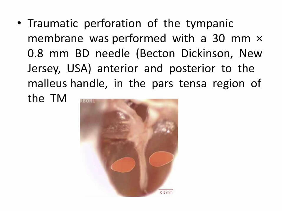
• Traumatic perforation of the tympanic membrane was performed with a 30 mm ×0.8 mm BD needle (Becton Dickinson, New Jersey, USA) anterior and posterior to the malleus handle, in the pars tensa region of the TM

• For histological evaluation, 3 animals were euthanized
• 3 days after the perforation (5 bullae), • 4 animals after 5 days (5 bullae),• 5 animals after 7 days (5 bullae),• 3 animals after 10 days (3 bullae) and • 3 animals after 14 days (4 bullae). • One animal (1 bulla) with intact TM was
assessed as control. • The animals were euthanized with an
intraperitoneal injection of an overdose of • thiopental (Thionembutal, Abbot, São Paulo,• Brazil).

• The bullae were removed from the animals and fixed for 24h in 10% formalin diluted in phosphate buffer solution and then decalcified in an aqueous solution of 4.13 g EDTA and 0.55 g NaOH for approximately 50 days.
• After decalcification, the samples were dehydrated ethanol, xylene and embedded in paraffin.
• Samples were stained with hematoxylin and eosin

instruments
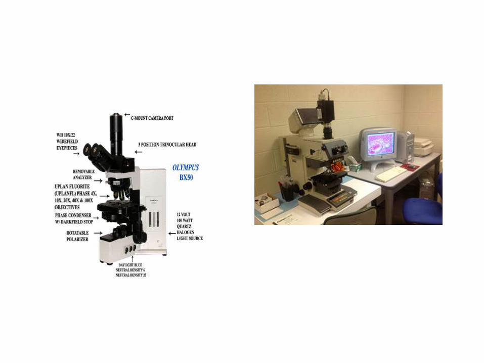
• Histological evaluation was performed with an Olympus BX50 microscope (Olympus America, Inc., Pennsylvania,
• digital high-resolution images were acquired with Spot RT3 camera (Diagnostic Instruments, Inc. Michigan,
• The thicknesses of the outer epithelial layer, lamina propria and mucosa were measured using the Image-Pro-Plus® program, release
• 7.0 (Media Cybernetics Inc., MD, USA)

Results
Intact tympanic membrane control
• The thickness of the TM was approximately 9.6 µm, having as reference the distance of 500 µm from the malleus handle.
• At a distance of 250 µm from the malleushandle, the TM thickness was 15.8 µm,
• and at 250 µm from the tympanic annulus, its thickness was 26.6 µm

Tympanic membrane 3 days after traumaticperforation
• The mean thickness of TM was approximately 39 µm
• Three days after the traumatic perforation, there was a more proliferative and hyperplasticepithelial layer, with approximately three to four rows of epithelial cells, both near the malleus handle and the tympanic annulus
• The mean epithelial layer thickness was 18.9µm
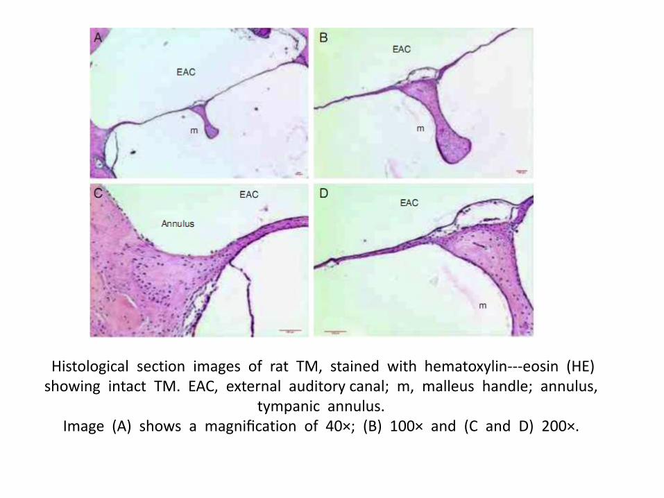
Histological section images of rat TM, stained with hematoxylin---eosin (HE)showing intact TM. EAC, external auditory canal; m, malleus handle; annulus,
tympanic annulus. Image (A) shows a magnification of 40×; (B) 100× and (C and D) 200×.

• The middle layer of the TM showed the presence of cells with basophilic nucleus, compatible with fibroblasts. These occasional disorganized fibroblasts did not overcome the
• limits of the ruptured collagen fibers in the TM perforation. There was edema in the loose subepithelial connective and submucosal tissue near the malleus handle and in the tympanic annulus.

• There was a predominance of inflammation with recruitent of polymorphonuclear cells located in the perivascular portion, and loose subepithelial and submucosal connective tissue.
• Blood vessels with plethora or turgescence were present, close to the malleus handle and the tympanic annulus region
• The mean lamina propria thickness was 14.3µm Hyperplasia was observed in the mucosal tissue near the region of the perforation borders.
• A row of mucosal tissue with hyperplastic cells and a mean thickness of 5.8 µm was identified.
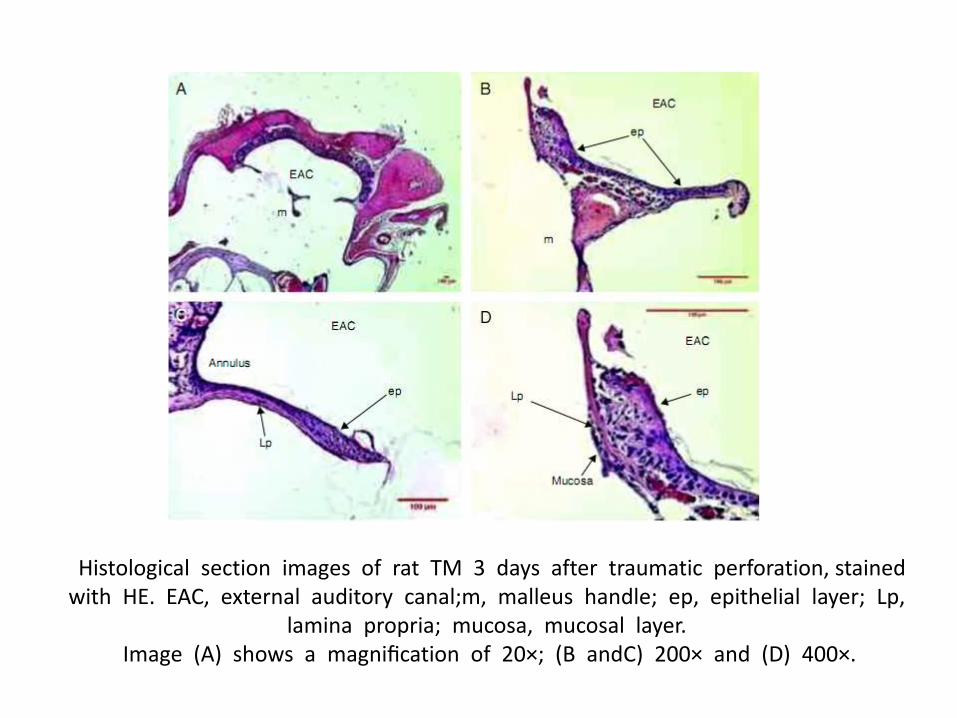
Histological section images of rat TM 3 days after traumatic perforation, stained with HE. EAC, external auditory canal;m, malleus handle; ep, epithelial layer; Lp,
lamina propria; mucosa, mucosal layer. Image (A) shows a magnification of 20×; (B andC) 200× and (D) 400×.


Tympanic membrane 5 days after traumatic perforation

Tympanic membrane 7 days after traumatic perforation

Tympanic membrane 10 days after traumatic perforation

Closure of the tympanic perforation
• The closure of the tympanic membrane occurred around 7-10 days after traumatic perforation, and the healing process was complete on the 14th day.

Discussion
• After the perforation, the healing process of the TM is typically described as occurring in three distinct phases,but temporally overlapping: inflammatory, proliferative and remodeling. 3 -7
• In experimental skin studies, the inflammatory phase begins immediately after tissue injury and lasts for 4 –6 days.10,11
• This phase consists of a disarray of blood vessels with increased vascular permeability, leakage of serum proteins, platelets and coagulation factors.
• After 5 or 6 h of tissue injury, polymorphonuclearneutrophils are recruited to the wound, while monocytes are recruited after 48-96 h.


Thank you