ossicular disruption with intact tympanic membrane
TRANSCRIPT

Page 1/12
Endoscopic Ear Surgery for the Management of TraumaticOssicular Disruption with Intact Tympanic MembraneChunlin Zhang ( [email protected] )
A�liated Hospital of Zunyi Medical University https://orcid.org/0000-0001-7825-0497Dan Long
Zunyi Medical UniversityYuan Deng
A�lated hospital of Zunyi Medical UniversityMei Ynag
A�lated hospital of Zunyi Medical UniversityDandan Guo
A�lated hospital of Zunyi Medical UniversityZhaohui Liu
A�lated hospital of Zunyi Medical University
Research article
Keywords: Endoscopic ear surgery, traumatic ossicular disruption, facial nerve paralysis, ossiculoplasty, clinical e�cacy.
Posted Date: November 12th, 2020
DOI: https://doi.org/10.21203/rs.3.rs-103560/v1
License: This work is licensed under a Creative Commons Attribution 4.0 International License. Read Full License
Page 2/12
AbstractBackground Traumatic ossicular disruption (TOD) usually had a severe conductive hearing loss, the exploratory tympanotomy iscritical for the diagnosis and improve hearing. Endoscopic ear surgery (EES) is becoming popular in the last decade, we conducteda retrospective study to explore the e�cacy of EES for management of TOD and the accompanied injuries.
Methods A retrospective study was performed on 18 ears (16 patients) of TOD with intact TM from May 2017 to Jun 2019 in ourdepartment. EES was conducted to check the ossicular chain anomalies, and to perform the ossiculoplasty and facial nerve (FN)decompression depending on the intraoperative �ndings. Hearing outcomes and surgical complications were assessed at 6 monthspostoperatively.
Results The incus injury was the most common type of TOD, which was observed in 14 ears (77.8%), stapes suprastructure fracturewas observed in 4 ears (22.2%). FN injury was found in 4 out of 13 ears with temporal bone fracture (TBF), the injury sites weremainly located in the perigeniculate area and the tympanic segment of FN. It showed the postoperative average pure-tone average(PTA) gain was 22.9 ± 9.5 dB, and the average ABG closure was 22.2 ± 8.3 dB, ABG closure to 20 dB or less and ABG closure to 10dB or less were achieved in 18 ears (100%) and 14 ears (77.8%), respectively. The facial function achieved favorable recovery ofHouse-Brackmann (H-B) grade (3 ears) and grade (1 ear) in all the 4 cases in 6 months after surgery. No iatrogenic FN paralysisand signi�cant sensorineural hearing loss were observed.
Conclusions ESS was effective in diagnosis and management of TOD and the accompanied otologic injuries, such as FN paralysis,it showed favorable surgical outcomes. ESS provides an alternative method to manage TOD with the advantage of excellent visionand less invasion.
BackgroundTraumatic ossicular disruption (TOD) is usually caused by a head trauma or direct and indirect injuries from auditory canal [1-3].TOD patients usually had a severe conductive hearing loss, which is induced by tympanic membrane (TM) perforation, ossicularinjury, or a combination [3]. Perforations of the TM could naturally heal and then present as an intact one, which is prone to beingmisdiagnosed [3]. Though high-resolution CT (HRCT) scans and modern imaging technology improve the diagnosis sensitivitybefore surgery, some TOD cases with the minor abnormalities were still missed in clinical practice [4]. The patients with a persistentsevere conductive hearing loss after head or ear trauma, though an intact tympanic membrane upon examination, TOD should besuspected, the exploratory tympanotomy is critical for the diagnosis, and ossicular reconstruction could be performed at the sametime according to the intraoperative �ndings.
Endoscopic ear surgery (EES) is becoming popular in the last decade, which is identical with microscopic ear surgery (MES),However, EES showed some obvious advantages, such as a wider surgical view and less invasive incision from the externalauditory canal [5-7], the advantages of EES seem to be especially remarkable in minimally invasive middle ear surgery, such asossiculoplasty [6, 8]. However, up to date, most TOD were treated by ossiculoplasty under microscope [1, 2, 9]. Moreover, TOD isusually accompanied with temporal bone fracture (TBF) and facial nerve (FN) injury in some patients [10, 11], whether the exclusiveESS could be quali�ed to manage these complex injuries is unknown. In order to explore the e�cacy of EES for management ofTOD and the accompanied injuries, we retrospectively collected and analyzed a series of TOD cases with intact TM which wereperformed EES for ossiculoplasty and FN decompression in a portion of cases accompanied with FN injury.
Methods1.1 Clinical material
We retrospectively analyzed a series of patients who visited our hospital due to hearing loss after trauma between May 2017 andJul 2019. The cases met the following criteria were included in this study: (1) a persistent conductive hearing loss after head or eartrauma; (2) an open external auditory canal and intact TM. Exclusion criteria: (1) TOD combined with otitis media. (2) Severe bonyfracture of skull base, CSF leakage, or meningoencephalocele. (3) the surgery under microscope with a post-auricular incision ormastoidectomy was performed. (4) preoperative radiologic evidence suggested the FN injury involvement was not limited from the

Page 3/12
geniculate ganglion to pyramidal segment. (5) the case lost to follow-up and postoperative hearing outcome was not obtained. Thisstudy was approved by the Institutional Ethics Review Board of our institution and informed consent was obtained for our studyfrom all participating patients.
A total of 85 ears were reviewed in this period, including 58 ears of traumatic TM perforation without TOD, 9 ears of traumatic TMperforation with TOD, 18 ears of TOD with intact TM. Finally, a total of 18 ears (16 cases) were included, the clinical characteristicsand hearing status of these patients were summarized and analyzed (Table 1). The patient population was composed of 12 males(13 ears) and 4 females (5 ears), with an age range of 7-54 years and a mean age of 36.6 ± 13.6 years. Fourteen patients showedunilateral TOD, and 2 patients showed bilateral TOD. The most common trauma etiology was fall down injury (9 cases), followed bytra�c accident injury (5 cases) and blast injury (2 cases, Table 2). The average interval between trauma onset and surgery was 2.7months (range: 0.5–24). Eight patients had complex injuries and were �rstly treated in other departments, such as cerebral surgery,orthopedics department, maxillofacial department and so on.
Temporal bone HRCT scans and 3-D reconstruction of ossicular chain were routinely performed before surgery, TBF was observedin 13 ears, including 9 patients of longitudinal TBF, 3 patients of transverse TBF, and 1 patient of oblique TBF. Four patients (P3, P5,P13 and P15) presented with FN paralysis immediately after trauma onset, including 2 cases of House-Brackmann (H-B) grade (P5, P13), 1 case of H-B grade (P15), and 1 case of H-B grade (P3), all the cases were lack of response to corticosteroid therapyat least 10 days and radiologic evidence suggested FN injury. The preoperative pure-tone average (PTA) of four frequencies (0.5,1.0, 2.0, and 4.0 kHz) ranged from 30 to 75 dB, the preoperative PTA and ABG were 48.1 ± 13.4 dB, and 32.4 ± 9.1 dB, respectively.
1.2 Surgical methods
All the EES procedures were conducted by the same senior surgeon (Z.C.) under general anesthesia via the transcanal approachwith 0° and 30° endoscopes (2.7 mm of diameter and 11 cm of length) (Karl Storz, Germany). A posterior tympanomeatal �ap wasmade and elevated to expose the tympanic cavity. The smaller amount scutum was removed using a curette for exposing the 3ossicles, oval window and round window. The integrity and connection of the ossicular were explored. Various treatment strategies,including ossiculoplasty and FN decompression were applied depending on the intraoperative �ndings: (1) a partial titaniumossicular replacement prosthesis (PORP) was used in the case of incus injury with normal stapes; (2) a total titanium ossicularreplacement prosthesis (TORP) was used in the case of stapes suprastructure injury with a normal stapes footplate; (3) a titaniumpiston prosthesis was used in the case of stapes footplate injury with a normal incus; (4) FN decompression was performed in thecase of FN paralysis induced by injury from the geniculate ganglion to pyramidal segment. A small piece of tragal cartilage washarvested and placed on the PORP/TORP prosthesis. The operation time and amount of bleeding were recorded. Two weeks aftersurgery, the external auditory canal was cleaned and the TM was examined with ear endoscopy in our outpatient department.Interval hearing status was assessed by pure-tone audiometry. All patients were followed-up at least 6 months.
1.3 Statistical methods
A GraphPad Prism 7 statistical software was used for statistical analyses. A one-sample Kolmogorov-Smirnov test was used toanalyze the normality of the data distribution. A paired-samples t-test or Wilcoxon signed rank test were used for comparisonsbefore and after surgery. A P value < 0.05 was considered to indicate statistical signi�cance.
Results2.1 Intraoperative �ndings
A comprehensive description of the clinical characteristics, expletory �ndings and treatment strategies were shown in Table 1 andFigure 1. It revealed that the incus injury was observed in 14 ears (77.8%), which was the most common type in this study, including11 ears of incus dislocation involved both IMJ dislocation and ISJ dislocation, 2 ears of isolated ISJ dislocation, and 1 ear of thelong process necrosis. Stapes suprastructure fracture was observed in 4 ears (22.2%), including 2 ears of stapes suprastructurefracture with normal footplate, 1 ear of suprastructure fracture with injured footplate, and 1 ear of suprastructure fractureaccompanied with IMJ dislocation. All of the cases had edema mucus or �brous tissue enveloped the injured ossicular. In the caseswith FN paralysis, the injury sites were consistent with the suggestions of preoperative CT examination, including 3 ears of

Page 4/12
perigeniculate ganglion injury and 1 ear of tympanic segment injury. We also observed a case with partial missing lateral bony wallof external auditory canal (P7), which was constructed by a piece of tragus cartilage.
2.2 Surgical results of EES
The operation time ranged from 45 to 95 min, the average of operation time was 62.4 ± 15.5 min. The TM remained intact aftersurgery in all ears. An interval serial assessment of the hearing status was conducted postoperatively, a mean follow-up period was18.4 months (range: 6.5-35.3). The mean PTA gain was 22.9 ± 9.5 dB, and the ABG was 22.2 ± 8.3 dB. The mean postoperative PTAwas signi�cantly reduced, and the mean postoperative ABG was obviously closed, respectively (P < 0.001) (Table 3). The ABGclosure to 20 dB or less and ABG closure to 10 dB or less were achieved in 18 ears (100%) and 14 ears (77.8%), respectively. Theaverage bone conduction was not changed signi�cantly (P = 0.236). (Table 3). The FN function recovered very well in all the 4patients with FN paralysis, it showed 3 cases of H-B grade , and 1 case of H-B grade in 6 months after surgery.
According to the involved ossicles, the TOD cases were divided into two subgroups, incuse injury with normal stapes and stapesinjury with or without incus injury, the preoperative PTA and ABG showed no statistical differences, respectively (P=0.126, P=0.386).Accordingly, the hearing reconstruction adopted PORP implantation for incus injury, and TORP implantation or stapedotomy andpiston prosthesis implantation for stapes injury, respectively, no statistical differences were found in postoperative PTA and ABG,respectively (P=0.975, P=0.790) (Table 4 and Fig 2).
2.3 Complications
The operative bleeding was less than 20 ml in all ears. A post-auricula approach, canalplasty or mastoidectomy was not necessaryin any case. However, the chorda tympani nerve was transfected in 1 case (right ear of Case 6), in which the chorda tympani nervewas tightly adhered with the dislocated incus and the �brous tissues surrounding it. No major complication, such asiatrogenic facial nerve paralysis, signi�cant sensorineural hearing loss (≥ 15 dB) was observed.
DiscussionOssiculoplasty was the main treatment for TOD, which was traditionally performed under microscope [1, 2, 9]. In recent years, EESfor tympanoplasty and ossiculoplasty has increased annually, showing some obvious advantages, such as a wider and multiangle�eld of view which allowed the surgeons to accurately assess and then reconstruct the ossicular chain [5-8]. However, ESS for themanagement of TOD have not been well studied, up to date, only Kim [12] reported 15 cases of TOD treated by ESS in 2019, theTOD cases with TM perforation were also included in their study, the diagnose of these cases was relatively easier, which could beobserved directly using the endoscope through the perforation. As previously reported, the traumatic TM perforation of some TODcases could be healed spontaneously in one or several weeks [13], which could lead to the diagnosis of TOD more di�cult. To thebest of our knowledge, this is the �rst study focused on employing totally ESS for the management of TOD with an intact TM. Ourresearch showed the ABG closure to 20 dB or less and ABG closure to 10 dB or less were achieved in 18 cases (100%) and 14 cases(77.8%), respectively, which was well in accordance with the study of Kim [12], and better than those of MES, Ghonim [2] reported 42cases of TOD with intact TM, which were performed MES, the postoperative PTA and ABG were signi�cantly improved to 20.2 ± 9.8dB and 4.2 ± 7.3 dB, respectively. However, so far, there is absent of a study on direct comparison between ESS and MES for TOD,further studies employing a larger cohort and including MES are needed for validation. Moreover, we also analyzed thepostoperative hearing outcomes between incus injury reconstrued by PORP implantation and stapes injury mainly reconstructed byTORP or piston implantation, no signi�cant differences were found, the results were different from our previous study of ossicularchain malformation [8], the possible reason was that the piston reconstruction was also adopted for stapes injury with footplateanomaly (P4), as many studies reported, the piston prosthesis usually obtained a better hearing result [2, 14, 15].
In this study, head trauma through fall from a height or tra�c accident was the most common cause of TOD, which was well inaccordance with previous studies [2]. Incus is prone to be involved in injury as it was suspended in the tympanic cavity between the�rmly anchored malleus and stapes [10], the injury types included isolated ISJ dislocation, isolated IMJ dislocation, and a complexof both. Isolated malleus handle and long process of incus fractures were rarely reported in previous reports [16-18], we observed acase of isolated long process necrosis (right ear of P16), and a case of stapes suprastructure fracture which was still suspendedupon the stapes footplate (P14), both the ears was misdiagnosed by preoperative examination. HRCT is the preferred modality due
Page 5/12
to its ability to demonstrate the details of TOD and other associated injuries, such as TBF and FN injury. In this study, the diagnosissensitivity of HRCT was 88.9% for TOD, which was in according with the previous reports [4], some TOD cases with the minorabnormalities were still missed in clinical practice. Therefore, exploratory tympanotomy is critical for the diagnosis, and couldsimultaneously perform ossiculoplasty to improve hearing.
TOD cases usually experienced a relatively long period of delayed diagnosis and treatment, it was still under controversy when toperform the tympanic expletory [19], suggestions of previous studies were from immediate exploration to 3 months, in this researchthe average interval between trauma onset and surgery was 2.7 months (range: 0.5 - 24). Grant [19] reported conservativemanagement was effective in traumatic TM perforation and hemotympanum, however, some TOD cases showed a worsen hearing,therefore, exploratory tympanotomy should be performed for the TOD cases with de�nite radiologic evidences. Moreover, we foundthat the dislocated ossicles were usually enveloped with �brous tissues, forming soft connection between the ossicles, whichinduced a relatively small average preoperative ABG of 32.4 ± 9.1 dB, compared with a larger average ABG of 63.2 ± 11.7 dB in thecases of isolated middle ear malformation in our precious study [8]. On the other hand, the �brous tissue may induce the ventilationblockage and provoke a dysventilation of the middle ear [20]. Therefore, the surgery should be considered once HRCT scansuggested the disruption of ossicular with an ABG of 30 dB or more.
TOD was usually accompanied with TBF, perilymphatic �stula, or FN injury [10]. The TOD cases accompanied with severe otologiccomplications should be treated timely, However, whether EES could be quali�ed to manage these associated anomalies at thesame time is yet unknown. To the best of our knowledge, this is the �rst study to explore this question for TOD. According toprevious reports, perilymphatic �stula commonly occurred in the stapes and round window [21], though after carefully examination,no occurrence of perilymphatic �stula was observed in our cases series, EES could provide a clear and magni�ed view for checkingthese areas, and reconstruction if necessary [22], the characteristic of endoscopic view ensured the surgical procedures beingminimally invasive. TBF was found in 13 ears in this research, 4 ears were accompanied with FN injury, the perigeniculate area andtympanic segment was most commonly involved in the injury, which was in according with previous researches [4, 11]. Though thetiming of treatment of traumatic FN paralysis was under controversy [23], the case of traumatic FN paralysis combined with TODwas a good indicator for timely surgery, especially for the cases with severe immediate-onset facial paralysis and lack of responseto the corticosteroid therapy. A few approaches can be used for FN decompression depending on the injury sites, includingtransmastoid approach and the middle cranial fossa approach, however, it is a challenging to control these areas throughtraditional surgical approaches [11]. In this research, it showed EES was effective in the management of some selected FN injury,transcanal approach could allow the surgeons to control the whole tympanic segment, geniculate ganglion and lateral aspect of thelabyrinthine portion, obviating the need of excessive removal of tympanic scute, canaloplasty, or a post-auricle incision andmastoidectomy, the FN function recovered very well in 6 months after surgeries in all cases. However, when radiologic evidencesuggested the injury involved mastoid segment, the transmastoid approach should be conducted, 5 cases (excluded from thisresearch) were performed transmastoid approach under microscope at the same period in our department, carefully preoperativeimaging study was critical for selecting TOD patients with FN paralysis for EES exclusively or combined with transmastoidapproach.
ConclusionIn conclusion, this research showed EES was effective in diagnosis and management of TOD with intact TM, and someaccompanied otologic injuries. It showed favorable surgical results. EES provides an alternative method to manage TOD, with theadvantage of excellent vision and less invasion, which could be promoted in clinical.
AbbreviationsAC: air conduction; ABG: air-bone gap; EES: endoscopic ear surgery; FN: facial nerve; MES: microscopic ear surgery; PORP: partialossicular replacement prosthesis; TM: tympanic membrane; TBF: temporal bone fracture; TOD: traumatic ossicular disruption;TORP: total ossicular replacement prosthesis.
Declaration

Page 6/12
Ethical approval and consent to participate
This study was approved by the Institutional Ethics Review Board of the A�liated Hospital of Zunyi Medical University and writteninformed consent was obtained for our study from all participating patients.
Consent for publication
All authors have agreed to publish this article.
Availability of data and materials
Data is available upon request by contacting the corresponding author.
Competing interests
The authors declare that they have no competing interests.
Funding
No.
Authors’ contributions
ZC and LZ participated in the design of the study. LD, DY, YM and GD performed the statistical analysis, drafted the manuscript, andZC revised the manuscript. All the authors have approved the manuscript as submitted.
Acknowledgements
Not applicable.
References1. Hakuba N, Iwanaga M, Tanaka S et al (2010) Ear-pick injury as a traumatic ossicular damage in Japan. Eur Arch
Otorhinolaryngol 267(7):1035-1039.
2. Ghonim MR, Shabana YK, Ashraf B, Salem MA (2016) Traumatic ossicular disruption with intact tympanic membrane:treatment modalities in 42 patients: Our experience. Clinical Otolaryngol 41(2):176-179.
3. Yetiser S, Hidir Y, Birkent H, Satar B, Durmaz A (2008) Traumatic ossicular dislocations: etiology and management. Am JOtolaryngol 29(1):31-36.
4. Rajati M, Pezeshki Rad M, Irani S, Khorsandi MT, Motasaddi Zarandy M (2014) Accuracy of high-resolution computedtomography in locating facial nerve injury sites in temporal bone trauma. Eur Arch Otorhinolaryngol 271(8):2185-2189.
5. Tseng CC, Lai MT, Wu CC, Yuan SP, Ding YF (2017) Comparison of the e�cacy of endoscopic tympanoplasty and microscopictympanoplasty: A systematic review and meta-analysis. Laryngoscope 127(8):1890-1896.
�. Iannella G, De Vincentiis M, Greco A et al (2019) Endoscopic approach in second stage ossicular chain reconstruction. Am JOtolaryngol 40(5):735-742.
7. Lade H, Choudhary SR, Vashishth A (2014) Endoscopic vs microscopic myringoplasty: a different perspective. Eur ArchOtorhinolaryngol 271(7):1897-1902.
�. Zhang C, Mi J, Long D, Deng Y, Sun Q, Liu Z (2020) Endoscopic Ossiculoplasty for the Management of Isolated CongenitalOssicular Chain Malformation: Surgical Results in 16 Ears. Ear Nose Throat J:145561320931218. doi:10.1177/0145561320931218.
9. Delrue S, Verhaert N, Dinther JV (2016) Surgical Management and Hearing Outcome of Traumatic Ossicular Injuries. J Int AdvOtol 12(3):231-236.
10. Hasso AN, Ledington JA (1988) Traumatic injuries of the temporal bone. Otolaryngol Clin North Am 21(2):295-316.

Page 7/12
11. Alicandri-Ciufelli M, Fermi M, Di Maro F, Soloperto D, Marchioni D, Presutti L (2020) Endoscopic facial nerve decompression inpost-traumatic facial palsies: pilot clinical experience. Eur Arch Otorhinolaryngol 2020 Apr 30.doi: 10.1007/s00405-020-05997-
12. Kim MS, Chung J, Kang JY, Choi JW (2020) Transcanal endoscopic ear surgery for traumatic ossicular injury. Acta Otolaryngol140(1):22-26.
13. Kristensen S, Juul A, Gammelgaard NP, Rasmussen OR (1989) Traumatic tympanic membrane perforations: complications andmanagement. Ears. Ear Nose Throat J 68(7):503-516.
14. Dhooge I, Desmedt S, Maly T, Loose D, Van Hoecke H (2018) Long-term hearing results of stapedotomy: analysis of factorsaffecting outcome. Eur Arch Otorhinolaryngol 275(5):1111-1119.
15. Shabana YK, Abu-Samra M, Ghonim MR (2009) Stapes surgery for post-traumatic conductive hearing loss: how we do it. ClinOtolaryngol 34(1):64-66.
1�. Delrue S, De Foer B, van Dinther J (2015) Handling an Isolated Malleus Handle Fracture: Current Diagnostic Work-up andTreatment Options. Ann Otol Rhinol Laryngol 124(3):244-249.
17. Cavada MN, Patel N (2019) Isolated Traumatic Fracture of the Malleus Handle Causing Hearing Fluctuation. Otol Neurotol40(3):e244-e247.
1�. Blanchard M, Abergel A, Vérillaud B, Williams MT, Ayache D (2011) Isolated malleus-handle fracture. Auris Nasus Larynx38(4):439-443.
19. Grant JR, Arganbright J, Friedland DR (2008) Outcomes for conservative management of traumatic conductive hearing loss.Otol Neurotol 29(3):344-349.
20. Marchioni D, Mattioli F, Alicandri-Ciufelli M (2010) Endoscopic evaluation of middle ear ventilation route blockage. Am JOtolaryngol 31(6):453-466.
21. Ikezono T, Shindo S, Sekine K (2011) Cochlin-tomoprotein (CTP) detection test identi�es traumatic perilymphatic �stula due topenetrating middle ear injury. Acta Otolaryngol 131(9):937-944.
22. Volkenstein S, Dazert S (2017) Recent surgical options for vestibular vertigo. GMS Curr Top Otorhinolaryngol Head Neck Surg16:Doc01. doi: 10.3205/cto000140.
23. Xie S, Wu X, Zhang Y, Xu Z, Yang T, Sun H (2016) The timing of surgical treatment of traumatic facial paralysis: a systematicreview. Acta Otolaryngol 136(12):1197-1200.
TablesTable 1 Patients clinical characteristics, intraoperation �ndings, surgery strategies and hearing outcomes

Page 8/12
Case Age(y)
/Sex
Side Traumaetiology
Intraoperative�ndings
Operationtime
Surgicalstrategies
Preoperative dB Postoperative
dB
PTA BC ABG PTA BC ABG
P1 34/M L Blastinjury
Stapessuprastructurefracture withnormalfootplate
55 TORP 63 23 40 39 21 18
P2 45/M L Falldowninjury
Incusdislocation
53 PORP 41 10 31 20 11 9
P3※
51/F L Falldowninjury
Incusdislocation; H-Bgrade
92 PORP + FND 46 11 35 18 10 8
P4 54/M R Falldowninjury
Stapessuprastructurefracture with
injuredfootplate
65 Piston 55 21 34 31 22 9
P5※
43/M R Tra�cinjury
Incusdislocation, H-Bgrade V
95 PORP + FND 55 23 32 31 23 8
P6
21/M R Tra�cinjury
Incusdislocation
50 PORP 43 13 30 20 12 8
L Incusdislocation
45 PORP 35 16 19 22 15 7
P7 7/M L Falldowninjury
Incusdislocation,partial missinglateral bonywall of EAC
65 PORP+ EACreconstruction#
30 6 24 15 7 8
P8 34/M R Blastinjury
Incudostapedialjoint dislocation
55 PORP 45 13 32 20 10 10
P9 34/M L Falldowninjury
Incusdislocation
57 PORP 63 10 53 16 8 8
P10 47/M L Falldowninjury
Stapessuprastructurefracture andincudomalleolarjoint dislocation
58 TORP 75 33 42 48 32 16
P11 41/M R Tra�cinjury
Incusdislocation
56 PORP 71 25 46 28 18 10
P12 11/F L Tra�cinjury
Incusdislocation
62 PORP 46 10 36 30 12 18
P13※
41/M R Falldowninjury
Incusdislocation, H-Bgrade V
88 PORP + FND 55 20 35 32 20 12
P14 36/M R Falldowninjury
Stapessuprastructurefracture withnormalfootplate
52 TORP 36 8 28 20 12 8
P15※
36/F R Falldown
Incusdislocation, H-B
78 PORP + FND 36 11 25 18 10 8
Page 9/12
injury grade
P16 51/F R Tra�cinjury
Incus longprocessnecrosis
52 PORP 39 16 23 26 16 10
L Incudostapedialjoint dislocation
46 PORP 32 14 18 20 12 8
ABG: air-bone conduction gap; EAC: external auditory canal; FND: facial nerve decompression; H-B: House-BrackmanGrading System; EAC: external auditory canal;
PORP: partial titanium ossicular replacement prosthesis; TORP: total titanium ossicular replacement prosthesis;
Incus dislocation involved both incudomalleolar joint and incudostapedial joint dislocation;
※ the cases accompanied with facial paralysis.
# the tragus cartilage was adopted to repair the EAC.
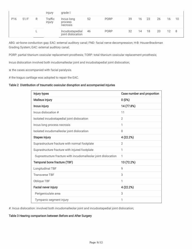
Table 2 Distribution of traumatic ossicular disruption and accompanied injuries
Injury types Case number and proportion
Malleus injury 0 (0%)
Incus injury 14 (77.8%)
Incus dislocation # 11
Isolated incudostapedial joint dislocation 2
Incus long process necrosis 1
Isolated incudomalleolar joint dislocation 0
Stapes injury 4 (22.2%)
Suprastructure fracture with normal footplate 2
Suprastructure fracture with injured footplate 1
Suprastructure fracture with incudomalleolar joint dislocation 1
Temporal bone fracture (TBF) 13 (72.2%)
Longitudinal TBF 9
Transverse TBF 3
Oblique TBF 1
Facial never injury 4 (22.2%)
Perigeniculate area 3
Tympanic segment injury 1
#: Incus dislocation: involved both incudomalleolar joint and incudostapedial joint dislocation;
Table 3 Hearing comparison between Before and After Surgery

Page 10/12
Outcome Preoperative Postoperative Statistical value P value
PTA, mean (SD), dB 48.1 ± 13.4 25.2 ± 8.7 t=10.22 0.001
BC, mean (SD), dB 15.7 ± 7.1 15.1± 6.5 t=1.23 0.236
ABG, mean (SD), dB 32.4 ± 9.1 10.2 ± 3.5 t=11.29 0.001
ABG ≤ 20 dB, % 2 (11.1%) 18 (100%) - 0.001*
ABG ≤10 dB, % 0 (0%) 14 (77.8%) - 0.001*
Hearing Gain, Mean (SD), dB - 22.9 ± 9.5 - -
ABG closure (SD), dB - 22.2 ± 8.3 - -
ABG: air-bone gap; BC: bone-conduction; PTA: the pure-tone average;
*: calculated by Fisher’s exact test
Table 4 Hearing Comparison between incus and stapes injury
Outcome Incus injury Stapes injury※ Statistical value P
Preoperative PTA, mean (SD), dB 45.5 ± 11.9 57.3 ± 16.4 t=1.162 0.126
Preoperative BC, mean (SD), dB 14.1 ± 5.4 21.3 ± 10.3 t=1.903 0.075
Preoperative ABG, mean (SD), dB 31.4 ± 9.7 36.0 ± 6.3 t=0.892 0.386
hearing Gain, Mean (SD), dB 22.9 ± 10.6 22.8 ± 4.7 t=0.032 0.975
ABG closure (SD), dB 21.9 ± 9.4 23.2 ± 2.8 t=0.271 0.790
ABG closure ≤10 dB, % 12 (85.7%) 2 (50.0%) - 0.197#
SNHL 15 dB, % 0 0 - -
ABG: air-bone gap; BC: bone-conduction; PTA: the pure-tone average;
SNHL: sensorineural hearing loss
#: calculated by Fisher’s exact test
※: included the case involved both stapes and incudomalleolar joint dislocation.
Figures

Page 11/12
Figure 1
Various traumatic ossicular disruption types under endoscopic view FN: facial nerve; MH: malleus handle; In: incus; St: stapes. A:white arrow showed the necrosis of the incus long process, which was instead by the �brous connection and appeared as normal;red arrow showed the minor missing of the incus long process after removal of the �brous tissues. B: white arrow showed thedisrupted incus which was enveloped by the edam mucus; red arrow showed the dislocated of incus which involved both theincudomalleolar joint and incudostapedial joint dislocation. C: red arrow showed the dislocation of incus, the FN nerve wasdepressed by the long process; white arrow showed the tissues connection between the lenticular process and the stapes. D: whitearrow showed the normal connection of incudostapedial joint and the fractured stapes suprastructure which was suspended overthe footplate; red arrow showed the complete fracture of stapes suprastructure after removal the dislocated incus.
Page 12/12
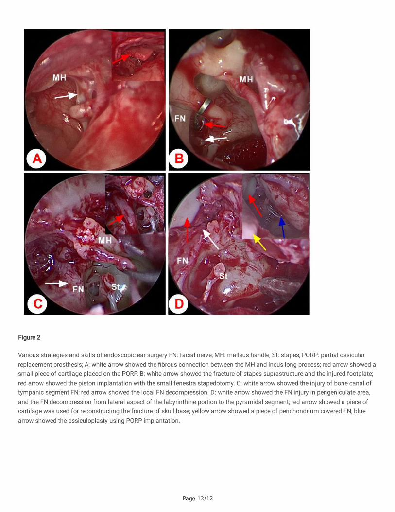
Figure 2
Various strategies and skills of endoscopic ear surgery FN: facial nerve; MH: malleus handle; St: stapes; PORP: partial ossicularreplacement prosthesis; A: white arrow showed the �brous connection between the MH and incus long process; red arrow showed asmall piece of cartilage placed on the PORP. B: white arrow showed the fracture of stapes suprastructure and the injured footplate;red arrow showed the piston implantation with the small fenestra stapedotomy. C: white arrow showed the injury of bone canal oftympanic segment FN; red arrow showed the local FN decompression. D: white arrow showed the FN injury in perigeniculate area,and the FN decompression from lateral aspect of the labyrinthine portion to the pyramidal segment; red arrow showed a piece ofcartilage was used for reconstructing the fracture of skull base; yellow arrow showed a piece of perichondrium covered FN; bluearrow showed the ossiculoplasty using PORP implantation.