tula-2, a novel histidine phosphatase, regulates bone ... · research article tula-2, a novel...
TRANSCRIPT

RESEARCH ARTICLE
TULA-2, a novel histidine phosphatase, regulates bone remodelingby modulating osteoclast function
Steven H. Back • Naga Suresh Adapala • Mary F. Barbe •
Nick C. Carpino • Alexander Y. Tsygankov •
Archana Sanjay
Received: 18 September 2012 / Revised: 16 October 2012 / Accepted: 18 October 2012 / Published online: 13 November 2012
� Springer Basel 2012
Abstract Bone is a dynamic tissue that depends on the
intricate relationship between protein tyrosine kinases
(PTK) and protein tyrosine phosphatases (PTP) for main-
taining homeostasis. PTKs and PTPs act like molecular on
and off switches and help modulate differentiation and the
attachment of osteoclasts to bone matrix regulating bone
resorption. The protein T cell ubiquitin ligand-2 (TULA-2),
which is abundantly expressed in osteoclasts, is a novel
histidine phosphatase. Our results show that of the two
family members, only TULA-2 is expressed in osteoclasts
and that its expression is sustained throughout the course of
osteoclast differentiation, suggesting that TULA-2 may
play a role during early as well late stages of osteoclast
differentiation. Skeletal analysis of mice that do not
express TULA or TULA-2 proteins (DKO mice) revealed
that there was a decrease in bone volume due to increased
osteoclast numbers and function. Furthermore, in vitro
experiments indicated that bone marrow precursor cells
from DKO mice have an increased potential to form
osteoclasts. At the molecular level, the absence of TULA-2
in osteoclasts results in increased Syk phosphorylation at
the Y352 and Y525/526 residues and activation of phos-
pholipase C gamma 2 (PLCc2) upon engagement of
immune-receptor-tyrosine-based-activation-motif (ITAM)—
mediated signaling. Furthermore, expression of a phospha-
tase-dead TULA-2 leads to increased osteoclast function.
Taken together, these results suggest that TULA-2 negatively
regulates osteoclast differentiation and function.
Keywords Syk � ITAM � Fc receptor � PLCc �Tyrosine kinase
Introduction
Osteoclasts are large multinucleated cells whose major
function in the body is to resorb bone under physiological
and pathological conditions. These cells are of monocyte-
macrophage lineage, and require macrophage colony
stimulating factor (M-CSF) and receptor activator of NFjB
ligand (RANKL), factors produced by bone marrow stro-
mal cells for their differentiation and maturation [1].
Reversible phosphorylation of tyrosine residues is a central
regulator of cellular functions and is controlled by the
opposing actions of tyrosine kinases and tyrosine phos-
phatases [2]. Many signaling pathways in osteoclasts
depend crucially on tyrosine phosphorylation. Defective
signaling by specific tyrosine kinases is implicated in
several osteopetrotic mice, e.g., c-Src-/-, Pyk2-/- mice
[3, 4]. The normal function of these signaling mechanisms
Electronic supplementary material The online version of thisarticle (doi:10.1007/s00018-012-1203-2) contains supplementarymaterial, which is available to authorized users.
S. H. Back � M. F. Barbe � A. Sanjay
Department of Anatomy and Cell Biology, Temple University
School of Medicine, Philadelphia, PA, USA
A. Y. Tsygankov
Department of Microbiology and Immunology, Temple
University School of Medicine, Philadelphia, PA, USA
N. C. Carpino
Department of Molecular Genetics and Microbiology, Stony
Brook University, Stony Brook, NY, USA
N. S. Adapala � A. Sanjay (&)
Department of Orthopaedic Surgery, New England
Musculoskeletal Institute, University of Connecticut Health
Center, Farmington, CT, USA
e-mail: [email protected]
Cell. Mol. Life Sci. (2013) 70:1269–1284
DOI 10.1007/s00018-012-1203-2 Cellular and Molecular Life Sciences
123
requires regulated tyrosine phosphorylation of effector
proteins, both in terms of time and amplitude, by dephos-
phorylation catalyzed by protein tyrosine phosphatases
(PTPs). Consequently, dysregulation of PTPs produces
skeletal abnormalities, as discussed briefly below.
Osteoclasts express several different types of tyrosine
phosphatases [5]. It has been shown that mice carrying the
moth-eaten mutation that inactivates the Src homology 2
(SH2) domain-containing tyrosine phosphatase 1 (SHP-1)
have reduced bone mass due to the increased number and
hyperactivity of osteoclasts [6]. This suggests that this PTP
is a negative regulator of osteoclastogenesis and possibly
osteoclast bone resorbing activity. PTP-PEST is a non-
receptor phosphatase that is also expressed by osteoclasts,
and inhibition of PTP-PEST with RNAi reduces pit for-
mation indicating it has a positive role in osteoclast
function [7]. Mice lacking PTPe exhibit a mild bone phe-
notype suggesting compensatory activity by other family
members that have been reported in osteoclasts [8]. In
general, however, the role of phosphatases in bone biology
is not well known [5].
The TULA/UBASH3/STS family members, TULA/
UBASH3A/STS-2 and TULA-2/UBASH3B/p70/STS-1
encoded on different chromosomes, were discovered a
few years ago by several groups, including ours [9–12].
Notably, TULA-family proteins belong to the superfamily
of histidine phosphatases, sharing a conserved catalytic
core centered on a reactive histidine residue [13]. T cell
ubiquitin ligand proteins clearly differ from classical
cysteine PTPs, such as SHP-1, which is involved in the
regulation of osteoclasts. TULA-family proteins exhibit a
unique architecture, featuring the ubiquitin-associated
(UBA), Src-homology 3 (SH3), and phosphatase domains.
In spite of a substantial homology (*60 % of iden-
tity ? similarity), TULA-1 and TULA-2 are quite
different [14–16]. First, TULA-2 is ubiquitously expres-
sed in mammalian cells [9, 10], whereas TULA-1 is
expressed mostly in lymphocytes [10, 11] and possibly,
in mast cells [17]. Second, TULA-2 is an active PTP,
while the phosphatase activity of TULA-1 is drastically
lower [18–20].
It has previously been shown that the lack of TULA-
family proteins renders T lymphocytes hyper-reactive [10].
This finding indicates an immunomodulatory effect of
TULA-family proteins. Protein tyrosine phosphatases
activity of TULA-2 is essential for this effect [20].
Although the mechanism by which TULA modulates T cell
reactivity is less clear, it may act as a PTP or through other
mechanisms [11, 21, 22]. Protein tyrosine phosphatases
activity is also essential for the regulatory role TULA-2
plays in platelets, in which the lack of TULA-2 facilitates
activation in response to signaling induced through the
GPVI receptor for collagen [23].
In this report, we set out to determine the role of TULA-
2, a novel phosphatase in osteoclast differentiation and
function. Our results show that the absence of TULA
proteins in mice results in decreased bone volume due
increased osteoclast numbers and function. We also dem-
onstrate that TULA-2 regulates Syk dephosphorylation in
osteoclasts and that the absence of TULA-2 in osteoclasts
results in increased osteoclast bone resorption possibly
through increased Syk phosphorylation and Syk-mediated
signaling events.
Materials and methods
Mice
Generation of mice deficient in both TULA and TULA-2
(DKO) are previously described [10]. Mice were obtained
from Dr. Nick Carpino, SUNY NY. Both DKO and the
counterpart wild-type mice were maintained on a mixed
C57BL/6JX129SvJ background. All mice-related experi-
ments were performed in compliance with the Institutional
Animal Care and Use Committee at University of Con-
necticut Health Center.
Materials
M-CSF and RANKL were purchased from R&D systems
(Minneapolis, MN). Bacterial collagenase and dispase were
purchased from Calbiochem (San Diego, CA). Anti-
TULA-2 antibodies were raised in rabbits against synthetic
peptide corresponding to either a N- or C-terminal
sequence of human TULA-2: REELYSKVTPRRNRQQR
PGT or GPTGGFNWRETLLQE, respectively. (The
N-terminal peptide starts at the residue four of TULA-2.) It
has been confirmed that both mouse and human TULA-2
react with both antibodies; this is consistent with the
identity (C-terminal) or near identity (90 %) of the corre-
sponding sequences for mouse and human TULA-2 are
identical. Each antibody was affinity purified on the cor-
responding antigenic peptide. Both antibodies were used
for Western-blot analysis. Antibody against phospho-tyro-
sine 4G10 was purchased from Millipore. Antibodies to
phosphoTyr525/526Syk, phosphoTyr352Syk, phospho-
Tyr1217PLCc2, PLCc2 and GAPDH were purchased from
Cell Signaling Technology (Danvers, MA). Antibodies
against Syk and 1,25-dihydroxyvitamin D3 and prosta-
glandin E2 and the leukocyte acid phosphatase kit for
tartrate-resistant acid phosphatase (TRAP), reagents
required for differentiation and characterization of osteo-
clasts were obtained from Sigma. Collagen gel was
obtained from Nitta Gelatin Co., Osaka, Japan. Osteo
1270 S. H. Back et al.
123
Assay tissue culture plates for pit forming assay were
purchased from Corning Life Sciences.
Skeletal analysis
High-resolution microcomputed tomography (microCT)
was performed as described previously [24]. Briefly, ima-
ges of long bones from 8-week-old male mice were
acquired using a Skyscan 1172, 12 MPix model (Micro-
photonics, Allentown, PA). Proximal tibiae and distal
femora were scanned with a source voltage of 59 kV, a
source current of 167, an image pixel size of approximately
5.75 lm, and a 0.5-mm aluminum filter. Imaging started at
the distal end of the femur or the proximal end of the tibiae
and included approximately 7 mm (1,335 slices) of the
total bone length. Using the CTAn software, trabecular
bone was separated from cortical bone with a region of
interest tool. Trabecular morphometric traits were com-
puted from binarized images using direct 3D techniques
that do not rely on prior assumptions from the underlying
structures. The volume of interest for trabecular microar-
chitectural variables was bounded to the endocortical
margin, starting 1.5 mm from the proximal tibial condyles
in the direction of the metaphysis, and then extending from
this position for 250 slices (1.5 mm). Upper and lower
thresholds of 255 and 80 were used to delineate each pixel
as ‘‘bone’’ or ‘‘non-bone,’’ and trabecular bone volume
per total volume (BV/TV), mean trabecular thickness
(Tb.Th.), mean trabecular number (Tb.N.), and mean tra-
becular separation (Tb.Sp.) indices were computed.
For histological and histomorphometric analysis,
12-week-old mice were used. To measure dynamic bone
formation parameters, mice were injected with calcein
(30 mg/kg body weight) 10 and 3 days before sacrifice.
Tibiae and femora were dissected and fixed in 3.7 %
formaldehyde in phosphate-buffered saline, preserved in
70 % ethanol, and embedded in methyl methacrylate resin.
Sections (5 lm) were deplasticized and stained with von
Kossa or were left unstained for the measurement of cal-
cein labeling. Some sections were processed for TRAP
staining as per manufacturer’s instructions (Sigma). For
histomorphometric analysis, to assess changes in bone
structure and remodeling, tibial sections were measured in
the proximal metaphysis beginning 340 lm below the
chondro-osseous junction of the secondary spongiosa using
image analysis software (BIOQUANT Osteo II, Bioquant
Image Analysis Corp., Nashville, TN) as described by
Parfitt et al. [25]. Osteoblast numbers per bone surface
(Ob.N./B.S.) on trabecular surfaces was determined in
toluidine blue-stained sections. Osteoclast numbers per
bone surface (Oc.N./B.S.; TRAP? cells) and osteoclast
surfaces on bone surfaces (Oc.S./B.S.) were determined on
trabecular surfaces in the secondary spongiosa of TRAP-
stained sections. Bone formation rate was calculated from
calcein labeled sections.
Determination of serum collagen telopeptide
Serum was prepared from blood collected by cardiac
puncture. Concentrations of C-telopeptide (CTX), a deg-
radation product of type I collagen, in serum of 12-week-
old mice were determined using the Rat Laps ELISA
(Osteometer BioTech A/S, Herlev, Denmark).
Adenoviral constructs
Adenovirus vectors carrying the reporter gene encoding
green fluorescent protein (GFP; AxGFP) and the FLAG-
tagged TULA-2 phosphatase-dead mutant (AxTULA-
2H391A) mutant were generated, as previously described
[26, 27].
Analysis of osteoclast precursors by fluorescence
activated cell-sorter
Antibodies used for flow cytometric analysis were as the
following: anti-mouse CD3 APCe780, anti-mouse B220
APCe780, anti-mouse CD11b FITC. Anti-mouse CD115-
biotin was used in combination with streptavidin–phyco-
erythrin (PE). All antibodies were purchased from
eBiosciences (San Diego, CA). To obtain cells, tibiae and
femora of 8-week-old mice were flushed with staining
medium (19 Hanks balanced salt solution (HBSS), 10 mM
HEPES, 2 % newborn calf serum) using 25-gauge needle.
After washing cells, red blood cells were lysed with
ammonium chloride and cells were filtered through a
40-lm cell strainer to remove cellular debris. Cells were
incubated with primary antibodies for 45 min on ice. After
washing, cells were incubated with secondary antibodies,
when necessary, for 45 min time on ice. Cells were washed
and resuspended in staining medium containing propidium
iodide (1 lg/ml) and populations of interest were analyzed
by using LSRII flow cytometer (BD Biosciences) exclud-
ing the dead cells. For some experiments cells were first
gated on CD45R, CD3, and NK1.1 negative (Triple Neg-
ative) population and then separated as CD11b-/low
CD115? population. To measure LSK (Sca1?, c-Kit?
Lin?) population, cells were labeled with biotinylated
mouse lineage panel antibodies (BD Bioscience) followed
by PerCP-conjugated streptavidin and stem cell markers
Sca-1 (FITC-conjugated) and c-Kit (APCe780-conjugated).
A lineage negative (Lin-) population was gated for further
analysis of stem cell phenotypes (Sca1?, c-Kit? Lin-).
TULA-2 regulates Syk phosphorylation in osteoclast signaling pathways 1271
123
Colony-forming unit assay
Colony-forming assays were performed, as previously
described [28]. Briefly, bone marrow cells (1 9 105 cells)
were cultured in a-MEM containing 3 % methylcellulose,
20 % FBS, 1 % bovine serum albumin (BSA) and
recombinant murine GM-CSF (1 ng/ml). Cells were plated
in volume of 1 ml in 35-mm culture dishes and incubated
at 37 �C in a humidified atmosphere of 5 % CO2-air for
7 days. Colonies composed of 50 or more cells were
counted after 7 days using an inverted microscope.
Cell culture
For generation of osteoclast-like cells (OCLs), bone mar-
row was isolated from tibia and femur of 4 to 6-week-old
mice. Following overnight incubation, the non-adherent
cells were plated at 2.5 9 105/cm2 in a-MEM medium
containing 10 % FBS and 20 ng/ml M-CSF. Subsequently,
cells were treated with M-CSF (20 ng/ml) and RANKL
(50 ng/ml) for additional 5–6 days. For some experiments,
OCLs were also generated by the co-culture method as
previously described [29, 30]. Briefly, mouse primary
osteoblastic cells were obtained from 1-day-old mouse
calvaria by enzymatic digestion. Bone marrow cells
(105 cells/cm2) were co-cultured with calvarial cells
(2 9 104 cells/cm2) on tissue culture plates or collagen
gel-coated plates in the presence of 10 nM 1,25-dihydroxy
vitamin D3 and 1 lM prostaglandin E2 (Sigma). For some
experiments, osteoclasts were infected with adenoviruses
bearing TULA-2 mutants as described previously [29, 30].
For the generation of the bone marrow macrophages
(BMMs), cells isolated from long bones were plated onto
polystyrene culture plates and cultured a-MEM containing
10 % FBS, and 50 ng/ml M-CSF for 72 h. Cells were then
trypsinized, counted and plated onto six-well culture plate
(106 cells/well).
RAW 264.7 cells were cultured in Dulbecco’s modified
essential medium supplemented with 10 % FBS. Cell
lysates were prepared as described below.
RT-PCR analysis
The expression levels of osteoclast differentiation and
fusion markers were analyzed by quantitative real-time
PCR. Sequences for the primers and details of the proce-
dure are described previously [31].
Pit formation assay
Functionally active OCLs were formed in co-cultures, as
described above. An aliquot of the crude OCL preparation
was transferred onto hydroxyapatite-coated dishes and
cultured for an additional 48 h. The resorbed area was
measured using an image analysis system linked to a light
microscope (Bioquant Osteo II). A similar aliquot was
cultured onto a 24-well dish and cells were TRAP stained
after 6 h.
Survival assay
Following differentiation with MCSF and RANKL on day
5, one set of 96-well plate was fixed with 10 % formal-
dehyde in PBS. Other sets were either kept in a-MEM or
were treated with RANKL (50 ng/ml) for 24 h. Cells were
fixed and TRAP stained using a commercial kit (Sigma, St.
Louis). Total number of TRAP ? multinucleated cells
were counted and expressed as percentage of the number of
cells at the start of the experiment on day 5.
Preparation of cell lysates and immunoprecipitation
For M-CSF and RANKL stimulation, cells were serum
starved for 1 h and stimulated with either M-CSF (50 ng/
ml) or RANKL (50 ng/ml) for the indicated time period.
For Fc receptor stimulation, cells were washed once with
serum-free medium and incubated with 10 lg/ml mouse
IgG primary antibody (Jackson ImmunoResearch, West
Grove, PA) for 30 min at 4 �C. Cells were washed once
with serum-free medium and incubated for additional
15 min at 37 �C. Cells were then stimulated by cross-
linking with goat anti-mouse secondary antibody (Jackson
ImmunoResearch) for the indicated time period at 37 �C.
Stimulation of cells was terminated by washing the cells
once with ice-cold PBS and flash freezing with liquid N2.
Flash-frozen samples were thawed on ice and lysed in
lysis buffer containing 20 mM HEPES (pH 7.4), 150 mM
NaCl, 0.05 % Nonidet P-40, 10 % glycerol, 10 mM
EDTA, 1 mM Na3VO4, 10 lg/ml leupeptin, 10 lg/ml
aprotinin, and 1 mM phenylmethylsulfonyl fluoride. After
30 min on ice, lysates were cleared by centrifugation at
12,0009g for 20 min. Lysates were then used for analysis
of proteins by Western blotting as previously described.
For immunoprecipitation, lysate (750 lg) was incubated
with primary antibody (1 lg) for 30 min on ice. Washed
Sepharose beads were resuspended in PBS and at a ratio
of 1:5 and 40 ll of the suspended beads were added to
the lysate/antibody mixture and incubated at 4 �C for 1 h
on end-on-end rocking shaker. Beads were then washed
three times in the above lysis buffer. Proteins bound to
beads were eluted by incubating beads with equal volume
of 10 mM glycine (pH 3.0) at room temperature for
30 min. Eluted proteins were mixed with equal volume of
SDS loading buffer and boiled for 5 min and loaded on to
SDS-PAGE gel.
1272 S. H. Back et al.
123
Statistics
Each experiment was repeated at least three times, unless
indicated otherwise. The results obtained from a typical
experiment were expressed as the mean ± standard devi-
ation. Statistical analysis was performed using GraphPad
Prism 4. Significant differences were determined using
students t test; p \ 0.05 was considered significant.
Results
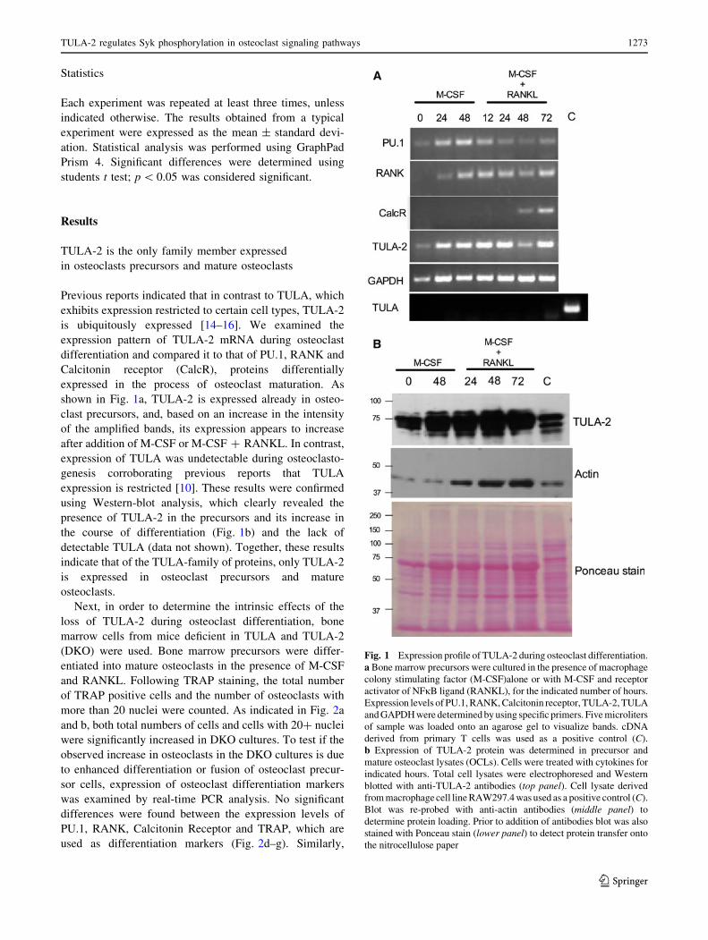
TULA-2 is the only family member expressed
in osteoclasts precursors and mature osteoclasts
Previous reports indicated that in contrast to TULA, which
exhibits expression restricted to certain cell types, TULA-2
is ubiquitously expressed [14–16]. We examined the
expression pattern of TULA-2 mRNA during osteoclast
differentiation and compared it to that of PU.1, RANK and
Calcitonin receptor (CalcR), proteins differentially
expressed in the process of osteoclast maturation. As
shown in Fig. 1a, TULA-2 is expressed already in osteo-
clast precursors, and, based on an increase in the intensity
of the amplified bands, its expression appears to increase
after addition of M-CSF or M-CSF ? RANKL. In contrast,
expression of TULA was undetectable during osteoclasto-
genesis corroborating previous reports that TULA
expression is restricted [10]. These results were confirmed
using Western-blot analysis, which clearly revealed the
presence of TULA-2 in the precursors and its increase in
the course of differentiation (Fig. 1b) and the lack of
detectable TULA (data not shown). Together, these results
indicate that of the TULA-family of proteins, only TULA-2
is expressed in osteoclast precursors and mature
osteoclasts.
Next, in order to determine the intrinsic effects of the
loss of TULA-2 during osteoclast differentiation, bone
marrow cells from mice deficient in TULA and TULA-2
(DKO) were used. Bone marrow precursors were differ-
entiated into mature osteoclasts in the presence of M-CSF
and RANKL. Following TRAP staining, the total number
of TRAP positive cells and the number of osteoclasts with
more than 20 nuclei were counted. As indicated in Fig. 2a
and b, both total numbers of cells and cells with 20? nuclei
were significantly increased in DKO cultures. To test if the
observed increase in osteoclasts in the DKO cultures is due
to enhanced differentiation or fusion of osteoclast precur-
sor cells, expression of osteoclast differentiation markers
was examined by real-time PCR analysis. No significant
differences were found between the expression levels of
PU.1, RANK, Calcitonin Receptor and TRAP, which are
used as differentiation markers (Fig. 2d–g). Similarly,
Fig. 1 Expression profile of TULA-2 during osteoclast differentiation.
a Bone marrow precursors were cultured in the presence of macrophage
colony stimulating factor (M-CSF)alone or with M-CSF and receptor
activator of NFjB ligand (RANKL), for the indicated number of hours.
Expression levels of PU.1, RANK, Calcitonin receptor, TULA-2, TULA
and GAPDH were determined by using specific primers. Five microliters
of sample was loaded onto an agarose gel to visualize bands. cDNA
derived from primary T cells was used as a positive control (C).
b Expression of TULA-2 protein was determined in precursor and
mature osteoclast lysates (OCLs). Cells were treated with cytokines for
indicated hours. Total cell lysates were electrophoresed and Western
blotted with anti-TULA-2 antibodies (top panel). Cell lysate derived
from macrophage cell line RAW297.4 was used as a positive control (C).
Blot was re-probed with anti-actin antibodies (middle panel) to
determine protein loading. Prior to addition of antibodies blot was also
stained with Ponceau stain (lower panel) to detect protein transfer onto
the nitrocellulose paper
TULA-2 regulates Syk phosphorylation in osteoclast signaling pathways 1273
123

expression of DC-STAMP and OC-STAMP, fusion mark-
ers, was comparable between the WT and DKO cells
(Fig. 2h, i).
An increase in osteoclast numbers could be due to
enhanced ability of cells to survive. Therefore, we next
examined the ability of mature osteoclasts to survive in
cultures in response to MCSF or RANKL. Two sets of
osteoclast cultures were derived from bone marrow, as
described above. Upon the appearance of large multinu-
cleated cells, one set was fixed and the second set was
incubated in serum-free media supplemented MCSF or
RANKL for an additional 24 h. Cells were then TRAP-
stained and the percent survival was calculated by
normalizing the number of TRAP-stained osteoclasts
counted after the additional 24-h incubation period to the
number of osteoclasts fixed immediately after the appear-
ance of osteoclasts. Comparable survival was observed
between WT and DKO cultures under all conditions
(Supplementary Figure 1).
Numbers of osteoclast precursors in the bone marrow
are increased in the absence of TULA proteins
Osteoclasts are of hematopoietic origin and share the same
lineage as macrophages. It has been reported that bone
marrow (BM) cells that negative for markers of
Fig. 2 Absence of TULA
proteins results in increased
osteoclast numbers in ex vivo
cultures. a Non-adherent bone
marrow precursors were
cultured in the presence of
M-CSF for 2 days and for
additional 3 days in the
presence of M-CSF and
RANKL. TRAP staining
showed increased in numbers of
TRAP? cells in DKO cultures
on day 5. Photomicrographs
show TRAP-stained OCLs at
49 (upper panels) and 209
(lower panels) magnification.
b The numbers of TRAP?
multinucleated osteoclasts
(MNCs) in the DKO cultures
(black bars) were significantly
greater than in WT cultures
(white bars) at day 5. c Cells
with more than 20–50 nuclei
were increased in numbers
DKO cultures. d–i. Bone
marrow cells were cultured in
the presence of MCSF (20 lg/
ml) and RANKL (50 lg/ml) for
5 days and expression of the
following osteoclast
differentiation markers d PU.1;
e RANK; f CalcR; and g TRAP;
and fusion markers h DC-
STAMP and i OC-STAMP was
determined by real-time PCRs
1274 S. H. Back et al.
123

B-lymphocytes (CD45R, also known as B220), and mac-
rophages (CD11b), but positive for M-CSF receptor c-Fms
(CD115), are osteoclast precursors [32, 33]. These popu-
lations are distinct from hematopoietic stem cells because
of their lack of reactivity to Sca-1 antibody. This popula-
tion represents less than 2 % of the fresh BM preparations
and has the highest levels of in vitro osteoclastogenic
activity [33]. Our FACS analysis results comparing the WT
and DKO BM cells demonstrated that the numbers of
CD11b? cells were comparable, while the number of
CD45-CD3-CD11b-/low CD115? cells were doubled in
the DKO mice, as compared to the WT mice (Fig. 3a–d).
The granulocyte macrophage progenitor (CFU-GM) is the
earliest identifiable osteoclast precursor and can become
Fig. 3 Numbers of osteoclast
precursors are increased in
DKO mice. Bone marrow cells
obtained from long bones were
analyzed for the expression of
CD11b and CD115 by FACS
analysis. Dot plotsdemonstrating gating
parameters and frequencies of
different subpopulations for a
representative WT (a) and DKO
(b) mouse is shown. Numbersrepresent percentages of parent
population. c Bar graphsrepresents percentage of total
live cells in bone marrow for
CD45R-, CD3-, NK1.1- (TN),
CD11b? cells. d Bar graphsrepresents percentage of total
live cells in bone marrow
CD45R-, CD3-, NK1.1- (TN),
CD11b-/low CD115?.
n = 3 mice/group, mean ± SD;
**p \ 0.0001 compared to WT.
e BM cells (1 9 105 cells/
culture) were prepared for
colony-forming assays as
described in methods. The
cultures were incubated in
humidified chamber for 7 days.
Clusters of 50 or more cells
were scored as a colony. Bargraph shows the number of
colonies formed. n = 7
samples, mean ± SD shown;
**p \ 0.0001 compared to WT.
The experiment was repeated
twice
TULA-2 regulates Syk phosphorylation in osteoclast signaling pathways 1275
123

osteoclasts in addition to macrophages or granulocytes
[28]. Colony-forming unit assays showed that in the pres-
ence of GM-CSF DKO BM cells formed twice as many
colonies than WT cells (Fig. 3e), suggesting that DKO
mice have more osteoclast precursors.
DKO mice have decreased bone volume due
to increased numbers and functions of osteoclasts
Gross radiological analysis of long bones from 12-week-
old DKO mice showed decreased bone density in the femur
and tibia as compared to the age-matched control WT mice
(Supplementary Figure 2). MicroCT analysis of long bones
of the hind limb indicate that bone volume was signifi-
cantly decreased in the DKO mice (Fig. 4). In the DKO
long bones, although trabecular separation was not affec-
ted, a significant decrease in trabecular thickness and
number was observed, compared to WT samples (Fig. 4b,
d). Histological examination of von Kossa-stained sagittal
sections of the proximal tibia from age-matched WT and
DKO mice indicated a significant decrease in mineralized
trabecular bone in DKO mice as compared to WT
(Fig. 5a). This result was confirmed with histomorphome-
try of von Kossa-stained bone sections (data not shown).
Also, no differences were observed in cortical thickness
(data not shown). A decrease in bone volume can result
from either a decrease in bone formation by osteoblasts or
an increase in bone resorption by osteoclasts. Therefore, in
order to determine which cell type contributed to the
decreased bone volume in DKO mice, we compared the
number and activity of both osteoblasts and osteoclasts in
DKO and WT mice. Toluidine blue staining of the long
bones indicated that the numbers of osteoblasts were
comparable in WT and DKO samples (Fig. 5c). Similarly,
bone formation rate as measured by calcein double labeling
was also comparable between the WT and DKO mice
(Fig. 5d). On the other hand, quantification of TRAP-
stained cells in the cancellous bone indicated that there was
a twofold increase in osteoclast numbers and osteoclast
surface/bone surface, in DKO mice as compared to WT
(Fig. 5b, e, f).
We next examined the effect of absence of TULA-2 on
osteoclast function. Serum levels of the C-terminal colla-
gen telopeptide (CTX), a serum biomarker of osteoclast
activity, were increased in DKO mice as compared to WT
mice (Fig. 6a). To confirm this defect of DKO osteoclast
function, we then examined bone resorption using in vitro
pit formation assay. DKO and WT OCLs were generated
by co-culture with osteoblasts on collagen gel, as described
previously [29]. After 5 days in culture, a portion of the
crude OCL preparation was placed on hydroxyapatite-
coated tissue culture plates for an additional 48 h. The
resorbed area was quantified using image analysis and
normalized to the number of OCLs. DKO OCLs resorbed
more surface area/cell than the WT OCLs (Fig. 6b, c),
confirming the cell autonomous nature of the observed
defect in osteoclast function. Thus, the decreased bone
volume in the adult DKO mice and increased bone
resorption in vivo and in vitro is, at least in part, due to
impaired osteoclast function. Taken together, these results
suggest that bone resorption under basal conditions is
affected in DKO mice.
Syk phosphorylation and Syk-mediated signaling is
augmented in DKO cells
The increased bone resorption by osteoclasts in the absence
of TULA-2 (Fig. 6) suggested that osteoclast function is
modified by DKO, because signaling events are perturbed
by the absence of TULA-2. Therefore, we next determined
the effects of the loss of TULA-2 on response of BMMs
and osteoclasts to M-CSF and RANKL, key physiological
stimuli of this cell lineage (Fig. 7a, data not shown). Upon
stimulation with RANKL, no significant increase in total
tyrosine phosphorylation was evident in either WT or DKO
osteoclasts. However, an increase in protein phosphoryla-
tion was observed both in WT and DKO cells upon
treatment with M-CSF. Notably, both the M-CSF-caused
increase and the basal tyrosine phosphorylation were ele-
vated, albeit modestly, in DKO cells as compared to WT
cells (Fig. 7a).
Syk is a known substrate of TULA-2 [19] and a key
factor in transmitting the ITAM-mediated signaling
downstream of FccR in several cell types, including BMMs
[34]. Downstream of ITAM-mediated signaling Syk is also
required for osteoclast development and function [35–37].
Therefore, we next examined the phosphorylation events
upon engagement of FccR in BMMs. After Fc receptor
crosslinking, there was a robust increase in total tyrosine
phosphorylation, which was higher in DKO BMM than in
WT BMM (Fig. 7b). Tyrosine phosphorylation of proteins
migrating at approximately 75 kD and above was enhanced
in DKO cells as compared to WT cells. To reduce back-
ground caused by cross-reactivity, we enriched tyrosine-
phosphorylated proteins by immunoprecipitating them
using anti-pTyr antibody and then immunoblotted the
obtained immunoprecipitates for total tyrosine phosphory-
lation and Syk. An increase in tyrosine phosphorylation
was observed in the DKO as compared to WT lysates.
Multiple tyrosine-phosphorylated proteins were detected in
BMM starting at 72 kDa and above it with approximately
10 kDa increments; the number of discernable bands is
much higher in DKO cells than in WT cells (Fig. 7c). Total
anti-pTyr reactivity normalized to the amount of Syk is
increased by *1.8-fold in DKO vs. WT cells (Fig. 7c).
1276 S. H. Back et al.
123

Fig. 4 Absence of TULA
proteins results in decreased
bone volume. Micro-CT
analysis of tibia (a) and femur
(c) from WT and DKO mice.
a and c shows representative
sagittal sections of tibia and
femur, respectively. Histograms
show the trabecular parameters
of tibia (b) and femur (d). n = 4
per genotype, 8-week-old male
mice, mean ± SD shown;
*p \ 0.05, **p \ 0.01
compared to WT
TULA-2 regulates Syk phosphorylation in osteoclast signaling pathways 1277
123
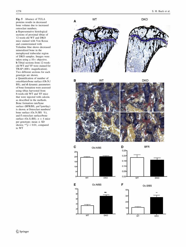
Fig. 5 Absence of TULA
proteins results in decreased
bone volume due to increased
osteoclast numbers.
a Representative histological
sections of proximal tibiae of
12-week-old WT and DKO
mice stained with Von Kossa
and counterstained with
Toluidine blue shows decreased
mineralized bone in the
metaphyseal trabecular region
of DKO samples. Images were
taken using a 109 objective.
b Tibial sections from 12-week-
old WT and YF were stained for
TRAP (4009 magnification).
Two different sections for each
genotype are shown.
c Quantification of number of
osteoblasts/bone surface (Ob.N./
BS), and d dynamic parameters
of bone formation were assessed
using tibias harvested from
8-week-old WT and YF mice
that were injected with calcein.
as described in the methods.
Bone formation rate/bone
surface (BFR/BS, lm2/lm/day)
is shown. e Osteoclast numbers/
bone surface (Oc.N./BS %),
and f osteoclast surface/bone
surface (Oc.S./BS). n = 4 mice
per genotype; mean ± SD
shown; **p \ 0.01, compared
to WT
1278 S. H. Back et al.
123

Given that Syk has several tyrosine residues that can
be differentially phosphorylated in DKO and WT cells
and may exhibit differential reactivity to a total anti-pTyr
antibody, we next analyzed the phosphorylation status of
individual tyrosine residues of Syk using specific anti-
phosphotyrosine site antibodies. For this analysis we
selected Y352 and Y525/Y526; the former is the site
known to preferentially dephosphorylated by TULA-2
[19], while the latter is a site of known importance for
Syk function [38, 39]. As expected, Western-blot analysis
showed an increase in tyrosine phosphorylation for both
Y525/526 and Y352 sites in both WT and DKO cells.
However, tyrosine phosphorylation in DKO cells was
elevated as compared to that in WT cells by *1.5- to
2-fold (Fig. 7d). We next examined tyrosine phosphory-
lation of PLCc2, a key signaling protein involved in Syk-
mediated signaling, in WT and DKO cells. Although, in
the absence of stimulation, basal phosphorylation of
PLCc2 tyrosine 1,217 was higher in DKO than in WT, it
was substantially increased in response to Fc receptor
crosslinking (Fig. 7e).
To further elucidate the involvement of Syk in these
events, we treated cells in our experiments with piceatan-
nol, a Syk specific inhibitor [37]. Phosphorylation of the
Syk sites examined did not change profoundly, while the
observed increase in PLCc2 tyrosine 1,217 phosphoryla-
tion was dramatically decreased (Fig. 7e). This suggests
that phosphorylation of Syk Y352 and Syk Y525/526 in
this cellular context is largely independent of Syk enzy-
matic activity (it can be carried out by Src-family kinases,
for example), while phosphorylation of PLCc2 is clearly
Syk-dependent. Thus, differential phosphorylation of Syk
caused by differential activity of TULA-2 is expected to
exert a strong effect on PLCc2 phosphorylation, which is a
key event of signaling via FcRc and other osteoclast/
macrophage receptors and other cells of immune system
[36, 40, 41].
TULA-2 phosphatase activity is needed for osteoclast-
mediated bone resorption
TULA-2 is highly expressed in osteoclasts (Fig. 1); its
absence in osteoclasts resulted in increased phosphoryla-
tion of Syk (Fig. 7d). To investigate the role of TULA-2
phosphatase activity in osteoclast function, we constructed
adenoviruses containing FLAG-tagged phosphatase-dead
TULA-2 (AxTULA-2H391A). We used a replication-
deficient adenovirus vector that contains a reporter gene
encoding GFP (AxGFP) as a control vector. Expression of
TULA-2 in OCLs isolated from co-cultures infected with
the recombinant viruses was verified after 3 days of
infection using Western blotting with anti-FLAG and anti-
TULA-2 antibodies (data not shown). To investigate the
functional consequences of overexpressing phosphatase-
deadTULA-2, the bone-resorbing activity of OCLs was
quantified by measuring the areas of pits formed on
hydroxyl apatite-coated resorption surface. The activity of
OCLs phosphatase-dead TULA-2 was significantly aug-
mented when compared to control and GFP-expressing
osteoclasts (Fig. 8a, b). This indicates that TULA-2 phos-
phatase activity is required for bone resorption.
Fig. 6 Absence of TULA-2 in osteoclasts results in increased
activity both in vivo and in vitro. a Measurement of serum collagen
telopeptide (CTX) demonstrated increased osteoclast activity in the
DKO mice (n = 4 male mice per genotype; mean ± SD shown;
**p \ 0.01, compared to WT). b Photomicrographs of the resorbed
area. c Bar graph demonstrating pit formation activity of DKO
osteoclasts. Osteoclasts were generated by co-culture method, as
described in the methods. After removing adherent cells, resorbed
area was quantified and normalized for the number of osteoclasts.
Data are presented as mean SE (n = 9). **p \ 0.001 compared to the
WT samples
TULA-2 regulates Syk phosphorylation in osteoclast signaling pathways 1279
123

Discussion
We have shown in the present study that the absence of
TULA-2, a novel PTP that belongs to the histidine
phosphatase superfamily, leads to the decreased bone
volume in TULA-2-deficient/null mice (Figs. 4, 5). This is
apparently due to both the marked increase in the ability of
the hematopoietic precursor cells to differentiate into
1280 S. H. Back et al.
123

osteoclasts (Fig. 2a, b) and the enhanced ability of osteo-
clasts to resorb bone both in vivo and in vitro (Fig. 6). At
the molecular level, our studies show that in osteoclast
precursors, TULA-2 mediates dephosphorylation of Syk,
which is known to regulate osteoclast differentiation and
function via ITAM signaling [36, 42].
Both members of the TULA family have been proposed
to be negative regulators of signaling. Deletion of both
TULA and TULA-2 is required for causing dramatic hy-
peractivation of T cells [10]. Re-expression of WT, but not
inactivated TULA-2 in cells that lack both TULA-family
members results in substantial reversal of the phenotype,
indicating that TULA-2 PTP activity is responsible, at least
in part, for the hyper-responsiveness of T cells [20]. In the
present study, we show that only TULA-2 is expressed in
developing and mature osteoclasts to a measurable extent
(Fig. 1). This finding is in line with the observation that
TULA has limited expression [14, 15]. Therefore, any
effect seen in osteoclasts in the DKO mice can be attrib-
utable to TULA-2 deficiency.
The overall effect of the lack of TULA-2 on the bone/
bone phenotype is due to increased osteoclast numbers
in vivo (Fig. 5e) and in vitro (Fig. 2a–c) and increased
osteoclast function (Fig. 6). We cannot, however, rule out
the possibility that the absence of TULA proteins also
affects the osteoblast lineage and this effect of TULA/
Fig. 7 Loss of TULA-2 leads to enhanced signaling down stream of
ITAM-mediated co-stimulatory pathway. Bone marrow macrophages
(BMMs) were generated from the bone marrow of WT and DKO
mice. a BMMs were stimulated by with M-CSF (50 ng/ml) or
RANKL (50 ng/ml) at indicated time points, shown in minutes. Blots
were probed with anti-phosphotyrosine antibodies (upper panels) to
visualize phosphorylated proteins. Blots were then reprobed with anti-
GAPDH antibodies to determine protein loading. b BMMs were
stimulated by cross-linking Fc receptor to activate the signaling
pathway downstream of ITAM receptors. Blot was probed with anti-
phosphotyrosine antibodies (upper panel). Asterisk indicates location
of proteins that are hyper-phosphorylated upon stimulation in DKO
samples. Blots were reprobed with anti-GAPDH antibodies to
determine protein loading. c BMMs were stimulated by cross-linking
Fc receptor and immunoprecipitation with anti-PTyr was performed
as described in the methods. Blot was probed with anti-PTyr
antibodies (upper panel) and reprobed with anti-Syk antibodies
(lower panel). The amounts of total proteins in individual bands were
quantified by using Odyssey Infrared Imaging Systems software 2.1
(LICOR Biosciences). The ratio of tyrosine phosphorylation to
amount of total protein is shown at the bottom of the panel. d,
e BMMs were pretreated with either 50 lg/ml of the Syk inhibitor
piceatannol (PICE) or DMSO (VEH) and were then stimulated to
engage the ITAM receptors as described above. d Blot was probed
with anti pY525/526 Syk or pY352Syk as indicated. Blots were then
probed with anti-Syk antibodies to determine loading. e To determine
PLCc2 phosphorylation blots were probed with anti-pY1217PLCc2
antibodies and then reprobed with anti-PLCc2 antibodies to deter-
mine protein loading. The amounts of total proteins in individual
bands were quantified by using Odyssey Infrared Imaging Systems
software 2.1 (LICOR Biosciences). The ratio of tyrosine phosphor-
ylation to amount of total protein is shown at the bottom of the panel
Fig. 8 TULA-2 phosphatase
activity is required for proper
osteoclast function. Osteoclasts
were either left un-infected, or
were infected at 500 MOI with
an adenoviral GFP (AxGFP) or
adenoviral construct expressing
phosphatase-dead form of
TULA 2 (AxTULA-2H380A)
for 48 h. Infected cells were
replated onto the resorbable
tissue culture surface and
incubated for 48 h as described
in the methods section.
a Photomicrographs show of
resorbed surface for each
condition. b After removing
adherent cells, resorbed area
was quantified and normalized
for the numbers of OCLs. Data
are presented as mean SE
(n = 9). **p \ 0.001 compared
to the uninfected control
samples. The results are
representative of two
experiments
b
TULA-2 regulates Syk phosphorylation in osteoclast signaling pathways 1281
123

TULA-2 deficiency on osteoblasts contributes to the oste-
openic phenotype. This possibility is supported by a
decrease in trabecular thickness and trabecular numbers in
DKO mice, when compared to the WT mice (Fig. 4).
Although further studies would be needed to elucidate how
loss of TULA proteins might affect the function of osteo-
blasts, it is likely that this effect would be indirect, being
mediated, for example, by changes in cytokine production.
The finding that an increase in osteoclastogenesis in DKO
mice is intrinsic to the hematopoietic lineage excludes the
possibility that the observed increase is due to an increase
in osteoprotegerin ligand (OPGL) or a decrease in osteo-
protegerin production by cells of the stromal lineage or
osteoblasts [43, 44].
Given the distinct sequential and essential effects by
M-CSF on osteoclast precursor monocyte proliferation and
survival, and by RANKL on osteoclast differentiation [1],
we attempted to dissect the roles of these cytokines in the
enhanced osteoclastogenesis that accompanies TULA-2
deficiency. A modest increase in phosphorylation was seen
in the DKO osteoclast precursors or mature osteoclasts as
compared to their WT counterparts stimulated with M-CSF
(Fig. 7a). Furthermore, a significant increase in phosphor-
ylation of several proteins, including Syk, was observed in
DKO cells as compared to WT cells when ITAM-mediated
signaling was engaged (Fig. 7b, c, e). Although the exact
molecular mechanisms responsible for TULA-2-modulated
regulation of osteoclastogenesis remain unresolved, we
postulate that decreased TULA-2 activity elevates tyrosine
phosphorylation of Syk and, hence, of its substrates, thus
leading to increased osteoclastogenesis in TULA-2-defi-
cient mice, analogous to the osteopenia observed in SHP-1-
deficient mev/mev mice [6]. Under normal remodeling
conditions, bone resorption is thought to stimulate an
equivalent level of bone formation. Osteoclast and osteo-
blast ‘‘coupling’’, is fine-tuned through variations in local
cytokine expression or release, from cell to cell, or from
cell to matrix induced signals [45]. However, our studies in
DKO mice show that increased osteoclastogenesis occurs
independently of osteoblastogenesis, consistent with other
studies where osteoclast resorptive activity appears dis-
pensable for coupling [46]. Thus, the increase in osteoclast
levels and activity that we observed histologically, func-
tionally, and independently of bone formation contribute to
the significant loss of trabecular bone volume in DKO
mice. Therefore, overall bone-remodeling rates appear to
be reduced arguing further for uncoupling of osteoclast and
osteoblast functions in the DKO mice.
Although the total numbers of bone marrow cells and
hematopoietic progenitors (Lin-Sca-1?c-kit? cells)
remained unchanged in the DKO mice, the numbers of
osteoclast precursors (CD11b-/low, CD115?, CD117?)
were significantly increased (Fig. 3 and data not shown).
This finding is consistent with increased osteoclastogenesis
wherein DKO osteoclast precursor cells differentiate into
osteoclasts (Fig. 2) and have increased resorption (Fig. 6).
In addition to bone formation, bone remodeling is also
tightly linked to hematopoiesis. Indeed, osteoclasts appear
to have a unique, direct role in hematopoietic progenitor
mobilization [47]. Thus, our findings also suggest the
possibility that enhanced osteoclastogenesis could have a
positive feedback effect on hematopoietic progenitors and
drive their differentiation toward different lineages, due in
part to release of soluble factors from the osteoid matrix
degraded by the increased osteoclast activity in the DKO
mice. Overall, the absence of TULA proteins and, in par-
ticular TULA-2, since only TULA-2 is expressed in
osteoclasts, leads to bone loss.
Taken together, these results indicate that, under normal
physiological conditions TULA-2 could down-regulate sig-
naling in response to factor(s) that activate differentiation of
osteoclasts and possibly osteoclast function. Although the
exact mechanisms mediating the role of TULA-2 remain to
be elucidated further, the obtained results allow us to propose
a molecular explanation for the positive effect of the loss of
TULA-2 activity on osteoclastogenesis and bone resorption
in DKO mice. Considering that the protein tyrosine kinase
Syk as a key element of the signaling mechanism in mac-
rophages and osteoclasts [36, 40, 41] and that Syk has been
identified as a bona fide substrate of TULA-2 [18, 19], it is
likely that the lack of TULA-2 facilitates phosphorylation of
Syk. Indeed, our results indicate that phosphorylation of Syk
and, in particular, phosphorylation of Tyr 525/526 and Tyr
352 sites is increased in TULA-2-deficient cells. It has been
shown previously that the elevated phosphorylation of these
sites is indicative of elevated enzymatic activity of Syk [34].
The hyper phosphorylation of Syk demonstrated in this
report is similar to that reported for TULA-2-deficient
platelets [23].
Likewise, we have shown that tyrosine phosphorylation
of PLCc2 is also elevated in the osteoclast precursors
derived from DKO mice as compared to WT precursors.
An increase in PLCc2 phosphorylation in DKO cells is
likely due to the increase in Syk activity caused by the lack
of TULA-2, since we (Fig. 7e) and others have shown that
PLCc2 phosphorylation is Syk-dependent [23]. Direct
dephosphorylation of PLCc2 pTyr 1,217 by TULA-2 is less
likely, since this site lacks some critical specificity deter-
minants defining it as a substrate of TULA-2 [19].
In addition to Syk, Src-family kinases (SFKs) may also
be targets of TULA-2 phosphatase activity. Several SFKs
also regulate bone remodeling by directly affecting osteo-
clast differentiation and function. While absence of c-Src
does not affect osteoclast differentiation, it severely affects
osteoclast function, thus causing osteopetrosis in mice [4].
In contrast, while the lack of Lyn does not impact the
1282 S. H. Back et al.
123

activity of mature osteoclasts under basal condition, Lyn-
null mice undergo accelerated osteoclastogenesis and bone
loss in response to RANKL [48]. Similarly, while Fyn-
deficient mice show no overt changes in the skeletal phe-
notype, the absence of Fyn results in decreased osteoclast
differentiation in culture [49]. Thus, if TULA-2 regulates
kinase activities of SFKs, its absence in osteoclast may
result in dysregulation of SFKs and could lead to the
increased numbers of osteoclasts and an increase in
osteoclastic activity.
In conclusion, the absence of TULA-2 leads to osteo-
penia accompanied by increased bone resorption. Both
osteoclast differentiation and osteoclast activity appear to
be increased in the absence TULA-2, suggesting that this
tyrosine phosphatase is a negative regulator of osteo-
clastogenesis and, possibly, of osteoclast resorbing activity.
Acknowledgments This work was supported by a grant from the
Pennsylvania Department of Health to S. B and by the National
Institute of Health Grant (AR055601) to A. S. The authors also thank
Dr. Hector Aguila (Department of Immunology, University of Con-
necticut Health Center) for antibodies and help with FACS analysis.
We also acknowledge the support of the MicroCT Core Facility at
Temple University.
References
1. Teitelbaum SL, Ross FP (2003) Genetic regulation of osteoclast
development and function. Nat Rev Genet 4:638–649
2. Hunter T (1995) Protein kinases and phosphatases: the yin and
yang of protein phosphorylation and signaling. Cell 80:225–236
3. Gil-Henn H, Destaing O, Sims NA, Aoki K, Alles N, Neff L,
Sanjay A, Bruzzaniti A, De Camilli P, Baron R, Schlessinger J
(2007) Defective microtubule-dependent podosome organization
in osteoclasts leads to increased bone density in Pyk2(-/-) mice.
J Cell Biol 178:1053–1064
4. Soriano P, Montgomery C, Geske R, Bradley A (1991) Targeted
disruption of the c-src proto-oncogene leads to osteopetrosis in
mice. Cell 64:693–702
5. Granot-Attas S, Elson A (2008) Protein tyrosine phosphatases in
osteoclast differentiation, adhesion, and bone resorption. Eur J
Cell Biol 87:479–490
6. Aoki K, DiDomenico E, Sims NA, Mukhopadhyay K, Neff L,
Houghton A, Amling M, Levy JB, Horne WC, Baron R (1999) The
tyrosine phosphatase SHP-1 is a negative regulator of osteoclasto-
genesis and osteoclast resorbing activity: increased resorption and
osteopenia in mev/mev mutant mice. Bone 25:261–267
7. Chellaiah MA, Kuppuswamy D, Lasky L, Linder S (2007)
Phosphorylation of a Wiskott–Aldrich syndrome protein-associ-
ated signal complex is critical in osteoclast bone resorption.
J Biol Chem 282:10104–10116
8. Chiusaroli R, Knobler H, Luxenburg C, Sanjay A, Granot-Attas
S, Tiran Z, Miyazaki T, Harmelin A, Baron R, Elson A (2004)
Tyrosine phosphatase epsilon is a positive regulator of osteoclast
function in vitro and in vivo. Mol Biol Cell 15:234–244
9. Carpino N, Kobayashi R, Zang H, Takahashi Y, Jou ST, Feng J,
Nakajima H, Ihle JN (2002) Identification, cDNA cloning, and
targeted deletion of p70, a novel, ubiquitously expressed SH3
domain-containing protein. Mol Cell Biol 22:7491–7500
10. Carpino N, Turner S, Mekala D, Takahashi Y, Zang H, Geiger
TL, Doherty P, Ihle JN (2004) Regulation of ZAP-70 activation
and TCR signaling by two related proteins, Sts-1 and Sts-2.
Immunity 20:37–46
11. Feshchenko EA, Smirnova EV, Swaminathan G, Teckchandani
AM, Agrawal R, Band H, Zhang X, Annan RS, Carr SA, Tsy-
gankov AY (2004) TULA: an SH3- and UBA-containing protein
that binds to c-Cbl and ubiquitin. Oncogene 23:4690–4706
12. Wattenhofer M, Shibuya K, Kudoh J, Lyle R, Michaud J, Rossier
C, Kawasaki K, Asakawa S, Minoshima S, Berry A, Bonne-Ta-
mir B, Shimizu N, Antonarakis SE, Scott HS (2001) Isolation and
characterization of the UBASH3A gene on 21q22.3 encoding a
potential nuclear protein with a novel combination of domains.
Hum Genet 108:140–147
13. Rigden DJ (2008) The histidine phosphatase superfamily: struc-
ture and function. Biochem J 409:333–348
14. Tsygankov AY (2008) Multidomain STS/TULA proteins are
novel cellular regulators. IUBMB Life 60:224–231
15. Tsygankov AY (2009) TULA-family proteins: an odd couple.
Cell Mol Life Sci 66:2949–2952
16. Tsygankov AY (2012) TULA-family proteins: a new class of
cellular regulators. J Cell Physiol 228(1):43–49
17. de Castro RO, Zhang J, Groves JR, Barbu EA, Siraganian RP
(2012) Once phosphorylated, tyrosines in carboxyl terminus of
protein-tyrosine kinase Syk interact with signaling proteins,
including TULA-2, a negative regulator of mast cell degranula-
tion. J Biol Chem 287:8194–8204
18. Agrawal R, Carpino N, Tsygankov A (2008) TULA proteins
regulate activity of the protein tyrosine kinase Syk. J Cell Bio-
chem 104(3):953–964
19. Chen X, Ren L, Kim S, Carpino N, Daniel JL, Kunapuli SP,
Tsygankov AY, Pei D (2010) Determination of the substrate
specificity of protein-tyrosine phosphatase TULA-2 and identifi-
cation of Syk as a TULA-2 substrate. J Biol Chem
285:31268–31276
20. Mikhailik A, Ford B, Keller J, Chen Y, Nassar N, Carpino N
(2007) A phosphatase activity of Sts-1 contributes to the sup-
pression of TCR signaling. Mol Cell 27:486–497
21. Smirnova EV, Collingwood TS, Bisbal C, Tsygankova OM,
Bogush M, Meinkoth JL, Henderson EE, Annan RS, Tsygankov
AY (2008) TULA proteins bind to ABCE-1, a host factor of HIV-
1 assembly, and inhibit HIV-1 biogenesis in a UBA-dependent
fashion. Virology 372:10–23
22. Collingwood TS, Smirnova EV, Bogush M, Carpino N, Annan
RS, Tsygankov AY (2007) T-cell ubiquitin ligand affects cell
death through a functional interaction with apoptosis-inducing
factor, a key factor of caspase-independent apoptosis. J Biol
Chem 282:30920–30928
23. Thomas DH, Getz TM, Newman TN, Dangelmaier CA, Carpino N,
Kunapuli SP, Tsygankov AY, Daniel JL (2010) A novel histidine
tyrosine phosphatase, TULA-2, associates with Syk and negatively
regulates GPVI signaling in platelets. Blood 116:2570–2578
24. Brennan T, Adapala NS, Barbe MF, Yingling V, Sanjay A (2011)
Abrogation of Cbl-PI3K interaction increases bone formation and
osteoblast proliferation. Calcif Tissue Int 89:396–410
25. Parfitt AM, Drezner MK, Glorieux FH, Kanis JA, Malluche H,
Meunier PJ, Ott SM, Recker RR (1987) Bone histomorphometry:
standardization of nomenclature, symbols, and units. Report of
the ASBMR Histomorphometry Nomenclature Committee.
J Bone Miner Res 2:595–610
26. Miyazaki T, Takayanagi H, Isshiki M, Takahashi T, Okada M,
Fukui Y, Oda H, Nakamura K, Hirai H, Kurokawa T, Tanaka S
(2000) In vitro and in vivo suppression of osteoclast function by
adenovirus vector-induced csk gene. J Bone Miner Res 15:41–51
27. Miyazaki T, Katagiri H, Kanegae Y, Takayanagi H, Sawada Y,
Yamamoto A, Pando MP, Asano T, Verma IM, Oda H, Nakamura
TULA-2 regulates Syk phosphorylation in osteoclast signaling pathways 1283
123

K, Tanaka S (2000) Reciprocal role of ERK and NF-jB pathways
in survival and activation of osteoclasts. J Cell Biol 148:333–342
28. Menaa C, Kurihara N, Roodman GD (2000) CFU-GM-derived
cells form osteoclasts at a very high efficiency. Biochem Biophys
Res Commun 267:943–946
29. Miyazaki T, Sanjay A, Neff L, Tanaka S, Horne WC, Baron R
(2004) Src kinase activity is essential for osteoclast function.
J Biol Chem 279:17660–17666
30. Sanjay A, Miyazaki T, Itzstein C, Purev E, Horne WC, Baron R
(2006) Identification and functional characterization of an Src
homology domain 3 domain-binding site on Cbl. FEBS J
273:5442–5456
31. Adapala NS, Barbe MF, Langdon WY, Nakamura MC, Tsygan-
kov AY, Sanjay A (2010) The loss of Cbl-phosphatidylinositol
3-kinase interaction perturbs RANKL-mediated signaling,
inhibiting bone resorption and promoting osteoclast survival.
J Biol Chem 285:36745–36758
32. Arai F, Miyamoto T, Ohneda O, Inada T, Sudo T, Brasel K,
Miyata T, Anderson DM, Suda T (1999) Commitment and dif-
ferentiation of osteoclast precursor cells by the sequential
expression of c-Fms and receptor activator of nuclear factor
kappaB (RANK) receptors. J Exp Med 190:1741–1754
33. Jacquin C, Gran DE, Lee SK, Lorenzo JA, Aguila HL (2006)
Identification of multiple osteoclast precursor populations in
murine bone marrow. J Bone Miner Res (the official J Am Soc
Bone Miner Res) 21:67–77
34. Mocsai A, Ruland J, Tybulewicz VL (2010) The SYK tyrosine
kinase: a crucial player in diverse biological functions. Nat Rev
Immunol 10:387–402
35. Faccio R, Zou W, Colaianni G, Teitelbaum SL, Ross FP (2003)
High dose M-CSF partially rescues the Dap12-/- osteoclast
phenotype. J Cell Biochem 90:871–883
36. Mocsai A, Humphrey MB, Van Ziffle JA, Hu Y, Burghardt A,
Spusta SC, Majumdar S, Lanier LL, Lowell CA, Nakamura MC
(2004) The immunomodulatory adapter proteins DAP12 and Fc
receptor gamma-chain (FcRgamma) regulate development of
functional osteoclasts through the Syk tyrosine kinase. Proc Natl
Acad Sci USA 101:6158–6163
37. Zou W, Kitaura H, Reeve J, Long F, Tybulewicz VL, Shattil SJ,
Ginsberg MH, Ross FP, Teitelbaum SL (2007) Syk, c-Src, the
alphavbeta3 integrin, and ITAM immunoreceptors, in concert,
regulate osteoclastic bone resorption. J Cell Biol 176:877–888
38. Lupher ML, Rao N, Lill NL, Andoniou CE, Miyake S, Clark EA,
Druker B, Band H (1998) Cbl-mediated negative regulation of the
Syk tyrosine kinase. A critical role for Cbl phosphotyrosine-
binding domain binding to Syk phosphotyrosine 323. J Biol
Chem 273:35273–35281
39. Rao N, Ghosh AK, Ota S, Zhou P, Reddi AL, Hakezi K, Druker
BK, Wu J, Band H (2001) The non-receptor tyrosine kinase Syk
is a target of Cbl-mediated ubiquitylation upon B-cell receptor
stimulation. EMBO J 20:7085–7095
40. Epple H, Cremasco V, Zhang K, Mao D, Longmore GD, Faccio
R (2008) Phospholipase C gamma 2 modulates integrin signaling
in the osteoclast by affecting the localization and activation of Src
kinase. Mol Cell Biol 28:3610–3622
41. Mocsai A, Zhou M, Meng F, Tybulewicz VL, Lowell CA (2002)
Syk is required for integrin signaling in neutrophils. Immunity
16:547–558
42. Koga T, Inui M, Inoue K, Kim S, Suematsu A, Kobayashi E,
Iwata T, Ohnishi H, Matozaki T, Kodama T, Taniguchi T,
Takayanagi H, Takai T (2004) Costimulatory signals mediated by
the ITAM motif cooperate with RANKL for bone homeostasis.
Nature 428:758–763
43. Lacey DL, Timms E, Tan H-L, Kelley MJ, Dunstan CR, Burgess
T, Elliott R, Colombero A, Elliott G, Scully S, Hsu H, Sullivan J,
Hawkins N, Davy E, Capparelli C, Eli A, Qian Y-X, Kaufman S,
Sarosi I, Shalhoub V, Senaldi G, Guo J, Delaney J, Boyle WJ
(1998) Osteoprotegerin ligand is a cytokine that regulates
osteoclast differentiation and activation. Cell 93:165–176
44. Simonet WS, Lacey DL, Dunstan CR, Kelley M, Chang M-S,
Luthy R, Nguyen HQ, Wooden S, Bennett L, Boone T, Shi-
mamoto G, DeRose M, Elliott R, Colombero A, Tan H-L, Trail
G, Sullivan J, Davy E, Bucay N, Renshaw-Gegg L, Hughes TM,
Hill D, Pattison W, Campbell P, Sander S, Van G, Tarpley J,
Derby P, Lee R, Boyle WJ (1997) Osteoprotegerin: a novel
secreted protein involved in the regulation of bone density. Cell
89:309–319
45. Teitelbaum SL (2000) Bone resorption by osteoclasts. Science
289:1504–1508
46. Karsdal MA, Martin TJ, Bollerslev J, Christiansen C, Henriksen
K (2007) Are nonresorbing osteoclasts sources of bone anabolic
activity? J Bone Miner Res 22:487–494
47. Kollet O, Dar A, Shivtiel S, Kalinkovich A, Lapid K, Sztainberg
Y, Tesio M, Samstein RM, Goichberg P, Spiegel A, Elson A,
Lapidot T (2006) Osteoclasts degrade endosteal components and
promote mobilization of hematopoietic progenitor cells. Nat Med
12:657–664
48. Kim HJ, Zhang K, Zhang L, Ross FP, Teitelbaum SL, Faccio R
(2009) The Src family kinase, Lyn, suppresses osteoclastogenesis
in vitro and in vivo. Proc Natl Acad Sci USA 106:2325–2330
49. Kim HJ, Warren JT, Kim SY, Chappel JC, DeSelm CJ, Ross FP,
Zou W, Teitelbaum SL (2010) Fyn promotes proliferation, dif-
ferentiation, survival and function of osteoclast lineage cells.
J Cell Biochem 111:1107–1113
1284 S. H. Back et al.
123