transcatheter ablation ectopic atrial tachycardia in...
TRANSCRIPT

1138
Transcatheter Ablation of Ectopic AtrialTachycardia in Young Patients Using
Radiofrequency Current
Edward P. Walsh, MD; J. Philip Saul, MD; J. Edward Hulse, MD; Larry A. Rhodes, MD;Allan J. Hordof, MD; John E. Mayer, MD; and James E. Lock, MD
Background. Ectopic atrial tachycardia (EAT) is a reversible cause of cardiomyopathy but may be quitedifficult to control with conventional therapy. Transcatheter ablation with radiofrequency current wastested as an alternative to medical or surgical treatment of this condition.Methods and Results. Twelve young patients (aged 10 months to 19 years) with drug-resistant EAT were
treated with direct transcatheter ablation of the ectopic focus using radiofrequency (RF) energy. All haddepressed left ventricular contractility by echocardiographic criteria, involving shortening fractions of10-26% (median, 20o; normal, 28-35%). The EAT was mapped to the left atrium in seven cases and tothe right atrium in five. Local atrial activation at the ectopic site preceded the onset of the surface P waveby 20-60 msec (median, 42 msec). Tachycardia terminated 0.5-13.0 seconds (median, 2.0 seconds) intoa successful RF application. The ablation effectively eliminated EAT in 11 of 12 patients (92%), all ofwhom were discharged in sinus rhythm without medications after a median hospital stay of 48 hours.Ablation was unsuccessful in one patient with diffuse dysplasia of the anterior right atrium, whoeventually did well after surgical resection of abnormal atrial tissue. Transient depression of sinus nodefunction was noted in one patient who had successful ablation of an EAT focus in close proximity to thesinus node, although normal sinus node function returned within 72 hours. No other complications wereencountered. During follow-up (3-21 months; median, 13 months), one patient had recurrence of a slowerand less-sustained EAT that was successfully eliminated at a second ablation session. All others remainedin sinus rhythm, and all 12 subjects recovered normal ventricular function.
Conclusions. RF ablation appears to be a safe and effective therapeutic option for drug-resistant ectopicatrial tachycardia and may be the preferred first-line therapy for those patients with depressed ventricularfunction. (Circulation 1992;86:1138-1146)KEY WoRDs * catheters * cardiomyopathy * supraventricular tachycardia
Ec ctopic atrial tachycardia (EAT) is an uncommonform of chronic supraventricular tachycardiathat can ultimately lead to cardiomyopathy if
unrecognized or uncontrolled."2 While it is seen mostfrequently in the pediatric age group,3 cases have beendocumented in the adult population as well.4 Thecellular genesis of this arrhythmia is not completelyunderstood, although the gross clinical behavior seemsmost consistent with a mechanism of abnormal automa-ticity5 arising from a single nonsinus atrial focus. EATmay resolve spontaneously in a small number of pa-tients6; however, the majority will require therapy,particularly if ventricular function is compromised. Un-fortunately, treatment for this condition has been noto-riously difficult. Chronic pharmacological suppression,when effective, often necessitates the use of class Ic7 or
From the Departments of Cardiology and Cardiovascular Sur-gery, Children's Hospital, Departments of Pediatrics and Surgery,Harvard Medical School, Boston.
Supported by the Sean Ray Johnson Memorial Heart Fund toChildren's Hospital, Boston.Address for reprints: Edward P. Walsh, MD, Department of
Pediatric Cardiology, Children's Hospital, 300 Longwood Avenue,Boston, MA 02115.
Received March 17, 1992; revision accepted June 24, 1992.
class III agents with the attendant risk of drug sideeffects. Surgical excision of EAT has also been used,8-1"with generally good results for left atrial foci but incon-sistent success with right atrial sites.10"'
See page 1339Transcatheter ablation techniques have been ex-
plored as alternative therapy for EAT in selected pa-tients, although the experience has been limited to date.While some initial procedures involving direct current(DC) fulguration'2 were promising, the technique didnot gain wide acceptance due to the risks of acutemyocardial damage inherent to DC methodology. Withthe advent of radiofrequency (RF) current as an energysource for cardiac ablation, interest in transcathetertherapy for EAT has resurfaced, heralded by recentattempts in both adult13 and pediatric4 patients. Thisreport details our clinical experience and technique forRF ablation of ectopic atrial tachycardia in the pediatricage group.
MethodsPatientsThe diagnosis of EAT was initially suspected on the
basis of characteristic findings on ECG and Holter
by guest on June 27, 2018http://circ.ahajournals.org/
Dow
nloaded from

Walsh et al RF Ablation for Ectopic Atrial Tachycardia 1139
TABLE 1. Profiles of Patients With Ectopic Atrial Tachycardia
Age PriorPatient (years) Symptoms drug trials EAT pattern EAT rate (bpm)
1 18 Moderate 5 Incessant 100-1702 14 None 10 Incessant 120-2003 9 None 1 Incessant 90-1804 8 Moderate 6 Episodic 100-2805 0.8 Severe 3 Incessant 160-2306 8 Moderate 2 Incessant 120-1707 7 Moderate 2 Incessant 140-2208 11 Moderate 2 Episodic 160-1809 13 Severe 2 Incessant 120-19010 17 Severe 6 Episodic 100-18011 14 Moderate 4 Incessant 100-23012 19 None 5 Episodic 160-210
EAT, ectopic atrial tachycardia; bpm, beats per minute.
recordings,15 including 1) prolonged episodes of rapidatrial rhythm at inappropriate rates for age, 2) widevariation in atrial rate (largely proportional to auto-nomic state) exhibiting "warm-up" at initiation and"cool-down" at termination, 3) abnormal axis and/ormorphology for the P wave, and 4) episodic Mobitz Iatrioventricular block without interruption of the pri-mary atrial tachyarrhythmia. Historical features werealso considered, such as 1) prior failed external cardio-version and 2) atypical response to conventional drugtherapy. If the diagnosis remained uncertain, intracar-diac electrophysiological study was used to demonstratethe generally accepted criteria16 for EAT, including 1)inability to initiate or terminate the atrial tachyarrhyth-mia with programmed stimulation techniques, 2) resetbehavior in response to premature beats or prolongedrapid pacing similar to the sinus node, 3) atrial activa-tion sequence supporting a nonsinus origin for initialdepolarization, and 4) elimination of accessory atrio-ventricular pathways or atrioventricular nodal reentryas diagnostic possibilities.
Patients were considered eligible for attempted RFablation if they had failed one or more trials of phar-macologic control and had EAT that was either inces-sant or accounted for >50% of the cardiac rhythm overa 24-hour period. The procedure was performed undera protocol approved in January 1990 by the ClinicalInvestigation Committee at Children's Hospital, Bos-ton. Written informed consent was obtained from par-ents and all participants over age 12 years and includeddiscussion of alternate therapy with additional drugtrials or surgical excision.Twelve patients who fulfilled criteria were studied
between June 1990 and January 1992 (Table 1). Six ofthe 12 had previously undergone diagnostic catheteriza-tion with preliminary electrophysiological testing. Pa-tients ranged in age from 10 months to 19 years (medi-an, 12 years) with weights between 10 and 66 kg(median, 38 kg). The diagnosis of EAT had beenrecognized for periods of 2 weeks to 8 years (median, 4years), during which time all subjects had failed trials ofpharmacological control using one to 10 agents (medi-an, 3.5). The EAT was incessant in eight patients.Tachycardia was intermittent in the remainder butpresent throughout 50-90% of the day. Ectopic focus
rates ranged from a minimum of 90 min' to a maximumof 280 min', with an average rate of 167 min-l.
Congenital anatomic cardiac defects were not de-tected in any patient on the preprocedure echocardio-gram (n = 12) nor at a prior diagnostic cardiac catheter-ization (n=6). Left ventricular function (Table 2) wasevaluated with echocardiography (n= 12), radioisotopeejection fraction (n =1), or angiographic ejection frac-tion (n =1). In addition, measurements of pulmonarycapillary wedge pressure (n =12) and cardiac index(n=8) were obtained at prior diagnostic catheterizationand/or at time of ablation. All patients had depressedleft ventricular shortening fractions on echocardiogra-phy, ranging from 10% to 26% (median, 20%; normal,28-35%). Four patients had a dilated left ventricle, withan end-diastolic dimension exceeding the 97th percen-tile when corrected for body surface area. Pulmonarycapillary wedge pressure was elevated above 10 mm Hgin seven subjects.Three patients denied any symptoms, whereas six had
moderate symptomatology (e.g., episodic dizziness, pal-pitations, exercise intolerance). Three patients had se-vere symptoms related to ventricular dysfunction, in-cluding a 10-month-old infant (patient 5) with lowcardiac output and mitral regurgitation from left ven-tricular dilation and one teenager (patient 9) initiallyconsidered for cardiac transplantation before EAT wasrecognized.
TechniqueThe procedure was performed on-site at Boston
Children's Hospital for 11 of 12 patients. In one case(patient 9), the ablation was done in the laboratory ofthe referring institution (Columbia University BabiesHospital, New York). Before study, patients underwenta baseline cardiac evaluation consisting of physicalexamination and history review, ECG, 24-hour Holter,echocardiogram with Doppler, and chest radiograph.Antiarrhythmic drugs were discontinued for five half-lives in all except one patient who was receiving amio-darone until 1 week before the ablation attempt.
Sedation involved intravenous fentanyl and midazo-lam in the five oldest patients; however, all youngersubjects were administered general anesthesia with in-tubation and mechanical ventilation. Two or more ves-
by guest on June 27, 2018http://circ.ahajournals.org/
Dow
nloaded from

1140 Circulation Vol 86, No 4 October 1992
TABLE 2. Left Ventricular Function of Patients With Ectopic Atrial Tachycardia
Echocardiogram Catherization Other
Patient LVEDD (%) LVESD SF (%) Wedge CI EF (%)1 6.9 (99) 6.0 12 8 3.22 4.6 (65) 3.8 17 11 4.1 -3 3.7 (91) 2.7 26 6 4.34 4.3(71) 3.4 20 8 4.2 -5 3.5 (99) 2.6 24 14 1.4 -6 3.2 (12) 2.5 22 11 - -7 3.5(2) 2.9 21 18 - -8 4.0 (49) 3.0 24 6 -
9 5.4 (99) 4.4 15 16 2.0 3010 4.4 (17) 3.9 10 15 - -
11 4.1 (10) 3.4 17 8 3.0 3612 6.0 (99) 4.5 25 20 2.7 -
LVEDD, left ventricular end-diastolic dimension (cm) (percentile corrected for body surface area);LVESD, left ventricular end-systolic dimension (cm); SF, shortening fraction; Wedge, pulmonarycapillary wedge pressure (mm Hg); CI, cardiac index (1/min/m2); EF, ejection fraction.
sels were cannulated percutaneously, after which pa-tients were anticoagulated using 100 IU/kg intravenousheparin (maximum, 5,000 IU) with repeat doses guidedby periodic measurement of activated clotting time(aiming for values approximately >50% of control).From two to five electrode catheters were then inserted,including at least one 6F or 7F deflectable-tip catheterwith 2-mm spacing on the distal electrode pair and a4-mm large-tip electrode (Mansfield/Webster, Water-town, Mass.) for detailed mapping and ablation. Thesimple two-catheter study used for the smallest patientinvolved a His-bundle recording and a single mapping/ablation catheter that was moved throughout both atria.For older patients, catheters were typically positionedat the His-bundle area, right ventricular apex, rightatrium, left atrium, and, occasionally, coronary sinus.Intracardiac signals were recorded and filtered between40 and 400 Hz for display on a 16-channel system(Bloom Associates, Reading, Pa.) along with four-surface ECG leads.
In 10 of 12 patients, EAT was present spontaneouslyupon arrival in the laboratory. Low-dose isoproterenol(0.01 pg/kg/min) was administered to two of these 10 inwhom the EAT was only intermittent, which simplifiedmapping by converting the rhythm to incessant EAT.Two other patients (patients 4 and 10) were in sinusrhythm at the start of the procedure. Standard atrialextrastimulus testing (up to S3 and burst pacing) failedto induce tachycardia in these two subjects, althoughsporadic episodes of spontaneous EAT were eventuallyseen after beginning an infusion of isoproterenol.With the patient in EAT, limited atrial stimulation
involving single premature extrastimuli and burst pacingwas performed to examine the gross electrophysiologi-cal behavior of the tachycardia. In no instance was EATterminated with this stimulation. The reset response oftachycardia to these maneuvers was in agreement withthe pattern previously described for EAT.5,16Mapping of the ectopic focus was then undertaken by
examining right and left atrial activation sequence. Theleft atrium was mapped directly in 11 of 12 patients (viaa patent foramen ovale in four and via transeptalpuncture using the Brockenbrough technique17 in sev-
en). For one patient (patient 9) in whom the EAT focushad been clearly mapped to the right atrial appendageat a prior electrophysiology study, a coronary sinus wirewas used to reflect left atrial activation time, and directentry to the left atrium was not attempted. Local atrialactivation times recorded from the distal electrode pairof the mapping/ablation catheter were indexed againstthe onset of the surface P wave to generate the map. IfP wave onset was indistinct or obscured by T waveactivity, a stable intracardiac signal (e.g., low septalright atrium) was initially used as the reference. Cath-eter manipulation in the right atrium was not difficult;however, left atrial mapping was often facilitated by theuse of long curved vascular sheaths (USCI, Tewksbury,Mass. or Cordis, Miami, Fla.), even in patients with apatent foramen. These sheaths ensured left atrial accessduring catheter changes and improved torque transmis-sion and overall catheter steering ability. Catheterpositions were examined with biplane fluoroscopy, gen-erally using the anteroposterior/lateral projections forleft-sided foci and right and left anterior oblique pro-jections for right-sided foci. During eight of the proce-dures, atrial angiograms were performed to clarifyanatomic landmarks.A general target area for possible ablation was iden-
tified when the local atrial activation preceded the onsetof the surface P wave by .20 msec. Precision mappingwas then performed in that region to localize the site ofearliest atrial activity, at which point ablation wasattempted. Radiofrequency electrical current (500 kHz)was generated from an electrosurgical device (ModelRFG-3C, Radionics Inc., Burlington, Mass.) and wasdelivered in a unipolar fashion from the distal electrodeof the mapping/ablation catheter to an adhesive patchelectrode positioned on the patient's leg. During eachRF application, generator output was monitored forvoltage (V), power (W), current (mA), impedance (fQ),and duration (seconds). Initial generator output was setfor 20-25 W, and RF application was commenced withconstant rhythm monitoring. If the tachycardia was notaffected within 10 seconds, RF discharge was termi-nated, and the catheter was repositioned for a repeatattempt. If EAT terminated or changed rate dramati-
by guest on June 27, 2018http://circ.ahajournals.org/
Dow
nloaded from

Walsh et al RF Ablation for Ectopic Atrial Tachycardia 1141
(Numbers identifypatent) IvC
FIGURE 1. Diagrammatic map ofectopic atrial tachycardiafoci locations (circles). Numbers identify the 12 studypatientsas listed in Table 1.
cally during the 10-second "test application," RF dis-charge was continued at the target area for 40-60seconds, with the power increased slightly to 25-30 W.
After a successful lesion, patients were observed inthe laboratory for 30 minutes, after which they werechallenged with a infusion of isoproterenol (0.01-0.04gag/kg/min). If no residual ectopic focus activity wasobserved, catheters were removed.
Postablation Evaluation and Follow-upContinuous rhythm monitoring and a heparin infusion
were maintained for 18-24 hours after the procedure.Chest radiograph, ECG, and echocardiogram were ob-tained within 6 hours of return from the laboratory. Earlysubjects had measurements of serum creatine phosphoki-nase (CPK) immediately after ablation and again 12 hoursafter the procedure, but this testing was later omitted fromthe protocol. Patients were observed for a minimum of 24hours in the hospital and underwent acute follow-uptesting with Holter monitoring and/or repeat isoprotere-nol challenge before discharge.
Outpatient follow-up visits were scheduled for 1, 6, and12 months after the procedure. Routine testing includedECG, Holter monitoring, and repeat echocardiogram.
ResultsMapping and Ablation ResultsThe EAT focus was mapped to the left atrium in
seven patients and to the right atrium in five (Figure 1).Left atrial foci tended to cluster near the pulmonaryveins but were also seen on the anterior atrial roof andnear the orifice of the left atrial appendage. Right atrialfoci were usually mapped toward the right atrial ap-pendage, except for one case (patient 5) where the EATarose from the high posterior roof of the right atrium inclose proximity to the sinus node. "Sinus node reentry"seemed quite unlikely in this patient based on the widerate variation of the clinical arrhythmia and failure toterminate tachycardia with attempted overdrive pacing.
Atrial mapping data (Table 3) identified local electri-cal activity at the EAT focus that preceded the onset ofthe surface P wave by 20-60 msec (median, 42 msec).The signals were otherwise unremarkable and con-tained no evidence of early extra potentials, fraction-ation, or continuous electrical activity (Figures 2A and
2B). In two cases, there was transient EAT terminationfrom local catheter pressure at the target site duringprecision mapping, which correctly predicted the site forsuccessful ablation. In all other instances, activationtime alone was used to locate the focus.The number of 10-second "test lesions" used for this
series ranged from 0 to 61 (median, 7), and the numberof full duration applications ranged from 1 to 11 (me-dian, 2). No impedance rises were encountered. Asnoted in Table 2, the initial response of the EAT focusto a successful RF lesion was somewhat variable butalways rapid. On eight occasions, the EAT simplystopped abruptly during the initial few seconds of RFapplication (Figure 3A). In two patients, there wastransient EAT acceleration before the focus was extin-guished (Figure 3B); in one case, the EAT slowed overa few beats before termination (Figure 3C). The timebetween onset of RF output and initial rate changevaried from 0.5 to 6.0 seconds (median, 2.0 seconds),and the time to EAT termination ranged from 0.5 to13.0 seconds (median, 2.0 seconds).Procedure results are summarized in Figure 4. For 11
of 12 patients (92%), the EAT was successfully elimi-nated using RF energy. Two ablation sessions wererequired in one patient (patient 4), who was receivingamiodarone until 1 week before study and arrived in thelaboratory for the initial procedure with sinus rhythm.High-dose isoproterenol was used to initiate the EAT,but tachycardia was quite intermittent, thereby compli-cating both mapping accuracy and assessment of effica-cy. Although the ablation initially appeared successful,recurrent EAT (at slower rates compared with baseline)was noted 2 months later by follow-up Holter. Thispatient returned for a second ablation session at whichtime the focus was more active and could be accuratelymapped and ablated in a 1.5-hour procedure. Oneadditional patient (patient 10), who was on sotalol until1 week before the procedure, had no spontaneous EATand minimal response to isoproterenol at an initialcatheterization performed under general anesthesia.Accurate mapping was not possible. The focus wassuccessfully ablated at a second procedure performedwith light intravenous sedation 1 week later when EATwas nearly incessant.
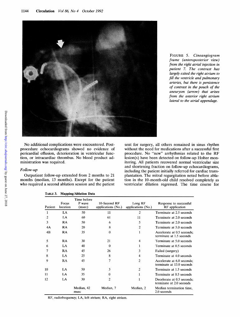
Ablation failed despite prolonged efforts in only onepatient (patient 7). The EAT focus in this instance waswell mapped to the orifice of the right atrial appendage,where local activation preceded the P wave by 45 msec.Of interest, signals recorded from adjacent atrial tissuein this area were noticeably fractionated and low am-plitude. Several RF applications were attempted butresulted only in acceleration of the EAT, which ulti-mately degenerated to atrial fibrillation. After cardio-version, EAT was still present at a faster rate with amore irregular pattern compared with baseline. At thispoint, an atrial angiogram was performed and revealedthe presence of a large saccular aneurysm at the ante-rior right atrium (Figure 5) that had not been appreci-ated on the preprocedure echocardiogram due to itsretrosternal location. A decision was made to proceedto surgery, where diffuse fibrous dysplasia of the ante-rior right atrium was noted in addition to the aneurysm.Abnormal atrial tissue was resected, and the patient didwell with normal sinus rhythm thereafter.
by guest on June 27, 2018http://circ.ahajournals.org/
Dow
nloaded from

1142 Circulation Vol 86, No 4 October 1992
1- F, ixF-JFi1J- ..I.k. i
..
A
...
VA....... ..........
..........
........
...
.. ..... ..... ..... .. .... ..... ..... , .. ........
...
....... ....... ..... ...... ... ......
--------------
....
C~NNEL 0~.. ......
..
.... ...1.................
CHM4NEI- *W..........
..... ........Ed
....
..
.............
1... I A'-, 2 A::;~~~~~~~~~~~~~~~~~~~~~~~.......: .. ...... ..
Vi. a~~~~~~~~~~~~~~~~~~~ i;,1J
t W 3. t r r iTir : T t' , '"'',
Trb ,
HBmT AAA
RVAB V. 2T-I
FIGURE 2. Intracardiac electro-grams and surface ECG recorded dur-ing mapping of a left-sided (A) andright-sided (B) ectopic atrial focus. Inboth examples, the distal electrode pairof the mapping/ablation catheter(MAPd) is located at a site wheresuccessful ablation was achieved. Lo-cal atrial activation precedes the onsetof the P wave (dotted line) by morethan 20 msec.
. . g aA i. ..bL....-4- --b.^* 1 - - - 2P.4 m.
by guest on June 27, 2018http://circ.ahajournals.org/
Dow
nloaded from

Walsh et al RF Ablation for Ectopic Atrial Tachycardia 1143
...~~~ ~ ~~~ ..
................... ...........
..
.......................................
.................. ........ .. ..... .............. . .
.. ...~~~~~~~~~~~~~~~.
..
............ C ....y..;......~~...... .......... ............. ....... ......~~~~~~~.T ... ..................1...........................
"E-! st ...Xl M" 1,r J
.I
... .. .... .. ....
- ....
C L---...................................................:..................................C.20 .........................
........................................... ...........~~~~~............ ..
'''''''''''''''''''''''-'''''''''' ------..... 2Ow ----............................................... ......... .. _
WAT.TS-J
-VL S .... .......
C:: (B):--...................................
nri"7j' ... ....--.. ..
..j,,,,....... .. ...................
.................. iiii,,!,,,,l,,!ii., .., j,l,,i,
~~~~~~~~~~~~~~~~~~~~~~~~~......... itii...
..
i'''"-"!'''''"~~~ ~~~ 1 -;
I *1f1 I -i ---i-*-i--iii-i-r' ''''
i i .........
..... ......~~~~~~~................. ...... ............. ...... ...... .................. ........ ....
.Ii~~~~~~~~~~~~~~~~.................i...
-'=. .........
I (C')..''
FIGURE 3. Examples of the response patterns observed during successfulRF applications for EAT: (A) abrupt termination, (B)transient acceleration followed by termination, and (C) transient slowing followed by termination.
Procedure times ranged from 1.5 to 7.0 hours (median,4.0 hours) with fluoroscopy times of 37-157 minutes(median, 77 minutes). Total hospital stays (excluding thepatient sent to surgery) ranged from 36 hours to 4 days(median, 48 hours).
FIGURE 4. Flow chart summarizing acute and follow-upresults for the study group.
Measurements of serum CPK were available from thefirst six patients in this series. Maximum values for totalCPK varied between 22 and 464 mU/ml with an MBfraction of 6-17%. No clear correlation was found be-tween CPK data and RF lesion number or procedureduration. This testing was not performed in later patients.
ComplicationsOne 10-month-old child (patient 5) had EAT with an
atrial rate in excess of 200 min' mapped to the rightatrium just anterior to the sinus node. Although sinusnode damage was considered possible with ablation inthis region, the patient was quite ill with severe mitralregurgitation and low cardiac output (mixed venoussaturation, 31%) such that the risk was deemed justi-fied. The EAT was successfully ablated, although theinitial recovery rhythm was slow sinus alternating with ajunctional escape. After 3 days of observation, sinusrhythm at physiological rates returned, and he thereaf-ter remained in normal rhythm.
J
im
-- ---J - - ---.- -- ---
by guest on June 27, 2018http://circ.ahajournals.org/
Dow
nloaded from
1144 Circulation Vol 86, No 4 October 1992
FIGURE 5. Cineangiogramframe (anteroposterior view)from the right atrial injection inpatient 7. The contrast haslargely exited the right atrium tofill the ventricle and pulmonaryarteries, but there is persistenceof contrast in the pouch of theaneurysm (arrow) that arisesfrom the anterior right atriumlateral to the atrial appendage.
No additional complications were encountered. Post-procedure echocardiograms showed no evidence ofpericardial effusion, deterioration in ventricular func-tion, or intracardiac thrombus. No blood product ad-ministration was required.
Follow-upOutpatient follow-up extended from 2 months to 21
months (median, 13 months). Except for the patientwho required a second ablation session and the patient
sent for surgery, all others remained in sinus rhythmwithout the need for medications after a successful firstprocedure. No "new" arrhythmias related to the RFlesion(s) have been detected on follow-up Holter mon-itoring. All patients recovered normal ventricular sizeand shortening fraction on follow-up echocardiograms,including the patient initially referred for cardiac trans-plantation. The mitral regurgitation noted before abla-tion in the 10-month-old child resolved completely asventricular dilation regressed. The time course for
TABLE 3. Mapping/Ablation Data
10-Second RFapplications (No.)
1161680
210
2687
Long RFapplications (No.)
211521
4
34
2
Response to successfulRF application
Terminate at 2.5 secondsTerminate at 2.0 secondsTerminate at 2.0 secondsTerminate at 5.0 secondsAccelerate at 0.5 seconds;terminate at 1.5 secondsTerminate at 5.0 secondsTerminate at 0.5 secondsFailed (surgery)Terminate at 4.0 secondsAccelerate at 6.0 seconds;terminate at 13.0 seconds
2 Terminate at 1.5 seconds1 Terminate at 0.5 seconds1 Decelerate at 0.5 seconds;
terminate at 2.0 secondsMedian, 2 Median termination time,
2.0 seconds
RF, radiofrequency; LA, left atrium; RA, right atrium.
Patient
12
34A4B
56789
Focuslocation
LALARARA
RA
RALARALARA
Time beforeP wave(msec)
5060502035
3040452545
1011
12
LALALA
503530
302
Median, 7Median, 42msec
-
by guest on June 27, 2018http://circ.ahajournals.org/
Dow
nloaded from

Walsh et al RF Ablation for Ectopic Atrial Tachycardia 1145
improvement in ventricular performance was generallyrapid (within 1-2 months), although echocardiographicvariables did not normalize in two cases until 6 months(patient 10) and 9 months (patient 2) after the ablation.
DiscussionEAT is well recognized as a correctable cause of
cardiomyopathy. Although it is a rare form of supraven-tricular tachycardia in both children and adults, thehemodynamic consequences of poor rhythm controlmay ultimately necessitate aggressive therapy. Trans-catheter ablation is now evolving as a realistic alterna-tive to chronic pharmacological or surgical managementof this condition. There are two potential targets foreffective ablation. The first involves permanent inter-ruption of the normal atrioventricular conducting sys-tem, allowing the EAT to persist but controlling theventricular rate. This technique has been employedsuccessfully for EAT and other refractory atrial arrhyth-mias18 but creates pacemaker dependence. A moreattractive option is direct eradication of the EAT focus.Experience with this later method had been limiteduntil the introduction of RF current as an ablationenergy source, since the technique of DC fulgurationcarried a- potential-risk -of perforation through therelatively thin atrial wall.While RF ablation is currently very well established
as definitive therapy for tachyarrhythmias due to acces-sory atrioventricular pathways in patients of allages,19-22 this report represents the first large series todescribe its application to EAT. Unlike accessory path-ways, accurate localization of an ectopic atrial focusmust involve a three-dimensional map and is furtherconfounded by the absence of reliable electrogrammarkers (such as accessory pathway potentials) apartfrom local activation times. Despite these difficulties,the technique was successful in 92% of our patients,with the only failure occurring in a patient who haddiffuse atrial fibrosis requiring surgical excision. Allsubjects had prompt reversal of the ventricular dysfunc-tion caused by the tachyarrhythmia, and all remain insinus rhythm without the need for medications. Whilesome series describing surgical therapy for EAT reportarrhythmia recurrence after excision of right atrialfoci,'0"11 we detected recurrence in only one patient witha suboptimal map at initial study due to residual amio-darone effect. Successful ablation was later achieved inthis case when the focus was more active. Although thefollow-up for this group is short term (median, 13months), the initial results remain quite encouragingand suggest that the technique is at least as effective asa surgical procedure.The mapping/ablation features that best predicted
successful EAT elimination included 1) local electricalactivity preceding onset of the surface P wave by 20-60msec (median, 42 msec), 2) rapid resolution of EATwithin less than 10 seconds of beginning RF application,and, on two occasions, 3) transient EAT suppressiondue to local catheter pressure over the target site. Thecritical requirement for a high-quality map was thepresence of nearly incessant tachycardia, which occa-sionally required the administration of isoproterenol.As demonstrated by two patients in this series, residualantiarrhythmic drug effects and/or the deep level of
the EAT in the laboratory environment. Except forcases involving very young children (in whom we prefera general anesthetic), it may be advisable to performEAT ablation under light intravenous sedation when-ever possible.
Since this series marked our early experience withEAT ablation, we tended to be very compulsive with themapping technique. Direct left atrial recording was
preferred over a coronary sinus electrode, even if thisrequired transseptal puncture. In cases where the focuscan be clearly mapped to the right atrial appendage, thisdegree of intervention may not be necessary for an
accurate map. However, for EAT foci at all otherlocations, we found a coronary sinus recording to be lessthan adequate for localization. Indeed, many left atrialfoci produce a similar misleading signal on the coronarysinus electrode involving earliest activation near themouth of coronary sinus. There does not appear to be a
reliable substitute for direct left atrial mapping if theEAT involves any site other than the right atrialappendage.RF lesions at the atrial level have been shown to
produce transmural necrosis that heals with a wellorganized fibrous scar and have not been associatedwith significant risk for either early or late perforation.23The RF technique is also free of the barotrauma andcatheter fling seen with DC ablation, further decreasingthe likelihood of acute atrial trauma. RF current wouldthus appear well suited for atrial muscle ablation,including the fragile areas near the atrial appendages.This report supports the general safety of the techniquein children as young as 10 months.
Fluoroscopy times and procedure times encounteredin this series were longer than those for a typicaldiagnostic electrophysiology study but were not dissim-ilar to times reported for RF ablation of accessory
pathways.19'20 Innovations in mapping catheter designmay permit faster simultaneous mapping of multipleatrial sites and are under investigation at our center.Additionally, laboratory equipment is now availablewith the capacity for pulsed fluoroscopy at slow framerates (7.5 sec-1), which will reduce radiation exposure tothe patient and operator without significantly compro-mising catheter control. Overall procedure times stillrequire reduction but will likely shorten as more expe-
rience is gained with this technique.Since atrial RF lesions are transmural, one cannot
safely comment on whether the foci ablated in thisseries involved endocardium, epicardium, or atrial myo-cytes. However, in contrast to the aforementioned sur-
gical reports,10"' which suggested that some cases ofEAT may represent diffuse atrial disease, 11 of the 12patients treated in this series (including four of five withright atrial EAT) appeared to have a point source
arrhythmia that could often be eliminated by a singleRF lesion. The ultimate size of such lesions is unlikelyto be more than 10 mm in surface diameter. Further-more, early disappearance of EAT at a median of 2.0seconds into the RF application suggests that an EATfocus could actually be smaller then even 2 mm.24
In this early experience involving 12 patients withdrug-resistant EAT, catheter ablation with RF currentappeared to be an effective and safe therapeutic option.At our institution, RF ablation is now recommended as
sedation accompanying general anesthesia can suppress first-line therapy for any age child who has developed
by guest on June 27, 2018http://circ.ahajournals.org/
Dow
nloaded from

1146 Circulation Vol 86, No 4 October 1992
myocardial dysfunction from the tachyarrhythmia. Forpatients with well-preserved ventricular performance,an initial drug trial with ,B-blocker is still attempted,reserving ablation as second-line therapy in preferenceto more potent antiarrhythmic drugs or surgery.
References1. Gillette PC, Smith RT, Garson A, Mullins CE, Gutgesell HP, Goh
TH, Cooley DA, McNamara DG: Chronic supraventricular tachy-cardia: A curable cause of congestive cardiomyopathy. JAMA 1985;253:391-392
2. Gillette PC, Wampler DG, Garson A, Zinner A, Ott D, Cooley D:Treatment of atrial automatic tachycardia by ablation procedures.JAm Coll Cardiol 1985;6:405-409
3. Keane JF, Plauth WH, Nadas AS: Chronic ectopic tachycardia ofinfancy and childhood. Am Heart J 1972;84:748-757
4. Seals AA, Lawrie GM, Magro S, Lin HT, Pacifico A, Roberts R,Wyndham CR: Surgical treatment of right atrial focal tachycardiain adults. JAm Coil Cardiol 1988;11:1111-1117
5. Gillette PC, Crawford FC, Zeigler VL: Mechanisms of atrial tachy-cardias, in Zipes DP, Jalife J (eds): Cardiac Electrophysiology;From Cell to Bedside. Philadelphia, WB Saunders, 1990,pp 559-563
6. Mehta AV, Sanchez GR, Sacks EJ, Casta A, Dunn JM, DonnerRM: Ectopic automatic atrial tachycardia in children: Clinicalcharacteristics, management and follow-up. J Am Coll Cardiol1988;11:379-385
7. Kunze KP, Kuck KH, Schluter M, Bleifeld W: Effect of encainideand flecainide on chronic ectopic atrial tachycardia. J Am CollCardiol 1986;7:1121-1126
8. Gillette PC, Garson A, Hesslein PS, Karpawich PP, Tierney RC,Cooley DC, McNamara DG: Successful surgical treatment ofatrial, junctional, and ventricular tachycardia unassociated withaccessory connections in infants and children. Am Heart J 1981;102:984-991
9. Ott DA, Gillette PC, Garson A, Cooley DA, Reul GJ, McNamaraDG: Surgical management of refractory supraventricular tachycar-dia in infants and children. JAm Coll Cardiol 1985;5:124-129
10. Garson A, Moak JP, Friedman RA, Perry JC, Ott DA: Surgicaltreatment of arrhythmias in children. Cardiol Clin 1989;7:319-329
11. Garson A, Smith RT, Moak JP, Judd VE, Ott DA, Cooley DA:Atrial automatic ectopic tachycardia in children, in Touboul P,Waldo AL (eds): Atrial Arrhythmias; Current Concepts and Man-agement. St Louis, Mosby Year Book, 1990, pp 282-287
12. Silka MJ, Gillette PC, Garson A, Zinner A: Transvenous catheterablation of a right atrial automatic ectopic tachycardia. JAm CollCardiol 1985;5:999-1001
13. Margolis PD, Roman CA, Moulton KP, Calame J, Wang X, Laz-zara R, Jackman WM: Radiofrequency catheter ablation of leftand right ectopic atrial tachycardia. Circulation 1990;82(suppl III):111-718
14. Walsh EP, Saul JP, Hulse JE, Lock JE: Successful transcatheterablation of ectopic atrial tachycardia using radiofrequency energy.PACE 1991;14(suppl II):II-656
15. Koike K, Hesslein PS, Finlay CD, Williams WG, Izukawa T, Free-dom RM: Atrial automatic tachycardia in children. Am J Cardiol1988;61:1127-1130
16. Gillette PC, Garson A: Electrophysiologic and pharmacologiccharacteristics of automatic ectopic atrial tachycardia. Circulation1977;56:571-575
17. Brockenbrough EC, Braunwald E: A new technique for left ven-tricular angiocardiography and transseptal left heart catheteriza-tion. Am J Cardiol 1966;6:1062-1064
18. Langberg JJ, Chin MC, Rosenqvist M, Cockrell J, Dullet N, VanHare G, Griffin JC, Scheinman MM: Catheter ablation of theatrioventricular junction with radiofrequency energy. Circulation1989;80:1527-1535
19. Jackman WM, Wang X, Friday KJ, Roman CA, Moulton KP, Beck-man KJ, McClelland JH, Twidale N, Hazlitt HA, Prior MI, MargolisPD, Calame JD, Overhalt ED, Lazzara R: Catheter ablation of acces-sory atrioventricular pathways (Wolff-Parkinson-White syndrome) byradiofrequency current. N EnglJMed 1991;324:1605-1611
20. Calkins H, Sousa J, El-Atassi R, Rosenheck S, deBuitleir M, KouWH, Kadish AH, Langberg JJ, Morady F: Diagnosis and cure ofthe Wolff-Parkinson-White syndrome or paroxysmal supraventric-ular tachycardias during a single electrophysiologic test. N Engl JMed 1991;324:1612-1618
21. Van Hare G, Lesh MD, Scheinman M, Langberg JJ: Percutaneousradiofrequency catheter ablation for supraventricular arrhythmiasin children. JAm Coll Cardiol 1991;17:1613-1620
22. Dick M, O'Connor BK, Serwer GA, LeRoy S, Armstrong B: Useof radiofrequency current to ablate accessory connections in chil-dren. Circulation 1991;84:2318-2324
23. Huang SK: Advances in applications of radiofrequency current tocatheter ablation therapy. PACE 1991;14:28-42
24. WittkampfFHM, Hauer RNW, de Medina EOR: Control of radio-frequency lesion size by power regulation. Circulation 1989;80:962-968
by guest on June 27, 2018http://circ.ahajournals.org/
Dow
nloaded from

E P Walsh, J P Saul, J E Hulse, L A Rhodes, A J Hordof, J E Mayer and J E Lockradiofrequency current.
Transcatheter ablation of ectopic atrial tachycardia in young patients using
Print ISSN: 0009-7322. Online ISSN: 1524-4539 Copyright © 1992 American Heart Association, Inc. All rights reserved.
is published by the American Heart Association, 7272 Greenville Avenue, Dallas, TX 75231Circulation doi: 10.1161/01.CIR.86.4.1138
1992;86:1138-1146Circulation.
http://circ.ahajournals.org/content/86/4/1138the World Wide Web at:
The online version of this article, along with updated information and services, is located on
http://circ.ahajournals.org//subscriptions/
is online at: Circulation Information about subscribing to Subscriptions:
http://www.lww.com/reprints Information about reprints can be found online at: Reprints:
document. Permissions and Rights Question and Answer information about this process is available in the
located, click Request Permissions in the middle column of the Web page under Services. FurtherEditorial Office. Once the online version of the published article for which permission is being requested is
can be obtained via RightsLink, a service of the Copyright Clearance Center, not theCirculationpublished in Requests for permissions to reproduce figures, tables, or portions of articles originallyPermissions:
by guest on June 27, 2018http://circ.ahajournals.org/
Dow
nloaded from