topographical organization of attentional, social, and ...graziano/igelstrom_2016b.pdf ·...
TRANSCRIPT

Cognition and Behavior
Topographical Organization of Attentional, Social,and Memory Processes in the HumanTemporoparietal Cortex1,2,3
Kajsa M. Igelström, Taylor W. Webb, Yin T. Kelly, and Michael S. A. Graziano
DOI:http://dx.doi.org/10.1523/ENEURO.0060-16.2016
Princeton Neuroscience Institute and Department of Psychology, Princeton University, Princeton, New Jersey 08544
Abstract
The temporoparietal junction (TPJ) is activated in association with a large range of functions, including socialcognition, episodic memory retrieval, and attentional reorienting. An ongoing debate is whether the TPJ performsan overarching, domain-general computation, or whether functions reside in domain-specific subdivisions. Wescanned subjects with fMRI during five tasks known to activate the TPJ, probing social, attentional, and memoryfunctions, and used data-driven parcellation (independent component analysis) to isolate task-related functionalprocesses in the bilateral TPJ. We found that one dorsal component in the right TPJ, which was connected withthe frontoparietal control network, was activated in all of the tasks. Other TPJ subregions were specific forattentional reorienting, oddball target detection, or social attribution of belief. The TPJ components that partic-ipated in attentional reorienting and oddball target detection appeared spatially separated, but both wereconnected with the ventral attention network. The TPJ component that participated in the theory-of-mind taskwas part of the default-mode network. Further, we found that the BOLD response in the domain-general dorsalcomponent had a longer latency than responses in the domain-specific components, suggesting an involvementin distinct, perhaps postperceptual, computations. These findings suggest that the TPJ performs both domain-general and domain-specific computations that reside within spatially distinct functional components.
Key words: angular gyrus; blind source separation; data-driven fMRI analysis; superior temporal gyrus andsulcus; supramarginal gyrus
Significance Statement
The temporoparietal junction (TPJ) is a major communication hub in the human brain. The exact pattern ofoverlap and separation of function in the TPJ has been difficult to study due to the complexity of itsresponses during many different kinds of tasks. We studied the activity in the TPJ during five behavioraltasks associated with attention, memory retrieval, and social cognition. We found that one zone in the TPJwas active in all five tasks, whereas other zones were active in a more task-specific manner. Our findingssuggest that the TPJ is a site where multiple brain networks converge and interact, but that it also containsmore functionally specific subregions.
New Research
March/April 2016, 3(2) e0060-16.2016 1–12
IntroductionMany theories have been proposed to explain the mul-
titude of tasks that activate the temporoparietal junction(TPJ), which involve functions ranging from bottom-upattention to episodic memory retrieval and social cogni-tion. For example, it has been suggested that the episodicmemory activity in the TPJ is related to reflexive orientingto information retrieved from memory (Cabeza, 2008; Ci-aramelli et al., 2008; Cabeza et al., 2011), and that socialcognition in the TPJ might depend on similar low-levelinformation processing (Decety and Lamm, 2007). Othertheories have postulated that the TPJ is a zone of con-vergence and integration, in which internal models ofone’s environment, social context, or attentional state aremaintained and updated (Graziano and Kastner, 2011;Carter and Huettel, 2013; Geng and Vossel, 2013; Kellyet al., 2014; Webb and Graziano, 2015). Comparisons ofactivation patterns within single subjects have shownsome separation but also zones of overlap. For example,topographic overlap has been reported between theory-of-mind and attentional reorienting activity (Mitchell,2008); between memory retrieval and attentional reorient-ing (Cabeza et al., 2011); and among theory-of-mind,attentional reorienting, and biological motion (Lee andMcCarthy, 2016). However, functional heterogeneity hasalso been seen in both meta-analyses and within-subjectfMRI studies, manifesting as the physical separation ofprocesses, an ability of multivoxel pattern analysis todiscriminate different tasks within regions of overlap, anddistinct connectivity patterns of activation foci (Hutchin-son et al., 2009; Scholz et al., 2009; Cabeza et al., 2011,2012; Daselaar et al., 2013; Lee and McCarthy, 2016).
The great spatial variability of fMRI activations in theTPJ is perhaps not surprising given the intersubject het-erogeneity in the TPJ, the limitations of normalizing indi-vidual brains into a common space, and the limitations ofvoxelwise analysis. One useful way of addressing TPJfunction is careful within-subject analysis of task-relatedactivity in high-resolution fMRI scans (Mitchell, 2008;Scholz et al., 2009; Cabeza et al., 2011; Lee and McCar-thy, 2016). Another possibility, explored in this study, is touse multivariate data-driven methods to work around the
limitations of voxelwise analysis. We previously found thatlocalized independent component analysis (local-ICA),which decomposes the fMRI signal in the TPJ into a linearmixture of spatiotemporal source processes, could beused to parcellate the TPJ into five to six subdivisions perhemisphere (Igelström et al., 2015, 2016). These compo-nents included bilateral posterior (TPJp), anterior (TPJa),dorsal (TPJd), and ventral (TPJv) regions, and a centralright-biased region (TPJc). The time courses of the inde-pendent components (ICs) within the TPJ were correlatedwith distinct resting-state networks (Igelström et al., 2015,2016), indicating that they represented functionally dis-tinct processes. This method of local-ICA was robustacross multiple independent subject cohorts (Igelströmet al., 2015).
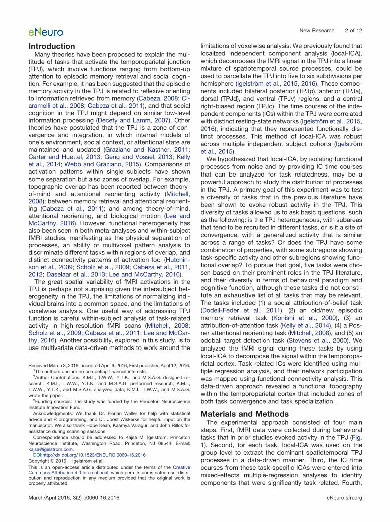
We hypothesized that local-ICA, by isolating functionalprocesses from noise and by providing IC time coursesthat can be analyzed for task relatedness, may be apowerful approach to study the distribution of processesin the TPJ. A primary goal of this experiment was to testa diversity of tasks that in the previous literature havebeen shown to evoke robust activity in the TPJ. Thisdiversity of tasks allowed us to ask basic questions, suchas the following: is the TPJ heterogeneous, with subareasthat tend to be recruited in different tasks, or is it a site ofconvergence, with a generalized activity that is similaracross a range of tasks? Or does the TPJ have somecombination of properties, with some subregions showingtask-specific activity and other subregions showing func-tional overlap? To pursue that goal, five tasks were cho-sen based on their prominent roles in the TPJ literature,and their diversity in terms of behavioral paradigm andcognitive function, although these tasks did not consti-tute an exhaustive list of all tasks that may be relevant.The tasks included (1) a social attribution-of-belief task(Dodell-Feder et al., 2011), (2) an old/new episodicmemory retrieval task (Konishi et al., 2000), (3) anattribution-of-attention task (Kelly et al., 2014), (4) a Pos-ner attentional reorienting task (Mitchell, 2008), and (5) anoddball target detection task (Stevens et al., 2000). Weanalyzed the fMRI signal during these tasks by usinglocal-ICA to decompose the signal within the temporopa-rietal cortex. Task-related ICs were identified using mul-tiple regression analysis, and their network participationwas mapped using functional connectivity analysis. Thisdata-driven approach revealed a functional topographywithin the temporoparietal cortex that included zones ofboth task convergence and task specialization.
Materials and MethodsThe experimental approach consisted of four main
steps. First, fMRI data were collected during behavioraltasks that in prior studies evoked activity in the TPJ (Fig.1). Second, for each task, local-ICA was used on thegroup level to extract the dominant spatiotemporal TPJprocesses in a data-driven manner. Third, the IC timecourses from these task-specific ICAs were entered intomixed-effects multiple-regression analyses to identifycomponents that were significantly task related. Fourth,
Received March 3, 2016; accepted April 6, 2016; First published April 12, 2016.1The authors declare no competing financial interests.2Author Contributions: K.M.I., T.W.W., Y.T.K., and M.S.A.G. designed re-
search; K.M.I., T.W.W., Y.T.K., and M.S.A.G. performed research; K.M.I.,T.W.W., Y.T.K., and M.S.A.G. analyzed data; K.M.I., T.W.W., and M.S.A.G.wrote the paper.
3Funding sources: The study was funded by the Princeton NeuroscienceInstitute Innovation Fund.
Acknowledgments: We thank Dr. Florian Weller for help with statisticaladvice and R programming, and Dr. Joost Wiskerke for helpful input on themanuscript. We also thank Hope Kean, Kaamya Varagur, and John Rillos forassistance during scanning sessions.
Correspondence should be addressed to Kajsa M. Igelström, PrincetonNeuroscience Institute, Washington Road, Princeton, NJ 08544. E-mail:[email protected].
DOI:http://dx.doi.org/10.1523/ENEURO.0060-16.2016Copyright © 2016 Igelström et al.This is an open-access article distributed under the terms of the CreativeCommons Attribution 4.0 International, which permits unrestricted use, distri-bution and reproduction in any medium provided that the original work isproperly attributed.
New Research 2 of 12
March/April 2016, 3(2) e0060-16.2016 eNeuro.sfn.org

all IC time courses were used for a functional connectivityanalysis to map the network participation of each IC.
Theory-of-mind taskThe theory-of-mind task was performed by 20 subjects(12 females; mean age, 22.6 � 0.8 years old). The studywas approved by the Princeton University InstitutionalReview Board. All subjects gave informed written con-sent, had normal or corrected-to-normal vision, and hadno history of psychiatric or neurological disorders.
We used a theory-of-mind localizer task from a study byDodell-Feder et al. (2011), which contrasts brain activa-tions during the attribution of beliefs to people (belieftrials) with activations during judgments about the con-tents of photographs, maps, or signs (photo trials; Fig.1A). The stimuli were provided by David Dodell-Feder,Nicholas Dufour, and Rebecca Saxe (http://saxelab.mit.edu/superloc.php). This task was chosen because of its
extensive use in theory-of-mind studies and its popularityas a TPJ localizer task (Saxe and Powell, 2006; Mitchell,2008; Scholz et al., 2009; Dodell-Feder et al., 2011). Ineach trial, the story was presented for 10 s, followed by atrue/false question for 4 s and an intertrial interval (ITI) of12 s. Participants responded to the questions using abutton box. The task consisted of two runs of 10 trialseach, and the order of stories was counterbalanced andequally distributed across the two runs. The BOLDresponse was modeled with a 14 s boxcar convolvedwith a standard hemodynamic response function (“wa-ver” function; AFNI). A contrast between belief trialsand photo trials is known to reveal activation in the TPJand other theory-of-mind regions (Dodell-Feder et al.,2011). The reaction time was not significantly differentin belief versus photo trials (mean � SEM reaction time,2.7 � 0.10 and 2.6 � 0.10 s, respectively; p � 0.18,paired t test).
Figure 1. Schematic representation of task designs. A, Theory-of-mind task. A story requiring either attribution of belief or reasoningabout a photo was shown for 10 s, followed by a true/false question for 4 s. There were 20 trials in total. B, Episodic memory retrievaltask. The task was divided over four runs, each consisting of an encoding phase (“E”) before the run began, and a retrieval phase (“P”;bottom). In the encoding-phase, subjects were asked to memorize words presented sequentially on the screen (33 words). In theretrieval run (25 old words intermixed with 50 new words and 25 fixation trials), subjects indicated with a button press whether a wordwas old or new. There were 400 trials in total. C, Attribution-of-attention task. An object with either negative or positive salience waspresented on the right or left for 1 s (this example shows a car fire). After a 0.5 s interval, the face of a cartoon character waspresented centrally for 2 s. Its gaze was directed either toward or away from the object (G� or G�), and its emotional expressioneither matched or mismatched the valence of the object (E� or E�). Subjects rated the character’s level of awareness of theobject on a scale of 1 (not aware) to 3 (very aware). Four trial types were possible: G�E�, G�E�, G�E�, and G�E�. The trialsin which the gaze and expression cues were inconsistent (G�E� and G�E�) were labeled as hard trials (for more details, seeMaterials and Methods). There were 384 trials in total. D, Attentional reorienting task. A central cue pointing right or left predictedthe location of a target in 75% of trials. Subjects were asked to indicate which side the target appeared on. There were 200 trialsin total. E, Target detection task. A visual standard stimulus (“OOOO”) was presented on the screen every 1.5 s. In 5% of trials,this was replaced by the target stimulus (“XXXX”). The subjects silently counted how many targets they saw. There were 480trials in total.
New Research 3 of 12
March/April 2016, 3(2) e0060-16.2016 eNeuro.sfn.org

Episodic memory retrieval taskThe episodic memory retrieval task was performed by 20subjects, 1 of whom was excluded due to excessivemovement (10 females; mean age, 22.3 � 1.0 years).Subjects had normal or corrected-to-normal vision and nohistory of psychiatric or neurological disorders.
The episodic memory task was an old/new task basedon a study by Konishi et al. (2000; Fig. 1B). The old/newparadigm was chosen because several studies havefound TPJ activation with this contrast, and it has beenpart of meta-studies of functional overlap (Hutchinsonet al., 2009, 2014; Cabeza et al., 2012). The task consistedof four runs. Each run comprised a memory-encodingphase and a memory-retrieval phase. In the encodingphase, subjects were asked to memorize 33 words, whichwere presented on the screen 1 word at a time (wordduration, 2 s; trial duration, 3 s). In the retrieval phase, arandomized collection of words was presented in thesame manner. These included 25 words from the learningphase and 50 new words. Twenty-five fixation trials werealso randomly interspersed in each run. Subjects wereasked to indicate with a button press whether or not theyremembered the word as being from the learning run.There was no overlap of words among the four runs. Thecontrast between successfully retrieved old words (oldtrials) and successfully rejected new words (new trials)has been shown to evoke TPJ activation (Konishi et al.,2000). The word list was derived from the SUBTLEXus1.00 word frequency database (Brysbaert and New, 2009)and consisted of five-letter words with a frequency of5–10 per million. The BOLD response was modeled with aboxcar time course convolved with a standard hemody-namic response function (waver function; AFNI). The av-erage accuracy did not differ between the old and newtrials (mean � SEM accuracy, 81.8 � 2.7% and 87.9 �2.9%, respectively; p � 0.13, paired t test). The reactiontime for new trials was slightly longer than that for oldtrials (mean � SEM reaction time, 993 � 27 and 927 � 23ms, respectively; p � 0.0013, paired t test).
Social attribution of attentionThe attribution-of-attention task was chosen because itwas previously found to evoke robust activity in the TPJeven in individual subjects (Kelly et al., 2014) and offers acontrasting approach to testing social cognition from thebelief attribution task. In the present study, we used thedata collected in our previous study (Kelly et al., 2014) andreanalyzed it for the present study. The task (Fig. 1C)required subjects to rate the perceived level of awarenessof a cartoon face (“Kevin”) for an object next to it. Thedirection of gaze of the face was manipulated (toward oraway from the location of the object), and the emotionalexpression of the face either matched the valence of theobject (e.g., a happy face paired with a cupcake or afrightened face paired with a house fire) or mismatchedthe valence of the object (e.g., a frightened face pairedwith a cupcake or a happy face paired with a house fire).Subjects rated Kevin’s level of awareness of the object ona scale of 1 (not aware) to 3 (very aware). When both thegaze and expression cues matched the object, subjects
tended to rate Kevin as very aware (rating of 3). Whenboth the gaze and expression cues mismatched the ob-ject, subjects tended to rate Kevin as unaware (rating of1). When the cues to Kevin’s state of awareness wereincompatible, with one cue suggesting awareness and theother suggesting unawareness, subjects tended to com-promise between the two cues and rate Kevin’s aware-ness as intermediate (rating of 2). In the previous study, itwas found that TPJ activity was significantly higher whensubjects compromised between two incompatible cues toKevin’s state of mind, presumably when the computationabout Kevin’s state of mind was more difficult (“Hardtrials”), and activity in the TPJ was significantly lowerwhen the subjects used two compatible cues to Kevin’sstate of mind, presumably when the computation aboutKevin’s state of mind was easier (“Easy trials”).
The details of the paradigm are described in a previouspublication (Kelly et al., 2014). Briefly, the behavioral taskconsisted of eight runs of 48 trials each. Each trial startedwith a fixation cross for 0.5 s, followed by a picture of anobject for 1 s. The fixation cross returned for 0.5 s, andthen was replaced by a cartoon face for 2 s. The objectwas presented either to the left or right and had eitherpositive or negative valence. The gaze of the cartoonfigure was either averted from or directed at the object,and the expression was either happy or alarmed. In thisway, the gaze could either match (Gaze�) or mismatch(Gaze�) the location of the object, and the expressioncould either match (Expr�) or mismatch (Expr�) the va-lence of the object (see examples in Fig. 1C). Subjectswere asked to indicate with one of three buttons whetherthe person was (1) not aware, (2) somewhat aware, or (3)very aware of the object. For the present study, we useddata from the first 20 subjects of the total of 50 subjectsfrom the previous study (Kelly et al., 2014; 8 females;mean age, 19.4 � 0.4 years). The reason for analyzing asmaller subset of subjects was a limitation in memory onthe compute cluster, which prevented a full 50-subjectgroup level-ICA. Therefore, we chose to analyze the first20 subjects in this task as an unbiased way of decreasingthe computing requirements. We used the same regres-sors and contrast as in the previous study (describedabove), convolving the hard and easy trial types with ahemodynamic response function (waver function; AFNI)and testing the contrast hard versus easy.
Attentional reorienting taskThis task was performed by 20 subjects with normal orcorrected-to-normal vision and no history of psychiatricor neurological disorders (11 females; mean age, 21.6 �0.6 years).
Attentional reorienting was tested using a Posner taskmodeled closely on a previous study (Mitchell, 2008; Fig.1D). This task was chosen to represent reorienting toinvalidly cued targets, which is thought to be a majorfunction of the TPJ (Corbetta and Shulman, 2002; Cor-betta et al., 2008; Mitchell, 2008; Geng and Vossel, 2013).The task consisted of five runs of 40 trials each. Subjectswere given one practice run inside the scanner before theexperiment started. The fixation screen consisted of a
New Research 4 of 12
March/April 2016, 3(2) e0060-16.2016 eNeuro.sfn.org

black background with a red central fixation plus sign (�)and two peripheral square boxes with white outlines. Theperipheral boxes were centered 7° from the fixation andwere 3° across. At the start of a trial, the central plus signturned green for a fixation period of 700 ms and wasreplaced by a central cue consisting of an arrow for 800ms. The arrow pointed either left or right, in a randomizedcounterbalanced order. After a pretarget period of 0.5–2s, the target (a white asterisk) was presented in one of thetwo peripheral boxes for 100 ms. The posttarget time wasselected to make the total trial duration 4 s, after whichthe fixation plus sign turned red again for a randomized ITIof 0.5–7.5 s. Subjects responded with a button press toindicate whether the target was on the left or on the right.The direction of the central cue predicted the side of thetarget in 75% of trials (valid trials) and was mismatched in25% of trials (invalid trials). Subjects were informed thatthe arrow would predict the target in the majority of trials.A contrast between invalid and valid trials has been re-ported to reveal activity in the right TPJ and the ventralattention network (Corbetta et al., 2000). The BOLD re-sponse was modeled by convolving the stimulus timingswith a gamma function. The reaction time for invalid trialswas significantly longer than that for valid trials (mean �SEM reaction time, 394 � 16 and 362 � 15 ms; p �0.00007, paired t test).
Oddball target detection taskThis task was performed by 20 subjects, 1 of whom wasexcluded due to poor performance (10 females; meanage, 22.9 � 1.0 years). The subjects had normal orcorrected-to-normal vision and no history of psychiatricor neurological disorders.
The target detection task was a simple visual oddballtask based on the study by Stevens et al. (2000; Fig. 1E).The oddball paradigm was chosen because it representsa nonspatial form of attentional reorienting that is oftenreported to cause TPJ activations (Corbetta et al., 2008;Cabeza et al., 2012). It consisted of four runs with 120trials each. The standard stimulus was the letters “OOOO”presented centrally for 500 ms. The rare target stimulusconsisted of the letters “XXXX” (4–7% of trials in each run)and was made task relevant by asking the subjects toreport on how many targets they had seen after each run.A contrast between the target (target trials) and the stan-dard stimuli (standard trials) was reported to evoke TPJactivity (Stevens et al., 2000). The BOLD response wasmodeled by convolving the stimulus timings with agamma function.
Distribution of subjects across the five tasksFor reasons of feasibility, every subject did not perform alltasks, which would have required a prohibitive number ofhours and sessions for each subject. Instead, each taskwas analyzed separately. In effect, we performed fiveindependent experiments. All statistical analyses wereperformed independently for each task, with no between-subjects statistical tests. The results are reported sepa-rately for each task and not used to draw conclusionsabout quantitative differences across tasks in the exactspatial location of ICs or the effect size of activity. In some
cases, for tasks that required less run time, the samesubjects participated in more than one task. Because ofthe separate analysis for each task, these overlaps in thesubject pools are not of direct relevance to the analysis,but are nonetheless reported here. Fourteen subjects per-formed both the theory-of-mind task and the attentionalreorienting task. Six subjects performed only the atten-tional reorienting task, and six subjects performed onlythe theory-of-mind task. Eighteen subjects performedboth the episodic memory retrieval task and the targetdetection task. One subject performed only the episodicmemory retrieval task, and one subject performed onlythe target detection task. For the social attribution-of-attention task, data from 20 subjects were used. In totalacross the five tasks, 66 subjects were tested.
Magnetic resonance imagingMRI images covering the whole cerebral cortex wereacquired with a 20-channel receiver head coil on a Sie-mens Skyra scanner. Functional imaging used a gradientecho, echoplanar pulse sequence with a 64 � 64 matrix[27 axial slices; 4 mm thick; in-plane resolution, 3 � 3 mm;TR, 1.5 s; TE, 28 ms; flip angle (FA), 64°; generalizedGRAPPA iPAT � 2. Anatomical imaging used anMP2RAGE sequence (256 � 240 matrix; TR, 5 s; TE, 2.98ms; FA, 4°; 1 mm2 resolution; GRAPPA iPAT � 3). Thereanalyzed data from the previous study (Kelly et al.,2014) were acquired on the same scanner. The functionaldata were acquired with a 64 � 64 matrix (35 axial slices;3 mm thick; in-plane resolution, 3 � 3 mm; TR, 2 s; TE, 30ms; FA, 77°), and the anatomical data were acquired withan MPRAGE sequence (256 � 224 matrix; TR, 2.3 s; FA,9°; and with 1 mm2 [TE, 2.98 ms], 0.9 mm2 [TE, 3.08 ms],or 1.1 mm2 [TE, 2.93 ms] resolution).
Preprocessing of fMRI dataPreprocessing was performed with AFNI (Cox, 1996) andFSL (Jenkinson et al., 2012). The functional data wereslice time corrected and motion corrected with FSL (Jen-kinson et al., 2002), and then detrended (linear and qua-dratic) with AFNI. The data were spatially normalized tothe FSL MNI-152 template with AFNI, and spatiallysmoothed with a Gaussian kernel (5 mm FWHM). We usedan ICA-based strategy for automatic removal of motionartifacts (ICA-AROMA; Pruim et al., 2015a). This toolboxruns single-session ICA with multivariate exploratorylinear decomposition into independent components(MELODIC, FSL); and classifies motion-related ICs byassessing their high-frequency content, correlation withmotion parameters, edge fraction, and cerebrospinal fluidfraction (Pruim et al., 2015a). ICA-AROMA removes noiseICs from the fMRI data by calling the FSL commandfsl_regfilt (Beckmann and Smith, 2004). Such ICA-baseddenoising is effective in removing aberrant connectivitymeasures resulting from subject motion (Power et al.,2012; Salimi-Khorshidi et al., 2014; Pruim et al., 2015b).
Group-level ICAFor each of the five tasks, the fMRI data were subjected toprobabilistic ICA applied on temporally concatenatedfMRI data, with all runs from all subjects concatenated
New Research 5 of 12
March/April 2016, 3(2) e0060-16.2016 eNeuro.sfn.org

into one matrix (MELODIC toolbox in FSL; Beckmann andSmith, 2004). We performed the ICA decomposition sep-arately for each task (instead of grouping the tasks intoone ICA) because we did not want to assume that the ICAdecomposition would be the same across task condi-tions. We applied the ICA to the voxels within a region ofinterest (ROI) mask that included the TPJ and surroundingcortex to ensure that all relevant ICs were detected in theirentirety. The mask was constructed from the standardsurface cvs_avg35_inMNI152 in Freesurfer, using mri_label2vol to combine multiple labels from the aparc.a2009satlas into one mask (G_pariet_inf-Supramar, G_pariet_inf-Angular, G_temp_sup-Plan_tempo, G_temp_sup-Lateral,G_temp_sup-G_T_transv,S_interm_prim-Jensen,S_temporal_sup, S_temporal_transverse) and trimming temporal cortexvoxels anterior to the postcentral sulcus. This mask al-lowed a parcellation of the whole temporoparietal region.The reason for using localized ICA is that it allows a finerparcellation of the region (Sohn et al., 2012; Beissneret al., 2014; Igelström et al., 2015, 2016), but the exactextent of the mask is not critical for the results. The fMRIdata were decomposed into 20 ICs, which isolates themajor functional processes in the region (Igelström et al.,2015). ICs were thresholded at z � 2.3 for visual inspec-tion (mixture-model threshold of p � 0.5) and at z � 4 forthe creation of winner-take-all maps for the figures. Timecourses for the figures were derived from the ICA mixingmatrix (demeaned and variance-normalized signal; arbi-trary y-axis; Beckmann and Smith, 2004). Event-relatedaveraged waveforms were first calculated for each sub-ject, and these were then averaged across subjects andpresented as the mean � SEM.
Task-related ICs were identified using a mixed-effectsmultiple regression (subjects as random effects) in R ver-sion 3.0.3 (nlme package version 3.1-113; Pinheiro et al.,2013; R Core Team, 2014), with the IC time courses asdependent variables and the predicted BOLD responsesfor each condition as independent variables (two trialtypes per task). The inclusion criteria for an IC to beaccepted as task related were as follows: (1) a significantpositive regression coefficient for the main condition ofinterest (“belief” trials in the theory-of-mind task; “old”trials in the episodic memory retrieval task; “hard” trials inthe attribution-of-attention task; “invalid” trials in the at-tentional reorienting task; and “target” trials in the targetdetection task); and (2) a significant positive contrast inthe general linear test (belief–photo; old–new; hard–easy;invalid–valid; target–standard). Any ICs located at the bor-der of the mask outside the ROI were excluded (anteriorsuperior temporal lobe, intraparietal sulcus, lateral fissure,postcentral sulcus, and anterior dorsal inferior parietallobule).
In fMRI experiments, one possible concern is that eyemovement might affect the measured cortical activity. TheTPJ is not typically active in relation to eye movement,unlike more ventral regions in the superior temporal sul-cus and more dorsal regions in the intraparietal sulcus.However, it is still important to ensure that the experimen-tal design minimizes the possibility of an eye movementconfound. In all five experimental designs, the analysis for
identifying task-related ICs relied on the correlation of theIC time courses to trial-specific models of the BOLDresponse. Because the trial types were matched andcounterbalanced with respect to visual features and pro-cessing demands (e.g., story/word length, left vs righttargets), eye movements should not have influenced theidentification of task-related ICs. All five tasks used afixation point where necessary to stabilize eye positionand were otherwise counterbalanced across the criticalcomparisons.
Functional connectivity analysisThe CONN toolbox 15.c in SPM 12 (http://www.nitrc.org/projects/conn; Whitfield-Gabrieli and Nieto-Castanon,2012) was used for seed-to-voxel connectivity analysis(Biswal et al., 1995) using the subject-specific IC timecourses as seed time courses. Conventional bivariatecorrelation analysis was used, with a voxelwise thresholdof p � 0.001 uncorrected and a cluster extent threshold ofp � 0.05 (false discovery rate corrected).
ResultsWe present the results from each of the five tasks sepa-rately and then discuss the comparison among the tasks.
Theory of mindThe theory-of-mind task activated a bilateral posterior ICresembling TPJp in our previous studies (Fig. 2A, red). Italso activated two lateralized dorsal components, in theregion of the right and left TPJd (Fig. 2A, TPJd-R, purple,TPJd-L, blue). The TPJp activity was specific for the belieftrials, with a large effect for the belief (“B”) condition (Fig.2B, red bar), and no significant activity related to thephoto (“P”) condition (Fig. 2B, gray bar). The activationpatterns for TPJd-R and TPJd-L were distinct from that ofTPJp, with lower specificity for the belief condition.TPJd-L showed some activity in photo trials, whereas theactivity of TPJd-R was negatively related to photo trials(Fig. 2B, gray bars).
We extracted belief-related BOLD time courses from thethree task-related ICs to examine the temporal properties ofthese processes. We found a latency difference between theposterior and dorsal components. While TPJp showed atypical BOLD response during the story stimulus (peak, 11s), TPJd-R and TPJd-L showed a later onset and peak(peaks, 17-18 s), indicating involvement in a more de-layed process compared with that of TPJp (Fig. 2C).
To further characterize these zones of activation andrelate them to previously reported TPJ subdivisions (Marset al., 2012; Bzdok et al., 2013; Igelström et al., 2015), weperformed a functional connectivity analysis using the ICtime courses as seed time courses. The most strongly andselectively recruited IC in the theory-of-mind task, TPJp,was connected with default-mode network regions, in-cluding precuneus, STS, and medial and lateral PFC (Fig.2D). This connectivity is similar to that of TPJp reported byothers and us (Mars et al., 2012; Bzdok et al., 2013;Igelström et al., 2015), and similar to regions activated bytheory-of-mind tasks (Dodell-Feder et al., 2011). TPJd-Land TPJd-R were connected with lateralized frontoparie-tal networks involving the inferior temporal lobe, lateral
New Research 6 of 12
March/April 2016, 3(2) e0060-16.2016 eNeuro.sfn.org

and superior frontal cortex, and precuneus (Fig. 2E,F).These connectivity patterns also agree with the networkparticipation of TPJd reported previously (Mars et al.,2012, termed “IPL”; Igelström et al., 2015).
Episodic memory retrievalThe episodic memory task activated an IC in the right angu-lar gyrus, which was also located in the region of TPJd-R(Fig. 3A). Its activity was specific for memory retrieval (Fig.3B), and it showed an activity peak at 7 s after stimulus onset(Fig. 3C). The IC was connected to the same right-lateralizedfrontoparietal network as the TPJd-R component activatedby the theory-of-mind task (compare Figs. 3D, 2F).
There was a left-lateralized component in a similar re-gion that did not reach our statistical criteria. This IC, inthe area of the TPJd-L, showed a significant old/newcontrast, but it did not pass the statistical threshold for theold condition (� � 0.50, p � 0.026). Therefore, we did notinclude it here as a significantly activated IC.
Attribution of attentionThe attribution-of-attention task activated two spatiallysimilar ICs in the region around the TPJd-R (Fig. 4A,TPJd-R, purple, TPJd-R 2, black). TPJd-R was morestrongly activated than TPJd-R 2 (Fig. 4B), but both wereconnected to the same right frontoparietal network (Fig.4D,E). The event-related time courses showed no differ-ence in latency or shape, suggesting that these processesreflected erroneous splitting of a single process. Thefrontoparietal network was similar to the networks con-nected with TPJd-R in the theory-of-mind and episodicmemory retrieval tasks. Figure 4C shows the time courses ofTPJd-R and TPJd-R 2 (peaks, 7 s). Because of the rapidpresentation of stimuli in this task, the time courseshowed activity from the previous trial dropping, and thenrising again in response to the current trial.
Attentional reorientingThe attentional reorienting task showed two significantICs (Fig. 5A,B). One was located in the region of theTPJd-R, and the other was located anterior to TPJd-R ina location close to TPJc reported previously (Igelströmet al., 2015). Event-related BOLD time courses forTPJd-R and TPJc again showed a long-latency re-sponse in TPJd-R (peak, 7 s) compared with TPJc(peak, 4 s; Fig. 5C).
The TPJd-R component was connected to the rightfrontoparietal control network (Fig. 5D). TPJc was con-nected with regions in the ventral attention network, in-
Figure 2. ICs activated in the theory-of-mind task. A, Location of significant ICs shown as a winner-take-all map, created from z-scoremaps thresholded at z � 4. B, Regression coefficients for the belief (B) and photo (P) conditions for the three significantly task-relatedICs. C, Event-related IC time courses for the belief condition during the 14 s story-plus-question block (black bar) for TPJp (red),TPJd-L (blue), and TPJd-R (purple). The y-axis is shown in arbitrary units. D–F, Connectivity patterns of the three task-related ICsobtained in the theory-of-mind task, as follows: TPJp (D), TPJd-L (E), and TPJd-R (F).
Figure 3. ICs activated during episodic memory retrieval. A,Location of the significant IC created from a z-score map thresh-olded at z � 4. B, Regression coefficients for the old (O) and new(N) conditions for the significantly task-related IC. C, Event-related IC time course for the old condition for TPJd-R. They-axis is shown in arbitrary units. D, Connectivity of TPJd-R.
New Research 7 of 12
March/April 2016, 3(2) e0060-16.2016 eNeuro.sfn.org

cluding the anterior cingulate cortex, anterior insula, andthe inferior frontal cortex, and showed a strong bias to-ward the right hemisphere (Fig. 5E).
Target detectionThe target detection task activated one IC matching theTPJd-R and one IC in the anterior supramarginal gyrus
Figure 4. ICs activated during social attribution of attention. A, Location of significant ICs shown as a winner-take-all map, createdfrom z-score maps thresholded at z � 4. B, Regression coefficients for the hard (H) and easy (E) conditions for the two significantlytask-related ICs. C, Event-related IC time courses for the hard condition for TPJd-R (purple) and TPJd-R2 (black). Due to the rapidtrial presentation in this task, the signal was still returning to baseline at the beginning of the trial. The y-axis is shown in arbitrary units.D, E, Connectivity of the task-related ICs, as follows: TPJd-R (D) and TPJd-R 2 (E).
Figure 5. ICs activated during the attentional reorienting task. A, Location of significant ICs shown as a winner-take-all map, createdfrom z-score maps thresholded at z � 4. B, Regression coefficients for the invalid (I) and valid (V) conditions for the two significantlytask-related ICs. C, Event-related IC time courses for invalid trials for the TPJd-R (purple) and TPJc (orange). The y-axis is shown inarbitrary units and the arrow shows stimulus onset. D, E, Connectivity of the task-related ICs, as follows: TPJd-R (D) and TPJc (E).
New Research 8 of 12
March/April 2016, 3(2) e0060-16.2016 eNeuro.sfn.org

in the region labeled TPJa in our previous study (Fig.6A,B). The location of TPJd-R appeared to be locatedmore posterior than in the other tasks (Fig. 6A); however,it was connected with the same right frontoparietal networkas the TPJd-R regions activated in the other tasks and in ourprevious study (Fig. 6D). TPJa was connected to regions ofthe ventral attention network, including the right inferior fron-tal gyrus, anterior insula, and anterior cingulate cortex (Fig.6E). As in the theory-of-mind and attentional reorientingtasks, the event-related BOLD time course of TPJd-Rshowed a longer-latency response (peak, 7 s) comparedwith the early peak of TPJa (peak, 4 s; Fig. 6C).
SummaryThe attentional reorienting and target detection tasks ac-tivated supramarginal regions (TPJc and TPJa, respec-tively) connected with the ventral attention network,whereas the theory-of-mind task uniquely activated theTPJp, which was connected with the default-modenetwork. All five tasks activated TPJd-R in the dorsalangular gyrus, which showed connectivity with the right-lateralized frontoparietal control network. The BOLD re-sponse of TPJd-R showed a longer latency than that ofthe other ICs activated in the same task.
DiscussionThe present findings show that the right dorsal TPJ isactive during a range of tasks, including social, atten-tional, and memory tasks, whereas other TPJ zones areactive in a more task-specific way (summarized in Fig. 7).The domain-specific TPJ processes in TPJp, TPJc, andTPJa showed a shorter-latency BOLD response com-pared with the domain-general process in TPJd, further
strengthening the suggestion that the computations per-formed by these areas are distinct from each other.
Separation of functions within the TPJThere was clear spatial separation of the theory-of-mindtask (activating the TPJp), and the attentional reorientingand target detection tasks (activating the TPJc and TPJa,respectively). These TPJ regions were also connected todistinct networks. The TPJp was connected with classictheory-of-mind/default-mode regions, including precu-neus and medial prefrontal cortex. The TPJc and TPJawere both connected with the ventral attention network.Thus, this study shows spatial separation of processesrelated to theory-of-mind and attentional reorienting. Thespatial relationship between theory-of-mind and atten-tional functions has been debated in previous studies, buta general pattern of a more posterior locus for theory ofmind has emerged (Decety and Lamm, 2007; Carter andHuettel, 2013; Geng and Vossel, 2013; Kubit and Jack,2013). The clear separation observed here is likely madepossible by linear decomposition of overlapping signalsusing ICA. The difference between TPJc and TPJa wasmore ambiguous, especially given their similar connectiv-ity patterns. Attentional reorienting tasks and target de-tection tasks have generally been grouped together inmeta-studies, but, when they were separated, a moreanterior location of target detection activity comparedwith activity in attentional reorienting tasks was seen(Kubit and Jack, 2013), which is similar to the presentfindings.
The dorsal location of activity in episodic memory re-trieval in the present experiment was similar to the dorsal
Figure 6. ICs activated in the oddball target detection task. A, Location of significant ICs shown as a winner-take-all map, created fromz-score maps thresholded at z � 4. B, Regression coefficients for the target (T) and standard (S) conditions for the two significantlytask-related ICs. C, Event-related IC time courses for the target condition for the TPJd-R (purple) and TPJa (green). The y-axis is shown inarbitrary units and the arrow shows stimulus onset. D, E, Connectivity of the task-related ICs, as follows: TPJd-R (D) and TPJa (E).
New Research 9 of 12
March/April 2016, 3(2) e0060-16.2016 eNeuro.sfn.org

activations seen in previous studies (Konishi et al., 2000;Wagner et al., 2005; Hutchinson et al., 2009). However,episodic memory retrieval activity in old/new tasks isoften dominantly expressed in the left hemisphere, incontrast to the right lateralization observed here. Ourresult does not exclude the possibility of relevant left-lateralized retrieval-related activity. First, the TPJd-Rcomponent showed strong functional connectivity to theleft TPJd (Fig. 3C), indicating involvement of the oppositehemisphere. Second, we observed a TPJd-L componentthat did not meet our strict inclusion criteria of a signifi-cant association with both the old condition and theold/new contrast. In a conventional analysis aiming toidentify activity significant for the old/new contrast, re-gardless of the significance of the old trials, TPJd-L wouldhave been identified as active.
A global role of the dorsal TPJThe TPJ has been suggested to be a hub or nexus inwhich multiple brain systems converge and communicate(Carter and Huettel, 2013; Geng and Vossel, 2013). In thecurrent study, not the whole TPJ, but specifically theTPJd-R, the subdivision connected to the right frontopa-rietal control network, was recruited in all five tasks. Thesefindings suggest that the TPJd may be a major site offunctional convergence and interaction.
An influential theory about TPJ function suggests that itis involved in postperceptual processes, such as the up-dating of internal models of the current context based onincoming sensory information (Geng and Vossel, 2013). Itwas suggested that stimuli that violate expectations insome way, such as invalidly cued targets, oddballs, orconflicting social cues, activate the TPJ for this reason(Geng and Vossel, 2013). In the memory domain, the TPJhas been suggested to be a buffer or convergence zoneto manipulate or bind episodic memories (Baddeley,2000; Vilberg and Rugg, 2008; Shimamura, 2011). Anintegrative function of the TPJ is also consistent with rolesof the TPJ in representing the subjective experience of
one’s own body (Tsakiris et al., 2008; Blanke et al., 2015)or one’s own state of awareness (Graziano and Kastner,2011; Kelly et al., 2014), functions that also rely on theintegration of external stimuli with internal models. TheTPJd is part of the frontoparietal control system, which isspatially interposed between the dorsal attention networkand the default-mode network (Vincent et al., 2008) andserves a regulatory role in maintaining a balance betweenthem (Spreng et al., 2013). This frontoparietal system hasbeen suggested to be a “flexible hub” that rapidly adaptsits brain-wide connectivity according to the current con-text and task demands (Addis et al., 2007; Christoff et al.,2009; Summerfield et al., 2010; Gerlach et al., 2011;Ellamil et al., 2012; Meyer et al., 2012; Cole et al., 2013).The TPJd is thus positioned to play a central role inmultiple interacting brain systems.
Limitations of findingsAn important limitation of this study is that it did notquantify the voxelwise differences across the five tasks inthe spatial location of the TPJ ICs. Our data-drivenmethod was effective in reducing TPJ fMRI data to themain spatiotemporal processes in the region, which al-lowed us to identify task-related activity with unprece-dented power and clean separation. However, the TPJheterogeneity and random starting parameters of the ICAalgorithm make the method unsuitable for quantifying andcomparing the exact coordinates of activity. The exactspatial configuration of group-level parcellations can dif-fer for each subject cohort, as observed in our previousstudy (Igelström et al., 2015). Therefore, it is not knownwhether the slight spatial variability between the taskswas caused by anatomical heterogeneity, task-relatedactivity, or variability of the ICA algorithm. For example,the heterogeneity observed in the localization of TPJd-Ramong different tasks may indicate that slightly differentTPJd regions interact with the frontoparietal control net-work in different tasks, may reflect anatomical differences
Figure 7. Simplified schematic summary of task activations. The task-related temporoparietal independent components from Figures2–6 are shown in their approximate locations with black outlines, and their network connectivity is marked with matching colors butno outlines (see Figures 2-6 for the exact distribution and connectivity of the independent components). All tasks activated regionswithin the TPJd-R (purple area), which was connected with the right-lateralized frontoparietal network. The theory-of-mind task alsoactivated TPJd-L (blue; connected with the left-lateralized frontoparietal network) and TPJp (red; connected with the default-modenetwork). The attentional reorienting task and the target detection task activated TPJc and TPJa, respectively (orange and green),which were connected with partially overlapping regions of the ventral attention network.
New Research 10 of 12
March/April 2016, 3(2) e0060-16.2016 eNeuro.sfn.org

in TPJ organization among subject cohorts, and mayreflect the random starting parameters of the ICA.
ConclusionsWe used localized ICA to study the TPJ during five be-havioral tasks known to activate the region. Localized ICAcan isolate spatiotemporal processes from each otherand from the background noise, distilling the noisy fMRIdata to a small number of processes and minimizingmultiple comparisons. The local ICs can then be placedinto brain-wide networks using functional connectivityanalysis. If two ICs in different subject cohorts showsimilar connectivity, they are likely to be, if not functionallyequivalent, at least very closely related. Using thismethod, we found that the TPJ contains both domain-specific and domain-general neural processes, which areseparable in space and show distinct temporal properties.Processes specific to attentional reorienting and targetdetection were located in the supramarginal gyrus, andwere associated with the ventral attention network. Aposterior TPJ component specifically contained theory-of-mind activity and was connected with default-moderegions. A right-lateralized dorsal TPJ zone within thefrontoparietal control network was activated across all thetested domains and showed a longer-latency BOLD re-sponse compared with the domain-specific processes.These findings strongly support the concept of the TPJ asa cognitive hub that mediates interactions among multiplebrain networks, but also show that more functionally spe-cific processes occur adjacent to the zone of conver-gence.
Note added in Proof - The last name of 4th author wasaccidentally left off this article that was published on-lineApril 12, 2016, as an Early Release. The author line hassince been corrected.
ReferencesAddis DR, Wong AT, Schacter DL (2007) Remembering the past and
imagining the future: common and distinct neural substrates dur-ing event construction and elaboration. Neuropsychologia 45:1363-1377. CrossRef Medline
Baddeley A (2000) The episodic buffer: a new component of workingmemory? Trends Cogn Sci 4:417-423. Medline
Beckmann CF, Smith SM (2004) Probabilistic independent compo-nent analysis for functional magnetic resonance imaging. IEEETrans Med Imaging 23:137-152. CrossRef Medline
Beissner F, Schumann A, Brunn F, Eisenträger D, Bär K-J (2014)Advances in functional magnetic resonance imaging of the humanbrainstem. Neuroimage 86:91-98. CrossRef Medline
Biswal B, Yetkin FZ, Haughton V, Hyde J (1995) Functional connec-tivity in the motor cortex of resting human brain using echo-planarMRI. Magn Reson Med 34:537-541. Medline
Blanke O, Slater M, Serino A (2015) Behavioral, neural, and compu-tational principles of bodily self-consciousness. Neuron 88:145-166. CrossRef Medline
Brysbaert M, New B (2009) Moving beyond Kucera and Francis: acritical evaluation of current word frequency norms and the intro-duction of a new and improved word frequency measure forAmerican English. Behav Res Methods 41:977-990. CrossRefMedline
Bzdok D, Langner R, Schilbach L, Jakobs O, Roski C, Caspers S,Laird AR, Fox PT, Zilles K, Eickhoff SB (2013) Characterization ofthe temporo-parietal junction by combining data-driven parcella-
tion, complementary connectivity analyses, and functional decod-ing. NeuroImage 81:381-392. CrossRef Medline
Cabeza R (2008) Role of parietal regions in episodic memory re-trieval: the dual attentional processes hypothesis. Neuropsycho-logia 46:1813-1827. CrossRef Medline
Cabeza R, Mazuz YS, Stokes J, Kragel JE, Woldorff MG, CiaramelliE, Olson IR, Moscovitch M (2011) Overlapping parietal activity inmemory and perception: evidence for the attention to memorymodel. J Cogn Neurosci 23:3209-3217. CrossRef Medline
Cabeza R, Ciaramelli E, Moscovitch M (2012) Cognitive contributionsof the ventral parietal cortex: an integrative theoretical account.Trends Cogn Sci 16:338-352. CrossRef Medline
Carter RM, Huettel SA (2013) A nexus model of the temporal–parietaljunction. Trends Cogn Sci 17:328-336. CrossRef Medline
Christoff K, Gordon AM, Smallwood J, Smith R, Schooler JW (2009)Experience sampling during fMRI reveals default network andexecutive system contributions to mind wandering. Proc Natl AcadSci U S A 106:8719-8724. CrossRef Medline
Ciaramelli E, Grady CL, Moscovitch M (2008) Top-down andbottom-up attention to memory: a hypothesis (atom) on the role ofthe posterior parietal cortex in memory retrieval. Neuropsycholo-gia 46:1828-1851. CrossRef Medline
Cole MW, Reynolds JR, Power JD, Repovs G, Anticevic A, Braver TS(2013) Multi-task connectivity reveals flexible hubs for adaptivetask control. Nat Neurosci 16:1348-1355. CrossRef Medline
Corbetta M, Shulman GL (2002) Control of goal-directed andstimulus-driven attention in the brain. Nat Rev Neurosci 3:201-215.CrossRef Medline
Corbetta M, Kincade JM, Ollinger JM, McAvoy MP, Shulman GL(2000) Voluntary orienting is dissociated from target detection inhuman posterior parietal cortex. Nat Neurosci 3:292-297. Cross-Ref Medline
Corbetta M, Patel G, Shulman GL (2008) The reorienting system ofthe human brain: from environment to theory of mind. Neuron58:306-324. CrossRef Medline
Cox RW (1996) AFNI: software for analysis and visualization offunctional magnetic resonance neuroimages. Comput Biomed Res29:162-173. Medline
Daselaar SM, Huijbers W, Eklund K, Moscovitch M, Cabeza R (2013)Resting-state functional connectivity of ventral parietal regionsassociated with attention reorienting and episodic recollection.Front Hum Neurosci 7:38. CrossRef
Decety J, Lamm C (2007) The role of the right temporoparietaljunction in social interaction: how low-level computational pro-cesses contribute to meta-cognition. Neuroscientist 13:580-593.CrossRef Medline
Dodell-Feder D, Koster-Hale J, Bedny M, Saxe R (2011) fMRI itemanalysis in a theory of mind task. Neuroimage 55:705-712. Cross-Ref Medline
Ellamil M, Dobson C, Beeman M, Christoff K (2012) Evaluative andgenerative modes of thought during the creative process. Neuro-image 59:1783-1794. CrossRef Medline
Geng JJ, Vossel S (2013) Re-evaluating the role of TPJ in attentionalcontrol: contextual updating? Neurosci Biobehav Rev 37:2608-2620. CrossRef Medline
Gerlach KD, Spreng RN, Gilmore AW, Schacter DL (2011) Solvingfuture problems: default network and executive activity associatedwith goal-directed mental simulations. Neuroimage 55:1816-1824.CrossRef Medline
Graziano MSA, Kastner S (2011) Human consciousness and itsrelationship to social neuroscience: a novel hypothesis. CognNeurosci 2:98-113. CrossRef Medline
Hutchinson JB, Uncapher MR, Wagner AD (2009) Posterior parietalcortex and episodic retrieval: convergent and divergent effects ofattention and memory. Learn Mem 16:343-356. CrossRef Medline
Hutchinson JB, Uncapher MR, Weiner KS, Bressler DW, Silver MA,Preston AR, Wagner AD (2014) Functional heterogeneity in poste-rior parietal cortex across attention and episodic memory retrieval.Cereb Cortex 24:49-66. CrossRef Medline
New Research 11 of 12
March/April 2016, 3(2) e0060-16.2016 eNeuro.sfn.org

Igelström KM, Webb TW, Graziano MS (2015) Neural processes inthe human temporoparietal cortex separated by localized indepen-dent component analysis. J Neurosci 35:9432-9445. CrossRefMedline
Igelström KM, Webb TW, Graziano MS (2016) Functional connectiv-ity between the temporoparietal cortex and cerebellum in neuro-typical and autistic children. Cereb Cortex, in press. CrossRef
Jenkinson M, Bannister P, Brady M, Smith S (2002) Improved opti-mization for the robust and accurate linear registration and motioncorrection of brain images. Neuroimage 17:825-841. Medline
Jenkinson M, Beckmann CF, Behrens TEJ, Woolrich MW, Smith SM(2012) FSL. Neuroimage 62:782-790. CrossRef
Kelly YT, Webb TW, Meier JD, Arcaro MJ, Graziano MSA (2014)Attributing awareness to oneself and to others. Proc Natl Acad SciU S A 111:5012–5017.
Konishi S, Wheeler ME, Donaldson DI, Buckner RL (2000) Neuralcorrelates of episodic retrieval success. Neuroimage 12:276-286.CrossRef Medline
Kubit B, Jack AI (2013) Rethinking the role of the rTPJ in attentionand social cognition in light of the opposing domains hypothesis:findings from an ALE-based meta-analysis and resting-state func-tional connectivity. Front Hum Neurosci 7:323. CrossRef Medline
Lee SM, McCarthy G (2016) Functional heterogeneity and conver-gence in the right temporoparietal junction. Cereb Cortex 26:1108-1116.
Mars RB, Sallet J, Schüffelgen U, Jbabdi S, Toni I, Rushworth MFS(2012) Connectivity-based subdivisions of the human right “tem-poroparietal junction area”: evidence for different areas participat-ing in different cortical networks. Cereb Cortex 22:1894-1903.CrossRef
Meyer ML, Spunt RP, Berkman ET, Taylor SE, Lieberman MD (2012)Evidence for social working memory from a parametric functionalMRI study. Proc Natl Acad Sci U S A 109:1883-1888. CrossRefMedline
Mitchell JP (2008) Activity in right temporo-parietal junction is notselective for theory-of-mind. Cereb Cortex 18:262-271. CrossRefMedline
Pinheiro J, Bates D, DebRoy S, Sarkar D, Heisterkamp S, VanWilligen B (2013) Package “nlme”: linear and nonlinear mixedeffects models: R package version 3.1-113. R Foundation forStatistical Computing. http://www.R-project.org.
Power JD, Barnes KA, Snyder AZ, Schlaggar BL, Petersen SE (2012)Spurious but systematic correlations in functional connectivity MRInetworks arise from subject motion. Neuroimage 59:2142-2154.CrossRef Medline
Pruim RHR, Mennes M, van Rooij D, Llera A, Buitelaar JK, BeckmannCF (2015a) ICA-AROMA: a robust ICA-based strategy for remov-ing motion artifacts from fMRI data. Neuroimage 112:267-277.CrossRef Medline
Pruim RHR, Mennes M, Buitelaar JK, Beckmann CF (2015b) Evalu-ation of ICA-AROMA and alternative strategies for motion artifactremoval in resting state fMRI. Neuroimage 112:278-287. CrossRefMedline
R Core Team (2014) R: a language and environment for statisticalcomputing. R Foundation for Statistical Computing. http://www.R-project.org.
Salimi-Khorshidi G, Douaud G, Beckmann CF, Glasser MF, GriffantiL, Smith SM (2014) Automatic denoising of functional MRI data:combining independent component analysis and hierarchical fu-sion of classifiers. Neuroimage 90:449-468. CrossRef Medline
Saxe R, Powell LJ (2006) It’s the thought that counts: specific brainregions for one component of theory of mind. Psychol Sci 17:692-699. CrossRef Medline
Scholz J, Triantafyllou C, Whitfield-Gabrieli S, Brown EN, Saxe R(2009) Distinct regions of right temporo-parietal junction are se-lective for theory of mind and exogenous attention. PLoS One4:e4869. CrossRef Medline
Shimamura AP (2011) Episodic retrieval and the cortical binding ofrelational activity. Cogn Affect Behav Neurosci 11:277-291. Cross-Ref Medline
Sohn WS, Yoo K, Jeong Y (2012) Independent component analysisof localized resting-state functional magnetic resonance imagingreveals specific motor subnetworks. Brain Connect 2:218-224.CrossRef Medline
Spreng RN, Sepulcre J, Turner GR, Stevens WD, Schacter DL (2013)Intrinsic architecture underlying the relations among the default,dorsal attention, and frontoparietal control networks of the humanbrain. J Cogn Neurosci 25:74–86. 10.1162/jocn_a_00281. Cross-Ref
Stevens AA, Skudlarski P, Gatenby JC, Gore JC (2000) Event-relatedfMRI of auditory and visual oddball tasks. Magn Reson Imaging18:495-502. Medline
Summerfield JJ, Hassabis D, Maguire EA (2010) Differential engage-ment of brain regions within a “core” network during scene con-struction. Neuropsychologia 48:1501-1509. CrossRef Medline
Tsakiris M, Costantini M, Haggard P (2008) The role of the righttemporo-parietal junction in maintaining a coherent sense of one’sbody. Neuropsychologia 46:3014-3018. CrossRef Medline
Vilberg KL, Rugg MD (2008) Memory retrieval and the parietal cortex:a review of evidence from a dual-process perspective. Neuropsy-chologia 46:1787-1799. CrossRef Medline
Vincent JL, Kahn I, Snyder AZ, Raichle ME, Buckner RL (2008)Evidence for a frontoparietal control system revealed by intrinsicfunctional connectivity. J Neurophysiol 100:3328-3342. CrossRefMedline
Wagner AD, Shannon BJ, Kahn I, Buckner RL (2005) Parietal lobecontributions to episodic memory retrieval. Trends Cogn Sci9:445-453. CrossRef Medline
Webb TW, Graziano MSA (2015) The attention schema theory: amechanistic account of subjective awareness. Front Psychol6:500.
Whitfield-Gabrieli S, Nieto-Castanon A (2012) Conn: a functionalconnectivity toolbox for correlated and anticorrelated brain net-works. Brain Connect 2:125-141. CrossRef Medline
New Research 12 of 12
March/April 2016, 3(2) e0060-16.2016 eNeuro.sfn.org