to intracellular ca2+ release in peptidergic neurons of aplysia
TRANSCRIPT
Journal of Physiology (1996), 494.3, pp.627-639
Ca2+ influx and activation of a cation current are coupledto intracellular Ca2+ release in peptidergic neurons
of Aplysia californica
Ronald J. Knox, Elizabeth A. Jonas, Lung-Sen Kao *, Peter J. S. Smitht,John A. Connor t and Leonard K. Kaczmarek
Department of Pharmacology, Yale University, New Haven, CT 06520, USA,*Institute of Biomedical Sciences, Academia Sinica, Taipei 11529, Taiwan, ROC,
tNIH National Vibrating Probe Facility, Marine Biological Laboratory, Woods Hole,MA 02543 and t The Lovelace Institute, Laboratory of Cell Biology, South East
Albuquerque, NM 87108, USA
1. Stimulation of inputs to bag cell neurons in the abdominal ganglion of Aplysia californicacauses an increase in their intracellular Ca2' concentration ([Ca2+]i). We have usedthapsigargin, a specific inhibitor of the endoplasmic reticulum Ca2' pump, to analyse theeffects of Ca2' released from intracellular stores on the electrophysiological responses of bagcell neurons.
2. Using digital imaging of fura-2-loaded isolated bag cell neurons we found that thapsigarginrapidly evoked an increase in [Ca2+]i in somata, with smaller increases in neurites.Thapsigargin-induced elevation of [Ca2+]i peaked at about 1 uSM within 5-10 min and thendecayed to basal levels by 30 min.
3. Placement of an extracellular vibrating Ca2+-selective microelectrode to within 1 utm ofsomata revealed a relatively large steady-state Ca2+ efflux. Thapsigargin produced a rapidincrease in Ca2P influx. Changes in Ca2+ flux were not detected at neurites.
4. Thapsigargin produced a small depolarization in isolated bag cell neurons in artificial seawater (ASW). Sometimes enhanced depolarizations were observed when extracellular Na+was replaced by TEA or Tris, but not N-methyl-D-glucamine (NMDG). The depolarizationwas not blocked by 100 stm tetrodotoxin (TTX), removal of extracellular Ca2P (0 5 mMEGTA) or addition of 10 mM Co2+ to the bath solution.
5. In voltage-clamp experiments, thapsigargin induced an inward current (ITg) that wasrecorded in Ca2+-free media containing TEA or Tris substituted for Na+. The apparentreversal potential of ITg was -16X8 + 1X2 mV in TEA-ASW. Induction of ITg was inhibitedin neurons that were microinjected with the Ca2+ chelator BAPTA-Dextran70 or treatedwith the membrane-permeant analogue BAPTA AM. Activation of ITg was not observedwhen Nae was replaced with NMDG. Manipulation of [Na']o and [K+]o produced shifts inthe reversal potential of ITg consistent with the underlying channels being permeable to bothNa+ and K+.
6. Thapsigargin did not alter the amplitude or kinetics of voltage-activated Ba2+ currents, butin some experiments it did increase the amplitude of a component of outward K+ current.
7. Thapsigargin neither induced bag cell neurons within the intact ganglion to depolarize andfire spontaneously, nor did it alter the frequency or duration of firing of an electricallystimulated bag cell after-discharge.
8. We conclude that thapsigargin-sensitive Ca2+ pools are present predominantly in the somataof bag cell neurons. Ca2P that is released from thapsigargin-sensitive Ca2P stores activates anon-selective cation current that may help sustain depolarization of the somata, but does notby itself trigger an after-discharge.
5325 627

R. J Knox antd others
Many important cellular events in excitable cells are
regulated by carefully controlled changes in their cytosolicfree-Ca2+ concentration ([Ca2+]j). For example, in neurons, a
change in the [Ca2+]i alters excitability, secretion of neuro-
transmitters and kinetic and morplhological properties ofgrowth cones (Connor, 1986; Carafoli, 1987; Tsien & Tsien,1990; Zheng, Felder, Connor & Poo, 1994). The importantroles that Ca2+ influx through voltage-gated Ca2+ channelsplay in the regulation of neuronal functions have beenextensively studied (Kaczmarek & Levitan, 1987; Hess,1990; Tsien & Tsien, 1990). However, Ca2+ can also enterneurons by other mechanisms, such as receptor-operatedchannels and ion exchangers (Blaustein, 1988). Once Ca2+reaches the inner surface of the plasma membrane it mayactivate exocytosis through activating synaptic vesicleproteins (Jahn & Sudhof, 1993), gate different types of ionchannels (Partridge & Swandulla, 1987; Tsien & Tsien,1990), stimulate the release of Ca2+ from intracellular stores(Neering & MeBurnev, 1984; Lipscombe, Madison, Poenie,Reuter & Tsien, 1988; Tsien & Tsien, 1990; Friel & Tsien,1992) or alter the activity of membrane transport proteins.
Intracellular Ca2+ stores have been classified on the basisof the different ligands that stimulate Ca2+ release. Twodominant stores in neurons are those regulated by inositol1,4,5,-trisphosphate (1P3) (Higashida & Brown, 1986;Payne & Fein 1987; Fink, Connor & Kaczmarek, 1988;Thayer, Perney & Miller, 1988) and caffeine (Lipscombe etat. 1988). Following stimulation of a neuron, severaldifferent patterns of changes in [Ca2+ ]i have been described.These include regenerative Ca2+ transients, Ca2+ waves andCa2+ oscillations. Each of these responses is shaped by theinterplay between Ca2+ reuptake into internal stores, Ca2+buffering by intracellular proteins and Ca2+ export across
the plasma membrane (Blaustein, 1988; Tsien & Tsien,1990). However, the precise functions of the multiple typesof intracellular Ca2+ stores in neuronal Ca2+ homeostasis andthe physiological role(s) of the Ca2+ that is discharged fromthese stores are not fully understood.
We have used thapsigargin, a Ca2 -ATPase inhibitor thatpromotes Ca2+ release from intracellular compartments(Thastrup, Cullen, Drobak, Hanley & Dawson, 1990), tostudy the actions of intracellular Ca2+ release in the bagcell neurons of Aplysia. In response to brief electricalstimulation of afferents, the bag cell neurons from theabdominal ganglion of Aplysia fire a prolonged burst(-30 min) of action potentials known as the after-discharge(Kaczmarek, Jennings & Strumwasser, 1978). This causes
the secretion of several neuroactive peptides, including egg-
laying hormone from the bag cell neurons which, in vivo,triggers a sequence of reproductive behaviours thatculminates in egg laying (Conn & Kaczmarek, 1989).
Activation of the after-discharge is associated with 1P3production (Fink et al. 1988) and recent experiments(Fisher, Levy & Kaczmnarek, 1994) have suggested that the
after-discharge triggers Cai+ release fromn intracellular stores.Little is known, however, about spatial and temporal
properties of intracellular Ca2+ release in bag cell neurons.WVe have now used intracellular and extracellular recordingsin combination with intracellular Ca2+ imaging to provideevidence that Ca2+ discharged from thapsigargin-sensitivecompartments stimulates a depolarizing non-selective cationconductance. Activation of such a non-selectivTe cation currentmay contribute to the maintenance and propagation ofaction potentials in the intact nervous system during anafter-discharge.
METHODSIsolation of bag cell neuronsAdult Aplysia californtica (200-250 g) were anaesthetized byinjection of isotonic AMgCl2 (50% of bodly weight) and theabdominal ganglia, along w!ith the pleuroabdo-minal connectives,Were excised. To make primary cultures of bag cell neurons, gangliawere incubated in a neutral protease solution (Dispase, 40 mg in3 ml water) for 18 h at 19-22 'C. Bag cell clusters were thendissected from their surrounding connective tissue and the neurons-ere plated using a Pasteur pipette into culture dishes containingartificial sea water (ASW). For experiments in which fluorescencemeasurements were required, the neurons were plated on #1 glassmicroscope coverslips coated with poly-lysine.Intracellular Ca2+ imaging with fura-2Fura-2 (Grynkiewicz, Poenie & Tsien, 1985) was microinjected intosomata (50-100 im) by pressure ejection from intracellularmicroelectrodes (electrical resistance > 30 MAi when filled with 3 MKCI). Injections requirecl 20-30 s and neurons were then allowed toequilibrate for at least 30 min. For electrical recording andstimulation, the neurons were repenetrated using microelectrodesfilled with 3 M KCI. Free [Ca2+]i was calculated from paired ratioimages of fura-2 fluorescence obtained using 340 and 380 nmnexcitation. Details of this method as well as the imaging apparatushave been described previously (Connor, 1986; Fink et at. 1988;Knox, Quattrocki, Connor & Kaczmarek, 1992). Acquisition timefor one frame pair was approximately 800 ms. Action potential-driven Ca2+ influx was incduced by injecting intracellular currentpulses (0 5-1 nA, 75 ms, 3 Hz). Correction for backgroundfluorescence and camera-dark current wsas carried out as follows.Proper focus of neurites and somata was first determined visuallyunder UV excitation and then the field of excitation (-250 mdliameter) was moved to a nearby, cell-free location and exposuresof the proper duration were taken at both 340 and 380 nmexcitation. These images were stored in computer RA.M andsubtracted from all subsequent cell images. With this correction, themean background signal measured in cell-free areas was less than1 arbitrary unit (a.u.) and the standard deviation of thisbackground was -0 5 a.u.. Minimum neurite fluorescence used inanalysis was always >5 a.u. and generally well above this value(20-100 a.u.). Because of the large discrepancy in size, somatafluorescence generally exceedled 1000 a.u.. Cell data were maskedsuch that areas where the 380 nm signal fell belows 5 a.u. during thepeak response did not appeall in any of the ratio images. In makingthe final ratio images, displayed individual 340 and 380 nm imageswere filteredI (low pass, recursive, with nearest neighbour pixelswseighted by 0 25) before clivision, enabling better use of colourdlisplay by reducing extr eime ratio pixel values during peakresponse. Data for tabulation were always clhecked for properbackground correction by measuiing residual signals in cell-freeareas of images near the structures of interest. When filling thecells, fura-2 was injected until the autofluorescence of the soIIma
J Physiol. 494.3628

Effect of intracellular Ca2" release on excitability
comprised no moie than about 5% of total fluorescence with380 nm excitation. For some experimients CaW+ stores in isolatedbag cell neurons were first artificially loaded with Ca2+ by exposureto KCI (30 mni) in normal ASW (containing (mM): 460 NaCl,10-4 KCl, II CaCl2, 55 MgCl2 and 15 Hepes; pH 7-8; Loechlner,Knox, Connor & Kaczmarek, 1992) for 2 min so as to increase theamount of Ca2+ within the stores. After Ca2+ loading, neurons werereturned to Ca2 -fiee solution (no added Ca2+, 0 5 mAi EGTA unlessotherwise stated).
Extracellular recording of Ca2+ fluxThe vibrating Ca2+ electrode Nwas used to measure changes inextracellular steady-state Ca2+ gradients maintained by the plasmamembrane (Smith, Sanger & Jaffe, 1994). Using video-enhancedmicroscopy for guidance, a microelectrode tip filled with a CW+-sensitive ionophore (FLUKA Ca2+ ionophore I-cocktail A,Roukoukoma, NY, USA) was moved to within 1 or 2 jtm of theplasma membrane. Using software control (DV'IS 10 software,National Vibrating Probe Facility, Alarine Biological Laboratory,Wroods Hole, MA, USA) of micromanipulator movement, theelectrode was moved 10 rum to and from the plasma membrane at afrequency of (0'3-0 5 Hz). The slow vibration frequency is necessaryso that a steady-state Cai+ concentration is attained at bothpositions. The voltage difference (d V') between the two positions isproportional to the Ca2+ gradient (8C) between the two positions.Differential voltages measured at the cell are compared writhbackground values taken approximately 40 pm from the plasmamembrane. Cai+ flux (J) values were computed from 8 V'measurements from the equation, J = -D8C1/r, where J is Ca2+flux in /tmol cm2s, 8r is the vibrational amplitude of theelectrode in cm and D is the aqueous diffusion constant for Ca`+(8 x 10⁻⁶ cm2 s-l). A derivation of the equation is described inSmith et al. (1994). Fast changes in Ca2+ flux, such as throughirregular channel openings, w-ill not be recorded by this technique,in part due to their sigmoidal activation but also because of theionophore response time (90% < 5 s; FLUKA specifications). Fluxvalues are therefore underestimated, being influenced by both theresponse time and the distance from the source.
Microelectrodes were pulled from 1P5 mm glass (TWr 150-4; WPIInc., New Haven, CT, USA) using a P-97 Flaming-Brown puller(Sutter Instrument Co., Novato, CA, USA) with a tip length of-1 mm and a tip diaineter of 2-4 utm. Electrodes were silanizedto stabilize the 30 ,tm ionophore column during recordings.Electrolyte (filtered 100 mM CaC12 in 0 5% agar gel) was front filledwith the ionophore. Electrodes were inserted into conventionalAg-AgCl pellet containing electrode holders. Electrode movementin x, y and z planes and vibrational angle were set by an orthogonalarray of stepper motors attached to the micromanipulator. Thissystem allows movement in the submicron range (minimumpractical value = 0 3 /um). Voltage was sampled at 1 KHz by anA/D board (DT-2800, I)ata Translations, Mlarlboro, MA, USA)with a dynamic range of +10 V. A 1000-fold gain amplifier wasinserted between the headstage and computer, which allowed lowmicrovolt changes in 8 V to be resolved. During the dwell time ofthe electrode at the twvo positions, data were averaged to yield tenmean values. The thlmee values measured immediately aftermovement were rejectecd. The remaining values were comparedwith the overall mean value from the preceding position. Tenseparate 8 V values were computed and fed into a running average.Values of a Vwere converted to flux values off-line.
Current- and voltage-clamp measurementsCurrent-clamp irecorclings were made wNTith microelectrodes(electrical resistance 7-12 AIQ when fille(d with 3 M KCI) pulled
with a Sutter Instruments P-87 puller. Neurons wvere impaled andmembrane potential recordings amplified using an Axoclamp 2A(Axon Instruments) controlled by a Coompaq 386/20e hostcomputer. Signals were acquired and analysed with pCLAM1P 5.0(Axon Instruments). Action potentials were stimulated by intra-cellular injection of depolarizing current pulses (0-2-05 nA;130-150 ins).
Single-electrode voltage-clamp (Axoclamp 2A, Axon Instruments)measurements were made with microelectrodes identical to thosedlescribed above. Thapsigargin-induced currents were measured ina variety of isosmotically substituted Ca2+-free media (containing2 mM EGTA), in which Na+ was replaced by TEA-Cl or Tris-Cl..Neurons were hyperpolarized from a holding potential of -40 or-60 mV to -90 mV and sequentially depolarized (2-3 s pulse) to+20 mV in 10 mV increments. Total current flowing at the end ofeach pulse was measured and used to construct I- V plots. Inducedcurrents were obtained by subtracting current amplitudes afterthlapsigargin application from control currents.
Voltage-activated BaP+ currents were measured in substituted ASWin which NaCl and KCl were replaced with 460 mm TEA-Cl and10-4 mm CsCl, respectively. Neurons were depolarized to testpotentials between -30 and +20 mV (300 ms pulse; interpulseinterval, 5 s) in 10 mV increments from a membrane holdingpotential of -60 mV. Peak currents were plotted against testpotential. Leak currents were subtracted from ionic currents on lineusing a P/4 pulse protocol. Data were digitized, stored andanalysed using pCLAMIP 5.0 (Axon Instruments). Voltage-activated K+ currents were recorded from neurons in CaW+-free(containing 2 mm EGTA) Tris-substituted ASW, in which NTa+ wasreplaced by Tris.
Drug applicationAll current- and voltage-clamp recordings were carried out in35 mm tissue culture dishes containing 2 or 3 ml of media.Thapsigargin (L. C. labs, Woburn, MA, USA) was bath applied tofinal concentrations ranging from 0 5 to 0 4uM (highest [DMSO],0 04%), which in control experiments had no effect on membranepotential or ionic currents. In some after-discharge experimentsthe higher thapsigargin concentration of 50 um was used.BAPTA-Dextran70 (BAPTA-free acid conjugated to Dextran(MW, 70000); Molecular Probes), which is excluded from intra-cellular membranous compartments, was injected into somata bypressure ejection (Picospritzer II, General Valve Corporation, EastHanover, NJ, USA) from intracellular microelectrodes (electricalresistance 30-50 M12 when filled with 3 M KCl). Microelectrode tipswere filled with a stock solution of BAPTA-Dextran70 (0-01 mg Ml-'deionized water). A small amount of 0-02% Lucifer Yellow wasmixed withl BAPIA-Dextran70 so that successful microinjectionscould be determined from visualization of intracellular fluorescence.For a 50 ,um diameter bag cell neuron, the estimated intracellular[BAPTA] is -800 nm. Assuming a simple bimolecular interactionbetween Ca2+ and BAPTA (Kd, 450 nM), this amount of BAPTAwould lower the resting [Ca2+]i to -80 nm and would attenuate thepeak [Ca2+]i following thapsigargin treatment to -340 nm.
RESULTSFura-2 measurementsIsolated bag cell neurons were injected with the fluorescentCa2P indicator, fura-2. Thapsigargin (2-7 yM) was bathapplied and produced a rise in the [Ca2+]C . This occurred inboth normal ASW (n = 10) and Ca2+-free media (n= 27),
J. Physiol.494.3 629

R. J Knox and others
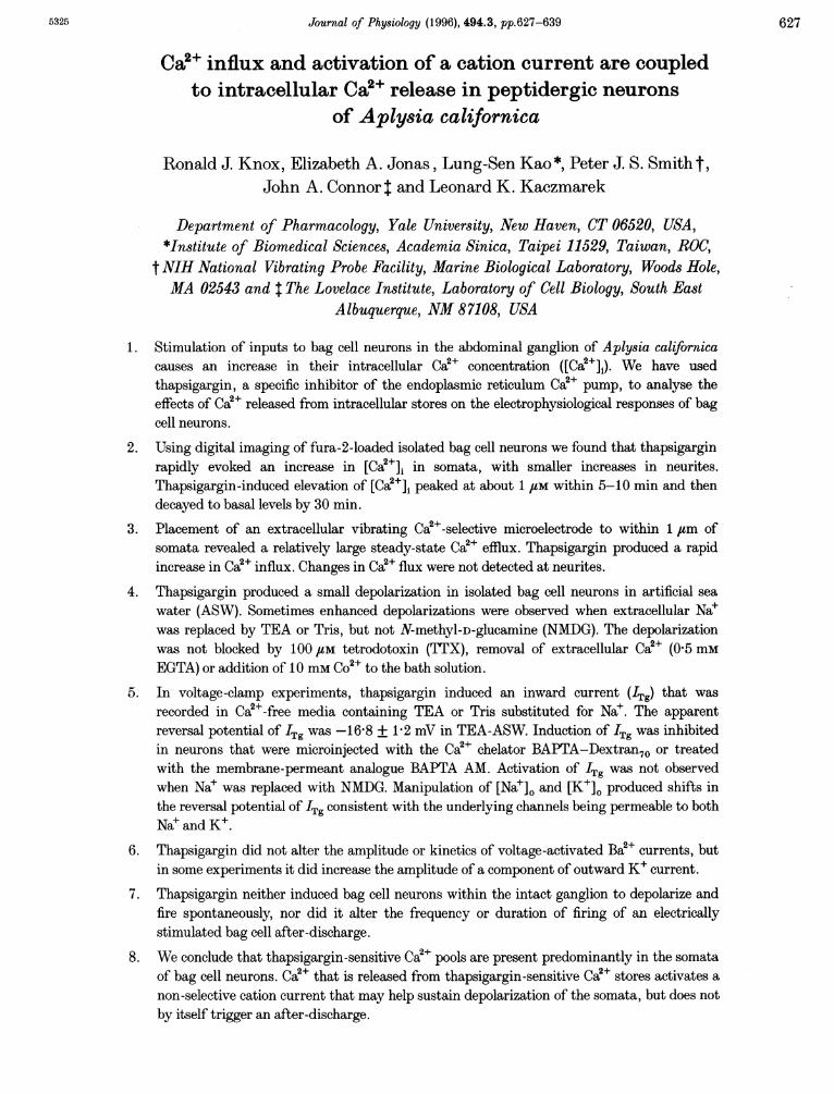
indicating that Ca21 is released from internal stores andnot from Ca2+ entry via plasma membrane pathways(Fig. 1A a). The onset of Ca2P release began within a minuteand reached maximum by about 3-5 min after the additionof thapsigargin. Thereafter the [Ca2+]1 declined towardscontrol levels within 30 min (Fig. 1 C).
The effect of thapsigargin on Ca2+ concentration thatoccurr-ed in neurites was generally smaller than it was insomnata. Measurement of intracellular Ca2± concentrations at
different locations revealed a standing Ca2+ gradient betweensomata and neurites. An example of such a Ca2+ gradient in aresting neuron is shown in Fig. IA and B, where [Ca2+]i inthe distal neurite was 32% of the corresponding [Ca2+]imeasured in the somata. When thapsigargin was added itproduced a smaller relative change in the [Ca2+]i in theneurite. In Ca2+-free ASW the mean control LCa2+]1 ofsomata was 315 + 10 nm (n = 20, mean + S.E.M.) and themean maximal [Ca2+]i after thapsigargin treatment was950 + 110 nM (n = 20). The corresponding values for
A
B 500
C
+cs
CU4U
50 100 15CDistance from tip (jim)
Thap
Neurite
0 10Time (min)
[Ca ](nM)
D12001100
1000
-S 900
- 800(U 700
600500Ann
20 30
TEA-containing ASW
-0- Soma
Thap
II
-10 0 10 20 30 40 50 60 70 80 90Time (min)
Figure 1. Fura-2 imaging of [Ca2+]i in somata and neurites of bag cell neurons duringthapsigargin stimulationA, sequence of ratio images of a neuron in normal AS"' (b), in Ca2+-free media before (c) and 10 min afterthe ad(lition of 4 ItM thapsigargin (d). A a shows a raw- fluorescence image of the cell evoked by 380 nmexcitation. Quantitative [Ca2+]i measurements were obtained from a 10 x 10 pixel box. The [Ca2+]i valuesdetermined at multiple cell locations between the two asterisks in A b are plotted in B. C and D show timecourses of the change in [Ca2]i in response to addition of thapsigargin in 2 neurons. The data shown in Cwere collected in normal ASW and the data in D were collected in Na+-free TEA-ASW.
I I I
50 275 500 700
C Na+-containing ASW800
700
-600
.S 500
400U
300
200
100_-10
J Physiol. 494.3630

Effect of intracellular Ca2" release on excitability
neurites were 175 + 27 nM before, and 305 + 52 nM afterthapsigargin treatment, respectively.
In experiments carried out in Ca2+-containing ASW,thapsigargin neither altered the time course nor the patternof action potential-driven Ca2P influx in somata or neurites(data not shown). Comparison with untreated cells showedthat thapsigargin did not alter the rate at which Ca2+ wasbuffered by reuptake and/or extruded from neuronsfollowing the large Ca2+ load that occurs during a train ofaction potentials (data not shown). These results suggestthat thapsigargin-sensitive Ca2+ stores probably contributelittle to the removal of Ca2+ that accumulates in thecytoplasm during action potentials.
Replacement of extracellular Nae with TEA changed thepattern of the thapsigargin response. Under these conditions,the increase in [Ca2+], did not decline to control values as itdid in Na+-containing ASW in the continuous presence ofthapsigargin, but continued to rise over 20-40 min (n = 6).However, as shown in Fig. 1D, when cells were returned toNae-containing ASW, [Ca2+]i began to fall with a similartime course to that observed in Nae-containing ASW. Thisobservation suggests that Ca2+ is extruded from thapsigargin-stimulated neurons by a mechanism that is dependent onextracellular Nae, possibly by a Na+-Ca2+ exchanger.
Collectively the fura-2 measurements directly demonstratethat release of Ca2+ from internal stores occurs in somataand neurites of thapsigargin-stimulated neurons and thatthe Ca2+ that accumulates in the cytoplasm is largelyremoved by a mechanism that is dependent uponextracellular Na+.
Ca2+ flux measurementsFigure 2A shows an example of a vibrating Ca2+-selectivemicroelectrode recording from an unstimulated neuron. Thesketch shows the two recording positions P1 and P2 close tothe cell body and a third position, P3, more distal from thecell. The recording system was configured such thatnegative flux values correspond to net Ca2+ efflux. Whenthe microelectrode was positioned more than 40 ,m awayfrom the plasma membrane a Ca2+ flux signal could not bedetected. Recording between P3 and P4, therefore,corresponds to the background (BG) signals as shown inFig. 2A and B. When microelectrodes were moved to theirrecording position (vibrating between P1 and P2), asindicated by the large arrows in Fig. 2A and B, steady-stateCa2+ efflux was recorded in all eight unstimulated neuronsexamined.
Typically, bath applications of thapsigargin to a finalconcentration of 7 uM caused a rapid reduction in thesteady-state measured efflux (7 of 8 cells). After 1 min nofurther reduction was observed as illustrated in Fig. 2B.After approximately 20 min, Ca2+ efflux steadily increasedto a value close to the pre-thapsigargin level.
There are two possible explanations for the reduction of theefflux value. As the probe technique generates a non-invasivedifferential measurement of Ca2+ activity at the tip of theelectrode, it only provides a net measure of Ca2+ flux betweenthese points. As, in these experiments, it is in proximity tothe plasma membrane, we can assume it measures net Ca2+movement across the surface in its immediate vicinity.Therefore, the suppression of net Ca2+ efflux reported here
BBG-_ _- - - -- --9 - -
0-5 r
_ -u-5nCY
-1*5
E0.
x -2-5
CYo -3.5
5 15 25 35Time (min)
45 55 65
r
-5
BG ___ BGBG4
| 7FM thapsigargin
A 44 a
4
AA1 A
5 15 25 35
Time (min)
Figure 2. Measurement of membrane CaO flux with a vibrating microelectrodeA, Ca2P flux recorded from a control, unstimulated cell and a sketch of the recording configuration. PI andP2 represent the two positions that the electrode vibrates between when recording membrane Ca2+ flux.The large arrows in both A and B indicate when the microelectrode was moved from background (BG)position to recording position. The smaller arrows indicate when the microelectrode was moved back fromthe recording position to the background position. BG signal was determined approximately 40 ,um fromthe plasma membrane as the electrode vibrated between P3 and P4. B, addition of 7 uM thapsigarginproduced a transient (- 1 min) enhancement of Ca2+ efflux followed by a sustained decline in efflux(-25 min). Subsequently, after a short overshoot, Ca2+ flux returned close to its initial value. Similar resultswere obtained from 6 other neurons.
HA
(0
E
E
CM0
0*5
-0-5
-1-5
-2-5
-3-5
-4-5-5
A
a
45 55 65
J Physiol.494.3 631
t%-c L

R. J Knox and others
could result from changes in either the activity of themembrane transport proteins, the Ca2+-ATPase and/orNa+-Ca2+ exchanger, or a general enhancement of Ca2+entry (Ca2+ influx channels) reducing the measured signalby simple subtraction. As thapsigargin is shown in thisstudy to greatly increase free cytosolic Ca2+, and as there isno evidence for a direct action of this compound on plasmamembrane transport proteins, the simplest explanation forthe observed response is a net increase in Ca2+ influx (seeDiscussion). The time course of this response closelymatches the thapsigargin-induced rise in cytosolic free Ca2P.The second phase of the response, the steady increase inefflux back to the pretreatment levels corresponding withthe return of the cytosolic free-Ca2+ level to control levels,may reflect the activities of either the plasma membraneCa2+-ATPase or the Nae-Ca2+ exchanger.
Thapsigargin depolarizes bag cell neuronsFigure 3A shows the effects of thapsigargin (1 uM) on themembrane potential of five isolated bag cell neurons indifferent ion-substituted media. In both normal andCa2P-free ASW (Fig. 3A a), thapsigargin caused a slowdepolarization. For each experiment described below (unlessotherwise stated) the extracellular solution was Ca2+ free.When Na+ was replaced with TEA or Tris (Fig. 3A b andA c), the amplitude of the depolarization was sometimeslarger (8-6 + 2-7 mV (n = 12) and 7-7 + 1 3 mV (n = 3),respectively) than it was in the presence of Nae(4 7 + 082 mV, n = 8). One explanation for this enhanceddepolarization is that removal of extracellular Nae inhibitsCa2+ efflux which, in turn, allows a higher local [Ca2+], toaccumulate at the site(s) where Ca2+ triggers thedepolarization. The depolarization in TEA-ASW is also
A 1 /uM thapsigargin
a ASW - - - - - .
b TEA-Cl - - - - - - - - - -
0 mvL
I min
d NMDG --
e TEA-Cl -_100 uM TTX
B5 uM thapsigargin
15 ; -5 m
2Figure 3. Thapsigargin-induced depolarization of bag cell neuronsA, records of membrane potential from 5 neurons under different pharmacological conditions. For each cell1 M thapsigargin was bath applied, as indicated by the bar. In e, 100 juM TTX was added to the bath40 min before the addition of thapsigargin. The resting potential of each of these neurons was between-52 and -67 mV. B, example of the thapsigargin-induced depolarization and the associated decrease inmembrane input resistance measured as the response to hyperpolarizing current pulses. The restingmembrane potential of the neuron was -54 mV.
_
c Tris-Cl - - - -_1I
632 J Physiol.494.3
§ ....

Effect of intracellular Ca
likely to be larger because of the block of Ca2+-activated K+channels by TEA. As shown in Fig. 3A d, when Na+ wasreplaced by N-methyl-D-glucamine (NMDG), addition ofthapsigargin caused a 3 mV hyperpolarization, presumablyby activating a Ca2+-dependent K+ conductance. In responseto thapsigargin application the mean change in membranepotential of neurons in NMDG was -2'4 + 0'23 mV (n = 4).When external Na+ was replaced with TEA-Br, thapsigargincaused depolarizations that were similar to those observedwith the Cl- salt, indicating that the depolarization did notdepend upon external Cl-.
Pretreatment of neurons with 100 UM TTX for at least40 min (Fig. 3A e) or the addition of the Ca2+ channelblocker CoC12 (10 mM) did not prevent the thapsigargin-induced depolarization, ruling out a mechanism involvingthe voltage-gated Na+ and Ca2+ channels in these neurons.In some experiments performed in Na+-containing mediathe depolarizations reversed when neurons were perfusedwith thapsigargin-free ASW. Thapsigargin also produced anincrease in membrane conductance measured by theamplitude of membrane potential responses to intra-cellularly injected hyperpolarizing current pulses. In theexample shown in Fig. 3B, a 9 mV depolarization wasassociated with a 42% decrease in input resistance of thecell 5 min after the addition of thapsigargin.
2+ release on excitability 633
Activation of a non-selective cation current (ITg)The nature of the ionic current(s) underlying thethapsigargin-induced depolarization was investigated usinga single-microelectrode voltage clamp. Figure 4A showssteady-state currents in Ca+-free ASW, in which NaCl wasreplaced by TEA-C1, flowing in an isolated neuron that washyperpolarized to -90 mV from a holding potential of-40 mV and then depolarized to +40 mV in 10 mV stepincrements before and 5 min after application of 2 FMthapsigargin. The third family of traces represents thedifference currents measured 5 min after the addition ofthapsigargin. The I-V relations of the measured currents(Fig. 4B a) and of the induced current ITg (Fig. 4Bb) showthe characteristics of a steady-state current that is relativelylinear over the voltage range -90 to +20 mV. An appreciableamount of ITg could be detected within 2 min and it usuallyreached a maximum amplitude (0'2-0A4 nA at -90 mV)within 5-10 min. The onset and development of ITgparalleled the general time course of the depolarizations aswell as the time course of the thapsigargin-stimulated risein [Ca2+]1 and changes in transmembrane Ca2+ flux. ITg wasnever observed in control experiments (bath application of30 ul of vehicle).The mean estimated reversal potential of ITg in Na+-containing ASW was -19-2 + 0-94 mV (n = 8), and in
BControl
5 min after 2 /lM thapsigargin
Difference current
---........fl...--I-_
1 nAL300 ms
"'.w 'Y-"---' - ' -40 mV-,.- -
a
Total current (nA)1-0 I
0-5
Test potential (mV)
-100 -80
In
b
-20
-0-5
-1-0-
duced current, ITg (nA)0.4
02
Test potential (mV)
-100 -80 A
A A -0.2
-0-4
Figure 4. Activation of an inward current by thapsigarginA, records of membrane currents at different voltages before and after the addition of thapsigargin, from a
neuron bathed in TEA-ASW. Difference currents represent thapsigargin-induced currents, and were
obtained by subtracting control currents from currents recorded 5 min after 2 /SM thapsigargin. Ba and bshows the corresponding I- Vrelations: 0, control and *, 5 min after 2 FM thapsigargin (Ba); A subtractedinduced current (Bb). The estimated reversal potential of the induced current in this example was -17-3 mV.
J Physiol. 494.3
A
20 40
20 40

R. J Knox and others
TEA- and Tris-ASW the estimated reversal potentialswere -16-8 + 1P2 mV (n = 8) and -18-3 + 1P4 mV (n = 4),respectively. ITg was not observed when Na+ was replacedby NMDG, which is consistent with the absence of athapsigargin-induced depolarization in NMDG substitutedmedia.
To test if ITg was activated directly in response to therelease of Ca2P from internal stores, BAPTA-Dextran70, acytoplasmic Ca2+ chelator, was microinjected into neuronsprior to application of thapsigargin. The size of the highmolecular weight conjugate (MW, 70000) excludes it fromintracellular membrane compartments. Figure 5B shows aNomarski image (a) and fluorescence image (b) of the samefield containing two neurons. The cell on the left wasinjected with BAPTA-Dextran70 plus a small amount of0-02% Lucifer Yellow, which was used to gauge successful
injections. The injected cells were allowed to equilibrate forat least 10 min before starting the voltage-clampexperiments. Figure 5A shows currents before and 10 minafter the addition of thapsigargin in a neuron injected withBAPTA-Dextran70. In contrast to the currents shown inFig. 4A, thapsigargin failed to induce ITg in the BAPTA-injected cells. Similar results were obtained in five otherneurons. In control experiments, microinjection ofBAPTA-Dextran70and Lucifer Yellow failed to induce ITg.These results, and those obtained with the membrane-permeant analogue BAPTA AM (acetoxymethyl ester formof BAPTA) (n = 4), strongly suggest that ITg is activateddirectly by Ca2P discharged from intracellular stores.
Collectively, the pharmacology of the depolarization and theITgand the fact that ITg reversed at approximately-20 mVsuggest that it is a voltage-independent non-selective cation
B
A
Control injected with BAPTA-Dextran70
I.
&1-- '' - ---'--
09 nA
300 ms
40 mV
Figure 5. Effect of BAPTA-Dextran70 on ITgA, microinjection of the Ca2' chelator, BAPTA-Dextran70, inhibited the activation of ITg. Lucifer Yellowwas included in the injection so that successful injections could be determined by observing intracellularfluorescence. B, Nomarski (a) and fluorescence images (b) of a field containing 2 cells. The cell on the leftwas microinjected with BAPTA-Dextran70 and a small amount of Lucifer Yellow. Scale bar, 45 #tsm.
J Physiol.494.3634

Effect of intracellular Ca2" release on excitability
current. We estimated the relative selectivity of ITg fromreversal potential measurements made in ion-substitutedASWs. Figure 6 shows I-V relations for ITg recorded inASWs containing different [Na+]. and [K+]e. The meanreversal potential of ITg in normal ASW was-19-2 + 0 94 mV (n= 8). Data from a single experimentare shown by filled circles in Fig. 6. When the driving forcefor K+ was decreased by increasing the extracellular [K+]from 104 to 50 mm, the reversal potential of ITg wasshifted to more positive potentials (19-7 + 1P3 mV, n = 8)(Fig. 6). When the [Na+]o was lowered to 46 mm, there wasa left shift in the I-V relation of ITg, as shown by thesquares in Fig. 6. The mean reversal potential of ITg underthese conditions was -29-3 + 1P8 mV (n= 8). These resultsdemonstrate that both Na+ and K+ contribute to ITg. Overallthe results suggest that Ca2+ released from internal storesdirectly stimulates a depolarizing non-selective cationconductance in somata of bag cell neurons. This may beimportant for maintaining depolarization of bag cell clustersin the abdominal ganglion. For example, activation of ITgcould facilitate the propagation of action potentialsthroughout the abdominal ganglion network during anafter-discharge (Kaczmarek et al. 1978).
We also tested for effects of thapsigargin on voltage-activated Ca2+ currents (Ba2+ was used as charge carrier toincrease the size of the inward current) and a K+ current. Inmost cells no apparent effect could be detected on inwardcurrents (6 of 8 cells). The small effect on inactivation ofBa+ current shown in Fig. 7A was, however, observed intwo of eight neurons, and may reflect Ca2+-inducedinactivation of Ca2+ current. The I- V plot for one of these
0-4
0-2
Test potential (mV)
experiments is shown in Fig. 7A (right). Thapsigargin alsoproduced only minor effects on K+ currents. Figure 7Bshows an experiment in which ITg was induced in Ca2+-free(0 5 mm EGTA) ASW in which Tris replaced Na+, andshows that thapsigargin produced a small increase in acomponent of K+ current, presumably due to intracellularCa2+ release acting on Ca2+-dependent K+ currents.
Lack of effect of thapsigargin on the after-dischargeIn the intact abdominal ganglion of Aplysia, briefstimulation of the pleuroabdominal connective nerveproduces a depolarization of the neurites of bag cell neurons(Kaczmarek et al. 1978). As a result, an after-dischargelasting approximately 30 min is generated within theneurites, and during this after-discharge action potentialspropagate towards the somata. To test if Ca2+ release fromintracellular stores induced by thapsigargin can influencethe characteristics of a normal after-discharge we stimulateddischarges in isolated abdominal ganglia in the presence orabsence of thapsigargin. Figure 8A shows the typical firingpattern of a bag cell after-discharge in response to a briefelectrical stimulus applied to the pleuroabdominalconnective nerve. Action potentials were recorded using anextracellular suction electrode (Loechner et al. 1992).Thapsigargin had no effect on baseline electrical activitybefore application of the electrical stimulus. Figure 8Bshows an electrically evoked after-discharge recorded fromthe ganglion 50 min after the addition of thapsigargin(50,UM), and as shown by comparison with theunstimulated preparation in Fig. 8A, thapsigargin did notalter the firing frequency or duration of the electricallyevoked after-discharge in any obvious way. The mean
luced current, ITg (nA)
, ITg-low Na+
Normal ASW
ITg-high K+
40
Figure 6. Reversal-potential measurements demonstrate ITg is a non-selective cation currentMean I- V relations for ITg obtained in isosmotic substituted ASW in which the driving forces for K+ (U)and Na+ (A) were respectively increased and decreased. The shifts in the reversal potentials observed underthese conditions relative to the reversal potentials measured in normal ASW (A) demonstrate that both Na+and K+ contribute to ITg.
635J Phy8iol.494.3

R. J Knox and others
Peak current (nA)
100 ms
1 nA
0 mV
L-60 mVJ
Test potential (mV)
-40
-3
40
8 min after 1 u/M thapsigargin
) ms
I1 nA
+30 mV
-60 mV , ,-60 mV
Figure 7. Effects of thapsigargin on voltage-activated Ca*' and K' currentsA, voltage-gated Ba2+ currents before and 10 min after treatment with thapsigargin and an I-V plot forthis type of experiment; A, control and *, 10 min after 1 /iM thapsigargin. Ba2+ was used as the chargecarrier to increase the size of the inward current. B, voltage-dependent outward K+ currents recorded inCa2+-free ASW before and after 1 /LM thapsigargin.
A
Pretreated with 50 /M thapsigargin
Electrical stimulus applied topleuroabdominal connective nerve
Figure 8. Lack of effect of thapsigargin on after-dischargeA, control bag cell after-discharge that lasted for 22 min. B, bath-applied thapsigargin (50 /M) did not alterin any way an after-discharge that was electrically stimulated 40 min after application of thapsigargin.Control after-discharges and those stimulated in the presence of thapsigargin had mean durations of35-2 + 1-8 min (n = 4) and 36-8 + 3-1 min (n = 4), respectively.
A
ControlB
+30 r
B
IM iniinitrlrrlrMl lm" ,I'f H .
636 J. Physiol. 494.3
1
ee.'-
VIIIUII I11I$II IIlILWIIILIIIII II lllrllllll1 11 lIlllNlliIIl1HMlI'lrll I

Effect of intracellular Ca2' release on excitability
durations of after-discharges in control and thapsigargin(50 /M)-treated preparations were 35-2 + 1P8 min and36 8 + 3-1 min, respectively (n = 4 for each group). Similarresults were obtained using 5/uM thapsigargin (n = 4).
DISCUSSIONDigital imaging of intracellular fura-2 fluorescence and theuse of Ca2P-selective vibrating microelectrodes on bag cellneurons has allowed us to measure spatial and temporalproperties of intracellular Ca2+ release, and its associatedplasma membrane Ca2+ flux. We have found thatthapsigargin-sensitive Ca+ stores are present in the bag cellneurons and that release from these Ca2+ stores activates adepolarizing non-selective cation conductance. We alsodemonstrated that release of intracellular Ca2+ stimulatesCa2+ influx across the plasma membrane, which is followedby extrusion of Ca2+ across the plasma membrane.
Initiation of the after-discharge occurs in bag cell neuritesand intracellular Ca2+ release has been shown to occurduring the after-discharge (Fisher et al. 1994). Our resultswith thapsigargin in bag cell neurons in intact abdominalganglia suggest that Ca2+ release from thapsigargin-sensitive sites is not likely to be responsible for triggeringnormal after-discharges. It is possible, however, that thedepolarization induced by Ca2+ release contributes to thenormal maintenance of the after-discharge and to the abilityof action potentials to propagate from the neurites, wherethe discharge is initiated, towards the somata. Maintaineddepolarization of somata may facilitate action potentialpropagation from bag cell neurons to target neuronsthroughout the Aplysia nervous system.
Effect of intracellular Ca2+ release on plasmamembrane Ca2+ fluxOur observations suggest that a significant component ofintracellular Ca2+ that accumulates during thapsigargintreatment is exported from the cells by a mechanismdependent on extracellular Na+, which differs from arecently described Nae-independent regulation of intra-cellular Ca2+ in voltage-clamped snail neurones (Kennedy &Thomas, 1995). Use of the Ca2P-selective vibrating-probetechnique allowed us to compare steady-state plasmamembrane Ca2P gradients in control and thapsigargin-stimulated neurons. The recordings revealed that intra-cellular Ca2+ release was followed by a sustained reductionof Ca2P efflux, the time course of which correlates with theincrease in cytoplasmic Ca2P levels observed in fura-2-loaded neurons. A likely explanation for the Ca2+ influx isthat thapsigargin induces Ca2P entry across the plasmamembrane. This Ca2P influx may be related to Ca2+depletion-activated currents, such as ICRAC (Ca2+ release-activated current) or IDAC(depletion-activated current), thatmay underlie capacitative Ca2+ entry in a variety ofinexcitable cell types (Putney, 1990; Luckhoff & Clapham,
1994). Since thapsigargin is the most reliable stimulant ofCa2P depletion-activated Ca2P entry, our data suggest thatan analogous Ca2+-repletion mechanism may be present inbag cell neurons. To date, a second messenger that regulatescapacitive Ca2P entry has not been identified, althoughRandriamampita & Tsien (1993) have partially characterizeda Ca2P influx factor substance (CIF) that is generated intra-cellularly when intracellular Ca2+ stores are depleted bythapsigargin.
Neurites versus somata: spatial and temporalproperties of thapsigargin-sensitive Ca2+ releaseFollowing treatment with thapsigargin, the elevation ofCa2+ reached in neurites of bag cell neurons is less than thatin somata. There may be several reasons for this. Forexample, the amount of thapsigargin-sensitive Ca2+ storesmay be less in neurites. Alternatively, because neurites havea smaller volume than somata, Ca2+ pumps or Na+-Ca2+exchangers (Levy & Tillotson, 1988) may extrude Ca2+ fromneurites at a rate that exceeds Ca2+-extrusion efficiency insomata, or there may be a higher density of Ca2+-sequestering proteins in neurites relative to somata. It isalso likely that the mechanisms that regulate Ca2+ levels inbag cell neurons following action potential-driven Ca2+influx differ from those in rat neocortical neurons, sinceCa2P transients in these neurons are prolonged by blockersof endoplasmic reticulum Ca2+-ATPase, thapsigargin andcyclopiazonic acid (Markram, Helm & Sakmann, 1995),whereas these drugs do not alter the decay time constant ofaction potential-induced transients in bag cell neurons(R. J. Knox, E. A. Jonas, L. K. Kaczmarek & J. A. Connor,unpublished observations).
Possible physiological functions of cation currentOur results have demonstrated that intracellular Ca2+release by thapsigargin produces a depolarization of bag cellneurons, probably because of the activation of a voltage-independent non-selective cation current ITg. This currentcould be recorded in extracellular solutions containing Na+,K+, TEA and Tris, but not in NMDG-substituted ASW orin neurons injected with the intracellular Ca2+ chelator,BAPTA-Dextran70.
Non-selective cation currents were first described in heart(Kass, Lederer, Tsien & Weingart, 1978), where they havebeen implicated in pacemaker activity, and may also beregulated by Ca2+ release from intracellular stores(Colquhoun, Neher, Reuter & Stevens, 1981; Reuter, 1984).Similar currents are thought to account for the burstingbehaviour of Helix neurons, where they provide amaintained depolarizing current that is critical topacemaking activity (Partridge & Swandulla, 1987). Otherfunctions have also been proposed for Ca2+-activated non-selective cation currents (Bevan, Gray & Ritchie, 1984; Lee,Dayanithi, Nordmann & Lemos, 1992). In some non-excitable cells, non-selective cation channels that are Ca2+
J. Physiol.494.3 637

638 R. J Knox anid others J Physiol.494.3
permeable are thought to provide one type of mechanismfor capacitativTe Ca2P entry following depletion of intra-cellular stores. For example, in mast cells, Ca2+ influx canoccur by two independent mechanisms: a Ca2+-selectivecurrent (ICRAC) that is activated by depletion of internalCa2+ stores (Hoth & Penner, 1993), and by non-selectivecation channels that are permeable to other divalent cations(Penner, Matthews & Neher, 1988). The latter channels alsocontribute to the plateau phase of elevated [Ca2+]1 followingreceptor-operated release of Ca2+ from internal stores.Following stimulation of the bag cell neurons, there is anelevation of intracellular calcium that can be attributed torelease from intracellular stores (Fisher et al. 1994). Thecation current, ITg, that is thereby activated may contributeto the depolarization that is maintained during the after-discharge, and may conceivably also contribute to therefilling of calcium stores.
BEVAN, S., GRAY, P. T. & RITCHIE, J. Al. (1984). A calcium-activatedcation-selective channel in rat cultured Schwann cells. Proceedingsof the Royal Society of London B 222, 349-355.
BLAUSTEIN, M. P. (1988). Calcium and synaptic function. In Handbookof Experimental Pharmacology, vol. 83, ed. BAKER, P. F.,pp. 275-304. Springer-Verlag, Berlin.
CARAFOLI, E. (1987). Intracellular calcium homeostasis. An nualReview of Biochemistry 56, 395-433.
COLQUHOUN, D., NEHER, E., REUTER, H. & STEVENS, C. F. (1981).Inward current channels activated by intracellular Ca in culturedcardiac cells. Nature 24, 752-754.
CONN, P. J. & KACZMAREK, L. K. (1989). A model for the study of themolecular mechanisms involved in the control of prolonged animalbehaviors. Molecular Neurobiology 3, 237-273.
CONNOR, J. A. (1986). Digital imaging of free calcium changes and ofspatial gradients in growing processes in single, mammalian centralnervous system cells. Proceedings of the National Academy ofSciences of the UTSA 83, 6179-6183.
FINK, L. A., CONNOR, J. A. & KACZMAREK, L. K. (1988). Inositoltrisphosphate releases intracellularly stored calcium and modulatesion channels in molluscan neurons. Journal of Neuroscience 8,2544-2555.
FISHER, T. E., LEVY, S. & KACZMAREK, L. K. (1994). Transientchanges in intracellular calcium associated with a prolongedincrease in excitability in neurons of Aplysia californica. Journal ofNeurophysiology 71, 1254-1257.
FRIEL, D. D. & TSIEN, R. W. (1992). A caffeine- and ryanodine-sensitive Ca2P store in bullfrog sympathetic neurones modulateseffects of Ca2+ entry on [Ca2+]i. Journal of Physiology 450,217-246.
GRYNKIEWICZ, G., POENIE, M. & TSIEN, R. Y. (1985). A newgeneration of calcium indicators with greatly improved fluorescentproperties. Journial of Biological Chemistry 260, 3440-3448.
HESS, P. (1990). Calciunm channels in vertebrate cells. Annnual Reviewof Neuroscience 13, 1337-1356.
HIG,ASHIDA, H. & BROWN, D. A. (1986). Membrane current responsesto intracellular injections of inositol 1,3,4,5-tetrakisphosphate andinositol 1,3,4-trisphosphate in NC 108-15 hybricl cells. FEBS Letters208, 283-286.
HOTH, Al. & PENNER, R. (1993). Calcium release-activated calciumcurrent in rat mast cells. Journal of Physiology 465, 359-386.
JAHN, R. & SUDHOF, T. C. (1993). PIoteins of synaptic vesiclesinvolved in exocytosis and membrane recycling. Neuron 6,665-677.
KA(CZMAREK, L. K., JENNINGS, K. & STRUMWASSER, F. (1978).Neurotransinitter modulation, phosphodiester inhibitor effects, andcyclic AMIP correlates of after-discharge in peptidergic neurites.Proceedings of the National Academy of Sciences of the USA 75,5200-5204.
KACZMAREK, L. K. & LEVITAN, I. (1987). Neurornodulation, theBiochemical Conitrol of Neuronal Excitability, ed. KACZMAREK,L. K. & LEVITAN, I. B. Oxford University Press, Oxford.
KASS, R. S., LEDERER, WV. J., TsIEN, R. W. & WEINGART, R. (1978).Role of calcium ions in transient inward currents and after-contractions induced by strophanthidin in cardiac Purkinje fibres.Journal of Physiology 281, 187-208.
KENNEDY, H. J. & THOMAS, R. C. (1995). Intracellular calcium and itssodium-independent regulation in voltage-clamped snail neurones.Journ2al of Physiology 484, 533-548.
KNOX, R. J., QUATTROCKI, E. A., CONNOR, J. A. & KACZMAREK, L. K.(1992). Recruitment of calcium channels by protein kinase C duringrapid formation of putative neuropeptide release sites in isolatedAplysia neurons. Neuron- 8, 883-889.
LEE, C. J., DAYANITHI, G., NORDMANN, J. J. & LEMOS, J. R. (1992).Possible role during exocytosis of a Ca2+-activated channel inneurohypophysial granules. Neuron 8, 335-342.
LEVY, S. & TILLOTSON, D. (1988). Effects of Nla~and Ca2 gradients onintracellular free Ca2P in voltage-clamped Aplysia neurones. BrainResearch 474, 332-342.
LIPSCOMBE, D., MADISON, D. V, POENIE, AM., REUTER, H. & TsIEN,R. W. (1988). Spatial distribution of calcium channels and cytosoliccalcium transients in growth cones and cell bodies of sympatheticneurons. Proceedings of the National Academy of Sciences of theUISA 85, 2398-2402.
LOECHNER, K. J., KNOX, R. J., CONNOR, J. A. & KACZMAREK, L. K.(1992). Hyperosmotic media inhibit peptide secretion via inhibitionof voltage-dependent calcium channels. Journal of MlembraneBiology, 128, 41-52.
LUCKHOFF, A. & CLAPHAM, D. E. (1994). Calcium channels activatedby depletion of internal calcium stores in A431 cells. BiophysicalJournal 67, 177-182.
MARKRAM, H., HELMI, P. J. & SAKMANN, B. (1995). Dendritic calciumtransients evoked by single back-propagating action potentials inrat neocortical pyramidal neurons. Journal of Physiology 485,1-20.
NEERING, I. R. & MCBURNEY, R. N. (1984). Role for microsomal Castorage in mammalian neurons? NTature 309, 158-160.
PARTRIDGE, D. L. & SWANDULLA, D. (1987). Single Ca-activatedcation channels in bursting neurons of Helix. Pfluigers Archiv 410,627-631.
PAYNE, R. & FEIN, A. (1987). Inositol I ,4,5-trisphosphate releasescalcium from specialized sites within Limulus photoreceptors.Journal of Cell Biology 104, 933-937.
PENNER, R., AIATTHEWS, G. & NEHER, E. (1988). Regulation ofcalcium influx by second messengers in rat mast cells. Nature 334,499-504.
PUTNEY, J. WV. (1990). Capacitative calcium entry revisited. CellCalcium 11, 611-624.
RANDRIAMAMIPITA, C. & TSIEN, R. Y. (1993). Emptying of intra-cellular Ca2+ stores r-eleases a novel smnall messenger that stimulatesCa2+ influx. NVature 364, 809-814.

Effect of intracellular Ca2+ release on excitability
REUTER, H. (1984). Ion channels in cardiac cell membranes. AtnnualReview of Physiology 46, 473-484.
SAH, P. & McLACHLAN, E. M. (1991). Ca2-activated K+ currentsunderlying the afterhyperpolarization in guinea pig vagal neurons:a role for Ca2P-activated Ca2P release. Neuron 7, 257-264.
SMITH, P. R., SANGER, R. H. & JAFFE, L. F. (1994). The vibratingcalcium electrode: a new technique for detecting plasma membraneregions of Ca2P influx and efflux. In A Practical Guide to the Studyof Calcium in Living Cells. Mlethods in Cell Biology, vol. 40, ed.NUCCITELLI, R., pp. 115-134. Academic Press, Inc., San Diego.
THASTRUP, O., CULLEN, P. J., DROBAK, B. K., HANLEY, MT. R. &DAwSON, A. P. (1990). Thapsigargin, a tumor promoter, dischargesintracellular Ca2P stores by specific inhibition of the endoplasmicreticulum Ca2+-ATPase. Proceedings of the National Academy ofSciences of the USA 87, 2466-2470.
THAYER, S. A., PERNEY, T. M. & MILLER, R. J. (1988). Regulation ofcalcium homeostasis in sensory neurons by bradykinin. Journal ofNeuroscience 8, 4089-4097.
TsIEN, R. W. & TsIEN, R. Y. (1990). Calcium channels, stores, andoscillations. Annlual review of Cell Biology 6, 715-760.
ZHENG, J. Q., FELDER, M., CONNOR, J. A. & Poo, M. M. (1994).Turning of nerve growth cones induced by neurotransmittersNature 368, 140-144.
AcknowledgementsThis work was supported in part by NIH grants to L.K.K.(NS-18492) and P.J.S.S. We thank Drs Benjamin White, MathewWhim and Peter Bell for helpful discussion of the work.
Author's email addressR. J. Knox: [email protected]
Received 2 January 1996; accepted 1 April 1996.
J. Physiol.494.3 639