thero/ssa autoantigen as an immunogen - the journal of
TRANSCRIPT

THE Ro/SSA AUTOANTIGEN AS AN IMMUNOGENSome Anti-Ro/SSA Antibody Binds IgG
BY MARK J. MAMULA, OWEN F. FOX, HAJIME YAMAGATA, ANDJOHN B. HARLEY
From the Departments ofMedicine, Microbiology, and Immunology,University ofOklahoma Health Sciences Center; Veterans Administration Medical Center ;and The Arthritis and Immunology Program, Oklahoma Medical Research Foundation,
Oklahoma City, Oklahoma 73104
Patients with connective tissue diseases produce autoantibodies to diversecellular components including nucleic acids, mitochondria, cell-surface antigens,and ribonucleoproteins . A small RNA-protein complex known as Ro/SSA bindsautoantibody from many patients with SLE and Sj6grens syndrome (1-4). Anti-Ro/SSA autoantibodies have been closely associated with the appearance ofnephritis, vasculitis, lymphadenopathy, and leukopenia in SLE patients (5, 6) .Furthermore, maternally acquired anti-Ro/SSA has been implicated in the im-munopathogenesis of congenital complete heart block and with the dermatitis ofneonatal lupus (7-10) .Ro/SSA was originally characterized as an antigen to which precipitating
autoantibodies were detectable in gel double diffusion (11, 12) and has onlyrecently been the subject of more definitive biochemical evaluation (13, 14). TheRo/SSA particle is composed of an acidic protein of 60 kD that in human cellsis associated with four distinct uridine-rich RNAs ranging from 80 to 112 bases(15). The antigenic reactivity of Ro/SSA is apparently independent of the RNAsince the precipitin activity is resistant to RNAse (11, 12, 16) and since theautoantibody binds the protein after denaturation and separation from the RNA(14) .
Clinical studies have shown that 70% of patients with Ro/SSA-precipitatingantibodies also have rheumatoid factor (17) . In primary Sj6gren's syndromepatients, the rheumatoid factor titer and the concentration of anti-Ro/SSA areclosely correlated (18) . In addition, previous data demonstrate that rheumatoidfactor and antinuclear antibody contribute to Ro/SSA precipitation by doublediffusion (19) . In other disorders, selected rheumatoid factors (some restrictedto particular idiotypes) have been shown to bind non-IgG cellular componentsincluding histones (20-22) and DNA (23, 24), as well as bacterial peptidoglycanand nitrophenyl groups (25, 26). This study compares the binding characteristicsThis study was supported by National Institutes of Health (Bethesda, MD) grants AM-34159 andAM-31133, the Veteran's Administration, the Oklahoma Chapter of the Arthritis Foundation, theOklahoma Lupus Association, and Sigma Xi-The Scientific Research Society . M. J. Mamula has beena recipient of a Gina Finzi Fellowship from the Lupus Foundation of America. J. B. Harley is anInvestigator of the Arthritis Foundation . Address correspondence to MarkJ. Mamula, Yale UniversitySchool of Medicine, 609 LCI, 333 Cedar St., New Haven, CT 06510.
Journal of Experimental Medicine - Volume 86/12/1889/13 $1 .00
1889
on January 6, 2019jem.rupress.org Downloaded from http://doi.org/10.1084/jem.164.6.1889Published Online: 1 December, 1986 | Supp Info:

1890
Ro/SSA AND IgG CROSSREACT
of'heteroimmune anti-Ro/SSA generated in a rabbit host with human autoim-mune anti-Ro/SSA . In this study, the data clearly show that both autoimmuneand heteroimmune anti-Ro/SSA not only bind the Ro/SSA protein but also reactwith IgG .
Materials and MethodsRo/SSA Antigen Preparation . Ro/SSA was purified according to a method adapted
from Yamagata et al . (14) . Briefly, bovine spleen was homogenized in an equal amount of(wt/vol) PBS (pH 7.2) supplemented with 2 mM DTT at 4°C . The cell lysate wascentrifuged at 10,000 g for 1 h . The supernatant was mixed with 40% (vol/vol) DE-52(Whatman Co., Clifton, NJ) for 4 h at 4°C . DE-52 was then washed thoroughly with PBS,pH 7.2, followed by extraction with 1 .0 M NaCl in 0.02 M phosphate buffer, pH 7 .2 .The extract was then applied to an anti-Ro/SSA affinity column . The affinity column wasmade by coupling purified IgG from an SLE patient with precipitating anti-Ro/SSA toCNBr-activated Sepharose 4B (Pharmacia Fine Chemicals, Piscataway, NJ) by conventionalmethods (27). The patient IgG fraction was negative for rheumatoid factor by the latexagglutination assay (28) but was additionally adsorbed with human IgG-Sepharose 4Bbefore the coupling reaction . Ro/SSA was eluted from the affinity column with 3 MMgCl2, pH 7 .0, and the eluate was dialyzed against 0.02 M Tris, 0.15 M NaCl, pH 7.2,and concentrated by Amicon filtration (Amicon Corp., Danvers, MA). Purified La/SSBwas similarly prepared by affinity chromatography using the anti-La/SSB IgG from anSLE patient coupled to Sepharose 4B (13) .
Enzyme-linked Immunosorbent Assay (ELISA) .
The solid-phase assay for anti-Ro/SSA oranti-La/SSB was modified from previously reported techniques (13, 14, 29) . Immulon 96-well microtiter plates (Dynatech Laboratories, Inc ., Alexandria, VA) were coated over-night at 4°C with 50 ul of antigen (human IgG, bovine IgG, or affinity-purified bovineRo/SSA or La/SSB) in carbonate buffer, pH 9.6 . Our preliminary studies demonstratedthat 5 ,ug/ml of each of these antigens was optimal for detection of antibody . Aftercoating, microtiter plates were washed with 0.05% Tween in PBS and unbound sites wereblocked with 0 .1% BSA in PBS. Sera were diluted in PBS containing 0.5% Tween and0.1% BSA and incubated on the plate for 12 h at 4 ° C . In some experiments, dilutions ofsera were preincubated with several concentrations ofbovine or human IgG (Cohn fraction11, Sigma Chemical Co., St . Louis, MO), total histones (purified by the method of Rubinet al . [301), or affinity purified bovine Ro/SSA or La/SSB for 4 h at room temperaturebefore application to the wells . Following serum incubation, the plates were washed anda saturating concentration of the IgG fraction of goat anti-human or anti-rabbit IgGconjugated to alkaline phosphatase (Sigma Chemical Co.) was applied to the plate . Theplates were allowed to incubate overnight at 4°C and again washed free of unboundconjugate . Paranitrophenylphosphate (PNPP)' was then added to the plates and chromo-phore development was measured at 405 nm with a MR 580 Microelisa Auto Reader(Dynatech Laboratories, Inc .)
Western Blot.
Western immunoblots were performed by a procedure modified fromTowbin et al . (31) . Purified Ro/SSA was denatured and reduced and then applied to7 .5% (wt/vol) polyacrylamide gels containing SDS and electrophoresed at 30 mA for 4 hat f 0°C . The proteins were either silver stained (Bio-Rad Laboratories, Richmond, CA)or electrophoretically transferred to nitrocellulose sheets in a Trans-Blot cell (Bio-RadLaboratories) at 200 mA for 4 h at 10 ° C . The nitrocellulose was blocked with 4% BSAin PBS for 4 h at room temperature . Appropriate dilutions of human au .
"mmune anti-Ro/SSA, rabbit heteroimmune anti-Ro/SSA, or IgG adsorption column eluates wereincubated with the nitrocellulose for 4 h at room temperature . The nitrocellulose waswashed with PBS for 30 min followed by a 3-h incubation with the IgG fraction of goatanti-human or anti-rabbit IgG conjugated to alkaline phosphatase . Nitrocellulose wasagain washed and then soaked in substrate solution consisting of 2.0 mM 0-naphthyl acid
'Abbreviation used in this paper:
PNPP .paranitrophenylphosphate .

MAMULA ET AL .
1891
phosphate (Sigma Chemical Co.), 1 .0 mM o-diansidine (Sigma Chemical Co.), and 4 .0 mMMgS04 in 0.07 M borate buffer (32) . The substrate reaction was then stopped withmethanol, water, and acetic acid (5:5:1) .
Preparation of Rabbit Anti-Ro/SSA .
Heteroimmune rabbit anti-Ro/SSA antisera wasmade by repeated immunization of New Zealand White rabbits with purified Ro/SSApreparations . Rabbits were initially immunized intramuscularly and subcutaneously with200,ug purified Ro/SSA in 1 .0 ml CFA . They were boosted at 2 wk with 200 kg Ro/SSAin 1 .0 m1 IFA and by ear vein at 4 wk and 200 ,ug Ro/SSA . They were bled 10 d later .Subsequent sera were collected after repeating the intravenous boosting .
Adsorption ofRabbit and Human Anti-Ro/SSA with IgG-Sepharose.
Bovine or human IgG(Cohn fraction II, Sigma Chemical Co.) was covalently bound to CNBr-activated Sepharose4B (Pharmacia Fine Chemicals) by the method of Axen et al . (27) . Columns wereequilibrated in PBS and rabbit heteroimmune anti-Ro/SSA serum was passed sequentiallythrough the bovine and human IgG columns . The columns were washed in 0.02 M Tris,0.15 M NaCl, pH 7 .4 (Tris-NaCI), until no evidence of protein was apparent in the wash(OD at 280 nm was <0.01) . The prototype human anti-Ro/SSA serum was similarlypassed through a human IgG affinity column . The columns were eluted with 3 M M9C12 ,pH 7 .2, and eluates were dialyzed against Tris-NaCl and concentrated . Both the rabbitand human sera were passed through control affinity columns consisting of ethanolamine-blocked Sepharose 4B to eliminate interference from nonspecific binding . Similarly,nonimmune rabbit and normal human serum were passed through bovine and humanIgG affinity columns as controls .
Anti-Ro/SSA-specific Antibody Purification of Immune Sera .
Affinity-purified Ro/SSAantigen was covalently attached to CNBr-activated Sepharose 4B and the heteroimmunerabbit or autoimmune human anti-Ro/SSA serum was passed through at 0.5 ml/min . Thecolumn was washed (Tris-NaCI), eluted with 3 M MgC12 , dialyzed, and concentrated asdescribed above .
Specific Activity Determination .
Rabbit and human anti-Ro/SSA and selected affinity-purified isolates were examined for specific anti-Ro/SSA or anti-human IgG activity . IgGconcentrations were determined by ELISA (33) . Goat anti-rabbit IgG or anti-humanIgG (Sigma Chemical Co.) was adsorbed to 96-well microtiter plates and unbound siteswere blocked with 0.1 % BSA in PBS . Rabbit or human IgG standard dilutions and testsera were added to the wells and detected by goat anti-rabbit IgG or anti-human IgGconjugated to alkaline phosphatase (Sigma Chemical Co.) . Upon addition of substrate (p-nitrophenylphosphate), IgG concentrations were calculated from IgG standard curves andcorrelated with anti-Ro/SSA or anti-human IgG ELISA OD values . Values are expressedas ELISA activity (OD4o5)/wg IgG .
Isolation of 7S-anti-Ro/SSA IgG .
The 7S IgG from the heteroimmune rabbit andautoimmune patient anti-Ro/SSA was isolated by sucrose density gradient centrifugationadapted from established methods (34) . 0.3 ml of serum was applied to a gradientconsisting of 1 .5 ml each of 16, 20, and 26% sucrose . Control gradients included 7S-I'2a-IgG and 11 S catalase were also applied in 0.3 ml . Gradients were centrifuged for 18 h at100,000 g. The 7S fraction was separated from the 11 S fraction and used for competitiveinhibition assays .
ResultsEvaluation of Immune Rabbit Serum for Anti-Ro/SSA Antibody.
The Ro/SSAimmune rabbit serum formed a precipitin line of identity with prototype patientanti-Ro/SSA serum when tested against purified Ro/SSA (Fig . 1) . Normal humanserum and nonimmune rabbit serum do not precipitate purified Ro/SSA. West-ern blot analysis revealed binding of the 60-kD Ro/SSA protein by both theprototype human anti-Ro/SSA and the immune rabbit serum at 10' dilutions(Fig . 2) . Control lanes of normal human and nonimmune rabbit serum fail to

1892
Ro/SSA AND IgG CROSSREACT
FIGURE 1 .
0.6% agarose double immunodiffusion gel of affinity-purified Ro/SSA againstundiluted rabbit anti-Ro/SSA (Rb) and prototype human anti-Ro/SSA (Hu).
FIGURE 2.
Western immunoblot analysis of normal human serum (NHS), normal rabbitserum (NRS), prototype human anti-Ro/SSA (Hu), and rabbit anti-Ro/SSA (Rb) . Affinity-purified Ro/SSA was electrophoresed under reducing conditions on a 7.5% SDS-polyacryl-amide gel, and was transferred to nitrocellulose strips . All samples were tested at a 10 -2dilution .
detect this protein by Western blotting, confirming the binding specificity of theimmune sera .A sensitive solid-phase immunoassay (ELISA) for the detection ofanti-Ro/SSA
antibody was used to characterize the immune rabbit serum. Titrations of therabbit serum were positive to a 10-6 dilution and subsequent assays used a 10-4dilution for analysis . At this dilution, preincubation of the serum with 10 Ag/mlpurified Ro/SSA inhibited 92% of the binding activity of the serum (Table I) .The specificity of the heteroimmune response to Ro/SSA was evaluated alongwith other autoantigens and proteins . Preincubation of the immune rabbit serumwith purified La/SSB, histones, or albumin failed to competitively inhibit bindingto the Ro/SSA protein (Table I) .
Loss of Anti-Ro/SSA Activity with IgG Adsorption .
The rabbit anti-Ro/SSAantiserum bound both human and bovine IgG in ELISA (Table 1I) . We per-

MAMULA ET AL.
TABLE I
Anti-Ro/SSA Activity ofHeteroimmune Rabbit Serum
A 10-4 dilution of rabbit anti-Ro/SSA was preincubated for 4 h at roomtemperature with selected inhibitors . Inhibition was calculated as 100 X(1 - inhibited sample OD405)/(uninhibited sample OD405) .
TABLE II
Rabbit Anti-Ro/SSA Adsorption with Human and Bovine IgG
1893
* Ligands were bound to the solid phase in ELISA for detection of antibody . Values expressed asOD at 405 nm (OD405).
$ Rabbit anti-Ro/SSA adsorbed over an ethanolamine-blocked Sepharose 4B control column .§ Rabbit anti-Ro/SSA adsorbed over bovine and human IgG bound to Sepharose 4B.Nonimmune rabbit serum.
formed adsorption ofanti-IgG activity to enrich the anti-Ro/SSA specific activityof the antisera and to prevent artifacts that might arise from anti-IgG contami-nating the anti-Ro/SSA serum. The rabbit antiserum was passed sequentiallythrough bovine and human IgG affinity columns . Adsorption reduced the anti-bovine and anti-human IgG activity by almost 1,000-fold . There was, however,a concomitant loss of nearly 99% of the anti-Ro/SSA activity with IgG adsorptionsince a 100-fold greater concentration of adsorbed serum was required to achievesimilar optical density measurements in this assay (Table II). Rabbit anti-Ro/SSAserum was also passed through an ethanolamine-blocked Sepharose 4B columnwith no loss of activity to Ro/SSA or IgG . Therefore, nonspecific binding ofanti-Ro/SSA to Sepharose 4B was not responsible for the loss of anti-Ro/SSAactivity . Nonimmune rabbit serum lacked any anti-Ro/SSA activity in this solid-phase assay and did not bind the IgG affinity columns .IgG Inhibits Anti-Ro/SSA Activity in ELISA .
The anti-Ro/SSA ELISA was usedto determine if the loss of anti-Ro/SSA activity after IgG adsorption was due tocrossreactive specificities of the Ro/SSA protein and IgG (Fig . 3) . A 10-4 serumdilution of either rabbit or prototype human anti-Ro/SSA was preincubated with10-5,000 Ag/ml bovine IgG . The human anti-Ro/SSA activity was reduced byup to 55% by bovine IgG, and the rabbit anti-Ro/SSA by as much as 75% .To assess the contribution of anti-IgG/anti-Ro/SSA complexes to these phe-
nomena, the 7S anti-Ro/SSA from both human and rabbit serum was isolated
Unadsorbed Control IgG adsorbed at dilution of-. 1Ligand* (10-3 adsorbed$ NRSI
dilution) dilution)10 -' 10-2 10-3
Ro/SSA 0 .72 0 .84 0.78 0.28 0.09 0.01Human IgG 0.77 0 .89 0.06 0.05 0.03 0.06Bovine IgG 0.53 0.69 0.09 0.01 0.04 0.11
Competitive inhibitor OD405Percent
inhibition- 0.74 -Ro/SSA (10 ug/ml) 0.06 92La/SSB (10 i+g/ml) 0.77 0Total histones (10 ug/ml) 0.79 0BSA (10 ug/ml) 0.80 0

1894
Ro/SSA AND IgG CROSSREACT
" Human anti-Ro/SSA
® Rabbit anti-Ro/SSA
. Human anti-La/SSB
e IgG 1LaBovine /SSB
Bovine IgG ( ug/ml)
( .ug/mll) (.u9/MI)
FIGURE 3.
(A) Bovine IgG inhibition of anti-Ro/SSA activity . A 10' dilution of rabbit anti-Ro/SSA or 3 x 10 -5 dilution of prototype human anti-Ro/SSA was preincubated with bovineIgG before addition to an anti-Ro/SSA-detecting ELISA. Percent binding to Ro/SSA iscalculated as (inhibited sample OD4u5/uninhibited sample OD4o5) x 100. (B) Bovine IgGinhibition of prototype human anti-La/SSB activity . A 10-5 dilution of prototype human anti-La/SSB serum was preincubated with 1,000 ag/ml bovine IgG or 10 fag/ml affinity-purifiedLa/SSB before determining the anti-La/SSB activity .
by sucrose density gradient centrifugation . The 7S anti-Ro/SSA fraction of therabbit and human serum was tested in competitive inhibition assays . IgG wasseen to inhibit 7S anti-Ro/SSA activity as effectively as it inhibited unfractionatedanti-Ro/SSA serum (data not shown) . These data suggest that rheumatoid factorbound to anti-Ro/SSA does not contribute to the apparent crossreactivity ofRo/SSA and IgG .To determine whether IgG inhibition was unique to the Ro/SSA-anti-Ro/SSA
interaction, we used a similar solid-phase immunoassay for the detection of arelated autoantibody, anti-La/SSB. Preincubation of a prototype human autoim-mune anti-La/SSB serum with up to 1,000 Mg/ml bovine IgG failed to inhibitbinding to La/SSB in ELISA (Fig . 3B), whereas preincubation of the anti-La/SSBserum with 10 jug/ml of purified La/SSB antigen reduced binding by 85%.Preincubation of human anti-histone antibody from a SLE patient with up to5,000 yg/ml bovine IgG also failed to inhibit binding to histories by ELISA (datanot shown) .Heteroimmune rabbit anti-Ro/SSA was also preincubated with human IgG at
concentrations from 10 to 5,000 Ag/ml. We saw similar IgG concentration-dependent inhibition with human IgG as was shown with bovine IgG in Fig. 3 .Human IgG inhibited up to 45% of the rabbit anti-Ro/SSA activity . The residualanti-Ro/SSA activity of the rabbit and human anti-Ro/SSA sera that had beenadsorbed with IgG-Sepharose was not inhibited by preincubation with IgG (datanot shown) . This phenomenon, as well as the previous data showing partialinhibition of anti-Ro/SSA with IgG, suggests that some epitopes on the Ro/SSA

(7
0
ermcdUda
MAMULA ET AL .
1895
. Binding to Human IgG
00/141 Binding to Bovine IgG
0 10 50 100 IgG BSA
Ro/SSA ( mg/tnl)FIGURE 4 .
Purified Ro/SSA inhibition of anti-IgG activity . A 10' dilution of rabbit anti-Ro/SSA was preincubated with purified Ro/SSA, 10 Ag/ml bovine or human IgG, or 100Ag/ml BSA and was assayed for anti-IgG activity by ELISA.
protein are identified by the polyclonal sera that are crossreactive with IgG whileother epitopes are not.Ro/SSA Inhibition of Anti-IgG Activity .
Since we observed that IgG is aninhibitor of anti-Ro/SSA activity, we performed reciprocal assays using purifiedRo/SSA to inhibit anti-IgG activity (Fig . 4) . A 10' serum dilution of heteroim-mune rabbit anti-Ro/SSA was incubated with 10-100 Ag/ml of affinity-purifiedbovine Ro/SSA before its addition to an anti-IgG-detecting ELISA. Maximalinhibition of anti-IgG activity by Ro/SSA was achieved by preincubation with100 Ag/ml purified Ro/SSA. The anti-bovine IgG and anti-human IgG activitiesof these anti-Ro/SSA sera were inhibited 75 and 63%, respectively . As a control,100 jug/ml BSA did not inhibit rabbit anti-Ro/SSA-binding to either bovine orhuman IgG. Preincubation of either the rabbit or the human anti-Ro/SSA withup to 20 tLg/ml purified La/SSB or total histones also failed to inhibit the anti-IgG activity of either sera (data not shown) . These data again suggest that theapparent crossreactivity found between Ro/SSA and IgG is not found with otherautoantigens .Anti-Ro/SSA Activity of IgG Affinity Column Eluates .
Rabbit anti-Ro/SSA wasadsorbed separately through human or bovine IgG affinity columns. Eluatesfrom both human and bovine IgG adsorbents had anti-Ro/SSA-binding activityby the solid-phase assay, which was more than 70% inhibited with Ro/SSA,bovine IgG, or human IgG. Antibody eluted from the IgG columns also boundthe 60-kD Ro/SSA protein in Western blot, while nonimmune rabbit serum didnot bind .A similar analysis was performed with prototype-human anti-Ro/SSA serum.
The antibody eluted from the human IgG-Sepharose column contained anti-Ro/SSA activity, which could be inhibited by >90% with Ro/SSA. Western blotanalysis demonstrated both the human anti-Ro/SSA serum and the human IgG-Sepharose eluate fraction of this serum-bound Ro/SSA .
Specific Anti-Ro/SSA Activity.
Heteroimmune rabbit anti-Ro/SSA was charac-

1896 Ro/SSA AND IgG CROSSREACT
TABLE III
Relative Specific Activity ofRabbit Anti-Ro/SSA Compared with itsAffinity-Isolated Anti-Ro/SSA and Anti-IgG Fractions
Anti- Anti-Fraction
Ro/SSA HumanIgG
Rabbit anti-Ro/SSA serum activity
1 .0
1 .0Human IgG column eluate
1 .8
3.4Ro/SSA column eluate
6.8
1 .5Rabbit anti-Ro/SSA sera were tested at 10-4 and 3 X 10 -5 dilutions,respectively . The rabbit anti-Ro/SSA was adsorbed to human IgG- orRo/SSA-Sepharose 4B, washed, eluted and concentrated . The relativespecific anti-Ro/SSA and anti-IgG activities were determined by ELISAand the normalized values for OD4o5/wg IgG were compared .
TABLE IV
Detection ofContaminating IgG in Ro/SSA by ELISA
Titrations of human or bovine IgG were added to purified Ro/SSA andadsorbed to microtiter wells . The sensitivity of IgG detection was meas-ured by the addition ofanti-human or anti-bovine alkaline phosphataseand the addition of paranitrophenylphosphate (PNPP)' as comparedwith uncontaminated Ro/SSA as the negative control . Values expressedas OD4o5.
* Purified Ro/SSA at 5 jug/ml was adsorbed to microtiter wells andincubated with anti-human or anti-bovine IgG alkaline phosphatasefollowed by the substrate PNPP . OD405 values are relative to 0.1 % BSAin PBS as the negative control .
terized for its relative specific anti-Ro/SSA and anti-IgG activity (Table III) . Thefraction ofthe rabbit anti-Ro/SSA serum that bound to human IgG had increasedrelative specific anti-IgG as well as anti-Ro/SSA activities . Likewise, both theanti-Ro/SSA and anti-IgG activities were concentrated in the fraction of thisserum-binding Ro/SSA . Normal rabbit serum failed to bind either the Ro/SSAor human IgG columns in quantities sufficient for analysis .
Affinity-purled Ro/SSA Is Not Contaminated by IgG.
Since interpretation ofthe crossreactivity data would be complicated if the Ro/SSA contained IgG, twoapproaches were used to assess possible Ig contamination of the purified Ro/SSA .First, by adding bovine and human IgG to the Ro/SSA preparation followed byseparation on 7.5% SDS-PAGE, silver staining was shown to detect ?2.0% (wt/wt)IgG in the purified Ro/SSA . In the purified Ro/SSA preparations, we saw nobands other than the 60-kD Ro/SSA in SDS-PAGE . Second, we used anti-IgGconjugated to alkaline phosphatase to detect IgG directly in the ELISA platescoated with Ro/SSA (Table IV) . By intentionally contaminating a Ro/SSA
Grams of IgG per ml Human IgG Bovine IgG10 -6 0.720 0.75510-7 0.124 0.11110 -8 0.041 0.18010 -Q 0.022 0.01710 -10 0.002 0.004
Ro/SSA (10 /Ag/ml)* 0.016 0.008
Specific binding activity
MAMULA ET AL .
1897
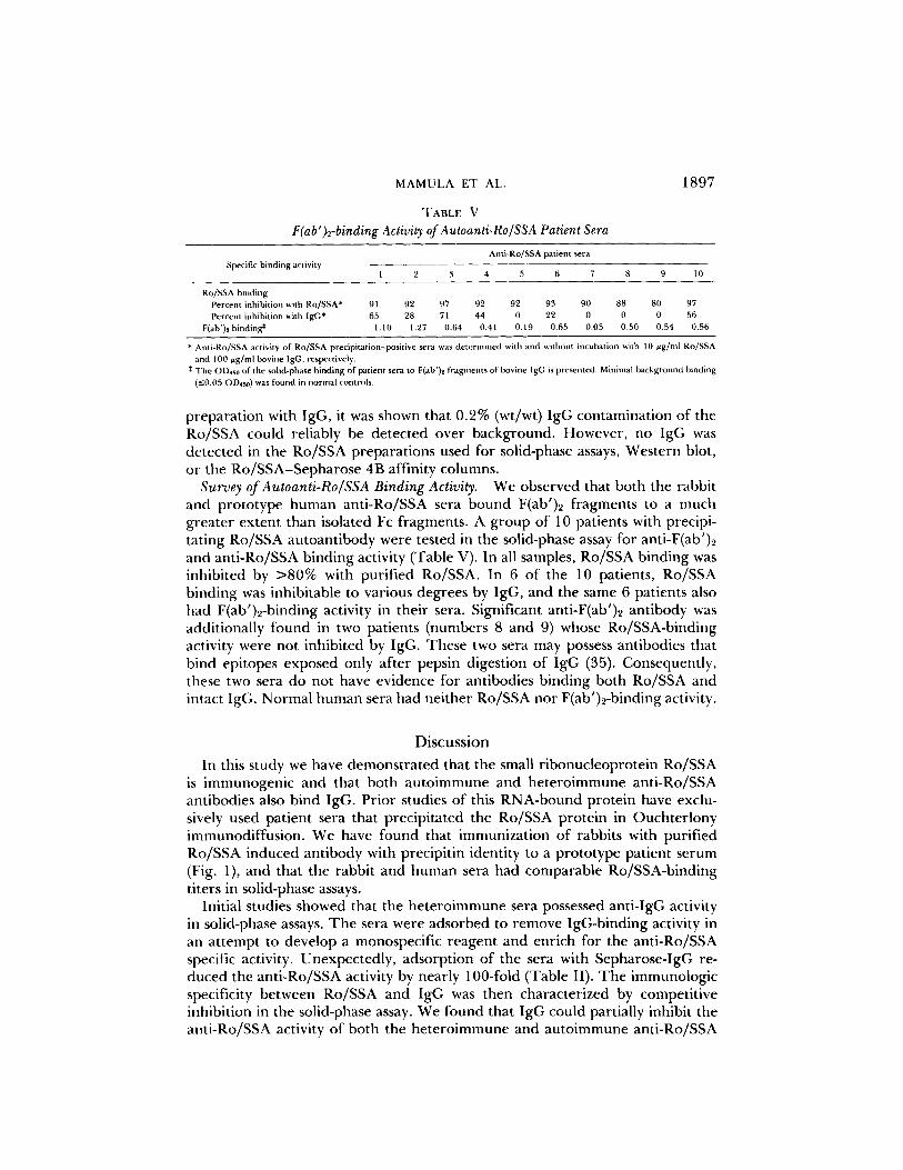
TABLE VF(ab')2-binding Activity of Autoanti-Ro/SSA Patient Sera
Discussion
Anti-Ro/SSA patient sera
I 2 3 4 5 6 7 8 9 10
Ro/SSA bindingPercent inhibition with Ro/SSA*
91
92
97
92
92
93
90
88
80
97Percent inhibition with IgG*
65
28
71
44
0
22
0
0
0
56F(ab')2 binding*
1 .10
1 .27
0.64
0.41
0.19
0.65
0.05
0.50
0.54
0.56
* Anti-Ro/SSA activity of Ro/SSA precipitation-positive sera was determined with and without incubation with 10 pg/ml Ro/SSAand 100 pg/ml bovine IgG, respectively.
$ The OD,s0 of the solid-phase binding of patient seta to F(ab')2 fragments of bovine IgG is presented . Minimal background binding( :50 .05 OD,sa ) was found in normal controls .
preparation with IgG, it was shown that 0.2% (wt/wt) IgG contamination of theRo/SSA could reliably be detected over background . However, no IgG wasdetected in the Ro/SSA preparations used for solid-phase assays, Western blot,or the Ro/SSA-Sepharose 4B affinity columns.Survey of Autoanti-Ro/SSA Binding Activity .
We observed that both the rabbitand prototype human anti-Ro/SSA sera bound F(ab')2 fragments to a muchgreater extent than isolated Fc fragments. A group of 10 patients with precipi-tating Ro/SSA autoantibody were tested in the solid-phase assay for anti-F(ab')2and anti-Ro/SSA binding activity (Table V) . In all samples, Ro/SSA binding wasinhibited by >80% with purified Ro/SSA . In 6 of the 10 patients, Ro/SSAbinding was inhibitable to various degrees by IgG, and the same 6 patients alsohad F(ab')2-binding activity in their sera . Significant anti-F(ab')2 antibody wasadditionally found in two patients (numbers 8 and 9) whose Ro/SSA-bindingactivity were not inhibited by IgG. These two sera may possess antibodies thatbind epitopes exposed only after pepsin digestion of IgG (35) . Consequently,these two sera do not have evidence for antibodies binding both Ro/SSA andintact IgG. Normal human sera had neither Ro/SSA nor F(ab')2-binding activity .
In this study we have demonstrated that the small ribonucleoprotein Ro/SSAis immunogenic and that both autoimmune and heteroimmune anti-Ro/SSAantibodies also bind IgG. Prior studies of this RNA-bound protein have exclu-sively used patient sera that precipitated the Ro/SSA protein in Ouchterlonyimmunodiffusion . We have found that immunization of rabbits with purifiedRo/SSA induced antibody with precipitin identity to a prototype patient serum(Fig. 1), and that the rabbit and human sera had comparable Ro/SSA-bindingtiters in solid-phase assays .
Initial studies showed that the heteroimmune sera possessed anti-IgG activityin solid-phase assays . The sera were adsorbed to remove IgG-binding activity inan attempt to develop a monospecific reagent and enrich for the anti-Ro/SSAspecific activity . Unexpectedly, adsorption of the sera with Sepharose-IgG re-duced the anti-Ro/SSA activity by nearly 100-fold (Table II) . The immunologicspecificity between Ro/SSA and IgG was then characterized by competitiveinhibition in the solid-phase assay. We found that IgG could partially inhibit theanti-Ro/SSA activity of both the heteroimmune and autoimmune anti-Ro/SSA

1898
Ro/SSA AND IgG CROSSREACT
sera (Fig. 3), and conversely, that Ro/SSA inhibits the anti-IgG activity of thesera (Fig . 4) . Histories, albumin, or a related ribonucleoprotein, La/SSB, failedto inhibit the binding ofanti-Ro/SSA to Ro/SSA (Table I) . We have also observedthat antibody eluted from the specific Ro/SSA band in Western blotting (by themethod of Smith et al . [36]) binds IgG in ELISA (data not shown), as doesaffinity-purified specific anti-Ro/SSA (Table III) . Finally, the IgG-binding frac-tion of the rabbit serum had increased relative anti-Ro/SSA specific activity ascompared with unadsorbed serum (Table III) .We performed additional experiments to rule out potential artifacts due to
IgG contamination of Ro/SSA (Table IV) or to immune complexes (Ro/SSA-IgG and IgG-IgG) . No IgG contamination of Ro/SSA preparations could bedetected nor was evidence found for immune complexes within either theheteroimmune or autoimmune sera . Gel diffusion experiments were also per-formed, but failed to define the immunologic relationship of Ro/SSA and IgG.Other investigators (Herrera-Esparza, R., and L. A . Diaz, unpublished observa-tions), however, have also immunized rabbits with Ro/SSA and have demon-strated partial immunologic identity between Ro/SSA and IgG in gel diffusion.
In a study of patients with Ro/SSA-precipitating antibody, we found that amajority of patients possessed Ro/SSA-binding activity that was inhibitable withIgG in the solid-phase assay (Table V). A relationship between Ro/SSA and IgGbinding antibody has been shown in clinical studies that may explain this obser-vation . In patients with SLE and Sjdgren's syndrome, rheumatoid factor is foundin ^-70% of those with Ro/SSA-precipitating antibody (17) . Additionally, thetiters of rheumatoid factor and anti-Ro/SSA antibody are directly associated inprimary Sjogren's syndrome (18) . In a study (19) designed to compare the solid-phase ELISA results with Ro/SSA precipitin data, it was found that higher levelsof rheumatoid factor were found in the sera that formed Ro/SSA precipitinswhen compared with sera with the same level of anti-Ro/SSA by solid phase-binding, but that failed to form a precipitin . The results suggest that rheumatoidfactor augments Ro/SSA precipitation . If some anti-Ro/SSA possess rheumatoidfactor-like activity, enhanced precipitation would result by increasing the com-plexity of lattice formation between the autoantibody and Ro/SSA .Two ligands that react with the same antibody will often share a common
structural component. The structural analog that leads anti-Ro/SSA to bind IgGis not known, although other preliminary data (Mamula M. J., andJ. B. Harley,unpublished observations) suggest that anti-Ro/SSA binds the H chain and theF(ab')z fragment of IgG. The binding to these sites on IgG raises the possibilitythat allotypes or particular idiotypes may be structurally important for thisinteraction . On the other hand, the effectiveness of both bovine and human IgGto inhibit binding to the Ro/SSA particle argues against contributions from IgGstructural properties that are not similar between species.
In the context of autoimmune disease, our findings may bear similarities tothe crossreactivity between IgG and histones that is found in some patients withrheumatoid arthritis who have positive antinuclear antibody (20) . In this regard,Hannestad et al . (37) were successful in removing both antinuclear antibody andrheumatoid factor activity by adsorption of serum with human IgG, just as anti-Ro/SSA and anti-IgG activity were lost with IgG adsorption . Rubin et al . (24)

MAMULA ET AL .
1899
have demonstrated monoclonal rheumatoid factors that also bound histories andsingle-stranded DNA. While IgG-binding activity is a common theme of severalstudies ofantibody multispecificity (20-26), its importance has yet to be exploitedin defining crossreactive epitopes of different ligands. Rheumatoid factor andantihistone antibodies appear to have similar idiotypes in some patients, thoughthe basis for the crossreactivity is perplexing since no obvious structural similar-ities exist between IgG and histories. Since no structural features of Ro/SSA areknown, the physical basis for crossreactivity of Ro/SSA and IgG cannot, as yet,be understood . Models of crossreactivity should not, however, be limited to thoseof similar structure since antibody can bind multiple ligands at different regionswithin the antibody-combining site (38) . Mechanisms of antigen crossreactivityappear to reach beyond explicit amino acid similarities and may rely on otherphysical properties, such as charge or spatial configuration . Nevertheless, multi-specificity of an antibody-binding site is one mechanism of generating diversitywithin the antibody repetoire.
This study adds the Ro/SSA ribonucleoprotein to the lengthening list ofsubstances that possess functional immunologic resemblance to IgG (20-26).The association of Ro/SSA with IgG maybe an important feature in the inductionand perpetuation of the autoimmune response .
SummaryThe rheumatic disease autoantigen, Ro/SSA, was immunogenic to a rabbit
host . The heteroimmune rabbit serum bound the Ro/SSA particle in immuno-blots and in an ELISA. Both the rabbit anti-Ro/SSA and a human prototypeanti-Ro/SSA serum also bound IgG; and moreover, IgG inhibited both rabbitand human anti-Ro/SSA activity . Anti-IgG activity of the rabbit and human anti-Ro/SSA sera bound Ro/SSA by Western blot and solid-phase assays . In addition,purified Ro/SSA inhibited the anti-IgG activity of the anti-Ro/SSA sera fromrabbit and man . Affinity purification of the IgG- and Ro/SSA-binding fractionsof the rabbit anti-Ro/SSA demonstrated that both the anti-Ro/SSA and anti-IgGactivities were concentrated in these fractions . These data show that Ro/SSAand IgG share epitopes that are bound by anti-Ro/SSA antibody . Inhibitionexperiments suggest that this antibody is found in most human anti-Ro/SSAautoimmune sera and that the epitope(s) are found in the F(ab')2 fragment ofIgG.
The authors gratefully acknowledge the advice of Drs. Richard M. Hyde and RobertaJacobs, and the technical assistance of Charles O'Brien.
Received for publication 4 June 1986 and in revisedform 28 August 1986.
References1 . Reichlin, M . 1981 . Current perspectives on serologic reactions in SLE patients . Clin .
Fxp. Immunol. 44 :1 .2 . Tan, E. M . 1982 . Autoantibody to nuclear antigens (ANA): their immunobiology
and medicine . Adv. Immunol. 33 :167 .3 . Wasicek, C. A., and M . Reichlin . 1982 . Clinical and serological differences between

1900
Ro/SSA AND IgG CROSSREACT
systemic lupus erythematosus patients with antibodies to Ro versus patients withantibodies to Ro and La . J. Clin . Invest. 69 :835 .
4. Reichlin, M., and C. A. Wasicek. 1983 . Clinical and biologic significance ofantibodiesto Ro/SSA . Hum. Pathol. 14:401 .
5 . Alexander, E. L., T. J. Hirsch, F. C. Arnett, T. T. Provost, and M. B. Stevens. 1983 .Associatio n ofanti-Ro(SSA) antibodies with vasculitis, hematologic abnormalities andserologic hyperreactivity . Ann. Intern . Med. 98:155 .
6. Maddison, P. J., and M. Reichlin . 1976 . The participation of antibodies to a solublecytoplasmic antigen in the nephritis of systemic lupus erythematosus. Clin. Res. 4:576 .(Abstr .)
7. Scott, J. S., P. J. Maddison, P. V. Taylor, E. Esscher, O . Scott, and R. P. Skinner.1983 . Connective-tissue disease, antibodies to ribonucleoprotein and congenital heartblock. N. Engl. J. Med. 309:209 .
8. Kephart, D. C., A. F. Hood, and T. T. Provost . 1981 . Neonatal lupus erythematosus:new serologic findings .J. Invest. Dermatol . 77 :331 .
9. Franco, H. L., W. L. Weston, C. Peebles, S. L. Forstot, and P. Phanaphak. 1981 .Autoantibodies directed against sicca syndrome antigens in the neonatal lupus syn-drome. J. Am . Acad . Dermatol . 4:67.
10 . Watson, R. M., A. T. Lune, M. K. Barnett, W. B. Bias, F. C. Arnett, and T. T.Provost. 1984 . Neonatal lupus erythematosus. A clinical, serological and immuno-genetic study with review of the literature . Medicine (Baltimore) . 63 :362 .
11 . Anderson, J. R., K. G. Gray, J. S. Bed, and W. I . Kinnear. 1961 . Precipitatingantibodies in Sj6gren's disease. Lancet . 2:456 .
12 . Clark, G. M., M. Reichlin, and T. B. Tomasi . 1968 . Characterization of a solublecytoplasmic antigen reactive with sera from patients with systemic lupus erythema-tosus. J. Immunol. 102 :117 .
13 . Harley, J. B., H. Yamagata, and M. Reichlin . 1984 . Anti-La/SSB antibody is presentin some normal sera and is coincident with anti-Ro/SSA precipitins in systemic lupuserythematosus. J. Rheumatol. 11 :309 .
14 . Yamagata, H ., J. B. Harley, and M. Reichlin . 1984 . Molecular properties of theRo/SSA antigen and enzyme-linked immunosorbent assay for quantitation of anti-body.J. Clin . Invest . 74 :625 .
15 . Wolin, S. L ., and J. A. Steitz . 1984 . The Ro small cytoplasmic ribonucleoproteins:identification of the antigenic protein and its binding site on the Ro RNAs . Proc.Natl. Acad . Sci . USA. 81 :1996.
16 . Venables, P. J . W., P. R. Smith, and R . N. Maini. 1983 . Purification and characteri-zation of the Sj6gren's syndrome A and B antigens . Clin . Exp. Immunol. 54:731 .
17 . Maddison, P., H. Mogavero, T. T. Provost, and M. Reichlin . 1979 . The clinicalsignificance of autoantibodies to a soluble cytoplasmic antigen in systemic lupuserythematosus and other connective tissue diseases . J. Rheumatol. 6:189 .
18 . Harley, J. B., E. L. Alexander, W. B. Bias, O . F. Fox, T. T. Provost, M. Reichlin, H.Yamagata, and F. C. Arnett . 1986 . Anti-Ro/SSA and anti-La/SSB in Sj6gren'ssyndrome . Arthritis Rheum. In press.
19 . Harley, J. B. 1985 . Autoantibodies in Sj6gren's syndrome : comparison of autoanti-body determination methods show that antinuclear antibody and rheumatoid factorare associated with Ro/SSA precipitin formation. Protides Biol. Fluids Proc. Colloq.33:343 .
20 . Agnello, V ., A. Arbetter, G. Ibanez de Kasep, R. Powell, E. M. Tan, and F. Joslin .1980 . Evidence for a subset of rheumatoid factors that cross react with DNA histoneand have a distinct cross idiotype . J. Exp. Med. 151 :1514 .
21 . Aitcheson, C . T., C . Pebbles, F. G. Joslin, and E. M . Tan. 1980 . Characteristics of

MAMULA ET AL .
190 1
antinuclear antibodies in rheumatoid arthritis. Reactivity of rheumatoid factor witha histone dependent nuclear antigen . Arthritis Rheum . 23:528 .
22 . Hannestad, K., and B. D . Stollar . 1978 . Certain rheumatoid factors react withnucleosomes . Nature (Lond.). 275:671 .
23 . Rauch, J ., H . Massicote, and H. Tannenbaum . 1985 . Hybridoma anti-DNA auto-antibodies from patients and rheumatoid arthritis and systemic lupus erythematosusdemonstrate similar nucleic acid binding characteristics . J. Immunol. 134:180 .
24 . Rubin, R . L ., R . S. Balderas, E . M . Tan, F . J. Dixon, and A. N . Theofilopoulos .1984 . Multiple autoantigen binding capabilities of mouse monoclonal antibodiesselected for rheumatoid factor activity . J. Exp. Med . 159:1429 .
25 . Bokish, V. A., J . W. Chaio, D. Bernstein, and R. M. Krause . 1973 . Homogeneousrabbit 7S anti-IgG with antibody specificity for peptidoglycan .J. Exp . Med. 138:1184 .
26 . Hannestad, K. 1969 . Monoclonal and polyclonal rheumatoid factors with anti-di andanti-trinitrophenyl activity . J. Exp . Immunol . 4 :555 .
27 . Axen, R., J . Porath, and S . Einback. 1967 . Chemical coupling ofpeptides and proteinsto polysaccharides by means of cyanogen halides. Nature (Lond.). 214:1302 .
28 . Singer, J . M., and C . M . Plotz . 1958 . Slide latex fixation test : a simple screeningmethod for the diagnosis ofrheumatoid arthritis .JAMA (J. Am. Med . Assoc.) . 168:180 .
29 . Engvall, E ., and P. Perlman . 1971 . Enzyme linked immunosorbent assay (ELISA)quantitative assay of immunoglobulin G. Immunochemistry. 8:871 .
30 . Rubin, R . L ., F . G. Joslin, and E . M . Tan . 1982 . A solid-phase radioimmunoassay foranti-histone antibodies in human sera : comparison with an immunofluorescence assay .Scand. J. Immunol . 15:63 .
31 . Towbin, H., T . Staehelin, and J . Gordon . 1979 . Electrophoresis transfer of proteinfrom polyacrylamide gels to nitrocellulose sheets : procedure and some applications .Proc. Natl . Acad. Sci. USA . 76:4350 .
32 . Dao, M. L . 1985 . An improved method of antigen detection in nitrocellulose : in situstaining of alkaline phosphatase conjugated antibody .J. Immunol. Methods. 82:225 .
33 . Volkman, D . J ., H . C . Lane, and A . C . Fauci . 1981 . Antigen-induced in vitro antibodyproduction in humans : a model for B cell activation and immunoregulation . Proc.Natl. Acad . Sci . USA . 78 :2528 .
34 . Theofilopoulos, A . N ., C. B. Wilson, and F . J . Dixon . 1976 . The Raji cell radioim-mune assay for detecting immune complexes in human sera . J. Clin . Invest. 57:169 .
35 . Osterland, C . K ., M . Harboe, and H. G. Kunkel . 1963 . Anti gamma globulin factorsin human sera revealed by enzymatic splitting ofanti-Rh antibodies . Vox. Sang. 8 :133 .
36 . Smith, D . E ., and P . A . Fisher . 1984. Identification, developmental regulation, andresponse to heat shock of two antigenically related forms ofa major nuclear envelopeprotein in Drosophila embryos : application of an improved method for affinitypurification of antibodies to polypeptides immobilized on nitrocellulose blots . I CellBiol. 99:20 .
37 . Hannestad, K., and A. Johannessen . 1976 . Polyclonal human antibodies to IgG(rheumatoid factors) which cross-react with cell nuclei . Scand . J. Immunol . 5:541 .
38 . Richards, F . F ., W. H . Konigsberg, R . W. Rosenstein, and J . M . Varga . 1975 . On thespecificity of antibody . Science (Wash. DC) . 187 :130 .