therapeutic strategies for spinal muscular atrophy: smn and … · majority of cases, associated...
TRANSCRIPT

REVIEW SPECIAL COLLECTION: NEURODEGENERATION
Therapeutic strategies for spinal muscular atrophy:SMN and beyondMelissa Bowerman1, Catherina G. Becker2, Rafael J. Yanez-Mun oz
3, Ke Ning4, Matthew J. A. Wood1,Thomas H. Gillingwater5, Kevin Talbot6,* and The UK SMA Research Consortium
ABSTRACTSpinal muscular atrophy (SMA) is a devastating neuromuscular disordercharacterized by loss of motor neurons and muscle atrophy, generallypresenting in childhood. SMA is caused by low levels of the survivalmotor neuron protein (SMN) due to inactivating mutations in theencoding geneSMN1. A second duplicated gene,SMN2, produces verylittle but sufficient functional protein for survival. Therapeutic strategies toincrease SMNare in clinical trials, and the firstSMN2-directed antisenseoligonucleotide (ASO) therapy has recently been licensed. However,several factors suggest that complementary strategies may be neededfor the long-term maintenance of neuromuscular and other functionsin SMA patients. Pre-clinical SMA models demonstrate that therequirement for SMN protein is highest when the structuralconnections of the neuromuscular system are being established, fromlate fetal life throughout infancy. Augmenting SMNmay not address theslow neurodegenerative process underlying progressive functionaldecline beyond childhood in less severe types of SMA. Furthermore,individuals receiving SMN-based treatments may be vulnerable todelayed symptoms if rescue of the neuromuscular system is incomplete.Finally, a large number of older patients living with SMA do not fulfill thepresent criteria for inclusion in gene therapy and ASO clinical trials, andmay not benefit from SMN-inducing treatments. Therefore, acomprehensive whole-lifespan approach to SMA therapy is requiredthat includes both SMN-dependent and SMN-independent strategiesthat treat theCNSand periphery. Here, we review the range of non-SMNpathways implicated in SMA pathophysiology and discuss how variousmodel systems can serve as valuable tools for SMA drug discovery.
KEY WORDS: Animal models, Cellular models, Combinatorialtherapies, Skeletal muscle, Spinal muscular atrophy, Survivalmotor neuron
IntroductionSpinal muscular atrophy (SMA) is the most common genetic diseaseresulting in death in infancy, affecting approximately 1 in 6000 to 1
in 10,000 births (Crawford and Pardo, 1996). This autosomalrecessive disorder, resulting from the loss-of-function of thesurvival motor neuron 1 (SMN1) gene, is characterized by loss ofspinal cord motor neurons, muscular atrophy, neuromuscularjunction (NMJ) denervation and paralysis (Crawford and Pardo,1996; Kariya et al., 2008; Kong et al., 2009; Lefebvre et al., 1995;Murray et al., 2008). SMN1 is highly conserved and present as asingle copy in the genome of all eukaryotic organisms (Bergin et al.,1997; Miguel-Aliaga et al., 1999, 2000; Paushkin et al., 2000). Inhumans, however, a genomic duplication has given rise to a secondgene, SMN2 (Lefebvre et al., 1995; Rochette et al., 2001). A crucialC-to-T substitution at position 6 of exon 7 in SMN2, which occurs inall individuals, leads to the aberrant splicing of exon 7 and thesubsequent production of an unstable SMNΔ7 protein (Lorson et al.,1999; Monani et al., 2000). An important sequence in intron 7 ofSMN2, termed intron splicing silence N1 (ISS-N1) has beendemonstrated to further favour the exclusion of exon 7 in thetranscript (Singh et al., 2006). Thus, the telomeric SMN1 copy givesrise to the full-length (FL) SMN protein while the centromericSMN2 copy predominantly produces the SMNΔ7 protein. However,the SMN2 gene always generates a small amount of functionalprotein, which maintains viability, as homozygous deletion ofSMN1 is uniformly lethal (Gennarelli et al., 1995; Lefebvre et al.,1995). Deletions or intragenic mutations in SMN1 are found in allforms of SMA, with SMN2 acting to modulate the disease severitythrough variation in its copy number (Gennarelli et al., 1995;Lefebvre et al., 1995) (Fig. 1). As the number of copies of SMN2increases, so does the quantity of stable FL-SMN protein produced.Thus, the variation in clinical severity seen in SMA is mostlyexplained by the total level of residual SMN protein.
SMA is clinically heterogeneous and has been categorized intofive types (0-IV) based on age of onset, severity of motor declineand life expectancy. The term ‘Type 0’, in which the minimalcomplement of one SMN2 gene is present, describes SMA with aclear in utero onset, arthrygryphosis (limited joint contracture) andcomplex motor and sensory nerve deficits, and death before or justafter birth. Type I SMA patients display the most severe symptoms,with death in infancy if invasive ventilation is not implemented.Types II and III have a later childhood onset and are associated withsurvival into adulthood, and the potential for a normal lifespan,albeit with considerable physical disability (Munsat and Davies,1992; Pearn, 1980). The clinical course of Type II and III patientsliving with SMA is characterized by long periods of relative stabilitywith superimposed periods of accelerated functional decline, forexample during the pubertal growth spurt, and a subsequent longperiod of slowly progressive age-related loss of motor function(Kaufmann et al., 2012). Advances in respiratory, musculoskeletaland nutritional care mean that greater numbers of patients with TypeI SMA are surviving beyond infancy. Most SMA Type II patientsare living full lives into adulthood and Type III SMA is, in the
1Department of Physiology, Anatomy and Genetics, University of Oxford,South Parks Road, Oxford OX1 3QX, UK. 2Euan MacDonald Centre for MotorNeurone Disease Research and Centre for Neuroregeneration, University ofEdinburgh, Edinburgh EH16 4SB, UK. 3AGCTlab.org, Centre for BiomedicalSciences, School of Biological Sciences, Royal Holloway, University of London,EghamHill, Egham, Surrey TW20 0EX, UK. 4Department of Neuroscience, SheffieldInstitute for Translational Neuroscience (SITraN), University of Sheffield, SheffieldS10 2HQ, UK. 5Euan MacDonald Centre for Motor Neurone Disease Research andCentre for Integrative Physiology, University of Edinburgh, Edinburgh EH8 9XD, UK.6Nuffield Department of Clinical Neurosciences, University of Oxford, JohnRadcliffe Hospital, Oxford OX3 9DU, UK.
*Author for correspondence ([email protected])
M.B., 0000-0002-3579-6403; K.T., 0000-0001-5490-1697
This is an Open Access article distributed under the terms of the Creative Commons AttributionLicense (http://creativecommons.org/licenses/by/3.0), which permits unrestricted use,distribution and reproduction in any medium provided that the original work is properly attributed.
943
© 2017. Published by The Company of Biologists Ltd | Disease Models & Mechanisms (2017) 10, 943-954 doi:10.1242/dmm.030148
Disea
seModels&Mechan
isms

majority of cases, associated with a normal lifespan (Rudnik-Schöneborn et al., 2001). The clinical and molecular features of thefive types of SMA are presented in Table 1. Given the range ofseverity and ages of onset, it will be necessary for any therapeuticstrategy to address the needs of all individuals affected by SMA,from infancy to adulthood.The SMN protein is ubiquitously expressed and is localized in the
cytoplasm (Liu and Dreyfuss, 1996), neuronal growth cones (Fan andSimard, 2002), neuronal extensions (Fallini et al., 2010), the nucleolus(Charroux et al., 2000; Wehner et al., 2002) and in punctate nuclearstructures called Gemini of coiled bodies (Gems) and Cajal bodies(Carvalho et al., 1999; Liu andDreyfuss, 1996). The SMNprotein hasthus been attributed several key regulatory cellular functions inneuronal cells, including roles in RNAmetabolism [specifically smallnuclear ribonucleoproteins (snRNPs)] (Li et al., 2014), actincytoskeleton dynamics (Hensel and Claus, 2017), mRNA transport(Donlin-Asp et al., 2016), ubiquitin homeostasis (Groen andGillingwater, 2015), bioenergetics pathways (Boyd et al., 2017) and
synaptic vesicle release (Kong et al., 2009) (Fig. 2). Importantly, todate, noneof these roleshas been identified as being solely responsiblefor SMA pathophysiology.
The most advanced therapies currently in clinical trials for SMAare aimed at increasing FL SMN either by exogenously expressingSMN1 or upregulating FL SMN2 production (d’Ydewalle andSumner, 2015). Unless these current SMN-dependent approachescan be given pre-symptomatically, when motor neuron dysfunctionmay still be reversible, and delivered with a very high level ofefficiency to drastically induce SMN levels in spinal cord motorneurons, it is likely that the progressive neurodegenerative processwill not be completely abrogated but simply slowed down. Thus,treated SMA patients may be vulnerable to a delayed deteriorationof the neuromuscular system. There are also a large number of olderchildren and adults living with SMA who do not fulfill the presentcriteria for inclusion (on the grounds of age and various clinicalparameters) in the ongoing clinical trials and for whom it is notcurrently clear that SMN-inducing treatments will be beneficial
Fig. 1. SMN1 and SMN2 contribute to spinal muscular atrophy(SMA). In healthy individuals, the survival motor neuron 1 (SMN1)gene produces 100% full length (FL) SMN protein while the SMN2gene produces ∼10% FL SMN and ∼90% of a non-functionalproduct that lacks exon 7 (SMNΔ7) due to aberrant alternativesplicing. In SMA patients, the SMN1 gene is lost due to mutations ordeletions. SMN2 remains and the small amount of FL SMN issufficient for survival. The number of SMN2 copies correlates withdisease severity, with a lower copy number being linked to moresevere types of SMA.
Table 1. Clinical and molecular features of SMA sub-types
Type of SMA
0 I II III IV
SMN2 copy number* 1 2 3 3-5 3-5
Age of onset In utero Majority by 6 months 6-12 months After 18 months (IIIa:<3 years, IIIb:>3 years)
Adulthood
Key clinical features Widespread motor andsensory neuronal loss
ContracturesHigh incidence ofcongenital cardiacdefects
Neonatal hypotoniaPoor feeding and headcontrol
Respiratory insufficiencyNever develop ability to rollor sit unaided
Sit unsupportedNever walkRespiratory muscleweakness
Walk unaided, even ifbriefly
Progressive proximalweakness
Lower limbpredominance
Natural history Peri-natal death 50% death by 12 months90% death by 24 monthswithout invasive ventilation
Life expectancy30-50 yearsdepending onrespiratory function
Loss of ambulation veryvariable (fromchildhood to late life)
Respiratory involvementuncommon
Life expectancy nearnormal
Slow progressionAmbulation
maintainedNormal lifespan
*All SMA patients, regardless of type have no functional copies ofSMN1; the number ofSMN2 copies in unaffected individuals (carriers or non-carriers) can rangefrom 2 to 5.
944
REVIEW Disease Models & Mechanisms (2017) 10, 943-954 doi:10.1242/dmm.030148
Disea
seModels&Mechan
isms

because of the significant and irreversible SMN-relatedneuromuscular decline that is already established. Furthermore, ithas become evident that SMA pathophysiology extends beyond theneuromuscular system, whereby numerous peripheral organs andtissues demonstrate pathological changes in pre-clinical models andpatients (Hamilton and Gillingwater, 2013). Therapies that improveneuromuscular function as well as maintain lifelong general healthof people living with SMA are therefore a major priority and anunmet clinical need.As the first successful SMN-targeted therapeutic approaches are
emerging into the clinical arena (Finkel et al., 2016; Gillingwater,2016), we review here how to best move forward in the developmentof combinatorial therapeutic approaches for SMA that, ideally,would target the CNS and the periphery, operating via SMN-dependent and SMN-independent processes. We will firstlyconsider the various existing and alternative experimental modelsthat could be used to identify novel SMA therapeutic targets. Wewill then discuss how the current SMN-specific compoundspresently in clinical trials inform the potential development oftreatments aimed at non-SMN targets in non-CNS tissues. Finally,we will expand on the idea of developing drug discovery anddelivery approaches that enable systemic delivery of therapies.
Advantages and limitations of current animal and cellmodelsAnimal modelsA range of in vivo model systems have been developed to aidunderstanding of the pathogenesis of SMA and to test the efficacyof therapies. The similarities in anatomy and physiology to thehuman neuromuscular system, coupled with the ease of geneticmanipulation, mean that the mouse has been an extremely valuablemodel for exploring the basic pathogenesis and evaluating potentialtreatments for SMA. Various mouse models have been developedover the years, displaying differing ranges of disease severity. Whilethe complete knockout (Smn−/−) is embryonic lethal (Schrank et al.,1997), the heterozygous animals (Smn+/−) do not develop a typicalSMA phenotype (Bowerman et al., 2014; Schrank et al., 1997),which is in accordancewith data demonstrating that loss of∼85% ofnormal Smn levels is required to reflect an SMA phenotype in mice(Bowerman et al., 2012a). Genetic modifications were thusintegrated in the Smn–/– mice to allow their survival, whilst stillretaining the criteria for an SMA model. The severely afflictedSmn–/–;SMN2 mice (Hsieh-Li et al., 2000; Monani et al., 2000)harbor a human SMN2 transgene that produces the typical∼15% FL
SMN2 transcript, whereas the Smn–/–;SMN2;SMNΔ7/Δ7 transgenicmice (Le et al., 2005) additionally express the partially functionalhuman SMNΔ7 protein, which confers an increase in survival. Boththese severe SMA mouse models typically do not survive past thefirst two post-natal weeks and therefore do not reflect the chronicphase of the disease, making them suitable for modelling onlysevere infantile SMA. This limitation means that these mice are alsounsuitable for the evaluation of non-SMN therapies that may benefitsome aspects of SMA pathology. In addition, severe SMA mousemodels do not allow for the long-term evaluation of extra-CNSdefects that might emerge over time in people living with Type II, IIIand IV SMA. To address this, animals that have a significantly longerasymptomatic phase have been developed. Smn2B/–mice (Bowermanet al., 2012a; Hammond et al., 2010) are an intermediate model withan average lifespan of 30 days. These mice carry an endogenousmutation within the murine Smn gene that mimics the human SMN2gene by principally producing SMNΔ7 transcripts. Recently, longer-lived intermediate models were generated by administration ofsub-optimal doses of exon 7 inclusion-promoting ISS-N1 antisense
Fig. 2. Localization of the survival motor neuron (SMN)protein in neuronal cells and associated general cellularfunctions. SMN regulates small nuclear ribonucleoprotein(snRNP) biogenesis, maturation and recycling in Gemini of coiledbodies (Gems) and Cajal bodies; ribosome biogenesis innucleolus; snRNP biogenesis and actin dynamics in thecytoplasm; mRNA transport in axons; actin dynamics and vesiclerelease in the synpase.
Box 1. GlossaryAAV: adeno-associated virus. A small DNA virus that infects humansand other primate species without causing disease.ASO: antisense oligonucleotide. A synthetic polymer, typically 15-20nucleotides long and complementary to the sense sequence of a targetmRNA.BBB: blood-brain barrier. A highly selective semi-permeable endothelialcell barrier separating the circulating blood from the brain in the centralnervous system.CPP: cell-penetrating peptide. A short peptide that facilitates cellularuptake of molecules.Discordant: in disagreement.FDA: Food and Drug Administration of the United States Department ofHealth and Human Services; responsible for protecting and promotingpublic health.Hypomorphic: genetic mutation causing partial loss of function.Intracerebroventricular: direct injection into the ventricular system(i.e. into the cerebral ventricles) of the brain.Intrathecal: direct injection into the sub-arachnoid space so that a drugreaches the cerebrospinal fluid.NMJ: neuromuscular junction. The chemical synapse between a motorneuron and a muscle fibre.SMN: survival motor neuron protein encoded by the human SMN1 andSMN2 genes that are ubiquitously expressed in most cells and tissueswith a range of cell-specific and housekeeping functions.
945
REVIEW Disease Models & Mechanisms (2017) 10, 943-954 doi:10.1242/dmm.030148
Disea
seModels&Mechan
isms

oligonucleotides (ASOs, Box 1) to human SMN2-carrying SMAmice (to promote an insufficient increase of FL-SMN for completerescue) (Hosseinibarkooie et al., 2016; Zhou et al., 2015). Mousemodels thus remain an important system for augmenting ourunderstanding of the SMA disease process and in the evaluation ofpotential therapeutic approaches.More targeted hypotheses about the role of SMN and its
interaction with specific proteins can be successfully explored ininvertebrates, particularlyDrosophila (fruit flies) (Chan et al., 2003;Chang et al., 2008) and Caenorhabditis elegans (Burt et al., 2006),and genetically tractable vertebrate model organisms that present adevelopmental phenotype, such as zebrafish (Hao et al., 2011;McWhorter et al., 2003). However, these models may displayphenotypes that differ greatly from what is observed in mousemodels and SMA patients. The locomotor and motility defectscharacterized in Drosophila larval SMA models (Praveen et al.,2012, 2014) can only have an indirect relationship to the disruptionsin neuromuscular function that occur in patients. In zebrafishmodels of SMA generated by antisense morpholinos or maternalzygotic genetic mutations (Hao et al., 2013; McWhorter et al.,2003), developing motor axons and dendrites display outgrowth andbranching defects, whereas in mouse and human, SMAmotor axonscorrectly reach the target muscle and form the NMJ, followed bydenervation of the muscle as the disease progresses (Ling et al.,2012). Nevertheless, such model systems are more efficient thanmammalian models for high-throughput screening and are likely tohave significant advantages if utilized efficiently and thoughtfully.Finally, there have also been initiatives to develop large animal
models for SMA, particularly for the evaluation of the delivery,benefits and toxicity of clinical-grade therapeutics. Specifically,endogenous and exogenous genetic modifications have beenintroduced in the pig to generate a porcine SMA model (Duqueet al., 2015; Towne et al., 2010). More work is needed to evaluatewhether the pig will become the pre-clinical model of choice fortherapeutic assessment.
Cellular modelsThere are obvious limitations in the extent to which mouse modelsand the other in vivomodels described above might be predictive ofeffects in humans, owing to inherent species-specific differences incellular function. The inability to directly study neurons inindividuals affected by a neurological disorder such as SMA hasbeen a key impediment to understanding basic pathologicalmechanisms, particularly those occurring early in the diseaseprocess. Recent developments in stem cell technology havesubstantially expanded the range of cellular models available inmotor neuron disease research by allowing the direct observation ofpathological mechanisms in neurons derived from inducedpluripotent stem cells (iPSCs) obtained from fibroblasts derivedfrom affected individuals. SMA was the first neurological disorderin which a disease-relevant phenotype was demonstrated iniPSC-derived motor neurons (Ebert et al., 2009).Beyond neurons, other cell types have been demonstrated to
contribute to SMA pathology, as described below. iPSCs can beconverted into pancreatic and cardiac cells, and thus represent an evenmore powerful tool, as these cell lines can be studied separately or inco-culture systems that mimic the dynamic interaction that exists inthe human body (De Vos et al., 2016). Furthermore, iPSCs can alsobe used to generate endothelial cells (Lippmann et al., 2014) to modelthe blood-brain barrier (BBB; Box 1) that protects the CNS, but alsolimits the entry of therapeutic compounds (Abbott, 2013; Pardridge,2005), thus allowing screening of novel and established drugs for the
ability to cross the BBB as well as compare the properties of SMAand healthy endothelial cells.
iPSC-derived motor neurons can be generated from families inwhich genotypically matched individuals are discordant for the SMAphenotype, to serve as a tool to identify disease modifiers (Boza-Morán et al., 2015). Similarly, iPSCs can be generated fromType I, IIand III patients, allowing the exploration of the impact of smallchanges in SMN expression at a cellular level. While iPSCs hold a lotof promise, experimental caveats include heterogeneity of the cellpopulations derived, as well as the limitations of studying cells inculture, isolated from other tissues within the context of the wholeorganism (Rouhani et al., 2014; De Filippis et al., 2017; Ebert et al.,2012). Nevertheless, iPSC-based models will give new insights intodisease mechanisms and will also serve as a screening and validationtool for potential therapies identified in model organisms, especiallyas part of an experimental workflow designed to identify novelmolecular targets anddrugs and evaluate their combinatorial potential.Indeed, testing of candidate therapies across multiple platforms willmost likely be key to the efficient and successful advancement ofcomplementary therapeutic approaches (Fig. 3).
Current SMN-targeted trials: early successesAt the forefront of SMA translational research are efforts nowentering clinical trials for therapies that promote SMN2 exon 7inclusion via ISS-N1 inhibition, that increase FL SMN2transcription (small molecules) or that directly replace SMN1(viral gene therapy) (Table 2) (d’Ydewalle and Sumner, 2015). Twodecades of basic research characterizing the molecular basis of exon7 splicing (Singh et al., 2017) have recently culminated in the firstsuccessful clinical trial of an ASO therapy (nusinersen, commercialname Spinraza) developed and commercialized by IonisPharmaceuticals and Biogen (Finkel et al., 2016; Gillingwater,2016). Not only did intrathecal delivery of nusinersen improve somedisease symptoms in Type I patients, but there was also evidencethat levels of FL SMN were increased in spinal motor neurons oftreated individuals. Although this was an open-label (unblinded)trial, treatment can be cautiously compared favourably to thedevastating natural history of Type I SMA patients where themajority of children have died or become ventilator-dependentbefore the age of 12 months. Whilst the results of an ongoing PhaseIII study in a larger cohort of patients are awaited, the drug wasapproved in December 2016 by the United States Food and DrugAdministration (US FDA) for all types of SMA based on thestrength of the existing data. Likewise, nusinersen wasrecommended for European Union approval by the EMA in April2017 and given marketing authorization in June 2017.
The ASO therapeutic approach, however, requires invasiveintrathecal and intracerebroventricular administration for adequatedelivery to the CNS, thus not addressing the issue of expressionlevels of FL SMN required in peripheral tissues, which are alsopotentially key contributors to SMA pathology (see below). A non-replicating adeno-associated virus (AAV9) vector has beendeveloped by AveXis to deliver a functional copy of a humanSMN1 gene. A potential advantage of this approach is that AAV9crosses the perinatal BBB, allowing a single intravenous dose toensure widespread systemic delivery. A Phase 1 clinical trial is nowunderway. Small molecules that increase FL SMN2 expression canalso be administered systemically but their ability to cross the BBBmay be limited and their exact mode of action needs to be decipheredto minimize potential off-target adverse effects. Small moleculestargeting SMN2 developed by Novartis Pharmaceuticals andHoffmann-La Roche are currently in clinical trials.
946
REVIEW Disease Models & Mechanisms (2017) 10, 943-954 doi:10.1242/dmm.030148
Disea
seModels&Mechan
isms
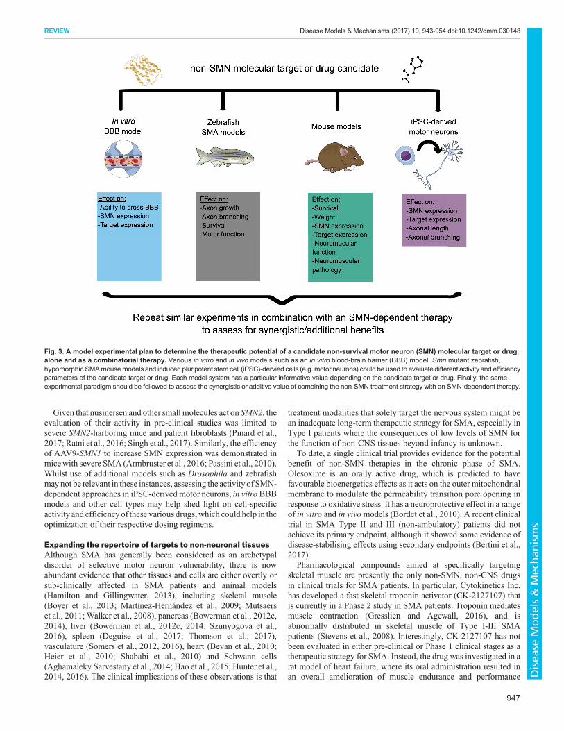
Given that nusinersen and other small molecules act on SMN2, theevaluation of their activity in pre-clinical studies was limited tosevere SMN2-harboring mice and patient fibroblasts (Pinard et al.,2017; Ratni et al., 2016; Singh et al., 2017). Similarly, the efficiencyof AAV9-SMN1 to increase SMN expression was demonstrated inmicewith severe SMA (Armbruster et al., 2016; Passini et al., 2010).Whilst use of additional models such as Drosophila and zebrafishmaynot be relevant in these instances, assessing the activity of SMN-dependent approaches in iPSC-derived motor neurons, in vitroBBBmodels and other cell types may help shed light on cell-specificactivity and efficiencyof these various drugs,which could help in theoptimization of their respective dosing regimens.
Expanding the repertoire of targets to non-neuronal tissuesAlthough SMA has generally been considered as an archetypaldisorder of selective motor neuron vulnerability, there is nowabundant evidence that other tissues and cells are either overtly orsub-clinically affected in SMA patients and animal models(Hamilton and Gillingwater, 2013), including skeletal muscle(Boyer et al., 2013; Martínez-Hernández et al., 2009; Mutsaerset al., 2011; Walker et al., 2008), pancreas (Bowerman et al., 2012c,2014), liver (Bowerman et al., 2012c, 2014; Szunyogova et al.,2016), spleen (Deguise et al., 2017; Thomson et al., 2017),vasculature (Somers et al., 2012, 2016), heart (Bevan et al., 2010;Heier et al., 2010; Shababi et al., 2010) and Schwann cells(Aghamaleky Sarvestany et al., 2014; Hao et al., 2015; Hunter et al.,2014, 2016). The clinical implications of these observations is that
treatment modalities that solely target the nervous system might bean inadequate long-term therapeutic strategy for SMA, especially inType I patients where the consequences of low levels of SMN forthe function of non-CNS tissues beyond infancy is unknown.
To date, a single clinical trial provides evidence for the potentialbenefit of non-SMN therapies in the chronic phase of SMA.Olesoxime is an orally active drug, which is predicted to havefavourable bioenergetics effects as it acts on the outer mitochondrialmembrane to modulate the permeability transition pore opening inresponse to oxidative stress. It has a neuroprotective effect in a rangeof in vitro and in vivomodels (Bordet et al., 2010). A recent clinicaltrial in SMA Type II and III (non-ambulatory) patients did notachieve its primary endpoint, although it showed some evidence ofdisease-stabilising effects using secondary endpoints (Bertini et al.,2017).
Pharmacological compounds aimed at specifically targetingskeletal muscle are presently the only non-SMN, non-CNS drugsin clinical trials for SMA patients. In particular, Cytokinetics Inc.has developed a fast skeletal troponin activator (CK-2127107) thatis currently in a Phase 2 study in SMA patients. Troponin mediatesmuscle contraction (Gresslien and Agewall, 2016), and isabnormally distributed in skeletal muscle of Type I-III SMApatients (Stevens et al., 2008). Interestingly, CK-2127107 has notbeen evaluated in either pre-clinical or Phase 1 clinical stages as atherapeutic strategy for SMA. Instead, the drug was investigated in arat model of heart failure, where its oral administration resulted inan overall amelioration of muscle endurance and performance
Fig. 3. A model experimental plan to determine the therapeutic potential of a candidate non-survival motor neuron (SMN) molecular target or drug,alone and as a combinatorial therapy. Various in vitro and in vivo models such as an in vitro blood-brain barrier (BBB) model, Smn mutant zebrafish,hypomorphic SMAmousemodels and induced pluripotent stem cell (iPSC)-dervied cells (e.g. motor neurons) could be used to evaluate different activity and efficiencyparameters of the candidate target or drug. Each model system has a particular informative value depending on the candidate target or drug. Finally, the sameexperimental paradigm should be followed to assess the synergistic or additive value of combining the non-SMN treatment strategy with an SMN-dependent therapy.
947
REVIEW Disease Models & Mechanisms (2017) 10, 943-954 doi:10.1242/dmm.030148
Disea
seModels&Mechan
isms

following exercise (Hwee et al., 2015). A first generation form of thecompound (tirasemtiv or CK-2017357) was also assessed in rodentneuromuscular models for nemaline myopathy (Lee et al., 2013),myasthenia gravis (Russell et al., 2012) and amyotrophic lateralsclerosis (Hwee et al., 2014), demonstrating a significantimprovement in muscle strength in each. Nevertheless, the FDAhas recently granted the Orphan Drug designation to CK-2127107for the treatment of SMA patients.Another interesting muscle target is the myostatin-follistatin
pathway, in which myostatin acts as a negative regulator of musclegrowth (McPherron et al., 1997) and is itself inhibited by follistatin(Lee and McPherron, 2001). Several therapeutics strategies aimedat modulating this signaling cascade to promote muscle masshave therefore been evaluated in pathologies characterized bymuscle atrophy, including SMA (Rodino-Klapac et al., 2009).Administration of recombinant follistatin to Smn–/–;SMN2;SMNΔ7/Δ7 mice resulted in significant improvement in musclemass, gross motor function and lifespan. However, inhibitionof myostatin by genetic (overexpression of follistatin) orpharmacological [soluble activin receptor IIB (ActRIIB-Fc) thatinhibits the myostatin-promoting receptor] interventions had noobvious beneficial effects on the phenotype of the same mousemodel (Sumner et al., 2009). Similar negative results were obtainedupon genetically deleting myostatin in severe SMA mice (Rindtet al., 2012). Conversely, the recent intraperitoneal administration ofa soluble form of ActRIIB encoded by an AAV2/8 viral vector in amilder model of SMA improved mass, contractile properties andsize of SMN-depleted muscles (Liu et al., 2016). Results from SMAmouse studies have therefore been varied and the discrepancies maybe due to the differing severities of the models used, thedevelopmental timing of the approach and the specific targeting/delivery strategy utilized. Several strategies based on the myostatin-follistatin pathway are currently in clinical trials for musclepathologies such as Duchenne muscular dystrophy (DMD),Becker muscular dystrophy and inclusion body myositis but have
yet to be initiated for SMA. However, a recent report shows thatserum and muscle biopsies from SMA patients display decreasedexpression of myostatin and increased levels of follistatin (Mariotet al., 2017), suggesting that additional mechanistic insight in therelevance of targeting this pathway for SMA therapy is required.
Taken together, these findings highlight an important need tobetter understand the intrinsic pathologies not only in SMA neuronsbut also in muscle and other non-CNS afflicted organs, so that cell-and tissue-specific treatments can be developed and eventually usedin combination with SMN- and CNS-targeted strategies.
Identifying non-SMN targets to develop combinatorialtherapeutic approachesSMN-dependent gene therapies will require administration as earlyas possible, even pre-symptomatically, to exert the maximum effect(Bevan et al., 2010; Foust et al., 2010; Valori et al., 2010), and atpresent can be expected to reduce disease severity rather than effecta complete cure. In the absence of a routine screening program fornewborns, the potential benefit may also be limited by the delay indiagnosis in milder forms, which generally have an insidious onset.An important step forward would be to develop therapeuticapproaches targeting pathways that reflect the chronic pathologicalprocess in SMA, facilitating treatments that are adjunctive to SMNreplacement therapy to improve and maintain neuromuscularintegrity and function throughout the life of the individual.
One of the first SMN-independent targets to prove beneficial withrespect to potential SMA therapy is the RhoA-ROCK pathway. Thesmall GTPaseRhoA and its downstream effector, the Ser/Thr proteinkinase ROCK, are key modulators of actin dynamics (Luo et al.,1997). It has been demonstrated that the RhoA-ROCK pathway isaberrantly upregulated in SMN-depleted rodent neuronal cells(Bowerman et al., 2007) and in the spinal cord and skeletal muscleof Smn2B/–mice (Bowerman et al., 2010, 2012b). Importantly, it wasshown that pharmacological inhibition of ROCK significantlyincreases lifespan and muscle pathology of Smn2B/– SMA mice
Table 2. SMN-dependent therapies in clinical trials*
Strategy Drug ClinicalTrials.gov identifier Clinical stage
SMN2 ISS-N1 targeting ASO Nusinersen/Spinraza NCT02386553NCT02052791NCT02865109NCT01780246NCT02462759NCT01703988NCT02193074NCT02292537NCT01839656NCT02594124NCT01494701
Active, Phase 2Completed, Phase 1Expanded accessCompleted, Phase 1Ongoing, Phase 2Completed, Phase 1 and 2Completed, Phase 3Completed, Phase 3Active, Phase 2Enrolling by invitation, Phase 3Completed, Phase 1
SMN2 targeting small molecules LMI070
RO7034067
Hydroxyurea
Celecoxib
NCT02268552
NCT03032172NCT02913482NCT02908685NCT02633709
NCT00485511NCT00568698NCT00568802
NCT02876094
Active, Phase 1 and 2
Recruiting, Phase 2Recruiting, Phase 2Recruiting, Phase 2Completed, Phase 1
Completed, Phase 2 and 3Completed, Phase 1 and 2Unknown, Phase 1 and 2
Not yet recruiting, Phase 2
SMN1 gene therapy AVXS-101 NCT02122952 Active, Phase 1
*As of June 2017.
948
REVIEW Disease Models & Mechanisms (2017) 10, 943-954 doi:10.1242/dmm.030148
Disea
seModels&Mechan
isms

(Bowerman et al., 2010, 2012b). Additional investigators havefurther confirmed the contribution of the RhoA-ROCK pathway toSMA in neuronal cells (Hensel et al., 2014), patient fibroblasts(Nölle et al., 2011) and glial cells (Caraballo-Miralles et al., 2012).The tumour suppressor protein PTEN is a member of the proteintyrosine phosphatase family that can regulate cell migration,spreading and growth (Lachyankar et al., 2000; Li et al., 2003;Sano et al., 1999; Tamura et al., 1999). Interestingly, PTEN isphosphorylated by ROCK (Bermúdez Brito et al., 2015), thusleading to increased PTEN inhibitory activity on neuronal survival.While PTEN activity in SMAmice has yet to be investigated, we canhypothesize that the increased activity of the RhoA-ROCK pathwayreported in SMA mice (Bowerman et al., 2010, 2012b) induces theincreased phosphorylation of PTEN. Concordantly, it has beenfound that suppressing PTEN in SMA mice through a gene therapyapproach led to improvements in NMJ pathology and a significantextension in lifespan (Little et al., 2015). Combined, these studieshave highlighted actin modulators as potential targets forcombinatorial therapeutic approaches for SMA.Further regulators of actin dynamics have emerged as potential
therapeutic targets for SMA. Plastin 3 (PLS3) is an actin-bundlingprotein that was identified as a modifier of disease severity in aninvestigation of discordant family members that carried the sameSMN1 mutations (Oprea et al., 2008). Additional analyses ofserum (Yanyan et al., 2014) and iPSC-derived motor neurons fromSMA patients have further supported the influence of plastin 3levels on disease progression in certain families but not others(Boza-Morán et al., 2015; Heesen et al., 2016). Indeed,overexpression of plastin 3 in a zebrafish model of SMAsignificantly rescues the axonal growth and branching defectscaused by smn1 gene depletion (Oprea et al., 2008). Furtheranalysis of smn1mutant zebrafish reveals that reduced SMN levelslead to decreased plastin 3 protein expression, NMJ defects andaberrant motor function, and these effects can be corrected byplastin 3 overexpression (Hao et al., 2012). More recently, studiesin mice have shown that increased expression of plastin 3 delaysaxonal degeneration and improves NMJ function (Ackermannet al., 2013) as well as ameliorates survival and neuromuscularphenotype (Kaifer et al., 2017), possibly through the modulationof endocytic pathways (Hosseinibarkooie et al., 2016). However, itmust be noted that a number of animal and patient studies do notreflect the suggested modifying powers of plastin 3 on SMApathogenesis (McGovern et al., 2015; Stratigopoulos et al., 2010),highlighting the complex relationship that may exist between SMNand plastin 3. The pathological relevance and therapeuticimportance of non-SMN targets can be highly dependent on theseverity of the disease (Kaifer et al., 2017) and as such, should beevaluated in hypomorphic models, whether transgenically orpharmacologically induced. Nonetheless, the studies on RhoA-ROCK, PTEN and plastin 3 have highlighted actin modulators aspotential targets for combinatorial therapeutic approaches forSMA.Subsequent work has also revealed other promising non-SMN
targets, including chondrolectin. It has been shown that thistransmembrane protein (encoded by Chodl) and its bindingpartners are potential modifiers of axonal integrity in SMA miceand that altered expression of Chodl is found in spinal motorneurons of SMA mice (Bäumer et al., 2009). Importantly,increasing the expression of chodl rescues motor neuronoutgrowth defects in a zebrafish model of SMA (Sleigh et al.,2014). Experiments in mouse models are underway to fully evaluatethe therapeutic potential of Chodl modulation.
As described above, one of the housekeeping functions of SMNis the regulation of RNA metabolism, particularly in the biogenesisof snRNPs (Li et al., 2014), essential components of the RNAsplicing machinery (Will and Lührmann, 2001). It has beendemonstrated that SMN depletion specifically impacts the activityof the spliceosome complex containing the U12 snRNP (Doktoret al., 2017; Gabanella et al., 2007) and that the Drosophilastasimon (stas) gene is a direct target of U12-dependent splicing(Lotti et al., 2012). Stasimon (also known as Tmem41b inmammals) plays a role in synaptic transmission in neuronalsynapses and its expression is significantly reduced and splicingsimilarly altered in motor and sensory neurons of SMA mice (Lottiet al., 2012). Importantly, overexpression of stas restoredneurotransmitter release in Drosophila smn mutants and rescuedmotor axon growth and branching defects in an SMA zebrafishmodel (Lotti et al., 2012).
The activity of cyclin-dependent kinase 5 (Cdk5) has been reportedto be upregulated in SMA mice and patient iPSC-derived motorneurons (Miller et al., 2015). The increased abundance of Cdk5 isresponsible for the pathological hyperphosphorylation of the tauprotein in SMN-depleted neuronal cells (Miller et al., 2015). Atransgenic approach was used to completely knockout Cdk5expression in SMA mice, which led to significant rescue of motorneuron synaptic stripping, motor neuron death and NMJ denervation(Miller et al., 2015). Interestingly, a recent unbiased RNA-sequencing-based assessment of global gene changes in SMN-depleted mouse tissues confirmed the specific missplicing of U12snRNP-dependent genes, several ofwhich areCa2+ channel genes andmay beupstream regulators ofCdk5activity (Doktoret al., 2017), thusfurther highlighting Cdk5 as a potential pathological effector in SMA.
Finally, the ubiquitin-like modifier activating enzyme Uba1(Groen and Gillingwater, 2015) and its downstream effectors[including the Wnt signaling effector β-catenin (Ctnnb1)], havebeen identified as major targets acting downstream of SMN toregulate neuromuscular and systemic pathology in SMA. Reducedlevels of Uba1 were reported in all tissues and organs investigatedfrom SMA mouse models (Aghamaleky Sarvestany et al., 2014;Wishart et al., 2014). Furthermore, β-catenin, which accumulates inneuromuscular tissues in SMA, has been uncovered as a keydownstream target of Uba1 deficiencies in SMA. Importantly,pharmacological inhibition of β-catenin dramatically amelioratedneuromuscular pathology in zebrafish, Drosophila and mousemodels of SMA (Wishart et al., 2014) while systemic Uba1 genetherapy increased survival and improved neuromuscular andperipheral pathology of SMA mice (Powis et al., 2016).
Thus, a range of pathways are already candidates for non-SMNtherapy approaches. As the list of molecular effectors grows, drug-screening approaches to identify pharmacological compounds thatcan modulate them will be essential. In addition, identifying noveltargets should combine proteomics and transcriptomics studies withgenome-based network analysis and drug-repositioning strategies.Combinatorial experimental paradigms should then be put in placeto evaluate the therapeutic potential of a ‘cocktail treatment’comprising SMN gene therapy and a non-SMN-targeting drug,optimally making use of the multiple in vitro and in vivo modelsdiscussed above (Fig. 3). While several non-SMN pathways andmolecular targets have been highlighted as being aberrantlyregulated in SMA models and display therapeutic potential, thesestudies remain, for the most part, in the pre-clinical discovery phase,in contrast to SMN-dependent strategies that are quickly dominatingthe clinical trial landscape. For non-SMN treatments to become apractical reality in the combinatorial approach paradigm, an
949
REVIEW Disease Models & Mechanisms (2017) 10, 943-954 doi:10.1242/dmm.030148
Disea
seModels&Mechan
isms

efficient strategic plan needs to be established to facilitate theirtransition to the clinic.
Improving systemic delivery of drugs to target CNS and non-CNS tissuesWhile motor neurons are undoubtedly the primary cellular target inSMA (Powis and Gillingwater, 2016), cumulative evidencehighlights the role of other cells and tissues that may be clinicallyor sub-clinically affected. However, most of these studies haveinvestigated these tissues or cells independently of the others. Thehierarchal contribution of each to Type 0-IV SMA therefore remainsunclear. While current gene therapies in clinical trials are promising,nusinersen (Ionis Pharmaceuticals/Biogen) delivery circumventsthe peripheral tissues and organs by being injected directly to theCNS, and, although AAV9-SMN1 gene therapy (AveXis) can bedelivered systemically, multiple rounds of administration might notbe possible due to immunogenicity (Basner-Tschakarjan andMingozzi, 2014). In both of these cases, this raises the risk ofincomplete rescue of SMN deficiency in peripheral organs and thepotential for development of non-CNS pathologies later in life.Development of novel therapeutic approaches targeting non-
SMN targets should therefore include careful consideration of bothCNS and systemic delivery methods. The optimal dosing regimenfor a pharmacological compound should balance its ability totarget all relevant tissues with the need to make therapy as non-invasive as possible. An option for systemic delivery of molecules isto conjugate them with a vehicle that can transport them across themembrane of multiple cell types. This has proven efficient for thedelivery of ASOs under the neutrally charged chemistry ofphosphorodiamidate morpholino (PMO) (Douglas and Wood,2013). Cell-penetrating peptides (CPPs) have previously beenshown to cross both plasma and endosomal membranes (Mitchellet al., 2000). One such peptide-conjugated PMO has beendeveloped, termed peptide nucleic acids/PMO internalizationpeptide 6a (Pip6a)-PMO, that efficiently modulates splicing invarious tissues of a DMD mouse model (Betts et al., 2012).
Importantly, it is delivered via a single intravenous (IV) injection.Recently, it has been reported that conjugation of Pip6a to the SMN2ISS-N1 PMO results in dramatic improvements in survival andneuromuscular phenotype associated with increased FL-SMNlevels in both CNS and peripheral tissues (Hammond et al.,2016). CPPs therefore have significant potential to facilitatetargeting of SMN to the whole body as well the eventual deliveryof therapeutic non-SMN targets or drugs in a similar fashion.
Concluding remarksGene therapy and ASO approaches to increase SMN levels are nowentering the clinical arena. In the severest form (Type I) of SMA,promising preliminary results must be balanced with a fullappreciation of the potential limitations of such strategies. Thevalue of SMN-based therapies in older Type II and III patients isunclear and it may be some time before these can be accuratelyevaluated. Translational research should therefore address thedevelopment of non-CNS and SMN-independent therapeuticapproaches to complement and enhance the benefits of CNS-directed and SMN-dependent therapies, taking into account theneed to maintain the neuromuscular system of an SMA patientthrough childhood and puberty, when there is maximal growth ofthe axial skeleton, and into adult life when the process of age-relatedattrition of motor units is likely to contribute to progressive loss ofmotor function (Fig. 4).
There remains a need for the use of various in vitro and in vivomodels as well as molecular high-throughput approaches forthe rapid identification of new targets and drugs. It will becrucial to develop tools to evaluate the effects of combinationpharmacological therapies at different disease stages. It is thereforeof utmost importance that the SMA research and clinicalcommunity, as well as those living with SMA, recognise the needto develop and test combinatorial therapeutic approaches that can beeffectively delivered systemically and target both SMN and non-SMNmolecular effectors. This will allow for a better understandingof the tissue requirements for SMN and non-SMN treatments and,
In utero 0-3 years 4-16 years
Gene therapy
Type 0 Type I
Type II Type III Type IV
Antisense oligonucleotides
Neuromusculardevelopment Pubertal growth
spurt
Age-relatedloss of motor units
Childhoodgrowth
CNS requirement for SMN
Drugs enhancing neuromuscular development Drugs promoting neuromuscular maintenance
Prevention of neurodegeneration
Adulthood
?
Fig. 4. Overview of the natural history of spinal muscular atrophy (SMA), major developmental milestones and treatment strategies. Although theprecise details of SMN expression in the developing human nervous system are difficult to study, evidence from animal models suggests that SMN levelspeak in the period of maximum neuromuscular development and then decline to a stable low level. This means that there are different windows of opportunityfor the various types of proposed therapies to be effectively employed. Whether combinatorial therapies might be particularly applicable in the more chronicphase of SMA or from the outset is a priority area for research. ASO therapy in utero will be dependent on an optimal route of delivery, which at presentremains unclear.
950
REVIEW Disease Models & Mechanisms (2017) 10, 943-954 doi:10.1242/dmm.030148
Disea
seModels&Mechan
isms

ultimately, provide the best therapeutic strategy for SMA. As withmost chronic progressive neurodegenerative disorders, it is likelythat, once loss of neuronal integrity has been initiated, combinatorialapproaches to therapy will be required to maintain neuromuscularhealth throughout life.
This article is part of a special subject collection ‘Neurodegeneration: fromModels toMechanisms to Therapies’, which was launched in a dedicated issue guest edited byAaron Gitler and James Shorter. See related articles in this collection at http://dmm.biologists.org/collection/neurodegenerative-disorders.
AcknowledgementsThe work of the UK SMA Research Consortium is funded by The SMA Trust. Fordetails of members of the UK SMA Research Consortium, see www.smatrust.org/research/uk-sma-research-consortium/meet-the-uk-sma-research-consortium-team/. We thank Lynn Ossher for graphical contributions. M.B. is an SMA TrustCareer Development Fellow.
Competing interestsThe authors declare no competing or financial interests.
FundingThis research received no specific grant from any funding agency in the public,commercial or not-for-profit sectors.
ReferencesAbbott, N. J. (2013). Blood-brain barrier structure and function and the challengesfor CNS drug delivery. J. Inherit. Metab. Dis. 36, 437-449.
Ackermann, B., Krober, S., Torres-Benito, L., Borgmann, A., Peters, M.,Hosseini Barkooie, S. M., Tejero, R., Jakubik, M., Schreml, J., Milbradt, J.et al. (2013). Plastin 3 ameliorates spinal muscular atrophy via delayed axonpruning and improves neuromuscular junction functionality.Hum. Mol. Genet. 22,1328-1347.
Aghamaleky Sarvestany, A., Hunter, G., Tavendale, A., Lamont, D. J., LlaveroHurtado, M., Graham, L. C., Wishart, T. M. and Gillingwater, T. H. (2014).Label-free quantitative proteomic profiling identifies disruption of ubiquitinhomeostasis as a key driver of Schwann cell defects in spinal muscular atrophy.J. Proteome Res. 13, 4546-4557.
Armbruster, N., Lattanzi, A., Jeavons, M., Van Wittenberghe, L., Gjata, B.,Marais, T., Martin, S., Vignaud, A., Voit, T., Mavilio, F. et al. (2016). Efficacy andbiodistribution analysis of intracerebroventricular administration of an optimizedscAAV9-SMN1 vector in a mouse model of spinal muscular atrophy. Mol. Ther.Methods Clin. Dev. 3, 16060.
Basner-Tschakarjan, E. and Mingozzi, F. (2014). Cell-mediated immunity to AAVvectors, evolving concepts and potential solutions. Front. Immunol. 5, 350.
Baumer, D., Lee, S., Nicholson, G., Davies, J. L., Parkinson, N. J., Murray, L. M.,Gillingwater, T. H., Ansorge, O., Davies, K. E. and Talbot, K. (2009). Alternativesplicing events are a late feature of pathology in amousemodel of spinal muscularatrophy. PLoS Genet. 5, e1000773.
Bergin, A., Kim, G., Price, D. L., Sisodia, S. S., Lee, M. K. andRabin, B. A. (1997).Identification and characterization of a mouse homologue of the spinal muscularatrophy-determining gene, survival motor neuron. Gene 204, 47-53.
Bermudez Brito, M., Goulielmaki, E. and Papakonstanti, E. A. (2015). Focus onPTEN regulation. Front. Oncol. 5, 166.
Bertini, E., Dessaud, E., Mercuri, E., Muntoni, F., Kirschner, J., Reid, C.,Lusakowska, A., Comi, G. P., Cuisset, J.-M., Abitbol, J.-L. et al. (2017). Safetyand efficacy of olesoxime in patients with type 2 or non-ambulatory type 3 spinalmuscular atrophy: a randomised, double-blind, placebo-controlled phase 2 trial.Lancet Neurol. 16, 513-522.
Betts, C., Saleh, A. F., Arzumanov, A. A., Hammond, S. M., Godfrey, C.,Coursindel, T., Gait, M. J. and Wood, M. J. A. (2012). Pip6-PMO, a newgeneration of peptide-oligonucleotide conjugates with improved cardiac exonskipping activity for DMD treatment. Mol. Ther. Nucleic Acids 1, e38.
Bevan, A. K., Hutchinson, K. R., Foust, K. D., Braun, L., McGovern, V. L.,Schmelzer, L., Ward, J. G., Petruska, J. C., Lucchesi, P. A., Burghes, A. H. M.et al. (2010). Early heart failure in the SMNDelta7 model of spinal muscularatrophy and correction by postnatal scAAV9-SMN delivery. Hum. Mol. Genet. 19,3895-3905.
Bordet, T., Berna, P., Abitbol, J.-L. and Pruss, R. M. (2010). Olesoxime(TRO19622): a novel mitochondrial-targeted neuroprotective compound.Pharm. Basel Switz. 3, 345-368.
Bowerman, M., Shafey, D. and Kothary, R. (2007). Smn depletion alters profilin IIexpression and leads to upregulation of the RhoA/ROCK pathway and defects inneuronal integrity. J. Mol. Neurosci. MN 32, 120-131.
Bowerman, M., Beauvais, A., Anderson, C. L. and Kothary, R. (2010). Rho-kinase inactivation prolongs survival of an intermediate SMAmouse model. Hum.Mol. Genet. 19, 1468-1478.
Bowerman, M., Murray, L. M., Beauvais, A., Pinheiro, B. and Kothary, R.(2012a). A critical smn threshold in mice dictates onset of an intermediate spinalmuscular atrophy phenotype associated with a distinct neuromuscular junctionpathology. Neuromuscul. Disord. NMD 22, 263-276.
Bowerman, M., Murray, L. M., Boyer, J. G., Anderson, C. L. and Kothary, R.(2012b). Fasudil improves survival and promotes skeletal muscle development ina mouse model of spinal muscular atrophy. BMC Med. 10, 24.
Bowerman, M., Swoboda, K. J., Michalski, J.-P., Wang, G.-S., Reeks, C.,Beauvais, A., Murphy, K., Woulfe, J., Screaton, R. A., Scott, F. W. et al.(2012c). Glucose metabolism and pancreatic defects in spinal muscular atrophy.Ann. Neurol. 72, 256-268.
Bowerman, M., Michalski, J.-P., Beauvais, A., Murray, L. M., DeRepentigny, Y.and Kothary, R. (2014). Defects in pancreatic development and glucosemetabolism in SMN-depleted mice independent of canonical spinal muscularatrophy neuromuscular pathology. Hum. Mol. Genet. 23, 3432-3444.
Boyd, P. J., Tu,W.-Y., Shorrock, H. K., Groen, E. J. N., Carter, R. N., Powis, R. A.,Thomson, S. R., Thomson, D., Graham, L. C., Motyl, A. A. L. et al. (2017).Bioenergetic status modulates motor neuron vulnerability and pathogenesis in azebrafish model of spinal muscular atrophy. PLoS Genet. 13, e1006744.
Boyer, J. G., Murray, L. M., Scott, K., De Repentigny, Y., Renaud, J.-M. andKothary, R. (2013). Early onset muscle weakness and disruption of muscleproteins in mouse models of spinal muscular atrophy. Skelet. Muscle 3, 24.
Boza-Moran, M. G., Martınez-Hernandez, R., Bernal, S., Wanisch, K., Also-Rallo, E., Le Heron, A., Alıas, L., Denis, C., Girard, M., Yee, J.-K. et al. (2015).Decay in survival motor neuron and plastin 3 levels during differentiation of iPSC-derived human motor neurons. Sci. Rep. 5, 11696.
Burt, E. C., Towers, P. R. and Sattelle, D. B. (2006).Caenorhabditis elegans in thestudy of SMN-interacting proteins: a role for SMI-1, an orthologue of humanGemin2 and the identification of novel components of the SMN complex. Invert.Neurosci. 6, 145-159.
Caraballo-Miralles, V., Cardona-Rossinyol, A., Garcera, A., Villalonga, P.,Soler, R. M., Olmos, G. and Llado, J. (2012). SMN deficiency attenuatesmigration of U87MG astroglioma cells through the activation of RhoA. Mol. Cell.Neurosci. 49, 282-289.
Carvalho, T., Almeida, F., Calapez, A., Lafarga, M., Berciano, M. T. and Carmo-Fonseca, M. (1999). The spinal muscular atrophy disease gene product, SMN: Alink between snRNP biogenesis and the Cajal (coiled) body. J. Cell Biol. 147,715-728.
Chan, Y. B., Miguel-Aliaga, I., Franks, C., Thomas, N., Trulzsch, B., Sattelle,D. B., Davies, K. E. and van den Heuvel, M. (2003). Neuromuscular defects in aDrosophila survival motor neuron gene mutant. Hum. Mol. Genet. 12, 1367-1376.
Chang, H. C.-H., Dimlich, D. N., Yokokura, T., Mukherjee, A., Kankel, M.W., Sen,A., Sridhar, V., Fulga, T. A., Hart, A. C., Van Vactor, D. et al. (2008). Modelingspinal muscular atrophy in Drosophila. PLoS ONE 3, e3209.
Charroux, B., Pellizzoni, L., Perkinson, R. A., Yong, J., Shevchenko, A., Mann,M. and Dreyfuss, G. (2000). Gemin4. A novel component of the SMN complexthat is found in both gems and nucleoli. J. Cell Biol. 148, 1177-1186.
Crawford, T. O. and Pardo, C. A. (1996). The neurobiology of childhood spinalmuscular atrophy. Neurobiol. Dis. 3, 97-110.
De Filippis, L., Zalfa, C. and Ferrari, D. (2017). Neural Stem Cells and Humaninduced pluripotent stem cells to model rare CNS diseases. CNS Neurol. Disord.Drug Targets doi:10.2174/1871527316666170615121753 [Epub ahead of print].
Deguise, M.-O., De Repentigny, Y., McFall, E., Auclair, N., Sad, S. and Kothary,R. (2017). Immune dysregulation may contribute to disease pathogenesis inspinal muscular atrophy mice. Hum. Mol. Genet 26, 801-819.
De Vos, J., Bouckenheimer, J., Sansac, C., Lemaître, J.-M. and Assou, S.(2016). Human induced pluripotent stem cells: a disruptive innovation. Curr. Res.Transl. Med. 64, 91-96.
Doktor, T. K., Hua, Y., Andersen, H. S., Brøner, S., Liu, Y. H., Wieckowska, A.,Dembic, M., Bruun, G. H., Krainer, A. R. and Andresen, B. S. (2017). RNA-sequencing of a mouse-model of spinal muscular atrophy reveals tissue-widechanges in splicing of U12-dependent introns. Nucleic Acids Res. 45, 395-416.
Donlin-Asp, P. G., Bassell, G. J. and Rossoll, W. (2016). A role for the survival ofmotor neuron protein in mRNP assembly and transport.Curr. Opin. Neurobiol. 39,53-61.
Douglas, A. G. L. and Wood, M. J. A. (2013). Splicing therapy for neuromusculardisease. Mol. Cell. Neurosci. 56, 169-185.
Duque, S. I., Arnold, W. D., Odermatt, P., Li, X., Porensky, P. N., Schmelzer, L.,Meyer, K., Kolb, S. J., Schumperli, D., Kaspar, B. K. et al. (2015). A largeanimal model of Spinal Muscular Atrophy and correction of phenotype. Ann.Neurol. 77, 399-414.
d’Ydewalle, C. and Sumner, C. J. (2015). Spinal muscular atrophy therapeutics:where do we stand? Neurother. J. Am. Soc. Exp. Neurother. 12, 303-316.
Ebert, A. D., Yu, J., Rose, F. F., Mattis, V. B., Lorson, C. L., Thomson, J. A. andSvendsen, C. N. (2009). Induced pluripotent stem cells from a spinal muscularatrophy patient. Nature 457, 277-280.
951
REVIEW Disease Models & Mechanisms (2017) 10, 943-954 doi:10.1242/dmm.030148
Disea
seModels&Mechan
isms

Ebert, A. D., Liang, P. and Wu, J. C. (2012). Induced pluripotent stem cells as adisease modeling and drug screening platform. J. Cardiovasc. Pharmacol. 60,408-416.
Fallini, C., Bassell, G. J. and Rossoll, W. (2010). High-efficiency transfection ofcultured primary motor neurons to study protein localization, trafficking, andfunction. Mol. Neurodegener. 5, 17.
Fan, L. and Simard, L. R. (2002). Survival motor neuron (SMN) protein: role inneurite outgrowth and neuromuscular maturation during neuronal differentiationand development. Hum. Mol. Genet. 11, 1605-1614.
Finkel, R. S., Chiriboga, C. A., Vajsar, J., Day, J. W., Montes, J., De Vivo, D. C.,Yamashita, M., Rigo, F., Hung, G., Schneider, E. et al. (2016). Treatment ofinfantile-onset spinal muscular atrophy with nusinersen: a phase 2, open-label,dose-escalation study. Lancet Lond. Engl. 388, 3017-3026.
Foust, K. D., Wang, X., McGovern, V. L., Braun, L., Bevan, A. K., Haidet, A. M.,Le, T. T., Morales, P. R., Rich, M. M., Burghes, A. H. M. et al. (2010). Rescue ofthe spinal muscular atrophy phenotype in a mouse model by early postnataldelivery of SMN. Nat. Biotechnol. 28, 271-274.
Gabanella, F., Butchbach, M. E. R., Saieva, L., Carissimi, C., Burghes, A. H. M.and Pellizzoni, L. (2007). Ribonucleoprotein assembly defects correlate withspinal muscular atrophy severity and preferentially affect a subset of spliceosomalsnRNPs. PLoS ONE 2, e921.
Gennarelli, M., Lucarelli, M., Capon, F., Pizzuti, A., Merlini, L., Angelini, C.,Novelli, G. and Dallapiccola, B. (1995). Survival motor-neuron gene transcriptanalysis in muscles from spinal muscular-atrophy patients. Biochem. Biophys.Res. Commun. 213, 342-348.
Gillingwater, T. H. (2016). Dawn of a new therapeutic era for spinal muscularatrophy. Lancet Lond. Engl. 388, 2964-2965.
Gresslien, T. and Agewall, S. (2016). Troponin and exercise. Int. J. Cardiol. 221,609-621.
Groen, E. J. N. and Gillingwater, T. H. (2015). UBA1: at the crossroads of ubiquitinhomeostasis and neurodegeneration. Trends Mol. Med. 21, 622-632.
Hamilton, G. and Gillingwater, T. H. (2013). Spinal muscular atrophy: goingbeyond the motor neuron. Trends Mol. Med. 19, 40-50.
Hammond, S. M., Gogliotti, R. G., Rao, V., Beauvais, A., Kothary, R. andDiDonato, C. J. (2010). Mouse survival motor neuron alleles that mimic SMN2splicing and are inducible rescue embryonic lethality early in development but notlate. PLoS ONE 5, e15887.
Hammond, S. M., Hazell, G., Shabanpoor, F., Saleh, A. F., Bowerman, M.,Sleigh, J. N., Meijboom, K. E., Zhou, H., Muntoni, F., Talbot, K. et al. (2016).Systemic peptide-mediated oligonucleotide therapy improves long-term survivalin spinal muscular atrophy. Proc. Natl. Acad. Sci. USA 113, 10962-10967.
Hao, L. T., Burghes, A. H. M. and Beattie, C. E. (2011). Generation andCharacterization of a genetic zebrafish model of SMA carrying the human SMN2gene. Mol. Neurodegener. 6, 24.
Hao, L. T., Wolman, M., Granato, M. and Beattie, C. E. (2012). Survival motorneuron affects plastin 3 protein levels leading to motor defects. J. Neurosci.Off. J. Soc. Neurosci. 32, 5074-5084.
Hao, L. T., Duy, P. Q., Jontes, J. D., Wolman, M., Granato, M. and Beattie, C. E.(2013). Temporal requirement for SMN in motoneuron development. Hum. Mol.Genet. 22, 2612-2625.
Hao, L. T., Duy, P. Q., Jontes, J. D. and Beattie, C. E. (2015). Motoneurondevelopment influences dorsal root ganglia survival and Schwann celldevelopment in a vertebrate model of spinal muscular atrophy. Hum. Mol.Genet. 24, 346-360.
Heesen, L., Peitz, M., Torres-Benito, L., Holker, I., Hupperich, K., Dobrindt, K.,Jungverdorben, J., Ritzenhofen, S., Weykopf, B., Eckert, D. et al. (2016).Plastin 3 is upregulated in iPSC-derived motoneurons from asymptomatic SMN1-deleted individuals. Cell. Mol. Life Sci. CMLS 73, 2089-2104.
Heier, C. R., Satta, R., Lutz, C. and DiDonato, C. J. (2010). Arrhythmia and cardiacdefects are a feature of spinal muscular atrophymodel mice.Hum.Mol. Genet. 19,3906-3918.
Hensel, N. and Claus, P. (2017). The actin cytoskeleton in SMA and ALS: how doesit contribute to motoneuron degeneration? Neurosci. Rev. J. Bringing Neurobiol.Neurol. Psychiatry. doi: 10.1177/1073858417705059 [Epub ahead of print].
Hensel, N., Stockbrugger, I., Rademacher, S., Broughton, N., Brinkmann, H.,Grothe, C. and Claus, P. (2014). Bilateral crosstalk of rho- and extracellular-signal-regulated-kinase (ERK) pathways is confined to an unidirectional mode inspinal muscular atrophy (SMA). Cell. Signal. 26, 540-548.
Hosseinibarkooie, S., Peters, M., Torres-Benito, L., Rastetter, R. H., Hupperich,K., Hoffmann, A., Mendoza-Ferreira, N., Kaczmarek, A., Janzen, E., Milbradt,J. et al. (2016). The power of human protective modifiers: PLS3 and CORO1Cunravel impaired endocytosis in spinal muscular atrophy and rescue SMAphenotype. Am. J. Hum. Genet. 99, 647-665.
Hsieh-Li, H. M., Chang, J.-G., Jong, Y.-J., Wu,M.-H., Wang, N.-M., Tsai, C. H. andLi, H. (2000). A mouse model for spinal muscular atrophy. Nat. Genet. 24, 66-70.
Hunter, G., Aghamaleky Sarvestany, A., Roche, S. L., Symes, R. C. andGillingwater, T. H. (2014). SMN-dependent intrinsic defects in Schwann cells inmouse models of spinal muscular atrophy. Hum. Mol. Genet. 23, 2235-2250.
Hunter, G., Powis, R. A., Jones, R. A., Groen, E. J. N., Shorrock, H. K., Lane,F. M., Zheng, Y., Sherman, D. L., Brophy, P. J. and Gillingwater, T. H. (2016).
Restoration of SMN in Schwann cells reverses myelination defects and improvesneuromuscular function in spinal muscular atrophy. Hum. Mol. Genet. 25,2853-2861.
Hwee, D. T., Kennedy, A., Ryans, J., Russell, A. J., Jia, Z., Hinken, A. C.,Morgans, D. J., Malik, F. I. and Jasper, J. R. (2014). Fast skeletal muscletroponin activator tirasemtiv increases muscle function and performance in theB6SJL-SOD1G93A ALS mouse model. PLoS ONE 9, e96921.
Hwee, D. T., Kennedy, A. R., Hartman, J. J., Ryans, J., Durham, N., Malik, F. I.and Jasper, J. R. (2015). The small-molecule fast skeletal troponin activator, CK-2127107, improves exercise tolerance in a rat model of heart failure. J. Pharmacol.Exp. Ther. 353, 159-168.
Kaifer, K. A., Villalon, E., Osman, E. Y., Glascock, J. J., Arnold, L. L.,Cornelison, D. D. W. and Lorson, C. L. (2017). Plastin-3 extends survival andreduces severity in mouse models of spinal muscular atrophy. JCI Insight 2,e89970.
Kariya, S., Park, G.-H., Maeno-Hikichi, Y., Leykekhman, O., Lutz, C., Arkovitz,M. S., Landmesser, L. T. and Monani, U. R. (2008). Reduced SMN proteinimpairs maturation of the neuromuscular junctions in mouse models of spinalmuscular atrophy. Hum. Mol. Genet. 17, 2552-2569.
Kaufmann, P., McDermott, M. P., Darras, B. T., Finkel, R. S., Sproule, D. M.,Kang, P. B., Oskoui, M., Constantinescu, A., Gooch, C. L., Foley, A. R. et al.(2012). Prospective cohort study of spinal muscular atrophy types 2 and 3.Neurology 79, 1889-1897.
Kong, L., Wang, X., Choe, D. W., Polley, M., Burnett, B. G., Bosch-Marce, M.,Griffin, J. W., Rich, M. M. and Sumner, C. J. (2009). Impaired synaptic vesiclerelease and immaturity of neuromuscular junctions in spinal muscular atrophymice. J. Neurosci. Off. J. Soc. Neurosci. 29, 842-851.
Lachyankar, M. B., Sultana, N., Schonhoff, C. M., Mitra, P., Poluha,W., Lambert,S., Quesenberry, P. J., Litofsky, N. S., Recht, L. D., Nabi, R. et al. (2000). A rolefor nuclear PTEN in neuronal differentiation. J. Neurosci. Off. J. Soc. Neurosci. 20,1404-1413.
Le, T. T., Pham, L. T., Butchbach, M. E. R., Zhang, H. L., Monani, U. R., Coovert,D. D., Gavrilina, T. O., Xing, L., Bassell, G. J. and Burghes, A. H. M. (2005).SMNDelta7, the major product of the centromeric survival motor neuron (SMN2)gene, extends survival in mice with spinal muscular atrophy and associates withfull-length SMN. Hum. Mol. Genet. 14, 845-857.
Lee, S.-J. and McPherron, A. C. (2001). Regulation of myostatin activity andmuscle growth. Proc. Natl. Acad. Sci. USA 98, 9306-9311.
Lee, E.-J., De Winter, J. M., Buck, D., Jasper, J. R., Malik, F. I., Labeit, S.,Ottenheijm, C. A. and Granzier, H. (2013). Fast skeletal muscle troponinactivation increases force of mouse fast skeletal muscle and amelioratesweakness due to nebulin-deficiency. PLoS ONE 8, e55861.
Lefebvre, S., Burglen, L., Reboullet, S., Clermont, O., Burlet, P., Viollet, L.,Benichou, B., Cruaud, C., Millasseau, P. and Zeviani, M. (1995). Identificationand characterization of a spinal muscular atrophy-determining gene. Cell 80,155-165.
Li, L., Liu, F. and Ross, A. H. (2003). PTEN regulation of neural development andCNS stem cells. J. Cell. Biochem. 88, 24-28.
Li, D. K., Tisdale, S., Lotti, F. and Pellizzoni, L. (2014). SMN control of RNPassembly: from post-transcriptional gene regulation to motor neuron disease.Semin. Cell Dev. Biol. 32, 22-29.
Ling, K. K. Y., Gibbs, R. M., Feng, Z. and Ko, C.-P. (2012). Severe neuromusculardenervation of clinically relevant muscles in a mouse model of spinal muscularatrophy. Hum. Mol. Genet. 21, 185-195.
Lippmann, E. S., Al-Ahmad, A., Azarin, S. M., Palecek, S. P. and Shusta, E. V.(2014). A retinoic acid-enhanced, multicellular human blood-brain barrier modelderived from stem cell sources. Sci. Rep. 4, 4160.
Little, D., Valori, C. F., Mutsaers, C. A., Bennett, E. J., Wyles, M., Sharrack, B.,Shaw, P. J., Gillingwater, T. H., Azzouz,M. andNing, K. (2015). PTEN depletiondecreases disease severity and modestly prolongs survival in a mouse model ofspinal muscular atrophy. Mol. Ther. J. Am. Soc. Gene Ther. 23, 270-277.
Liu, Q. and Dreyfuss, G. (1996). A novel nuclear structure containing the survival ofmotor neurons protein. EMBO J. 15, 3555-3565.
Liu, M., Hammers, D. W., Barton, E. R. and Sweeney, H. L. (2016). Activinreceptor type IIB inhibition improves muscle phenotype and function in a mousemodel of spinal muscular atrophy. PLoS ONE 11, e0166803.
Lorson, C. L., Hahnen, E., Androphy, E. J. and Wirth, B. (1999). A singlenucleotide in the SMN gene regulates splicing and is responsible for spinalmuscular atrophy. Proc. Natl. Acad. Sci. USA 96, 6307-6311.
Lotti, F., Imlach, W. L., Saieva, L., Beck, E. S., Hao, L. T., Li, D. K., Jiao, W.,Mentis, G. Z., Beattie, C. E., McCabe, B. D. et al. (2012). An SMN-dependentU12 splicing event essential for motor circuit function. Cell 151, 440-454.
Luo, L., Jan, L. Y. and Jan, Y.-N. (1997). Rho family small GTP-binding proteins ingrowth cone signalling. Curr. Opin. Neurobiol. 7, 81-86.
Mariot, V., Joubert, R., Hourde, C., Servais, L., Hanna, M. G., Maisonobe, T.,Muntoni, F., Feasson, L., Panse, R. L., Benvensite, O. et al. (2017). Myostatininhibition for neuromuscular disorders: defining the good candidate.Neuromuscul. Disord. 27, S8.
Martınez-Hernandez, R., Soler-Botija, C., Also, E., Alias, L., Caselles, L., Gich,I., Bernal, S. and Tizzano, E. F. (2009). The developmental pattern of myotubes
952
REVIEW Disease Models & Mechanisms (2017) 10, 943-954 doi:10.1242/dmm.030148
Disea
seModels&Mechan
isms

in spinal muscular atrophy indicates prenatal delay of muscle maturation.J. Neuropathol. Exp. Neurol. 68, 474-481.
McGovern, V. L., Massoni-Laporte, A., Wang, X., Le, T. T., Le, H. T., Beattie,C. E., Rich, M. M. and Burghes, A. H. M. (2015). Plastin 3 expression does notmodify spinal muscular atrophy severity in the Δ7 SMA mouse. PLoS ONE 10,e0132364.
McPherron, A. C., Lawler, A. M. and Lee, S.-J. (1997). Regulation ofskeletal muscle mass in mice by a new TGF-beta superfamily member. Nature387, 83-90.
McWhorter, M. L., Monani, U. R., Burghes, A. H. M. and Beattie, C. E.(2003). Knockdown of the survival motor neuron (Smn) protein in zebrafishcauses defects in motor axon outgrowth and pathfinding. J. Cell Biol. 162,919-931.
Miguel-Aliaga, I., Culetto, E., Walker, D. S., Baylis, H. A., Sattelle, D. B.and Davies, K. E. (1999). The Caenorhabditis elegans orthologue of thehuman gene responsible for spinal muscular atrophy is a maternal productcritical for germline maturation and embryonic viability. Hum. Mol. Genet. 8,2133-2143.
Miguel-Aliaga, I., Chan, Y. B., Davies, K. E. and van den Heuvel, M. (2000).Disruption of SMN function by ectopic expression of the human SMN gene inDrosophila. FEBS Lett. 486, 99-102.
Miller, N., Feng, Z., Edens, B. M., Yang, B., Shi, H., Sze, C. C., Hong, B. T., Su,S. C., Cantu, J. A., Topczewski, J. et al. (2015). Non-aggregating tauphosphorylation by cyclin-dependent kinase 5 contributes to motor neurondegeneration in spinal muscular atrophy. J. Neurosci. Off. J. Soc. Neurosci. 35,6038-6050.
Mitchell, D. J., Kim, D. T., Steinman, L., Fathman, C. G. and Rothbard, J. B.(2000). Polyarginine enters cells more efficiently than other polycationichomopolymers. J. Pept. Res. Off. J. Am. Pept. Soc. 56, 318-325.
Monani, U. R., Sendtner, M., Coovert, D. D., Parsons, D. W., Andreassi, C., Le,T. T., Jablonka, S., Schrank, B., Rossoll, W., Rossol, W. et al. (2000). Thehuman centromeric survival motor neuron gene (SMN2) rescues embryoniclethality in Smn(-/-) mice and results in a mouse with spinal muscular atrophy.Hum. Mol. Genet. 9, 333-339.
Munsat, T. L. andDavies, K. E. (1992). International SMA consortiummeeting. (26-28 June 1992, Bonn, Germany). Neuromuscul. Disord. NMD 2, 423-428.
Murray, L. M., Comley, L. H., Thomson, D., Parkinson, N., Talbot, K. andGillingwater, T. H. (2008). Selective vulnerability of motor neurons anddissociation of pre- and post-synaptic pathology at the neuromuscular junctionin mouse models of spinal muscular atrophy. Hum. Mol. Genet. 17, 949-962.
Mutsaers, C. A., Wishart, T. M., Lamont, D. J., Riessland, M., Schreml, J.,Comley, L. H., Murray, L. M., Parson, S. H., Lochmuller, H., Wirth, B. et al.(2011). Reversible molecular pathology of skeletal muscle in spinal muscularatrophy. Hum. Mol. Genet. 20, 4334-4344.
Nolle, A., Zeug, A., van Bergeijk, J., Tonges, L., Gerhard, R., Brinkmann, H., AlRayes, S., Hensel, N., Schill, Y., Apkhazava, D. et al. (2011). The spinalmuscular atrophy disease protein SMN is linked to the Rho-kinase pathway viaprofilin. Hum. Mol. Genet. 20, 4865-4878.
Oprea, G. E., Krober, S., McWhorter, M. L., Rossoll,W., Muller, S., Krawczak, M.,Bassell, G. J., Beattie, C. E. and Wirth, B. (2008). Plastin 3 is a protectivemodifier of autosomal recessive spinal muscular atrophy. Science 320, 524-527.
Pardridge, W. M. (2005). The blood-brain barrier: bottleneck in brain drugdevelopment. NeuroRx J. Am. Soc. Exp. Neurother. 2, 3-14.
Passini, M. A., Bu, J., Roskelley, E. M., Richards, A. M., Sardi, S. P., O’Riordan,C. R., Klinger, K. W., Shihabuddin, L. S. and Cheng, S. H. (2010). CNS-targeted gene therapy improves survival and motor function in a mouse model ofspinal muscular atrophy. J. Clin. Invest. 120, 1253-1264.
Paushkin, S., Charroux, B., Abel, L., Perkinson, R. A., Pellizzoni, L. andDreyfuss, G. (2000). The survival motor neuron protein of Schizosacharomycespombe. Conservation of survival motor neuron interaction domains in divergentorganisms. J. Biol. Chem. 275, 23841-23846.
Pearn, J. (1980). Classification of spinal muscular atrophies. Lancet Lond. Engl. 1,919-922.
Pinard, E., Green, L., Reutlinger, M., Weetall, M., Naryshkin, N. A., Baird, J.,Chen, K. S., Paushkin, S. V., Metzger, F. and Ratni, H. (2017). Discovery of anovel class of survival motor neuron 2 splicing modifiers for the treatment of spinalmuscular atrophy. J. Med. Chem. 60, 4444-4457.
Powis, R. A. and Gillingwater, T. H. (2016). Selective loss of alpha motor neuronswith sparing of gamma motor neurons and spinal cord cholinergic neurons in amouse model of spinal muscular atrophy. J. Anat. 228, 443-451.
Powis, R. A., Karyka, E., Boyd, P., Come, J., Jones, R. A., Zheng, Y.,Szunyogova, E., Groen, E. J. N., Hunter, G., Thomson, D. et al. (2016).Systemic restoration of UBA1 ameliorates disease in spinal muscular atrophy. JCIInsight 1, e87908.
Praveen, K., Wen, Y. and Matera, A. G. (2012). A Drosophila model of spinalmuscular atrophy uncouples snRNP biogenesis functions of survival motorneuron from locomotion and viability defects. Cell Rep. 1, 624-631.
Praveen, K., Wen, Y., Gray, K. M., Noto, J. J., Patlolla, A. R., Van Duyne, G. D.and Matera, A. G. (2014). SMA-causing missense mutations in survival motor
neuron (Smn) display a wide range of phenotypes when modeled in drosophila.PLoS Genet. 10, e1004489.
Ratni, H., Karp, G. M., Weetall, M., Naryshkin, N. A., Paushkin, S. V., Chen, K. S.,McCarthy, K. D., Qi, H., Turpoff, A., Woll, M. G. et al. (2016). Specific correctionof alternative survival motor neuron 2 splicing by small molecules: discovery of apotential novel medicine to treat spinal muscular atrophy. J. Med. Chem. 59,6086-6100.
Rindt, H., Buckley, D. M., Vale, S. M., Krogman, M., Rose, F. F., Garcia, M. L. andLorson, C. L. (2012). Transgenic inactivation of murine myostatin does notdecrease the severity of disease in a model of Spinal Muscular Atrophy.Neuromuscul. Disord. NMD 22, 277-285.
Rochette, C. F., Gilbert, N. and Simard, L. R. (2001). SMN gene duplication andthe emergence of the SMN2 gene occurred in distinct hominids: SMN2 is uniqueto Homo sapiens. Hum. Genet. 108, 255-266.
Rodino-Klapac, L. R., Haidet, A. M., Kota, J., Handy, C., Kaspar, B. K. andMendell, J. R. (2009). Inhibition of myostatin with emphasis on follistatin as atherapy for muscle disease. Muscle Nerve 39, 283-296.
Rouhani, F., Kumasaka, N., de Brito, M. C., Bradley, A., Vallier, L. and Gaffney,D. (2014). Genetic background drives transcriptional variation in human inducedpluripotent stem cells. PLoS Genet. 10, e1004432.
Rudnik-Schoneborn, S., Hausmanowa-Petrusewicz, I., Borkowska, J. andZerres, K. (2001). The predictive value of achievedmotor milestones assessed in441 patients with infantile spinal muscular atrophy types II and III. Eur. Neurol. 45,174-181.
Russell, A. J., Hartman, J. J., Hinken, A. C., Muci, A. R., Kawas, R., Driscoll, L.,Godinez, G., Lee, K. H., Marquez, D., Browne, W. F. et al. (2012). Activation offast skeletal muscle troponin as a potential therapeutic approach for treatingneuromuscular diseases. Nat. Med. 18, 452-455.
Sano, T., Lin, H., Chen, X., Langford, L. A., Koul, D., Bondy, M. L., Hess, K. R.,Myers, J. N., Hong, Y. K., Yung, W. K. et al. (1999). Differential expression ofMMAC/PTEN in glioblastoma multiforme: relationship to localization andprognosis. Cancer Res. 59, 1820-1824.
Schrank, B., Gotz, R., Gunnersen, J. M., Ure, J. M., Toyka, K. V., Smith, A. G. andSendtner, M. (1997). Inactivation of the survival motor neuron gene, a candidategene for human spinal muscular atrophy, leads to massive cell death in earlymouse embryos. Proc. Natl. Acad. Sci. USA 94, 9920-9925.
Shababi, M., Habibi, J., Yang, H. T., Vale, S. M., Sewell, W. A. and Lorson, C. L.(2010). Cardiac defects contribute to the pathology of spinal muscular atrophymodels. Hum. Mol. Genet. 19, 4059-4071.
Singh, N. K., Singh, N. N., Androphy, E. J. and Singh, R. N. (2006). Splicing of acritical exon of human Survival Motor Neuron is regulated by a unique silencerelement located in the last intron. Mol. Cell. Biol. 26, 1333-1346. doi: 10.1038/gt.2017.34 [Epub ahead of print].
Singh, N. N., Howell, M. D., Androphy, E. J. and Singh, R. N. (2017). How thediscovery of ISS-N1 led to the first medical therapy for spinal muscular atrophy.Gene Ther. doi: 10.1038/gt.2017.34 [Epub ahead of print].
Sleigh, J. N., Barreiro-Iglesias, A., Oliver, P. L., Biba, A., Becker, T., Davies,K. E., Becker, C. G. and Talbot, K. (2014). Chondrolectin affects cell survival andneuronal outgrowth in in vitro and in vivo models of spinal muscular atrophy. Hum.Mol. Genet. 23, 855-869.
Somers, E., Stencel, Z., Wishart, T. M., Gillingwater, T. H. and Parson, S. H.(2012). Density, calibre and ramification of muscle capillaries are altered in amouse model of severe spinal muscular atrophy. Neuromuscul. Disord. NMD 22,435-442.
Somers, E., Lees, R. D., Hoban, K., Sleigh, J. N., Zhou, H., Muntoni, F., Talbot,K., Gillingwater, T. H. andParson, S. H. (2016). Vascular defects and spinal cordhypoxia in spinal muscular atrophy. Ann. Neurol. 79, 217-230.
Stevens, L., Bastide, B., Maurage, C. A., Dupont, E., Montel, V., Cieniewski-Bernard, C., Cuisset, J. M., Vallee, L. and Mounier, Y. (2008). Childhood spinalmuscular atrophy induces alterations in contractile and regulatory protein isoformexpressions. Neuropathol. Appl. Neurobiol. 34, 659-670.
Stratigopoulos, G., Lanzano, P., Deng, L., Guo, J., Kaufmann, P., Darras, B.,Finkel, R., Tawil, R., McDermott, M. P., Martens, W. et al. (2010). Association ofplastin 3 expression with disease severity in spinal muscular atrophy only inpostpubertal females. Arch. Neurol. 67, 1252-1256.
Sumner, C. J., Wee, C. D., Warsing, L. C., Choe, D. W., Ng, A. S., Lutz, C. andWagner, K. R. (2009). Inhibition of myostatin does not ameliorate diseasefeatures of severe spinal muscular atrophy mice. Hum. Mol. Genet. 18,3145-3152.
Szunyogova, E., Zhou, H., Maxwell, G. K., Powis, R. A., Francesco, M.,Gillingwater, T. H. and Parson, S. H. (2016). Survival motor neuron (SMN)protein is required for normal mouse liver development. Sci. Rep. 6, 34635.
Tamura, M., Gu, J., Danen, E. H. J., Takino, T., Miyamoto, S. and Yamada, K. M.(1999). PTEN interactions with focal adhesion kinase and suppression of theextracellular matrix-dependent phosphatidylinositol 3-kinase/Akt cell survivalpathway. J. Biol. Chem. 274, 20693-20703.
Thomson, A. K., Somers, E., Powis, R. A., Shorrock, H. K., Murphy, K.,Swoboda, K. J., Gillingwater, T. H. and Parson, S. H. (2017). Survival of motorneurone protein is required for normal postnatal development of the spleen.J. Anat. 230, 337-346.
953
REVIEW Disease Models & Mechanisms (2017) 10, 943-954 doi:10.1242/dmm.030148
Disea
seModels&Mechan
isms

Towne, C., Schneider, B. L., Kieran, D., Redmond, D. E. and Aebischer, P.(2010). Efficient transduction of non-human primate motor neurons afterintramuscular delivery of recombinant AAV serotype 6. Gene Ther. 17, 141-146.
Valori, C. F., Ning, K., Wyles, M., Mead, R. J., Grierson, A. J., Shaw, P. J. andAzzouz, M. (2010). Systemic delivery of scAAV9 expressing SMN prolongssurvival in a model of spinal muscular atrophy. Sci. Transl. Med. 2, 35ra42.
Walker, M. P., Rajendra, T. K., Saieva, L., Fuentes, J. L., Pellizzoni, L. andMatera, A. G. (2008). SMN complex localizes to the sarcomeric Z-disc and is aproteolytic target of calpain. Hum. Mol. Genet. 17, 3399-3410.
Wehner, K. A., Ayala, L., Kim, Y., Young, P. J., Hosler, B. A., Lorson, C. L.,Baserga, S. J. and Francis, J. W. (2002). Survival motor neuron protein in thenucleolus of mammalian neurons. Brain Res. 945, 160-173.
Will, C. L. and Luhrmann, R. (2001). Spliceosomal UsnRNP biogenesis, structureand function. Curr. Opin. Cell Biol. 13, 290-301.
Wishart, T. M., Mutsaers, C. A., Riessland, M., Reimer, M. M., Hunter, G.,Hannam,M. L., Eaton, S. L., Fuller, H. R., Roche, S. L., Somers, E. et al. (2014).Dysregulation of ubiquitin homeostasis and β-catenin signaling promote spinalmuscular atrophy. J. Clin. Invest. 124, 1821-1834.
Yanyan, C., Yujin, Q., Jinli, B., Yuwei, J., Hong, W. and Fang, S. (2014).Correlation of PLS3 expression with disease severity in children with spinalmuscular atrophy. J. Hum. Genet. 59, 24-27.
Zhou, H., Meng, J., Marrosu, E., Janghra, N., Morgan, J. and Muntoni, F. (2015).Repeated low doses of morpholino antisense oligomer: an intermediate mousemodel of spinal muscular atrophy to explore the window of therapeutic response.Hum. Mol. Genet. 24, 6265-6277.
954
REVIEW Disease Models & Mechanisms (2017) 10, 943-954 doi:10.1242/dmm.030148
Disea
seModels&Mechan
isms