the wound debriding enzyme · ffective wound debridement has been widely used to remove necrotic...
TRANSCRIPT

Original research
242 WOUNDS® www.woundsresearch.com
Abstract: An important part of the wound healing process is the removal of necrotic tissue from a wound to promote healing. Enzymatic de-bridement is one of the widely used methods to accomplish this goal. Clostridium collagenase (C. collagenase) containing ointment is fre-quently used in clinics to debride wounds. In this work, the influence of metal salts and various types of surfactants on the enzymatic activ-ity of C. collagenase is tested. The relationship between charge and size of metal ions and surfactant structure is explained in the context of enzyme inhibition. Commonly used wound care products, such as cleansers, dressings, antimicrobial formulations, and silver dressings are tested with C. collagenase. The results are discussed in terms of enzyme compatibility with such materials, and recommendations for use of wound care accessories in conjunction with the debriding en-zyme are given, with the aim to help wound care providers make more educated choices towards accomplishing optimal therapy outcome.
WOUNDS 2012;24(9):242–253
From Healthpoint Biotherapeutics, Fort Worth, TX
Address correspondence to:Lei Shi, PhD Healthpoint BiotherapeuticsResearch and Development3909 Hulen StreetFort Worth, TX [email protected]
Effective wound debridement has been widely used to remove necrotic tissue from a wound to promote healing. Necrotic tissue present in a wound bed is undesirable because it prolongs the inflammatory stage
and may serve as a reservoir for bacterial growth, thus slowing the tissue regranulation necessary for wound repair.1 It is increasingly well-recognized that removal of nonviable tissue from a wound2 is an important step that may facilitate the healing process for a variety of wound types, especially burn wounds and various chronic wounds.3-5 Wound debridement may be performed in several different ways: surgical, autolytic, enzymatic, and me-chanical. Each of these has its own benefits and shortcomings, depending on the wound type and the condition of the patient.6 7 Enzymatic debridement can provide an effective methodology for various chronic ulcers, especially in patient populations not amenable to surgical debridement.
Currently, Collagenase Santyl® Ointment (Healthpoint Biotherapeutics, Fort Worth, TX) is the only Food and Drug Administration (FDA)-approved enzymatic debriding biological in the United States.8 The enzyme activly used in this drug is a bacterially derived collagenase from Clostridium histolyti-cum (C. collagenase). C. collagenase, a metalloproteinase with Zn2+ in the
The Influence of Metal Salts, Surfactants, and Wound Care Products on Enzymatic Activity of Collagenase, the Wound Debriding Enzyme
Aleksa Jovanovic, PhD; Ryan Ermis, MPH; Rachel Mewaldt, BS; Lei Shi, PhD; Dennis Carson, PhD, DABT
DO NOT D
UPLICATE

Jovanovic et al
Vol. 24, No. 9 September 2012 243
active site, contains 2 principal enzyme species: collage-nase I (Col H, 116 kDa, predominantly β-sheet structure) and collagenase II (Col G, 124kDa, predominantly α helix structure). Both enzymes specifically attack collagen in wound necrotic tissue, which contains mostly the dena-tured collagens.9
Frequently, the enzymatic debrider is used in conjunc-tion with various wound dressings10 to achieve multiple treatment goals simultaneously, including infection con-trol, pain control, and exudate management. Furthermore, wound cleansers are often used before or even alongside debriders to remove loosened tissue debris, bacteria, and other physicochemical contaminants that can seriously impede the wound healing process. Some dressings con-tain certain levels of metal elements (eg, silver) as princi-pal bactericides, while wound cleansers rely on the clean-ing power of various surfactants to remove the debris from the wound bed. The purpose of this work is to evalu-ate the influence of various metal salts and surfactants on C. collagenase enzymatic activity. Moreover, commercially available wound care accessories, such as cleansers, dress-ings, and antibacterial preparations, will also be tested for compatibility with the enzyme. The outcome of this study should help wound care professionals make appropriate choices with regards to dressings and cleansers (ie, com-monly used accessories in the treatment of hard-to-heal wounds) used alongside an enzymatic debrider to ensure the optimal therapy outcome.
Materials and MethodsC. collagenase was manufactured by Healthpoint Bio-
therapeutics (Fort Worth, TX). N-(3[2-Furyl]acryloyl)-Leu-Gly-Pro-Ala (FALGPA), a chromogenic substrate, was purchased from Bachem Americas, Inc (Torrance, CA). Po-loxamers 124, 188, and 407 were gifts from BASF (Floram Park, NJ). Cocamine oxide (Ammonyx®) was a gift from Stepan (Northfield, IL). Cocamidopropyl dimonium chlo-ride phosphate (Arlasilk™ PTC) was a gift from Croda, Inc (Edison, NJ). Collagen FITC was purchased from Elastin products Co, Inc. All other chemicals were purchased from Sigma Aldrich (St. Louis, MO) and used without any further purification.
All of the dressings, cleansers, and anti-bacterial acces-sories were purchased from the respective manufactur-ers or specialized stores (Tables 1-6).
Collagenase Activity Assays for Metal Salts and Sur-factants. The collagenolytic activity of C. collagenase in the presence of metal salts was measured using FALGPA.11 The concentrations of enzyme, substrate, and salts were
the following: C. collagenase 0.1 mg/ml, FALGPA substrate 1 mg/ml, and the metal salts at concentrations varying from 100-600 mM (refer to Table 1). The enzyme activ-ity was measured as decreased absorbance at 345 nm for the first 35 minutes at room temperature. The slope of the linear curve is used as the activity rate (Vmax). In the case of an insoluble salt, such as AgCl, the enzyme solution was mixed with the powder for no less than 30 minutes, followed by centrifugation and the analysis of the supernatant.
The activity of C. collagenase in the presence of sur-factants was measured in a slightly different way. The con-centrations of the enzyme, substrate, and surfactant were as follows: C. collagenase 0.1 mg/mL, FALGPA 1 mg/ml, and surfactant from 0.1-10 mg/mL (refer to Table 2). The enzyme and surfactant containing solutions were incubat-ed at room temperature for 30 minutes. The 5 millimolar (mM) FALGPA solution was prepared in the assay buffer (400 mM NaCl, 10 mM CaCl2, 50 mM Tricine, pH = 7.4), and mixed well prior to use to ensure complete solubili-zation. Using a 96-well microplate, 100 L of the enzyme solution was mixed with 150 L of the FALGPA solution. The kinetic reaction was monitored for 35 minutes, and kinetic rates (for enzymatic activity) were determined by recording the absorbance change at 345 nm. Rates were reported as Vmax milli-OD (1/1000 optical density unit) per minute. The results for the metal salts and surfactants are shown as percent enzymatic activity and are given by:
The results for influence of wound accessories on en-zyme activity are shown as a percent inhibition and are given by:
Collagenase activity assays for dressings and other wound care products. The collagenolytic activity of C. collagenase in presence of dressings was measured in similar fashion for the metal salts and surfactants. The only difference was that the dressing was soaked in the buffer for at least 3 hours, and this buffer was then used to solu-bilize the enzyme. The measurement parameters were the same as described above for salts and surfactants. Wound cleansers that are water-based solutions (eg, ALLCLENZ®, Healthpoint, Fort Worth, TX), were used as a solubilization media for the enzyme. Antimicrobial powders (eg, Poly-
( (% Inhibition = 100 − × 100Vmax tested article
Vmax enzyme control solution
× 100% Activity =Vmax tested article
Vmax enzyme control solution
DO NOT D
UPLICATE

Jovanovic et al
244 WOUNDS® www.woundsresearch.com
sporin®, Johnson and Johnson, New Brunswick, NJ) were freely soluble in buffered solutions. Ointments, gel dress-ings, and creams (eg, Bactroban®, GlaxoSmithKline, Re-search Triangle Park, NC) were mixed with assay-buffered solutions, followed by centrifugation and the analysis of the supernatant. The measurement parameters were the same as already described for salts and surfactants.
Collagenase activity assays for insoluble analytes. Several testing articles (eg, Iodoform) were insoluble in aqueous system, had similar absorption maximum as FAL-GPA substrate, and therefore could not be tested using the FALGPA method. In this case, an alternative method for determination of C. collagenase was employed: 20mg of Collagen-FITC was dispersed in enzyme solution (pos-itive control), buffer (negative control), analyte/enzyme solution, and analyte solution alone. The samples were incubated at RT for 90 minutes, followed by centrifu-gation and spectrofotometric analysis at 485 nm. The
result is shown as percent inhibition and is given by:
Statistical AnalysisEnzyme activity with metal salts and surfactants data
were averaged from a group of 3 samples (n=3). Standard deviation was calculated and displayed in the graphs. En-zyme inhibition data with wound care accessories were averaged from a group of 3 samples (n=3) and displayed in tables.
Results and Discussion Influence of metal salts on enzymatic activity of C.
collagenase. It is well documented that certain metal ions exhibit strong influence on the activity of metal-loproteins.12,13 In this work, the influence of metal salts
Figure 1. Influence of alkali and earth alkali metal chloride salts on C. collagenase enzymatic activity.
Figure 2. Influence of transition metal salts on C. collage-nase enzymatic activity.
Figure 3. Influence of Na and Mg sulfates on enzymatic ac-tivity of C. collagenase.
Figure 4. Influence of different concentrations of Na Ascor-bate and Acetate on enzymatic activity of C. collagenase.
( (% Inhibition = 100 − × 100Amax tested article
Amax enzyme control solution
DO NOT D
UPLICATE

Jovanovic et al
Vol. 24, No. 9 September 2012 245
(mostly chlorides) have been investigated on the enzy-matic activity of C. collagenase. Moreover, salts with a common cation (eg, sodium salts) having different anions (eg, sulfate and acetate) were tested as well. The results are summarized in Figures 1-5.
Chloride salts of alkali and earth alkali metals generally exhibit either no or slightly positive effect on enzymatic activity of C. collagenase (Figure 1). There is no apparent difference between 100 mM and 500 mM concentrations. It has been reported that NaCl markedly increases the catalytic activity of thermolysin,14 a member of the me-talloproteinase family. The reason behind this is that Kcat increases due to favorable electrostatic interactions be-tween the protein and the medium.15 Both of the alkali metals (ie, Na and K) increased the activity of C. collage-nase in a similar fashion. Therefore, it appears that the ion size and hydration are not the only factors that contribute to enzymatic activity increase.
It is known that enzymes of this family actually have binding sites for divalent cations other than Zn2+, such as Ca2+. These sites are responsible for the stabilization of the enzyme structure. Therefore, as expected, Ca2+ enhanced the activity of C. collagenase. A somewhat novel finding is that ions smaller and bigger than Ca2+, Mg2+, and Ba2+ behave in a similar way. All 3 of the aforementioned met-als have unoccupied d orbitals, unlike transition metals.
Transition metals, except Co2+ at lower concentrations, exhibit a profound negative influence on enzymatic ac-tivity of C. collagenase (Figure 2). It is known that the higher a concentration of Zn2+, although present in the active site of the enzyme, diminishes the enzyme activ-ity, probably due to steric hindrance of the active site (ie, more than one Zn2+ is in the active site).16 All other metals,
except for Ag+, were divalent cations and their influence on enzymatic activity was compared to Ca2+. The main dif-ference between the Ca2+ and the transition metal ions is the population of the d orbitals. Ca2+ has empty d orbitals, while transition metals have between 1 and 10 electrons in these orbitals. The presence of such electrons gives rise to a number of interesting properties such as paramag-netism and color. Moreover, d electrons can aid the com-plexation between the metal and the ligand (ie, enzyme), which can lead to conformational changes of the latter resulting in the decrease of enzymatic activity.17, 18
It is of special interest to investigate the influence of Ag+ on C. collagenase since the Ag-based wound dressings are often applied in conjunction with enzymatic wound debriders in clinics.19 Ag+ is a monovalent cation like Na+ or K+; however, the difference between these metals is the presence of d electrons in the case of Ag+. Similarly to divalent cations, it was observed that metallic monovalent ions with d electrons significantly inhibited the enzymat-ic activity of C. collagenase, compared to metals without such electrons (ie, alkali metals). It is interesting to note that silver inhibited the enzyme in the form of a soluble cation (AgNO3), as well as in the form of an insoluble salt
Figure 5. Influence of different concentrations of Na2EDTA on enzymatic activity of C. collagenase.
Figure 6. Influence of non-ionic surfactants (Tween 20 and Tween 80) on enzymatic activity of C. collagenase.
DO NOT D
UPLICATE

Jovanovic et al
246 WOUNDS® www.woundsresearch.com
(AgCl). Various silver dressings will be discussed later.We also tested the influence of metal salts (eg, Na+
and Mg2+) with a divalent anion, SO42-. The results showed
these salts have positive influence on enzymatic activity, regardless of concentration (Figure 3).
Two salts commonly used in physiological buffers, sodium acetate (NaAc), and sodium ascorbate (NaAsc), were tested at 3 different concentrations (Figure 4). A general trend towards an increase of enzymatic activity was observed with the increase in concentration.
Another sodium salt commonly found in many wound care products, disodium ethylenediaminetetraacetic acid (EDTA), a strong metal chelating agent, was tested for en-zyme compatibility. Results (Figure 5) suggest that EDTA at concentrations > 0.1 mM (0.0037 w%) inhibits the ac-tivity of the enzyme. The reason for this behavior is the complexation of metal ion (Zn2+) from the enzyme active site by EDTA.20
Influence of surfactants on enzymatic activity of C. collagenase. Based on the nature of their polar part (ie, “head”), surfactants can be divided into 4 groups: non-
ionic, anionic, cationic, and zwitterionic.Tween 20 and Tween 80 were chosen as representa-
tives of small non-ionic surfactants. As shown in Figure 6, small non-ionic surfactants exhibited none to minimal effect on the enzyme activity up to the concentration of 10 mg/mL.21 The concentration and physical state of the surfactant, in the form of micelles (at > 0.1 mg/ml) or individual molecules (at ≤ 0.1 mg/ml), seems to have no influence on enzymatic activity.
Poly (ethylene oxide) - poly (propylene oxide) - poly (ethylene oxide) (PEO-PPO-PEO) block copolymer-type surfactant (pluronics or poloxamers), and were used as models for polymeric non-ionic surfactants in this study (Figure 7). It was found that these surfactants actually have a positive effect on C. collagenase activity.22 This is true for both surfactants tested, poloxamer 124 with hy-drophilic-lipophilic balance (HLB) of 12-18, and the much more hydrophilic poloxamer 188 with HLB of > 24. Fur-thermore, the positive influence was independent of con-centration, at least in the range tested. This result appears to be in line with the previous findings by Johnston et al23
Figure 7. The influence of polymeric non-ionic surfactants (poloxamer 124 and poloxamer 188) on enzymatic activity of C. collagenase.
Figure 8. The influence of cationic surfactants (CTC and CEDB) on enzymatic activity of C. collagenase.
DO NOT D
UPLICATE

Jovanovic et al
Vol. 24, No. 9 September 2012 247
where poloxamer 407 had none to minimal influence on enzymatic activity up to concentrations of 50 mg/ml. It is likely that stabilizing and favorable solubilization effects of a surfactant are responsible for the preservation and enhancement of enzymatic activity.
Cationic surfactants tested in this study were cetyl pyridinium chloride (CPC) and cetyl ethyl dimethyl am-monium bromide (CEDB). Surfactants of this type greatly inhibit the enzyme activity (Figure 8), even at the lowest concentration.24 Cationic surfactants are able to electro-statically bind to negatively charged amino acid residues (acidic amino acids), those involved in the active site inter-acting with Zn2+ within the protein, and can also disrupt the enzyme native conformation through hydrophobic in-teraction by their non-polar tail. It is interesting to note that the inhibition happens at concentrations under the critical micelle concentration (CMC) (0.1 mg/ml), persists at the same level through 2 medium concentrations (0.5-1 mg/ml), and finally completely prevails at the highest concen-tration (10 mg/ml).
Cocamidopropyl dimonium chloride phosphate (Ar-lasilk™ PTC) and cocamine oxide (Ammonyx®), the zwit-
terionic surfactants, were the only class of surfactants that displayed linear concentration-dependent inhibition of C. collagenase (Figure 9). The linearity probably stems from very low CMC. Therefore, the surfactant structure in wa-ter will not change with increasing concentration; only the number of structures (ie, micelles) will change. Further-more, due to their dual electrostatic nature (ie, both charg-es on the same molecule), they can easily bind to the en-zyme and disrupt the physiological protein conformation.
Anionic surfactant, such as sodium dodecyl sulfate (SDS) had minimal influence on enzymatic activity when it was tested under the CMC (0.1 mg/ml). However, at CMC and above, SDS completely inhibited the enzyme (Figure 10). This behavior is expected since at least 1 C. collagenase (Collagenase G) has predominantly an α-helix structure as oppose to an SDS-resistant β-sheet.25 Moreover, as in the case of cationic surfactants, charge interactions are the pri-mary reason for the inhibition of enzymatic activity.
Influence of dressings and other wound care products on enzymatic activity of C. collagenase. In this work, sev-eral classes of drugs and devices used frequently in wound care settings have been tested for compatibility with C. collagenase. These include silver, iodine, polymeric (eg, col-lagen), and other type dressings; wound cleansers; antimi-crobial semi-solids (eg, creams and ointments); and antimi-crobial actives (eg, gentamicin sulfate). (Refer to Tables 1 through 6).
Silver dressings are considered standard of care for treat-ment and prevention of infections in clinics today. In this work the authors have tested several products containing various forms of silver with C. collagenase. Generally, silver products inhibit the enzymatic activity of C. collagenase, as shown in Table 1. However, products that contain ionic
Figure 9. The influence of amphoteric surfactants (Arlasilk CDM and Ammonyx C) on enzymatic activity of C. col-lagenase.
Figure 10. Influence of anionic surfactant (SDS) on enzymatic Activity of C. collagenase.
DO NOT D
UPLICATE

Jovanovic et al
248 WOUNDS® www.woundsresearch.com
silver (Products 4-9, Table 1) in the form of water-insoluble chloride salt tend to have a milder effect on the enzyme, while products with elemental or nanocrystalline silver (Products 1 and 2, Table 1) reduce the initial enzyme activity to half. The product that contains silver ionically bound to a sulfadiazine molecule (Product 3, Table 1) inhibits > 60% of the initial activity of the enzyme. It appears the enzyme inhibition of silver-containing products is a function of the silver release potential (ie, available concentration) and chemical reactivity of the various silver forms.26
Iodine containing products are also frequently used in clinics to treat wound infections or for sole wound-clean-ing applications. In this work, the authors have tested an iodine dressing (Product 10, Table 2), an iodine-containing gel (Product 11, Table 2), and iodine in the form of Iodo-form (Product 12, Table 2). Both iodine-containing prod-ucts almost completely inhibit the activity of the enzyme
(Table 2). This finding is in line with previously observed inhibition of matrix metalloproteinases (MMP) in chronic wounds by Povidone-Iodine.27 However, iodine in the form of Iodoform (Product 12, Table 2.) showed no inhibition of C. collagenase, probably due to the fact that it is very insoluble in the aqueous system.
The next set of products tested are various wound dressings made of polymeric materials (Products 13-16, 20-24, and 26-27, Table 3), semi-solid ointments (Products 17, 19, 25, and 28-31, Table 3), and porcine-derived extracel-lular matrix (ECM) (Product 18, Table 3). The results (Ta-ble 3) suggest these types of dressings generally will not cause any inhibition of the enzymatic activity of C. collage-nase. Few exceptions are Products 25 and Products 30-31, which moderately to severely inhibit the activity of the en-zyme. Product 25 inhibits the enzyme due to the presence of poly (hexamethylene) biguanide (PHMB) that has great
Table 1: Influence of silver dressings on enzymatic activity of C. collagenase.
No Product Inhibition (%) Type Manufacturer Description
1 Silverlon 44.3 Silver dressingArgentum Medical, LLC
Elemental Silver
2 Acticoat 52.4 Silver dressing Smith-Nephew Nanocrystalline silver
3 Silvadene (10%) 67.0 Silver dressingKeltman Pharmaceuticals
1% Silver sulfadiazine in cream base
4 Silvasorb 25.0 Silver dressing Medline Ionic silver
5 Algidex Ag 3.81 Silver dressing DeRoyalIonic silver alginate wound dressing
6 Maxorb Ag 18.0 Silver dressing MedlineIonic silver alginate wound dressing
7 Mepilex Ag 15.7 Silver dressing MolnlyckeIonic silver silicone dressing
8 Aquacel Ag 7.7 Silver dressing ConvaTecHydrofiber silver dressing
9 Allevyn Ag 7.6 Silver dressing Smith & Nephew Ionic silver urethane
Table 2: Influence of iodine dressings on enzymatic activity of C. collagenase.
No Product Inhibition (%) Type Manufacturer Description
10 Iodoflex 93.8 Iodine dressing Smith-Nephew Iodine dressing
11 Iodosorb 87.0 Iodine dressing Smith-Nephew Iodine gel
12 Iodoform 1.6 Iodine dressing InvacareIodoform-impregnated gauze
DO NOT D
UPLICATE

Jovanovic et al
Vol. 24, No. 9 September 2012 249
potential to bind the metal ion at the active site, thus ef-fectively diminishing the enzyme activity. Product 30, thick hydrophobic ointment, contains anionic surfactants (ie, ce-tyl esters) that can significantly inhibit the enzyme (Figure 10.). Product 31 is a powder that contains iodide, a known C. collagenase inhibitor.
Wound cleansers are formulations designated to remove
impurities, bacteria, and debris from the wound bed. Sev-eral representative examples of such formulations (Prod-ucts 32-50, Table 4) were tested with C. collagenase. Gener-ally these products will lower the enzyme activity due to the presence of cleansing agents (ie, surfactants necessary to clean the wound.) The enzyme is severely inhibited by products that contain strong zwitterionic and/or anionic
Table 3: Influence of various wound dressings on enzymatic Activity of C. collagenase.
No ProductInhibition
(%)Type Manufacturer Description
13 Xeroform 0.0 Wound dressing Kendall HealthcareBismuth in petrolatum gauze
14 Procellera 5.8 Wound dressing Vomaris Wound dressing
15 Fibracol 2.7 Wound dressing SystagenixCollagen-based Alginate dressing
16 Hydrofera 0.0 Wound dressing Hydrofera LLCDye-containing bacteriostatic dressing
17 Proshield 0.0 Wound dressingHealthpoint Biotherapeutics
PEG-based semi-solid dressing
18 Oasis 6.1 Wound dressingHealthpoint Biotherapeutics
SIS product
19 Multidex Gel < 5.0 Wound dressing DeRoyalMaltodextrin gel dressing
20 Aquasorb 0.0 Wound dressing DeRoyalHydrogel-polyurethane dressing
21 Multipad 0.0 Wound dressing DeRoyal Non-adherent dressing
22 Covaderm 0.0 Wound dressing DeRoyalAbsorbent-adhesive dressing
23 Sofsorb 0.0 Wound dressing DeRoyal Super Absorbent dressing
24 Transeal 0.0 Wound dressing DeRoyalTransparent film dressing (polyurethane)
25Prontosan Gel
(10%)96.5 Wound dressing B.Braun Medical PHMB-containing gel
26 Allevyn 0.0 Wound dressing Smith & Nephew Hydrocellular dressing
27 Mepilex 10.4 Wound dressing Molnlycke Soft silicone foam dressing
28 Carrasyn Hydrogel 0.0 Wound dressing MedlineCarbopol-based hydrogel
29 Medihoney 22.3 Wound dressing Derma Sciences Leptospermum Honey dressing
30Glucan Pro Cream
300069 Wound dressing Brennen Medical
Petrolatum-based dressing
31Bismuth Formic
Iodide (BFI)75.9 Wound dressing
Numark Laboratories
BFI in talc powder base
DO NOT D
UPLICATE

Jovanovic et al
250 WOUNDS® www.woundsresearch.com
surfactants such as those found in Products 32, 37, 40-41, 45, and 47 (Table 4). As discussed earlier (Figure 9), these types of surfactants can alter the structure of enzyme, thus inhibiting its activity. Furthermore, several products (Prod-ucts 42, 44, and 45; Table 4) contain EDTA, a strong chelat-ing agent that removes metal (ie, Zn2+) from the active site of the enzyme. Wound cleansers that are formulated with
antibacterial actives of cationic surfactant structure, such as Products 37 and 44, also inhibit the enzymatic activity as noted earlier (Figure 8). Products 33 and 38, although surfactant free, inhibit enzyme activity due to a lower-than-necessary pH value for optimal enzymatic performance of C. collagenase (pH = 6.6 versus pH =7 .2-7.6).28 Poloxamer-based wound cleansers, (eg, Products 39, 43, and 46, Ta-
Table 4: Influence of various wound cleansers on Eenzymatic Activity of C. collagenase.
No ProductInhibition
(%)Type Manufacturer Description
32 Allclenz 68.8 Wound dressingHealthpoint Biotherapeutics
Amphotheric surfactant-based cleanser
33Lactated Ringers Solution pH 6.7
25.0 Wound dressing Hospira Inc Sodium lactate based-cleanser
34 Anasept Spray 0.0 Wound dressingAnacapa T echnologies
Sodium hypochlorite-based cleanser
35 MicrocynRx Spray 0.0 Wound dressing OculusSodium hypochlorite-based cleanser
36Dakin's Solution
(4X diluted)0.0 Wound dressing
Century Pharmaceuticals
Sodium hypochlorite-based cleanser
37 Microklenz 100.0 Wound dressing MedlineAmphotheric surfactant-based cleanser with benzethonium chloride
383M Wound Cleanser
76.2 Wound dressing 3MPyridoxine- and Zn-salts-based cleanser
39 Biolex 0.0 Wound dressing BARDPoloxamine 908 and potassium -sorbate based cleanser
40 Gentell Wound cleanser
63.0 Wound dressing GentellAmphotheric surfactant-based cleanser
41 SAF-Clens AF 77.7 Wound dressing ConvatecAmphotheric surfactant-based cleanser
42 Seaclens 42.0 Wound dressing ColoplastNon-ionic surfactant- and EDTA-based cleanser
43 Restore 35.18 Wound dressing HollisterPoloxamer 188 and alkyl paraben- based cleanser
44Dermal Wound
Cleanser52.0 Wound dressing Smith & Nephew
Non-ionic surfactant and EDTA-based cleanser with benzethonium chloride
45 Skintegrity 70.0 Wound dressing MedlineAmphotheric surfactant- and EDTA-based cleanser
46 Shur-Clens 0.0 Wound dressing Convatec Poloxamer188-based cleanser
47 CarraKlenz 100.0 Wound dressing Medline Anionic surfactant-based cleanser
48VASHE wound
therapy0.0 Wound dressing Puricore Hypochlorite-based cleanser
49 Clorpactin 20.9 Wound dressingGuardian Laboratories
Hypochlorite-based cleanser
50 Dermaklenz 84.3 Wound dressingDermarite Industries LLC
Pyridoxine HCl- and alcohol-based cleanserDO N
OT DUPLIC
ATE
Jovanovic et al
Vol. 24, No. 9 September 2012 251
ble 4), are generally very compatible with C. collagenase, as noted earlier (Figure 7). The exception is Product 43, which significantly inhibits the enzyme, probably due to the high concentration of alkyl parabens present in the formulation. Finally, Products 34-36 and 48-49, which are based on sodium hypochlorite, are very compatible with C. collagenase, with exception of powdered Product 49 that contains unidentified residue after solubilization in water.
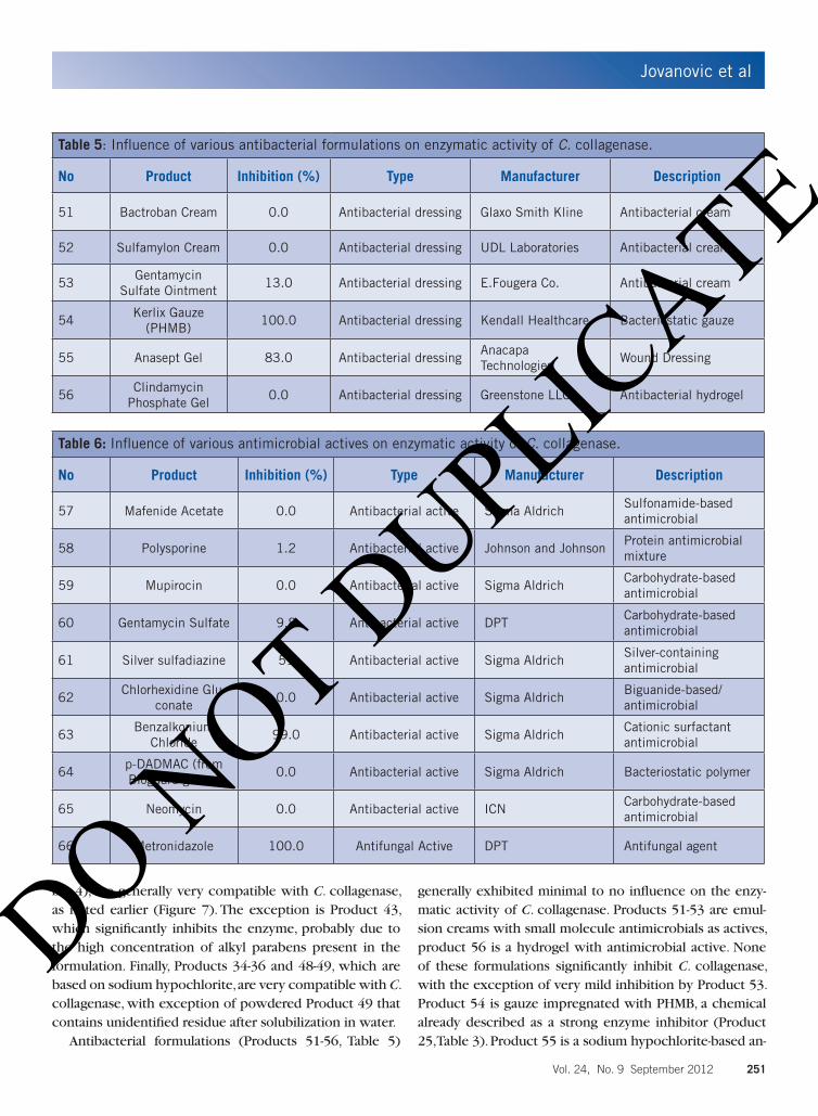
Antibacterial formulations (Products 51-56, Table 5)
generally exhibited minimal to no influence on the enzy-matic activity of C. collagenase. Products 51-53 are emul-sion creams with small molecule antimicrobials as actives, product 56 is a hydrogel with antimicrobial active. None of these formulations significantly inhibit C. collagenase, with the exception of very mild inhibition by Product 53. Product 54 is gauze impregnated with PHMB, a chemical already described as a strong enzyme inhibitor (Product 25, Table 3). Product 55 is a sodium hypochlorite-based an-
Table 5: Influence of various antibacterial formulations on enzymatic activity of C. collagenase.
No Product Inhibition (%) Type Manufacturer Description
51 Bactroban Cream 0.0 Antibacterial dressing Glaxo Smith Kline Antibacterial cream
52 Sulfamylon Cream 0.0 Antibacterial dressing UDL Laboratories Antibacterial cream
53Gentamycin
Sulfate Ointment13.0 Antibacterial dressing E.Fougera Co. Antibacterial cream
54Kerlix Gauze
(PHMB)100.0 Antibacterial dressing Kendall Healthcare Bacteriostatic gauze
55 Anasept Gel 83.0 Antibacterial dressingAnacapa Technologies
Wound Dressing
56Clindamycin
Phosphate Gel0.0 Antibacterial dressing Greenstone LLC Antibacterial hydrogel
Table 6: Influence of various antimicrobial actives on enzymatic activity of C. collagenase.
No Product Inhibition (%) Type Manufacturer Description
57 Mafenide Acetate 0.0 Antibacterial active Sigma AldrichSulfonamide-based antimicrobial
58 Polysporine 1.2 Antibacterial active Johnson and JohnsonProtein antimicrobial mixture
59 Mupirocin 0.0 Antibacterial active Sigma AldrichCarbohydrate-based antimicrobial
60 Gentamycin Sulfate 9.8 Antibacterial active DPTCarbohydrate-based antimicrobial
61 Silver sulfadiazine 51 Antibacterial active Sigma AldrichSilver-containing antimicrobial
62Chlorhexidine Glu-
conate0.0 Antibacterial active Sigma Aldrich
Biguanide-based/ antimicrobial
63Benzalkonium
Chloride99.0 Antibacterial active Sigma Aldrich
Cationic surfactant antimicrobial
64p-DADMAC (from Bioguard gauze)
0.0 Antibacterial active Sigma Aldrich Bacteriostatic polymer
65 Neomycin 0.0 Antibacterial active ICNCarbohydrate-based antimicrobial
66 Metronidazole 100.0 Antifungal Active DPT Antifungal agent
DO NOT D
UPLICATE

Jovanovic et al
252 WOUNDS® www.woundsresearch.com
timicrobial gel that strongly inhibits the activity of the en-zyme. This is somewhat contradictory with the earlier find-ings for Products 34-36 (Table 4), which contain the same concentration of sodium hypochlorite, yet without any in-fluence on enzyme activity. The difference is the presence of a gelling component in Product 55, sodium magnesium silicate, which is probably responsible for the inhibition of the enzyme activity. Anti-fungal Product 66 (Table 6) com-pletely inhibits the activity of the enzyme, probably due to complexation of the metal by metronidazole (the active molecule in the formula).
Finally, the authors tested drug actives commonly used in antimicrobial formulations (Drug Substances 57-66, Ta-ble 6). Drug Substance 58 is a free-flowing white powder, containing active peptide-based antimicrobials bacitracin and polymyxin B in a lactose base. An important finding is that these actives are compatible with C. collagenase (Table 6). Products 59, 60, and 65 (Table 6), carbohydrate-based antimicrobials, also have no influence on the enzy-matic activity of C. collagenase. Drug Substance 61 (Table 6), a silver-based antimicrobial showed significant inhibi-tory effect (refer to Table 1). Drug Substances 57, 62, and 64 have no negative effect on the enzymatic activity of C. collagenase, while cationic surfactant-like Drug Substance 63 strongly inhibited the activity of the enzyme. Prod-uct 66, an antifungal, exhibits the total inhibition of the C.collagenase.
ConclusionsIn summary, our results demonstrated the influence of
various metal salts and surfactants on the enzymatic activ-ity of C. collagenase. Alkali metal salts are chemical entities that generally have no to slight positive influence on enzy-matic activity. It was noted that the positive effect on enzy-matic activity is proportional to salt concentration, which implies the electrostatic interactions between the protein and salts are favorable for catalytic transformations. Earth alkali metal salts also exhibited minimal or mild positive influence on enzyme activity. However, divalent transition-al metals strongly inhibit the enzymatic activity of C. col-lagenase. The main difference between the 2 populations of divalent cations is the presence of electrons in the d orbitals. Transitional metals have partially filled d orbitals, and thus they are able to make donor-acceptor complexes with the enzyme, change the structure of the latter, and thus decrease its activity. The true nature of this interesting phenomenon was far beyond the scope of this text.
The influence of surfactants on the enzymatic activity of C. collagenase is primarily a function of the electrostatic
nature of the polar head. Non-charged surfactant molecules generally tend to have minimal or even slightly positive in-fluence on the enzymatic activity of C. collagenase. Large non-ionics have a particularly positive effect on enzymatic activity, probably due to favorable solubilization effects and very mild surfactantcy that is unable to unfold the protein structure. Charged surfactants, both cationic and anionic, generally inhibit enzymatic activity even at the lowest con-centration tested. This interesting difference was observed in the behavior of these 2 types of surfactants: cationics strongly inhibit the enzyme even at concentrations under the CMC, but do not completely diminish the activity of the enzyme until very high concentrations; anionics (eg, SDS) exhibited only mild inhibition at concentrations un-der CMC, but completely attenuate the activity of the en-zyme at any concentration above CMC. Amphoteric surfac-tants inhibit the activity of the enzyme proportionally with their concentration.
An important part of this work was to evaluate the com-patibility of various wound care products with C. collage-nase. Most silver-containing products are not compatible with the enzyme, significantly inhibiting its activity. How-ever, certain products containing silver in ionic form, em-bedded in polymeric matrix or foam, exhibit only mild in-fluence on enzymatic activity. Iodine-containing dressings strongly inhibit enzymatic activity regardless of the form (ie, foam dressing or gel). Commonly used wound dressings are generally compatible with the enzymatic activity of C. collagenase, leaving a plethora of choices for clinicians. Wound cleansers tend to inhibit the enzymatic activity of C. collagenase, mainly because of the presence of powerful ionic or zwitterionic surfactants. The other reasons for the enzyme inhibition are the presence of EDTA, inadequate pH of the formulations, or presence of cationic surfactant-like structures in the formulas. Wound cleansers contain-ing large block co-polymer surfactants (eg, poloxamers) or formulas with sodium hypochlorite are compatible for use with the collagenase debrider. If wound cleansers, for ex-ample, contain any other surfactant type, except non-ionic or sodium hypochlorite salts, it is strongly advised to rinse the cleanser from the wound bed with saline thoroughly before applying the collagenase debrider. Antimicrobial formulations as well as antimicrobial actives are generally compatible with C. collagenase.
This work was performed with the intention to help wound care professionals make more educated decisions with respect to what type of supporting products should be used along with a collagenase wound debrider.
DO NOT D
UPLICATE
Jovanovic et al
Vol. 24, No. 9 September 2012 253
Acknowledgements The authors wish to thank Renée Carstens for medical
writing contributions.
Reference1. Clark RA. Cutaneous tissue repair: basic biologic considerations.
I. J Am Acad Dermatol. 1985;13:701-725.
2. Schultz GS, Sibbald RG, Falanga V, et al. Wound bed preparation:
a systematic approach to wound management. Wound Repair
Regen. 2003;11 Suppl 1:S1-28.
3. Fowler E, van Rijswijk L. Using would debridement to help
achieve the goals of care. Ostomy Wound Manage. 1995;41(7A
Suppl):23S-35S.
4. Berger MM. Enzymatic debriding preparations. Ostomy Wound
Manage. 1993;39(5):61-69.
5. Steed DL, Donohoe D, Webster MW, Lindsley L. Effect of ex-
tensive debridement and treatment on the healing of dia-
betic foot ulcers. Diabetic Ulcer Study Group. J Am Coll Surg.
1996;183(1):61-64.
6. Wright J, Shi L. Accuzyme® papain urea debriding ointment: a
historical review. Wounds. 2010;15 (Supp. 4), 2-12. Ref Type: Ge-
neric.
7. Klasen HJ. A review on the nonoperative removal of necrotic
tissue from burn wounds. Burns. 2000;26(3):207-222.
8. Shi L, Carson D. Collagenase Santyl ointment: a selective agent
for wound debridement. J Wound Ostomy Continence Nurs.
2009;37(6 Suppl):S12-16.
9. Skrabut EM, Hebda PA, Samuels JA, et al. Removal of necrotic
tissue with an ananain-based enzyme-debriding preparation.
Wound Repair Regen. 1996;4(4):433-443.
10. Dryburgh N, Smith F, Donaldson J, Mitchell M. Debride-
ment for surgical wounds. Cochrane Database Syst Rev.
2008;16(3):CD006214.
11. Jackson RJ, Lim DV, Dao ML. Identification and analysis of a col-
lagenolytic activity in Streptococcus mutans. Curr Microbiol.
1997;34(1):49-54.
12. Gerlach RF, de Souza AP, Cury JA, Line SR. Effect of lead, cadmium
and zinc on the activity of enamel matrix proteinases in vitro.
Eur J Oral Sci. 2000;108(4):327-334.
13. Mallya SK, Van Wart HE. Inhibition of human neutrophil collage-
nase by gold(I) salts used in chrysotherapy. Biochem Biophys
Res Commun. 1987;144(1):101-108.
14. Inouye K, Kuzuya K, Tonomura B. Sodium chloride enhances
markedly the thermal stability of thermolysin as well as its cata-
lytic activity. Biochim Biophys Acta. 1998;1388(1):209-214.
15. Inouye K, Kuzuya K, Tonomura B. Effect of salts on the solubility
of thermolysin: a remarkable increase in the solubility as well
as the activity by the addition of salts without aggregation or
dispersion of thermolysin. J Biochem. 1998;123(5):847-852.
16. Hashida Y, Inouye K. Kinetic analysis of the activation-and-inhi-
bition dual effects of cobalt ion on thermolysin activity. J Bio-
chem. 2007;141(6):843-853.
17. Duffy B, Schwietert C, France A, et al. Transition metals as prote-
ase inhibitors. Biol Trace Elem Res.1998;64(1-3):197-213.
18. Krsti D,, Krinulovi K, Vasi V. Inhibition of Na+/K(+)-ATPase and
Mg(2+)-ATPase by metal ions and prevention and recovery of
inhibited activities by chelators. J Enzyme Inhib Med Chem.
2005;20(5):469-476.
19. Jain J, Arora S, Rajwade JM, Omray P, Khandelwal S, Paknikar
KM. Silver nanoparticles in therapeutics: development of an
antimicrobial gel formulation for topical use. Mol Pharm.
2009;6(5):1388-1401.
20. Varani J, Perone P, Inman DR, et al. Human skin in organ culture.
Elaboration of proteolytic enzymes in the presence and absence
of exogenous growth factors. Am J Pathol. 1995;146(1):210-
217.
21. Malek Alkasrawi, Eriksson T, Borjesson J, et al. The effect of
Tween-20 on simultaneous saccharification and fermentation
of softwood to ethanol. Enzyme and Microbial Technology.
2003;33(1):71-79
22. Johan Borjesson, Peterson R, Tjerneld F. Enhanced enzymatic
conversion of softwood lignocellulose by poly(ethylene glycol)
addition. Enzyme and Microbial Technology. 2007;40(4):754-
762.
23. Johnston TP, Palmer WK. Effect of poloxamer 407 on the activ-
ity of microsomal 3-hydroxy-3-methylglutaryl CoA reductase in
rats. J Cardiovasc Pharmacol. 1997;29(5):580-585.
24. Leung S, Gironella A, Trigo C, Bhushan A, Daniels CK, Lai JC. Cat-
ionic surfactants and other factors that affect enzymatic activi-
ties and transport. Proc Inst Mech Eng H. 2007;221(2):153-160.
25. Manning M, Colon W. Structural basis of protein kinetic stabil-
ity: resistance to sodium dodecyl sulfate suggests a central role
for rigidity and a bias toward beta-sheet structure. Biochemistry.
2004;43(35):11248-11254.
26. Shi L, Ermis R, Kiedaisch B, Carson D. The effect of various
wound dressings on the activity of debriding enzymes. Adv
Skin Wound Care. 2010;23(10):456-462.
27. Eming SA, Smola-Hess S, Kurschat P, Hirche D, Krieg T, Smola H.
A novel property of povidon-iodine: inhibition of excessive pro-
tease levels in chronic non-healing wounds. J Invest Dermatol.
2006;126(12):2731-2733.
28. Shi L, Ramsay S, Ermis R, Carson D. pH in the bacteria-contam-
inated wound and its impact on clostridium histolyticum col-
lagenase activity: implications for the use of collagenase wound
debridement agents. J Wound Ostomy Continence Nurs.
2011;38(5):514-521.
DO NOT D
UPLICATE