debridement: is there a limit? - npuap.org · 3/3/2017 2 objectives • describe the rationale for...
TRANSCRIPT
3/3/2017
1
©2016 National Pressure Ulcer Advisory Panel | www.npuap.org
Debridement: Is there a limit?
Lisa J. Gould, MD, PhD, FACS
Clinical Associate Professor
Brown University Department of Medicine
Faculty Disclosure
• Advisory board for Acelity
• Advisory board for Cardinal Health
• Past President of Wound Healing Society
• Plastic Surgeon with career emphasis on
difficult wounds and reconstruction
• No conflicts of interest related to this
presentation

3/3/2017
2
Objectives
• Describe the rationale for surgical
debridement of chronic wounds
• Discuss the benefits of surgical wound
debridement
• List 3 biomarkers that could be used to
determine the efficacy of wound debridement
• Describe currently available tools to
determine the margins of debridement
• Debride Necrotic Tissue
• Achieve Bacterial Balance
• Maintain Moisture Balance
• Optimize tissue perfusion
Basic Principles of Wound Bed Preparation

3/3/2017
3
What is Debridement?
• French: To remove a constraint
• Removal of foreign or unhealthy material
• Removal of Necrotic Tissue– Skin
– Fat
– Muscle/Tendon
• Removal of Slough– Fibrin
– Proteinaceous Material
– Neutrophils
– Bacteria
Why Do Wounds Need Debridement?
• Treat or prevent infection
– Decrease bacterial burden
– Reduce Inflammation
• Address Chronic (Hostile) Wound
Environment
– Remove senescent cells
– Altered signaling
• Elevated cytokines
• Inflammatory Mediators
• Lack of growth factor receptors
Brem et al. Mol Med 13(1-2) 30-33.
Lindley et al. Plast.Reconstr. Surg. 138: 18S, 2016.

3/3/2017
4
Debridement Promotes Wound Healing
JAMA Dermatol. 2013;149(9):1050-1058.
Ladder of Debridement
Surgical
Curettage
Hydrosurgery
Biological/ultrasonic
Mechanical
Enzymatic
Autolyticsimple
complex

3/3/2017
5
What is that yellow stuff?
Mature fibrin;
Traps critical
growth factors
needed for
healing
Increased MMPs
Chronic
Inflammation
Chronic Ulcer with Yellow Slough

3/3/2017
6
Fibronectin
Fibrinogen, Fibrin and Fibronectin
Fibrinogen Fibrin Vitronectin
Fibronectin Collagen type-I
Kubo et al. J Invest
Dermatol 117:1369±1381,
2001
Fibrinogen and Fibrin are Anti-Adhesive for Keratinocytes

3/3/2017
7
No Fibronectin With Fibronectin
Fibronectin Promotes Cell Attachment, Spreading and Migration
Fibroblasts
Endothelial Cells
J. Gailit and R.A.F. Clark, J Invest Dermatol 106:102-108, 1996
Herrick, Sloan, McGurk, Freak, McCollum and Ferguson.
Am J Pathol 141, 1992.
Fibronectin reappears (stable) as ulcer heals
Fibronectin is degraded in non-healing ulcer
Wysocki and Grinnell. Lab Invest 63:825, 1990
Pla
sm
a
Venous u
lcer
1
Venous u
lcer
2
Dia
betic u
lcer
1
Dia
betic u
lcer
2
Maste
cto
my F
luid
1
Maste
cto
my F
luid
2
Pla
sm
a +
CW
F 1
Pla
sm
a +
CW
F 2
Fibronectin in Chronic Wound Fluids and Tissue

3/3/2017
8
Surgical Strategy
• Operative Debridement and Irrigation– Debride to viable tissue
• Scalpel, Curette, Hydrosurgery
– Pulse lavage
– Tissue Biopsy
• Culture– Aerobes, Anaerobes
– Fungal, Atypical mycobacteria
• Pathology– Wound edge with intact skin
– Debrided tissue
• Dressing– Antimicrobial, antibiofilm
– Edema reducing
– Extracellular matrix
Debridement Decision parameters
– Pain
– Patient’s environment
– Patient’s choice and consent
– Biological age and comorbidities
– Quality of life
– Skill of the caregiver
– Resources of the caregiver
– Regulations
– GuidelinesEWMA, J Wound Care, 2013 ;22(1)

3/3/2017
9
Easy Decisions
• Massive burden of necrotic tissue
– +/- purulence
• Lack of access to wound bed
Presentation Clinic Debridement Operative
Debridement
4 weeks of NPWT
Rapid Removal of Necrotic Tissue

3/3/2017
10
Limited Access to Wound Bed
Wide Undermining
Cannot Access
Necrotic Tissue
Basic Principles: Wound Bed Preparation + Surgical Approach
• Debride necrotic tissue
• Irrigate
• Post-debridement, Post-lavage tissue biopsy
• Culture guided antibiotics
5 months
2 operations

3/3/2017
11
Small Skin Opening, Large Wound
Surgical Approach: Adapt to Patient
NPWT
Improve access to wound bed
Debride necrotic tissue
Post-debridement, post-lavage bone biopsy
Culture guided antibiotics for osteomyelitis

3/3/2017
12
Debridement considerations
• Patient comfort, positioning
• Bedside debridement– Lighting
– Bleeding
– Vital structures
– Tools
• Operative debridement– Lighting
– Bleeding
– Necrotic tissue• Extent
• Location
– Suspected Osteomyelitis
Ideal Patient
• New wound
– Known event
• Patient factors
– Nutrition
– Off-loading
– Few co-morbid illnesses, medically optimized
– Support structure
Early, Aggressive Approach

3/3/2017
13
DTPI and Unstageable: When and If?
• Bariatric Surgical Patient– Surgical complication
• Acute wound– Exact event unknown
• Probable hypotension
• Multiple operations, ICU, poor nutrition
• Appears to have demarcated
• Sensate, mobile
• Goals of care:– Reduce pain
– Prevent infection
– Rapid healing
Initial Debridement
• Nutrition marginal
• Impaired mobility
• Pain
• Wound characteristics– Patchy bleeding
– Gluteal muscle involved• Necrotic fibers
– No bone exposed
– No purulence
• Apparent excellent prognosis!
…but, not fully demarcated

3/3/2017
14
Serial Surgical Debridement
• Nutrition improving
• Mobility poor
• Bone exposed,
cortex intact
• Tunneling along
gluteal muscle
Rapid healing after Operative Excision of all Necrotic Tissue
6 weeks after operative
debridement
3 months after operative debridement

3/3/2017
15
Compare to Presence of Osteomyelitis
• 41 y/o s/p GBP with neuropathy due to spinal stenosis
• Fell on black ice in March. Noted some drainage in August and sought medical care
• Exam under anesthesia by colorectal surgeon referred to me for further care
• Appearance 7 months after initial injury
Operative Debridement
• Chronic wound
• Coccygeal fracture with osteomyelitis
• Surgical debridement with ostectomy
• Culture guided antibiotics x 12 weeks per ID
• Persistently elevated inflammatory markers
3/3/2017
16
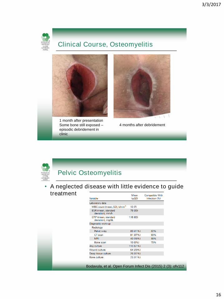
Clinical Course, Osteomyelitis
1 month after presentation
Some bone still exposed –
episodic debridement in
clinic
4 months after debridement
Pelvic Osteomyelitis
• A neglected disease with little evidence to guide
treatment
Bodavula, et al. Open Forum Infect Dis (2015) 2 (3): ofv112

3/3/2017
17
Which of these pressure injuries has osteomyelitis?
What is Osteomyelitis?
• An infection of the bone and bone marrow– Classification
• Acute– Necrotic bony spicules, inflammatory exudate
adhere to bony spicules
• Chronic– Lymphocytes, histiocytes, plasma cells
– Fibroblastic proliferation around bony spicules
– +/- new bone formation
– Long Bone
• Associated with pressure ulcer

3/3/2017
18
How Does Osteomyelitis Occur?
• Hematogenous seeding• Contiguous spread• Direct inoculation
• Healthy bone is resistant to infection• Bone injured or devitalized by trauma,
inflammation or vascular insufficiency is more susceptible
• Pressure ulcers: contiguous spread to bone damaged by decreased vascularity due to pressure
Diagnosis of Foot Osteomyelitis
• Bone biopsy for culture
• Positive probe to the bone: – PPV = 89% (JAMA (1995) 273:721-723)
• Imaging studies– Baseline foot X-ray (Sens=60%,Spec=50%)
– Triple phase Tc-MDP bone scan (Sens= 100%, Spec=56%)
– Dual isotope: In-111 leukocyte (Sens=93%, Spec=83%)
– MRI (Sens=70%, Spec=100%)• Deep abscess, septic joint, osteo, tendon rupture

3/3/2017
19
Diagnosis of Osteomyelitis in the Foot
Bone Biopsy Probe to Bone
Imaging of Osteomyelitis in the Foot

3/3/2017
20
Diagnosis of Pelvic Osteomyelitis
• Fever and presence of a wound
• WBC, ESR, pelvic X-ray: Sens 89%, Spec 88%
• Bone Scan
– Technetium
– Indium
• CT Scan
• MRI
• Bone biopsy
Pelvic Imaging Studies
• Pelvic X-ray
– Periosteal reaction, heterotopic new bone, cortical destruction
– Chronic: sclerotic bone, periosteal reaction
– Findings persist after successful treatment
– Sens 18%, Spec 100%
Lewis et al. Plast Reconstr Surg. 1988
Feb;81(2):229-32

3/3/2017
21
Bone Scan
• 99Tc: increased areas of bone turnover
– Sens 64%, Spec 57%
• Indium:
– WBC go to active infectionBone marrow activity may lead to inaccurate interpretation
– Sens 100%, Spec 75% or 50%
Melkun et al. Ann Plast Surg. 2005;54(6):633–636.
CT scan
– Extent of deep soft tissue injury
• Sens 11%, Spec 90%
Lewis et al. Plast Reconstr Surg. 1988 Feb;81(2):229-32
Santiago Restrepo et al. Rheum Dis Clin North Am. 2003;29(1):89–109.
Ma et al. Crit Rev Diagn Imaging. 1997;38(6):535–568.

3/3/2017
22
MRI
– Decreased T1 weighted, increased T2 weighted signal intensity in marrow
– Better than CT for bony changes
• Sens 98%, Spec 89%
Huang et al. J Comput Assist
Tomogr. 1998;22(3):437–443.
Can a Wound Culture Guide Therapy?
• Organisms in soft tissue do not correlate with bone
Khatri G et al, Am J Med Sci
2001;321(6):367-371
3/3/2017
23
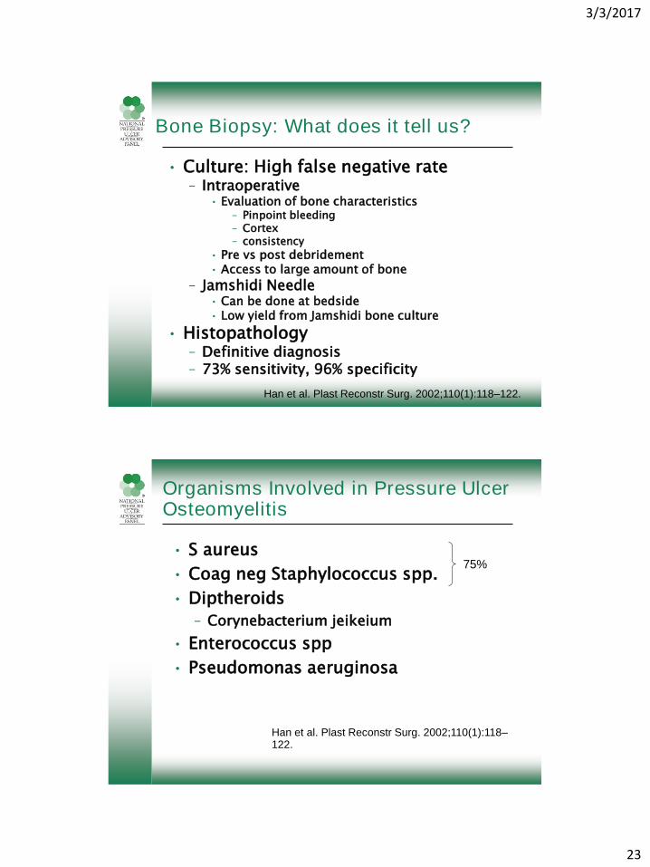
Bone Biopsy: What does it tell us?
• Culture: High false negative rate– Intraoperative
• Evaluation of bone characteristics– Pinpoint bleeding– Cortex– consistency
• Pre vs post debridement• Access to large amount of bone
– Jamshidi Needle• Can be done at bedside• Low yield from Jamshidi bone culture
• Histopathology– Definitive diagnosis– 73% sensitivity, 96% specificity
Han et al. Plast Reconstr Surg. 2002;110(1):118–122.
Organisms Involved in Pressure Ulcer Osteomyelitis
• S aureus
• Coag neg Staphylococcus spp.
• Diptheroids
– Corynebacterium jeikeium
• Enterococcus spp
• Pseudomonas aeruginosa
75%
Han et al. Plast Reconstr Surg. 2002;110(1):118–122.

3/3/2017
24
Why Treat Osteomyelitis?
Reduce post-operative complications
◦ Deep abscess
◦ Sinus tract formation
Reduce wound recurrence
Cost
◦ 1996 Incremental Cost (per pressure ulcer)
◦ Pressure ulcer treatment: $2731
◦ Associated with osteo: $59,000• Hirschberg J et al, Adv Skin Wound Care 2000;
13:25-9
Historical methods of Treatment
• 6 weeks of antibiotics
– Empiric
– Culture guided
• What is this based on?
– Experimental osteomyelitis, Norden
• J Infectious Disease 1971;124:565
• Surgical excision and flap coverage
– Antibiotic therapy

3/3/2017
25
Variability in Treatment of Pelvic Osteomyelitis
Bodavula, et al. Open Forum Infect Dis (2015) 2 (3): ofv112
• Retrospective Observational Study
220 patients with Pressure Ulcer and Pelvic
Osteomyelitis
How Do We Treat Surgically?
• Debridement
– Paint ulcer with methylene blue
– Excise wound
– Irrigate
• Send bone for pathology/microbiology
• Cover with muscle vs fasciocutaneous flap
• Antibiotic treatment guided for bone that remains, not what was taken out
Anghel et al. Plast. Reconstr. Surg. 138: 82S, 2016.

3/3/2017
26
How Should We Treat Medically?
• Pre-flap IV antibiotics
– Empiric
– Based on Culture from Jamshidi needle or operative debridement
– How long prior to surgery?
• One post-operative algorithm
– Culture negative, pathology negative: 5-7 days
– Culture negative, pathology positive: 2 weeks
– Culture positive, pathology positive: 6 weeks Han H, et al, Plast Reconstr Surg 110:118, 2002
Chronic OsteomyelitisOccult Source
26 y/o male with spina bifida had chronic drainage from right ischial
wound that was ‘healed’ for 2 years

3/3/2017
27
Small skin opening, large problem
• Presented with drainage from scrotum.
• CT scan: ‘fluid collection’ from perineum to right
thigh on CT scan.
• I&D via small incisions but persistent drainage.
Operative Debridement
9 days after I&D: 3 pieces of alginate at most distal aspect of track in thigh.
Bone exposed in ischium, positive for osteomyelitis

3/3/2017
28
Rapid Response
NPWT immediately after surgery
1 month post-debridement2 months post-debridement
5 months later, ischial wound still not healed.
Re-evaluate all potential modifiable risk factors for non-healing
Chronic Tissue Response

3/3/2017
29
Factors for Flap Reconstruction
• Reconstruction is ELECTIVE
• Ability to adhere to 6 weeks of convalescence (minimum)– Caregiver support
– Psychosocial factors
– Home vs Skilled Nursing Facility
– Understanding that additional procedures may be required
• Medical optimization– Spasms
– Contractures
– Respiratory status
– Nutrition
• Available support surfaces
Flap Reconstruction
10 days post-op
2 months post-op
(7 months after original I&D)

3/3/2017
30
NPUAP/EPUAP Palliative Care Wound Guidelines
• Manage the pressure ulcer and periwound area on a regular basis. (Strength of Evidence = B/C.)
• Debride the ulcer of devitalized tissue to control infection and odor, based on the individual’s overall quality of life (Martin et al., 1996; Pullen et al., 2002; WOCN, 2003). (Strength of Evidence = B.)
• Use conservative, non-surgical (autolytic, enzymatic) debridement of necrotic tissue as appropriate (AHCPR, 1994; WOCN, 2003; Hampton, 2006; Grocott, 2006). (Strength of Evidence = ?)
• Avoid sharp debridement with fragile tissue that bleeds easily. (Strength of Evidence = C.)
Standard vs Palliative Wound Care
STANDARD WOUND CARE
• Debride Necrotic Tissue
• Achieve Bacterial Balance
• Maintain Moisture Balance
• Optimize tissue perfusion
ALVAREZ protocol
• Stabilize the wound
• Prevent further wounds
• Eliminate odor
• Control pain
• Infection prophylaxis
• Advanced wound dressing
• Lessen dressing changes
Alvarez, et al. J PALLIATIVE MEDICINE,
2007:10(5)1161-1189

3/3/2017
31
Prevent or Treat Infection
Debridement
– Removes necrotic debris
– Prevents infection
– Eliminates odor
– Reduces exudate
– Avoid wet-to-dry gauze• Painful
• Labor intensive
• Impedes wound healing
– Combination approach• Enzymatic and sharp
ALVAREZ protocol
S tabilize the wound
P revent further wounds
E liminate odor
C ontrol pain
I nfection prophylaxis
A dvanced wound
dressing
L essen dressing
changes
Putting it all together: Patient #1
82 y/o presents to emergency room with fever,
elevated white count and altered mental status.

3/3/2017
32
Treatment intensity & prognosis
• Caregiver & family communication essential
• Consequences of decisions must be
communicated
• Hard decisions
– When to operate
– How much to operate
– Is amputation beneficial/desirable?
Putting it all together: Patient #2
• 84 y/o paraplegic due to spinal infarction, SNF resident
• Sacral pressure injury for at least 6 months
• Surgical debridement prior to presentation, down to bone but no bone cultures
• Treated empirically with IV antibiotics
• Presented to wound clinic, admitted due to new onset of seizure activity
• Operative debridement at request of infectious disease specialist
• Bone culture negative, pathology shows acute osteomyelitis
• Weight loss, no appetite, loss of strength, low albumin/prealbumin
• Son is very involved

3/3/2017
33
Putting it all together: Patient #3
15 months
70 y/o quadriplegic with h/o
COPD, resident of long-term care
facility with excellent wound care.
Developed sacral pressure injury
during respiratory exacerbation
3 months
Putting it all together: Patient #3 (cont)
Bilateral ischial pressure injuries were chronic but stable
Sent to Wound Center for worsening of right ischial wound
Pain with debridement despite topical analgesia
Returns for follow up with slough in the wound bed
Decision not to perform sharp debridement
Patient’s wishes
No evidence of infection
Debridement
in clinic
3/3/2017
34
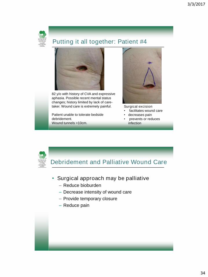
Putting it all together: Patient #4
82 y/o with history of CVA and expressive
aphasia. Possible recent mental status
changes; history limited by lack of care-
taker. Wound care is extremely painful.
Patient unable to tolerate bedside
debridement.
Wound tunnels >10cm.
Surgical excision
• facilitates wound care
• decreases pain
• prevents or reduces
infection
Debridement and Palliative Wound Care
• Surgical approach may be palliative
– Reduce bioburden
– Decrease intensity of wound care
– Provide temporary closure
– Reduce pain

3/3/2017
35
Putting it all together: Patient #5
85 y/o developed right heel blister after right hip
arthroplasty. History of PAD, ABI=0.68, vascular surgeon
states ‘not a candidate for revascularization’
Putting it all together: Patient #6
78 y/o diabetic had prolonged course after CABG.
Right heel is not painful.
Pulses are palpable
Eschar is dry
a

3/3/2017
36
Putting it all together: Patient #6 (cont)
b c d
Treatment must be adjusted based on wound
characteristics, patient functional status and medical status
Debridement: What is the Limit?
Strohal, R., Apelqvist, J., Dissemond, J. et al. EWMA Document:
Debridement. J Wound Care. 2013; 22 (Suppl. 1): S1–S52.

3/3/2017
37
Putting it all together: Patient #6 (cont)
Topical therapy and need for debridement is based on the
wound characteristics.
Wound healing was promoted by prior debridement
Debridement: Is there a limit?
• Surgical approach is
currently inexact and results
in either too much or too
little tissue removal

3/3/2017
38
The Future:Using Science to guide debridement
Taverna et al, J. Proteome Res., 2015, 14 (2), pp 986–996
The Future:Using Science to guide debridement
< Back
< Back
Lindley et al, Plastic and Reconstructive Surgery, 2016. 138(3S):
18S–28S
EGF-R
C-myc
B-catenin
MMPs

3/3/2017
39
Thank you! Questions?
This presentation is made possible by multiple contributions
from the Wound Healing Society Education Committee