the vascular endothelium, nutrients, and diseases
TRANSCRIPT

The Vascular Endothelium, Nutrients, and Diseases Suthipong Pongworn
26 March 2015

The vascular endothelium (a large endocrine organ)
ALBERTS, B., JOHNSON, A., LEWIS, J., RAFF, M., ROBERTS, K. & WALTER, P. 2008. Molecular Biology of Cell, United States of America, Garlan Science.
Rajendran, P., Rengarajan, T., Thangavel, J., Nishigaki, Y., Sakthisekaran, D., Sethi, G., & Nishigaki, I. (2013). The vascular endothelium and human diseases. International journal of biological
sciences, 9(10), 1057.
LUMEN
Tunica
adventitia Tunica
media
Tunica
intima
Blood Vessel Structure
The endothelium actively maintains approximately 60,000 miles of blood vessels in
the human body. The vast majority of endothelial cells are located in microvessels. So, our body systems rely on microvasculartory endothelial cells.

Functions of vascular endothelium (as a physical barrier and a source of a variety of regulatory substances)
Functions Fluid filtration
(Glomeruli of the kidneys)
Haemostatis
(헤모스테이티스)
The stoppage of bleeding or haemorrhage
Blood vessel tone
(혈관 톤)
Blood flow regulation
Growth of blood vessel
Neutrophil recruitment
(Platelet and leukocyte interaction)
Hormone trafficking
By membrane-bound receptors for numerous molecules - (Proteins, lipid-transporting particles, metabolites, and hormones. Specific junctional proteins and receptors that govern cell-cell and cell-matrix interaction.
Antithrombotic activity (anti-blood clotting)

Functions of vascular endothelium Haemostatic balance or Haemostasis Thrombosis and thrombolysis are the process that endothelium use to maintain blood fluidity.
Endothelial cells prevent thrombosis by means of different anticoagulant and antiplatelet mechanisms. Haemostatic pathways will limit clot formation to the areas where haemostasis is needed to restore vascular functions.
The breakdown of this complex balance (from genetic
or disturbance causes) may result in BLEEDING (출혈)or THROMBOSIS (혈전증).
Diversity (다양성) of endothelial cell The local environment elicits heterogeneous endothelial cell phenotypes determined by local needs. This heterogeneity also explains the diverse pathological responses to a disturbed vascular function. Localised manifestation of thrombosis in the exist of disturbance of systemic procoagulant systems depends on vascular bed-specific properties.
More detail about endothelial cell diversity :
Aird, W. C. (2012). Endothelial cell heterogeneity. Cold Spring Harbor perspectives in medicine, 2(1), a006429.

Functions of vascular endothelium Coagulant (응고제) mechanisms
Engelmann, B., & Massberg, S. (2013). Thrombosis as an intravascular effector of innate immunity. Nature Reviews Immunology, 13(1), 34-45.
The activity of numerous anticoagulant pathways are used to maintain
blood fluidity (healthy blood flow).
Recent studies suggest that similar changes in endothelial coagulant properties can be induced by advanced glycosylation end products, which are proteins modified by
glucose and accumulate in the vasculature at a rapid rate in diabetic subjects,
indicating the potential relevance of these mechanisms to diabetic vascular disease.

Functions of vascular endothelium Platelet and leukocyte (or white blood cells) interaction through P-selection, VWF, and other factors
http://www.birmingham.ac.uk/research/activity/mds/domains/cardio-resp-neuro/vascular-inflammation/leukocyte-trafficking/index.aspx
SMCs = inflammatory cytokines in smooth muscle cells VWF = Von Willebrand factor in Weibel-Palade bodies CCL2 / MCP-1 = Monocyte chemoattractant protein-1 CXCL4 = Platelet factor 4 TGFβ-1 = Transforming growth factor beta-1 L-TGFβ-1 = Latent form (or un-developed form)
1) Platelet adhesion to and leukocyte
rolling on the endothelium. 2) The leakage of white blood cells to
inflammation or infection sites. 3) Platelet–leukocyte interaction and
aggregation on a thrombogenic surface.
4) Vascular occlusion.
Detail in next page

Jackson, S. P. (2011). Arterial thrombosis [mdash] insidious, unpredictable and deadly. Nature medicine, 17(11), 1423-1436.
1) Platelet adhesion to and leukocyte rolling on the endothelium.
2) The leakage of white blood cells to inflammation or infection sites.
3) Platelet–leukocyte interaction and aggregation on a thrombogenic surface.
4) Vascular occlusion.
Functions of vascular endothelium Platelet and leukocyte (or white blood cells) interaction through P-selection, VWF, and other factors

Galley, H. F., & Webster, N. R. (2004). Physiology of the endothelium. British journal of anaesthesia, 93(1), 105-113.
Nitric oxide synthase (NOS) type III catalyses the production of nitric oxide from the cationic amino acid L-arginine. The enzyme is activated via changes in intracellular calcium in response to changes in shear
forces or via a receptor-mediated process. The released nitric oxide activates soluble guanylate cyclase (GC) in smooth muscle cells, converting GTP to cGMP. This activates a protein kinase which leads to the inhibition of calcium influx into the smooth muscle cell, and decreased calcium-calmodulin stimulation of
myosin light chain kinase. This in turn decreases the phosphorylation of myosin light chains, decreasing smooth muscle tension development and causing vasodilatation.
Functions of vascular endothelium Regulation of vascular tone
Detail in next page
Dysfunction of these endothelium-dependent regulatory systems may play a role in cardiovascular diseases, such as hypertension and atheroschlerosis.

ALBERTS, B., JOHNSON, A., LEWIS, J., RAFF, M., ROBERTS, K. & WALTER, P. 2008. Molecular Biology of Cell, United States of America, Garlan Science.
The role of nitric oxide (NO) in smooth muscle relaxation in a blood vessel wall. (A) Simplified drawing of an autonomic nerve contacting a blood vessel. (B) Acetylcholine released by nerve terminals in the blood vessel wall activates NO synthase in endothelial cells lining the blood vessel, causing the endothelial cells to produce NO from arginine. The NO diffuses out of the endothelial cells and into the neighboring smooth muscle cells, where it binds to and activates guanylyl cyclase to
produce cyclic GMP. The cyclic GMP triggers a response that causes the smooth muscle cells to relax, enhancing blood flow through the blood vessel.
NO acts only locally because it has a short half-life—about 5–10 seconds—in the extracellular space before oxygen and water convert it to nitrates and nitrites.
Functions of vascular endothelium

Galley, H. F., & Webster, N. R. (2004). Physiology of the endothelium. British journal of anaesthesia, 93(1), 105-113.
Cristofanilli, M., Charnsangavej, C., & Hortobagyi, G. N. (2002). Angiogenesis modulation in cancer research: novel clinical approaches. Nature Reviews Drug Discovery, 1(6), 415-426.
Vascular endothelial growth factor (VEGF) is an angiogenic factor produced by a variety of cells, including endothelial cells, with specific receptors on the endothelium. Angiogenesis - the formation of new blood vessels (or immature vessels) from pre-existing endothelium - is mediated by VEGF. VEGF contributes to the inflammatory response through stimulation of the release of adhesion molecules, metalloproteinases and nitric oxide, via the transcription factor activator protein-1 (AP-1).
Functions of vascular endothelium Growth of blood vessel (Angiogenesis)
Tumour cells release pro-angiogenic factors, such as vascular endothelial growth factor (VEGF), which diffuse into nearby tissues and bind to receptors on the endothelial cells of pre-existing blood vessels,
leading to their activation. Such interactions between endothelial cells and tumour cells lead to the secretion and activation of various proteolytic enzymes, such as matrix metalloproteinases (MMPs), which degrade the basement membrane and the extracellular matrix. Degradation allows activated endothelial cells — which are stimulated to proliferate by growth factors — to migrate towards the tumour.
Integrin molecules, such as v3-integrin, help to pull the sprouting new blood vessel forward. The endothelial cells deposit a new basement membrane and secrete growth factors, such as platelet-derived growth factor (PDGF), which attract supporting cells to stabilize the new vessel. PDGFR, PDGF receptor; VEGFR, VEGF receptor (Cristofanilli, Charnsangavej, & Hortobagyi, 2002).
Metalloproteinases = any protease enzyme whose catalytic mechanism involves a metal

The Vascular Endothelium and Human Diseases
Induced causes
•Obesity
•Smoking
•Sleep deprivation
•Acute microbial infections
•High glucose intake
•Exposure to metals or air pollutants
Free Radical
•Endothelial dysfunction can be caused by several conditions, including diabetes, or metabolic syndrome, hypertension, and physical inactivity.
Endothelial Dysfunction
•Endothelial cell damage
• Permeability allowing toxins to pass into body tissues
•Impaired cell signaling systems
Diseases
•Vascular leakage
•Atherosclerosis
•Stroke
•Heart disease
•Hypertension
•Coronary artery disease
•Chronic heart failure
•Peripheral vascular disease
•Diabetes and Insulin resistance
•Chronic kidney failure
•Cancer
•Severe infectious diseases
•Alzheimer’s disease
•Parkinson’s disease
Rajendran, P., Rengarajan, T., Thangavel, J., Nishigaki, Y., Sakthisekaran, D., Sethi, G., & Nishigaki, I. (2013). The vascular endothelium and human diseases. International journal of biological sciences, 9(10), 1057. Seshadri, S., Beiser, A., Selhub, J., Jacques, P. F., Rosenberg, I. H., D'Agostino, R. B., ... & Wolf, P. A. (2002). Plasma homocysteine as a risk factor for dementia and Alzheimer's disease. New England Journal of Medicine, 346(7), 476-483.0 McDowell, I. F., & Lang, D. (2000). Homocysteine and endothelial dysfunction: a link with cardiovascular disease. The Journal of nutrition, 130(2), 369S-372S.
Disrupt the balance of NO

Peripheral Vascular Disease (PAD)
Libby, P. (2002). Atherosclerosis in inflammation. Nature, 420, 868-874.
Schematic of the life history of an atheroma. The normal human coronary artery has a
typical trilaminar structure. The endothelial cells in contact with the blood in the arterial
lumen rest upon a basement membrane. The intimal layer in adult humans generally
contains a smattering of smooth muscle cells scattered within the intimal extracellular
matrix. The internal elastic lamina forms the barrier between the tunica intima and the
underlying tunica media. The media consists of multiple layers of smooth muscle cells,
much more tightly packed than in the diffusely thickened intima, and embedded in a
matrix rich in elastin as well as collagen. In early atherogenesis, recruitment of
inflammatory cells and the accumulation of lipids leads to formation of a lipid-rich
core, as the artery enlarges in an outward, ablumenal direction to accommodate the
expansion of the intima. If inflammatory conditions prevail and risk factors such as
dyslipidaemia persist, the lipid core can grow, and proteinases secreted by the
activated leukocytes can degrade the extracellular matrix, while pro-inflammatory
cytokines such as interferon-g (IFN-g) can limit the synthesis of new collagen. These
changes can thin the fibrous cap and render it friable and susceptible to rupture.
When the plaque ruptures, blood coming in contact with the tissue factor in the
plaque coagulates. Platelets activated by thrombin generated from the coagulation
cascade and by contact with the intimal compartment instigate thrombus formation.
If the thrombus occludes the vessel persistently, an acute myocardial infarction can
result (the dusky blue area in the anterior wall of the left ventricle, lower right).
The thrombus may eventually resorb as a result of
endogenous or therapeutic thrombolysis. However, a
wound healing response triggered by thrombin generated
during blood coagulation can stimulate smooth muscle
proliferation. Plateletderived growth factor (PDGF)
released from activated platelets stimulates smooth
muscle cell migration. Transforming growth factor-b (TFG-
b), also released from activated platelets, stimulates
interstitial collagen production. This increased migration,
proliferation and extracellular matrix synthesis by smooth
muscle cells thickens the fibrous cap and causes further
expansion of the intima, often now in an inward direction,
yielding constriction of the lumen. Stenotic lesions
produced by the lumenal encroachment of the fibrosed
plaque may restrict flow, particularly under situations of
increased cardiac demand, leading to ischaemia,
commonly provoking symptoms such as angina pectoris.
Advanced stenotic plaques, being more fibrous, may
prove less susceptible to rupture and renewed thrombosis.
Lipid lowering can reduce lipid content and calm the
intimal inflammatory response, yielding a more ‘stable’
plaque with a thick fibrous cap and a preserved lumen (centre).
Loss of nitric oxide endothelial dysfunction plaque rupture PAD and coronary artery disease
Atherosclerosis = A thicken wall as a
result of invasion and accumulation of white blood cells

Peripheral Vascular Disease (PAD)
Libby, P. (2002). Atherosclerosis in inflammation. Nature, 420, 868-874. Plaque rupture in the lower extremities (the end parts of your body such as hands and feet) would produce and acute
reduction in blood flow, and this mechanism has been proposed to contribute to the development of critical limb ischemia (not
enough blood flowing to a part of the body).
Furthermore, thrombus reorganization (혈액 응고 개혁) following subacute rupture has been proposed as a mechanism for lesion
progression (병변의 진행). Consistent with these potential mechanisms, several small cross-sectional studies have demonstrated the loss of nitric oxide bioavailability in patients
with PAD. Urine nitrate and cyclic GMP levels (Cyclic guanosine monophosphate levels) are reduced in patients with PAD,
suggesting decreased total body nitric oxide production ( NO production).
PAD is associated with increased production of endothelin (proteins that constrict blood vessels and raise blood pressure) and plasminogen activator inhibitor-1 (or endothelial plasminogen activator inhibitor, functions as anti-thrombolysis).
Loss of nitric oxide endothelial dysfunction (promoting) plaque rupture PAD and coronary artery disease
Atherosclerosis = A thicken wall as a
result of invasion and accumulation of white blood cells
Rajendran, P., Rengarajan, T., Thangavel, J., Nishigaki, Y., Sakthisekaran, D., Sethi, G., & Nishigaki, I. (2013). The vascular endothelium and human diseases. International journal of biological sciences, 9(10), 1057.

STROKE There is much evidence suggesting that endothelial dysfunction can play a role in the pathogenesis of ischemic stroke.
Vascular sources of ROS and BP (blood pressure)
• ( Salt diet)
• The superoxide-producing enzyme NADPH oxidase, xanthine oxidase, mitochondrial enzymes, and nitric oxide synthase (NOS, a state in which this enzyme generates superoxide instead of nitric oxide.)
Many causes
(Oxidative stress and vascular inflammation are major pathways of their bad effects on blood vessels.)
• BP = the most important risk factors for stroke.
• Many cardiovascular factors increase the production rate of reactive oxygen species ( ROS) and promote inflammation in systemic and cerebral blood vessels.
Many vascular diseases
• Stroke (cerebrovascular accidents) clearly represents a typical example of the potential role of a dysfunctional endothelium.
• With 30% incidence and mortality rate of stroke, it is related to death, long-term disability and suffering conditions.
Rajendran, P., Rengarajan, T., Thangavel, J., Nishigaki, Y., Sakthisekaran, D., Sethi, G., & Nishigaki, I. (2013). The vascular endothelium and human diseases. International journal of biological sciences, 9(10), 1057.
In spite of an improved control of blood pressure, the secular trend of stroke in well-controlled populations is increasing.
It remains to be determined how individual risk factors trigger the activation of one or both of these processes (oxidative stress and vascular inflammation).

Hypertension and Atherosclerosis (고혈압 과 동맥경화) It is not clear whether endothelial cell damage is the cause or the result of hypertension.
Endothelial dysfunction is the cause of….
2) On the other hand, another study , in 1993, found that treatment of hypertension did not improve endothelial function, arguing against endothelial dysfunction as being a consequence of hypertension.
3) In 2002, lowering blood pressure with beta-blockers does not improve endothelial function; whereas
4) Supporting the notion that endothelial dysfunction is one of the causes of hypertension is the finding of impaired endothelial function in the normotensive offspring of patients with essential hypertension(1998).
Endothelial dysfunction is the result of….
1) In 1978, Moncada and Vane suggested that endothelial dysfunction follows the course of a chronic increase in blood pressure and is therefore a consequence of hypertension.
3) In 2002, the treatment with angiotensin-converting enzyme inhibitors (ACEI’s - the treatment of hypertension) or angiotensin-receptor blockers (ARB’s) significantly improves endothelial function.
Rajendran, P., Rengarajan, T., Thangavel, J., Nishigaki, Y., Sakthisekaran, D., Sethi, G., & Nishigaki, I. (2013). The vascular endothelium and human diseases. International journal of biological sciences, 9(10), 1057.
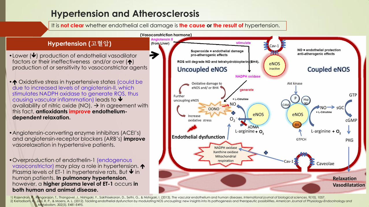
Hypertension and Atherosclerosis It is not clear whether endothelial cell damage is the cause or the result of hypertension.
Hypertension (고혈압)
•Lower () production of endothelial vasodilator factors or their ineffectiveness and/or over () production of or sensitivity to vasoconstrictor agents
• Oxidative stress in hypertensive states (could be due to increased levels of angiotensin-II, which stimulates NADPH oxidase to generate ROS, thus causing vascular inflammation) leads to availability of nitric oxide (NO). In agreement with this fact, antioxidants improve endothelium-dependent relaxation.
•Angiotensin-converting enzyme inhibitors (ACEI’s) and angiotensin-receptor blockers (ARB’s) improve vasorelaxation in hypertensive patients.
•Overproduction of endothelin-1 (endogenous vasoconstrictor) may play a role in hypertension. Plasma levels of ET-1 in hypertensive rats, But in human patients. In pulmonary hypertension, however, a higher plasma level of ET-1 occurs in both human and animal disease.
1) Rajendran, P., Rengarajan, T., Thangavel, J., Nishigaki, Y., Sakthisekaran, D., Sethi, G., & Nishigaki, I. (2013). The vascular endothelium and human diseases. International journal of biological sciences, 9(10), 1057. 2) Kietadisorn, R., Juni, R. P., & Moens, A. L. (2012). Tackling endothelial dysfunction by modulating NOS uncoupling: new insights into its pathogenesis and therapeutic possibilities. American Journal of Physiology-Endocrinology and Metabolism, 302(5), E481-E495.
(Vasoconstriction hormone)

Hypertension and Atherosclerosis It is not clear whether endothelial cell damage is the cause or the result of hypertension.
Hypertension (고혈압)
•Lower () production of endothelial vasodilator factors or their ineffectiveness and/or over () production of or sensitivity to vasoconstrictor agents
• Oxidative stress in hypertensive states (could be due to increased levels of angiotensin-II, which stimulates NADPH oxidase to generate ROS, thus causing vascular inflammation) leads to availability of nitric oxide (NO). In agreement with this fact, antioxidants improve endothelium-dependent relaxation.
•Angiotensin-converting enzyme inhibitors (ACEI’s) and angiotensin-receptor blockers (ARB’s) improve vasorelaxation in hypertensive patients.
•Overproduction of endothelin-1 (endogenous vasoconstrictor) may play a role in hypertension. Plasma levels of ET-1 in hypertensive rats, But in human patients. In pulmonary hypertension, however, a higher plasma level of ET-1 occurs in both human and animal disease.
1) Rajendran, P., Rengarajan, T., Thangavel, J., Nishigaki, Y., Sakthisekaran, D., Sethi, G., & Nishigaki, I. (2013). The vascular endothelium and human diseases. International journal of biological sciences, 9(10), 1057. 2) Fr Channick, R; Rubin, L. Endothelin Receptor Antagonism: A New Era in the Treatment of Pulmonary Arterial Hypertension. Advances in Pulmonary Hypertension. 2002; 1(1):13-17.
Fig. 1—Illustration of the actions
of endothelin-1 (ET-1) on vascular
smooth muscle cells. In addition
to contraction, ET-1 can mediate
smooth muscle cell relaxation
through release of PG2
(vasodilatory prostacyclin )and nitric oxide - NO
Abnormalities in the Endothelin
System in Pulmonary Hypertension Numerous studies have confirmed the prominent role of abnormalities in ET-1 in the pulmonary hypertensive process. Patients with primary pulmonary hypertension (PPH) have been shown to have elevated circulating levels of ET-1, with higher arterial than venous levels, suggesting increased pulmonary production.9 Some investigators have found that levels of ET-1 correlate with the severity of pulmonary hypertension.
2)
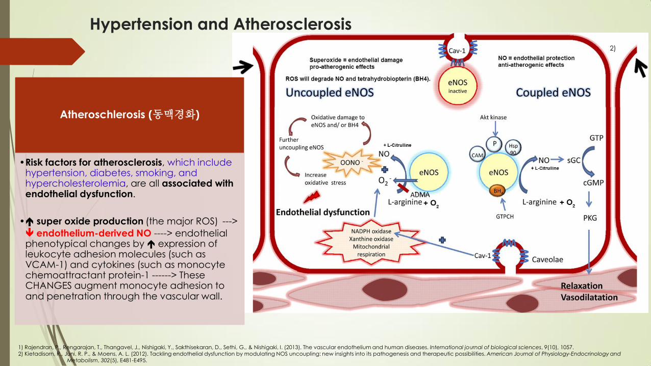
Hypertension and Atherosclerosis
1) Rajendran, P., Rengarajan, T., Thangavel, J., Nishigaki, Y., Sakthisekaran, D., Sethi, G., & Nishigaki, I. (2013). The vascular endothelium and human diseases. International journal of biological sciences, 9(10), 1057. 2) Kietadisorn, R., Juni, R. P., & Moens, A. L. (2012). Tackling endothelial dysfunction by modulating NOS uncoupling: new insights into its pathogenesis and therapeutic possibilities. American Journal of Physiology-Endocrinology and Metabolism, 302(5), E481-E495.
Atheroschlerosis (동맥경화)
•Risk factors for atherosclerosis, which include hypertension, diabetes, smoking, and hypercholesterolemia, are all associated with endothelial dysfunction.
• super oxide production (the major ROS) --->
endothelium-derived NO ----> endothelial phenotypical changes by expression of leukocyte adhesion molecules (such as VCAM-1) and cytokines (such as monocyte chemoattractant protein-1 ------> These CHANGES augment monocyte adhesion to and penetration through the vascular wall.
2)

Hypertension and Atherosclerosis
1) Rajendran, P., Rengarajan, T., Thangavel, J., Nishigaki, Y., Sakthisekaran, D., Sethi, G., & Nishigaki, I. (2013). The vascular endothelium and human diseases. International journal of biological sciences, 9(10), 1057. 2) Khan, F., Galarraga, B., & Belch, J. J. (2010). The role of endothelial function and its assessment in rheumatoid arthritis. Nature Reviews Rheumatology, 6(5), 253-261.
Atheroschlerosis (동맥경화)
•Risk factors for atherosclerosis, which include hypertension, diabetes, smoking, and hypercholesterolemia, are all associated with endothelial dysfunction.
• super oxide production (the major ROS) --->
endothelium-derived NO ----> endothelial phenotypical changes by expression of leukocyte adhesion molecules (such as VCAM-1) and cytokines (such as monocyte chemoattractant protein-1 ------> These CHANGES augment monocyte adhesion to and penetration through the vascular wall.
2)
Endothelin-1 (ET-1) 1) Can have a significant role in atherogenesis ( ET-1 in hyperlipidemia and early and
advanced atherosclerosis).
2) Enhances atherogenesis through several mechanisms.

Hypertension and Atherosclerosis
1) Rajendran, P., Rengarajan, T., Thangavel, J., Nishigaki, Y., Sakthisekaran, D., Sethi, G., & Nishigaki, I. (2013). The vascular endothelium and human diseases. International journal of biological sciences, 9(10), 105 2) Fr Channick, R; Rubin, L. Endothelin Receptor Antagonism: A New Era in the Treatment of Pulmonary Arterial Hypertension. Advances in Pulmonary Hypertension. 2002; 1(1):13-17.
2)
Mechanisms of Endothelin-1 (ET-1) in Atherogenesis
It is a strong chemoattractant that acts by stimulating ETB receptors on circulating monocytes.
ET-1 activates macrophages leading to over secretion of inflammatory mediators such as IL-6, IL-8, TNF, PGE2, and superoxide anion.
ET-1 stimulates smooth muscle cell migration and hypertrophy and the production of firoblast growth factor-2, making them hyper responsive to angiotensin-2.
ET-1 increases fibroblast proliferation, chemotaxis, and matrix biosynthesis.
ET-1 causes PKC activation and increases platelet adherence through increased expression of P-selectin.

Diabetes
Patients with diabetes invariably show impaired endothelium-dependent vasodilation.
Hypertension
Obesity
Dyslipidemia
(an abnormal amount of lipids in the blood)
Consume high-calorie diet rich in macronutrients.
Insulin resistance
Hyperglycemia
Low-grade systemic inflammation
Protein, lipid, and glucose loads
(High-fat meals)
ROS production Impaired endothelium-dependent vasodilation
Rajendran, P., Rengarajan, T., Thangavel, J., Nishigaki, Y., Sakthisekaran, D., Sethi, G., & Nishigaki, I. (2013). The vascular endothelium and human diseases. International journal of biological sciences, 9(10), 105 Sundell, J., & Knuuti, J. (2003). Insulin and myocardial blood flow. Cardiovascular Research, 57(2), 312-319.
Systemic low-grade inflammation, defined by a 2- to 3-fold
increase in plasma concentrations of cytokines and acute
phase proteins, is associated with chronic disease such as
atherosclerosis, the metabolic syndrome, and type 2 diabetes mellitus.
Expression
production
( Blood flow)
The interaction between insulin & the NO system.
re=receptor, eNOS=endothelial NO synthase,
GTP=guanosine triphosphate, sGC=soluble
guanylate cyclase, cGMP=cyclic guanosine monophosphate, Ca2+=calcium
The activation of
insulin-receptor substrate pathway

Rajendran, P., Rengarajan, T., Thangavel, J., Nishigaki, Y., Sakthisekaran, D., Sethi, G., & Nishigaki, I. (2013). The vascular endothelium and human diseases. International journal of biological sciences, 9(10), 1057.
Xu, J., & Zou, M. H. (2009). Molecular insights and therapeutic targets for diabetic endothelial dysfunction. Circulation, 120(13), 1266-1286.
Endothelial vs. Insulin resistance
Peripheral endothelial dysfunction
Insulin resistance & insulin resistance syndrome
Endothelial dysfunction & atherogenesis in the large
vessels.
more likely to generate
accelerates
(Phosphatidylinositol 3-kinase )
(insulin receptor substrate) Src [sarcoma]
Homology domain C-terminal
(phosphoinositide-dependent kinase-1)
(Protein kinase B)
(mitogen-activated protein kinase)
(mitogen-activated protein kinase kinase)
Functon
Hyperinsulinemia
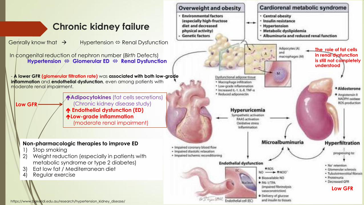
Chronic kidney failure
Genrally know that Hypertension Renal Dysfunction
The role of fat cells
in renal dysfunction
is still not completely understood
- A lower GFR (glomerular filtration rate) was associated with both low-grade
inflammation and endothelial dysfunction, even among patients with moderate renal impairment.
Low GFR
Adipocytokines (fat cells secretions) (Chronic kidney disease study) Endothelial dysfunction (ED)
Low-grade inflammation (moderate renal impairment)
Low GFR
In congenital reduction of nephron number (Birth Defects) Hypertension Glomerular ED Renal Dysfunction
https://www.bakeridi.edu.au/research/hypertension_kidney_disease/
Non-pharmacologic therapies to improve ED 1) Stop smoking 2) Weight reduction (especially in patients with
metabolic syndrome or type 2 diabetes) 3) Eat low fat / Mediterranean diet 4) Regular exercise

Cancer
Endothelial cell migration
Filopodia = membrane projections that contain long parallel actin filaments arranged in tight bundles.
Filopodia
Lamellipodia
cytoplasmic
Stress fibers = actin filaments of inverted polarity
linked by β-actinin and myosin and distributed along contractile fibers
Stress fibers
(Formation and protrusion of lamellipodia)
recycling of adhesive & signalling materials.
Rajendran, P., Rengarajan, T., Thangavel, J., Nishigaki, Y., Sakthisekaran, D., Sethi, G., & Nishigaki, I. (2013). The vascular endothelium and human diseases. International journal of biological sciences, 9(10), 1057.
Lamalice, L., Le Boeuf, F., & Huot, J. (2007). Endothelial cell migration during angiogenesis. Circulation research, 100(6), 782-794.
Tumors can give off chemical signals that stimulate
angiogenesis and can also stimulate nearby normal
cells to produce angiogenesis signaling molecules. The
resulting new blood vessels “feed” growing tumors with oxygen and nutrients.

Endothelial cell relocation during angiogenesis
Rajendran, P., Rengarajan, T., Thangavel, J., Nishigaki, Y., Sakthisekaran, D., Sethi, G., & Nishigaki, I. (2013). The vascular endothelium and human diseases. International journal of biological sciences, 9(10), 1057.
• The directional migration toward an increasing gradient of soluble chemoattractants.
• Chemotaxis is driven by growth factors such as VEGF and basic fibroblast growth factor (bFGF).
Chemotaxis
• The directional migration toward an increasing gradient of immobilized ligands.
• Haptotaxis is associated with increased endothelial cell migration activated in response to integrins binding to the extracellular matrix components.
Haptotaxis
• The directional migration generated by mechanical forces.
• Shear force in blood vessel contributes to the activation of migratory pathways.
Mechanotaxis
There are 3 major mechanisms involved in endothelial cell migration.

The endothelium plays a key role in the pathogenesis of coagulation disorders in infectious diseases, although the exact mechanisms are not yet clear in some cases. It is involved in both bacterial and non-bacterial infections and is important for the initiation and regulation of hemostasis.
KEY : The loss of the endothelium barrier and vascular leakage
Rajendran, P., Rengarajan, T., Thangavel, J., Nishigaki, Y., Sakthisekaran, D., Sethi, G., & Nishigaki, I. (2013). The vascular endothelium and human diseases. International journal of biological sciences, 9(10), 1057.
Vaheri, A., Strandin, T., Hepojoki, J., Sironen, T., Henttonen, H., Mäkelä, S., & Mustonen, J. (2013). Uncovering the mysteries of hantavirus infections. Nature reviews microbiology, 11(8), 539-550.
Severe infectious diseases
virus causes changes in vascular permeability without damaging the endothelium.
Hantavirus pulmonary syndrome

The endothelium plays a key role in the pathogenesis of coagulation disorders in infectious diseases, although the exact mechanisms are not yet clear in some cases. It
is involved in both bacterial and non-bacterial infections and is important for the initiation and regulation of hemostasis.
KEY : The loss of the endothelium barrier and vascular leakage
Rajendran, P., Rengarajan, T., Thangavel, J., Nishigaki, Y., Sakthisekaran, D., Sethi, G., & Nishigaki, I. (2013). The vascular endothelium and human diseases. International journal of biological sciences, 9(10), 1057.
Avirutnan, P., & Matangkasombut, P. (2013). Unmasking the role of mast cells in dengue. Elife, 2, e00767.
Severe infectious diseases
- dengue hemorrhagic fever
virus causes changes in vascular
permeability without damaging the endothelium.

It is conceivable that the therapeutic
correction of endothelial dysfunction may lead to an improvement of
prognosis in patients with PAD,
cardiovascular diseases, stroke,
chronic kidney failure, cancer or
infectious disease.
However, scant data are available
on this topic, and most of the
conclusions that can be draw are
highly speculative(based on a guess). Therefore, there is virtually no
available substance able to specifically target the endothelium.
Pharmacological remedies (Therapy goals)
Rajendran, P., Rengarajan, T., Thangavel, J., Nishigaki, Y., Sakthisekaran, D.,
Sethi, G., & Nishigaki, I. (2013). The vascular endothelium and human
diseases. International journal of biological sciences, 9(10), 1057.

Pharmacological remedies (Therapy goals)
Rajendran, P., Rengarajan, T., Thangavel, J., Nishigaki, Y., Sakthisekaran, D., Sethi, G., & Nishigaki, I. (2013). The vascular endothelium and human diseases. International journal of biological sciences, 9(10), 1057.
•Angiotensin-converting enzyme (ACE) inhibitors
•Statins
•Insulin sensitizers
•L-arginine
•Agents that target endothelial nitric oxide synthase (eNOS)
•Folates or tetrahydrobioterin
•Inhibitors of Rho-kinase, PARP [poly(ADP-ribose) polymerase], PTPase (Protein tyrosine phosphatase), geranyl transferase, transketolase
•Activators of Akt (Protein kinase B or PKB) and PKA (Protein kinase A)
Pharmacological interventions
(Medicinal uses)
To restore endothelial function
•Peripheral vascular disease
•Stroke
•Heart disease
•Diabetes
•Insulin resistance
•Chronic kidney failure
•Tumor growth
•Venous thrombosis
•Severe viral infectious diseases.
•Metastasis (the spread of a cancer or disease from one organ or part to another not directly connected with it)
Protect against diseases

Role of Vitamin C in the Function of the Vascular Endothelium
May, J. M., & Harrison, F. E. (2013). Role of vitamin C in the function of the vascular endothelium. Antioxidants & redox signaling, 19(17), 2068-2083.
Metabolism of vitamin C (ascobic acid, or ascorbate)
Ascorbate donates a single electron to become the ascorbate radical, which reacts with another ascorbate radical to form a molecule each of ascorbate and dehydroascorbate (DHA). The latter is unstable at physiologic pH and if not reduced back to ascorbate via GSH-dependent mechanisms, it will undergo irreversible ring opening and loss.
In buffers, DHA forms a hemiketal that has a molecular structure resembling that of glucose.
unstable form of a tri-
ketone lactone ring structure
Ascorbate Chemistry and Biochemical Functions
donates a single electron
Of 2 ascorbate radicals
1)
2)
Ascorbate radical is not very reactive with anything but Itself.

Role of Vitamin C in the Function of the Vascular Endothelium
May, J. M., & Harrison, F. E. (2013). Role of vitamin C in the function of the vascular endothelium. Antioxidants & redox signaling, 19(17), 2068-2083.
Humans cannot synthesize their own vitamin C, it
should be absorbed in the intestine and carried through the circulation to the various organs.
Ascorbate Uptake
Ascorbate (AA) is taken up from the intestine either on the
SVCT1 or as dehydroascorbate (DHA) on glucose
transporters (not shown). Once inside the intestinal
epithelium, it exits by an unknown mechanism on the
basolateral membrane into the interstitium and then into
nearby capillaries. Ascorbate in the bloodstream is taken
up by erythrocytes (either as DHA or as slow diffusion) and
by leukocytes and endothelial cells on the SVCT2. Plasma
ascorbate is distributed by the vascular tree to organ
beds. Interstitial ascorbate is then taken up by the SVCT2
on nucleated cells in the organs. In the central nervous
system, ascorbate enters the cerebrospinal fluid largely by
secretion from the choroid plexus (not shown).
SVCT1, sodium-dependent vitamin C transporter 1; SVCT2, sodium-dependent vitamin C transporter 2.
(sp
ac
e)
(leukocytes)
(capillaries)
Due to its low molecular weight, vitamin C is
freely filtered by the kidney, but reabsorbed in the renal proximal tubule, again by the SVCT1,
“ascorbate conserved mechanism”.
DHA ≤ 2μM

Role of Vitamin C in the Function of the Vascular Endothelium
May, J. M., & Harrison, F. E. (2013). Role of vitamin C in the function of the vascular endothelium. Antioxidants & redox signaling, 19(17), 2068-2083.
Superoxide (especially in response to excessive glucose metabolism in diabetes)
Endothelial cell ascorbate uptake and recycling.
Ascorbate (AA) enters endothelial cells largely on the SVCT2, although a small amount may come in as DHA on glucose transporters (GLUT), to be rapidly reduced to ascorbate in the cell. Once in the cell, ascorbate can donate an electron ferric
iron, superoxide (O2- ), and other radical species generated in
mitochondria or via activation of cell surface receptors, such as those for thrombin or advanced glycation end-products (AGE). The resulting ascorbate radical (AA - ) is mostly reduced directly back to ascorbate by NADH- and NADPH-dependent reductases. However, if the oxidative stress is overwhelming, the
ascorbate radical may dismutate to form ascorbate and DHA, with subsequent reduction of the latter back to ascorbate.
1) 2)
Plentiful NO will react at diffusion-limited rates with superoxide, generating the strong oxidant peroxynitrite. The major scavenger of superoxide in cells is likely to be superoxide dismutase (react in
vitro with superoxide 105 times faster than Ascorbate).
Low millimolar ascorbate concentrations in endothelial
cells may allow ascorbate to aid in scavenging both superoxide and peroxynitrite.
GLUT or SLC2A family are a membrane proteins that facilitate glucose transport across a plasma membrane and are found in most mammalian cells.

Role of Vitamin C in the Function of the Vascular Endothelium
May, J. M., & Harrison, F. E. (2013). Role of vitamin C in the function of the vascular endothelium. Antioxidants & redox signaling, 19(17), 2068-2083.
Ascorbate transfer across the endothelial barrier
Routes of transfer of ascorbate out of the vascular bed as represented by endothelial cells in culture on semiporous filters. Ascorbate (AA) or DHA added on the luminal side of endothelial cells cultured on semi-porous membranes enter the cells on the SVCT2 or GLUT-type
transporter, respectively. The resulting ascorbate is trapped with little exit on the basolateral side of the cells over a 90 min time-frame. Rather, most ascorbate passes between the cells by a paracellular route, which intracellular ascorbate tightens. There may also be some transit of ascorbate between the cells as sieving across tight junctional proteins.
SVCT2, sodium-dependent vitamin C transporter 2
Ascorbate could enter
through endothelial cells OR going between them.
enhanced by intracellular calcium
From microdialysis measurement of subcutaneous ascorbate concentration: - Interstitial ascorbate concentration = 1 mM = similar to the SVCT2 generation = 15-20 fold higher than present in blood.
- If correct the simple diffusion between microcapillary endothelial cells IS NOT reflect the situation in vivo.

Role of Vitamin C in the Function of the Vascular Endothelium
May, J. M., & Harrison, F. E. (2013). Role of vitamin C in the function of the vascular endothelium. Antioxidants & redox signaling, 19(17), 2068-2083.
Ascorbate function in endothelial cells
Antioxidant function: scavenging of
endogenous and exogenous radicals
Recycling of intracellular radicals of cellular
constituents and enzyme co-factors
Regulation of enzyme
phosphorylation
Enzyme co-factor function
(Glutathione)
Note: GSH can be synthesized by the cell.
(BH4)

Ascorbate recycling of BH4 (tetrahydrobiopterin) is especially important for the proper function of endothelial nitric oxide synthase (eNOS).
Role of Vitamin C in the Function of the Vascular Endothelium
May, J. M., & Harrison, F. E. (2013). Role of vitamin C in the function of the vascular endothelium. Antioxidants & redox signaling, 19(17), 2068-2083.
Recycling of intracellular radicals of cellular constituents and enzyme co-factors
efficiently reduced
Superoxide
peroxunitrite
Ascorbate recycling of BH4 and preservation of nitric oxide. Dimeric eNOS (eNOSd) attached to the endothelial cell plasma membrane
utilizes arginine, molecular oxygen, and BH4 to generate nitric oxide (NO) that subsequently activates endothelial and smooth muscle guanylate cyclase (G. cyclase). In the enzyme cycle, the trihydrobiopterin radical (BH3 ) is generated, which is recycled by ascorbate (AA). The resulting ascorbate radical (AA-) is recycled by various NAD(P)H-dependent reductases. Failure to recycle BH3 , or its formation due to BH4 oxidation
by reactive oxygen species (ROS), results in the formation of dihydrobiopterin (BH2), which competes with BH4 for the enzyme. This, and loss of BH4 uncouples eNOS, which then dissociates from the membrane into monomers (eNOSm) that generate superoxide (O2
- ) rather than NO. Reaction of O2
- with any available NO forms peroxynitrite, a strong nitrating oxidant. By initially recycling BH4,
ascorbate prevents loss of BH4 and sustains eNOS activity. BH4, tetrahydrobiopterin.

Role of Vitamin C in the Function of the Vascular Endothelium
May, J. M., & Harrison, F. E. (2013). Role of vitamin C in the function of the vascular endothelium. Antioxidants & redox signaling, 19(17), 2068-2083.
Regulation of enzyme phosphorylation – multiple mechanisms by which ascorbate preserves nitric oxide and tightens the endothelial permeability barrier.
In endothelial cells in which NADPH oxidase (NOX) is
activated by septic insult (or other mechanisms), the resulting superoxide (O2
- ) reacts with available nitric oxide (NO) to form peroxynitrite (ONOO- ), which nitrates and activates PP2A. The phosphatase then dephosphorylates occludin, causing it to pull away from the membrane and weaken tight junctional
structures. Ascorbate prevents the activation of PP2A in this pathway by inhibiting NOX function and scavenging O2
- and ONOO- . In unstimulated cells (with presumably low levels of ONOO- , ascorbate
also enhances nitric oxide generation by inhibiting PP2A by an unknown mechanism. This prevents PP2A from dephosphorylating and thus deactivating eNOS
itself, as well as the AMP-dependent kinase (AMPK). The resulting increase in eNOS phosphorylation is mediated at least in part by phosphorylation-dependent activation of AMPK, which activates eNOS to generate nitric oxide. This, along with the preservation of BH4 by ascorbate, increases
intracellular nitric oxide, which then generates cyclic GMP through the canonical pathway to eventually tighten the endothelial permeability barrier. PP2A, protein phosphatase type 2A.
PP2A = protein phosphatase type 2A ; AA = Ascorbate
un
kn
ow
n

May, J. M., & Harrison, F. E. (2013). Role of vitamin C in the function of the vascular endothelium. Antioxidants & redox signaling, 19(17), 2068-2083.
Enzyme co-factors function
1) The major effect of ascorbate is to stimulate de novo (from the beginning) collagen synthesis.
2) Ascorbate can affect gene expression by serving as a co-factor for demethylases of both DNA and histones.
3) Another well-established function of
ascorbate is to sustain the activity of mono- and dioxygenase enzymes. These enzymes vary in their substrates, tissue localizations, and mechanisms (Table 3).

Role of Vitamin C in the Function of the Vascular Endothelium
May, J. M., & Harrison, F. E. (2013). Role of vitamin C in the function of the vascular endothelium. Antioxidants & redox signaling, 19(17), 2068-2083.
Role of ascorbate in endothelial function
Ascorbate effects on endothelial cell proliferation and apoptosis
Ascorbate modulation of vascular tone Ascorbate-stimulated tightening of the
endothelial permeability barrier
Main References
Rajendran, P., Rengarajan, T., Thangavel, J., Nishigaki, Y., Sakthisekaran, D., Sethi, G., & Nishigaki, I. (2013). The vascular endothelium and
human diseases. International journal of biological sciences, 9(10), 1057.
May, J. M., & Harrison, F. E. (2013). Role of vitamin C in the function of the vascular endothelium. Antioxidants & redox signaling, 19(17),
2068-2083.