chapter 25 part 2. renal physiology. other factors affecting gfr nitric oxide – vasodilator...
TRANSCRIPT
Chapter 25
Part 2. Renal Physiology
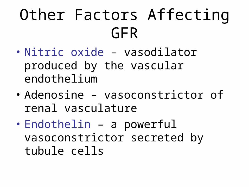
Other Factors Affecting GFR
• Nitric oxide – vasodilator produced by the vascular endothelium
• Adenosine – vasoconstrictor of renal vasculature
• Endothelin – a powerful vasoconstrictor secreted by tubule cells
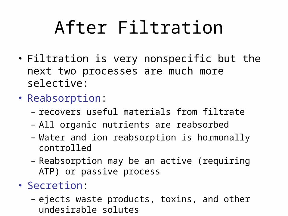
After Filtration
• Filtration is very nonspecific but the next two processes are much more selective:
• Reabsorption: – recovers useful materials from filtrate– All organic nutrients are reabsorbed– Water and ion reabsorption is hormonally controlled– Reabsorption may be an active (requiring ATP) or
passive process
• Secretion: – ejects waste products, toxins, and other undesirable
solutes
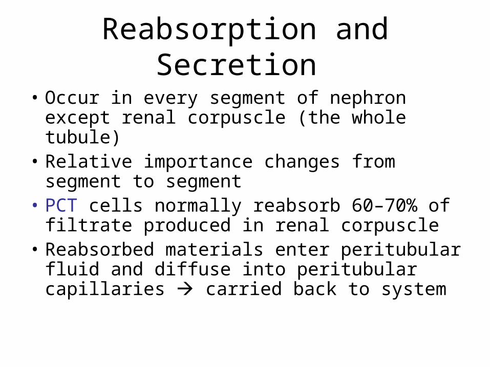
Reabsorption and Secretion
• Occur in every segment of nephron except renal corpuscle (the whole tubule)
• Relative importance changes from segment to segment
• PCT cells normally reabsorb 60–70% of filtrate produced in renal corpuscle
• Reabsorbed materials enter peritubular fluid and diffuse into peritubular capillaries carried back to system
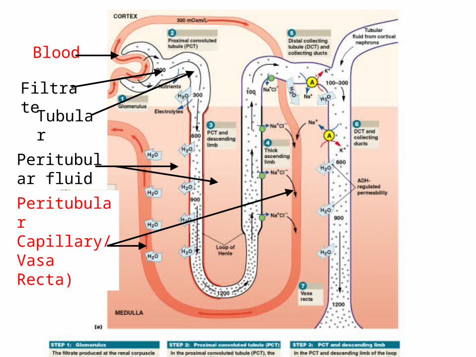
Figure 26–16a
Blood
Filtrate
Tubular fluid
Peritubular fluid
Peritubular Capillary/ Vasa Recta)
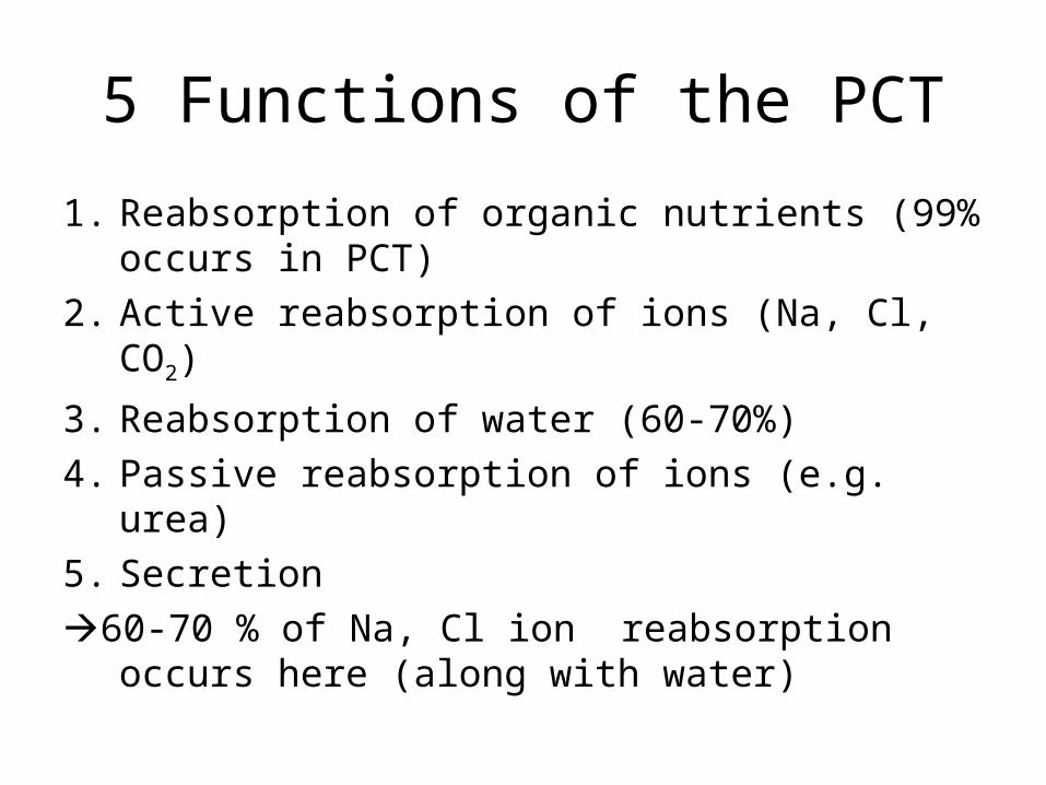
5 Functions of the PCT
1. Reabsorption of organic nutrients (99% occurs in PCT)
2. Active reabsorption of ions (Na, Cl, CO2)
3. Reabsorption of water (60-70%)
4. Passive reabsorption of ions (e.g. urea)
5. Secretion
60-70 % of Na, Cl ion reabsorption occurs here (along with water)
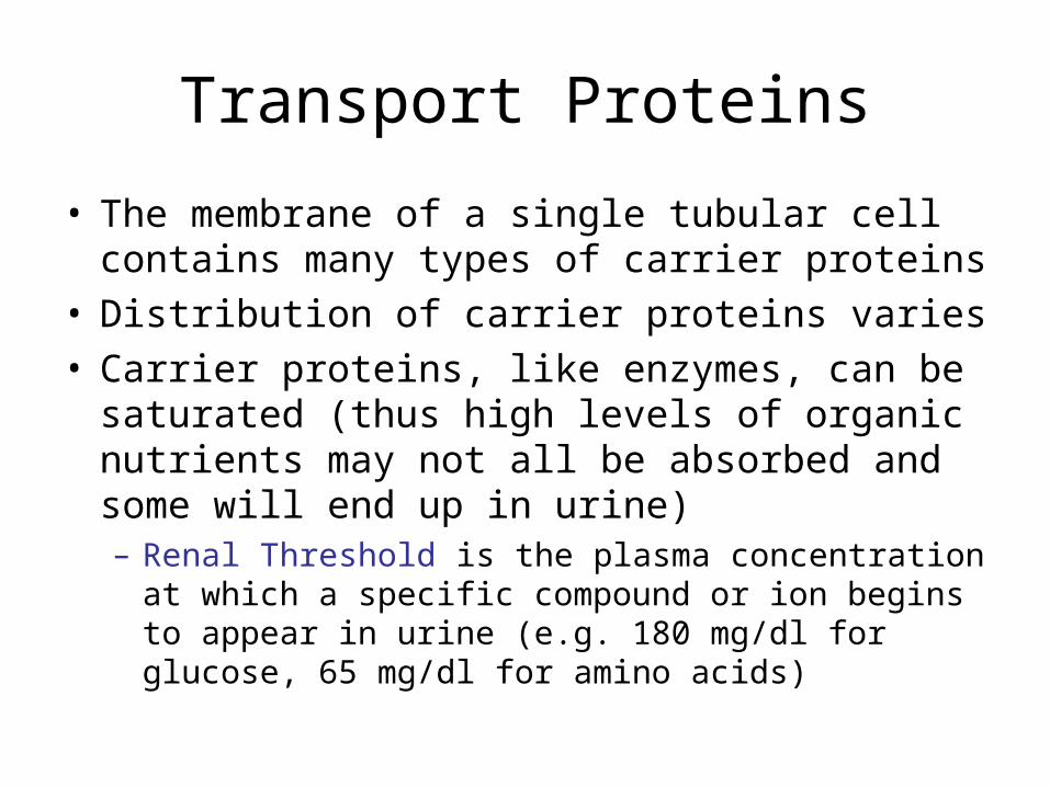
Transport Proteins
• The membrane of a single tubular cell contains many types of carrier proteins
• Distribution of carrier proteins varies • Carrier proteins, like enzymes, can be saturated
(thus high levels of organic nutrients may not all be absorbed and some will end up in urine)– Renal Threshold is the plasma concentration at which
a specific compound or ion begins to appear in urine (e.g. 180 mg/dl for glucose, 65 mg/dl for amino acids)
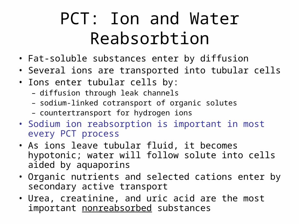
PCT: Ion and Water Reabsorbtion
• Fat-soluble substances enter by diffusion• Several ions are transported into tubular cells• Ions enter tubular cells by:
– diffusion through leak channels– sodium-linked cotransport of organic solutes– countertransport for hydrogen ions
• Sodium ion reabsorption is important in most every PCT process
• As ions leave tubular fluid, it becomes hypotonic; water will follow solute into cells aided by aquaporins
• Organic nutrients and selected cations enter by secondary active transport
• Urea, creatinine, and uric acid are the most important nonreabsorbed substances
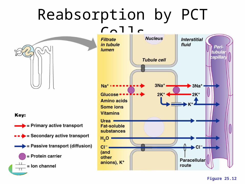
Reabsorption by PCT Cells
PLAYPLAY InterActive Physiology ®: Early Filtrate Processing, pages 3–15
Figure 25.12
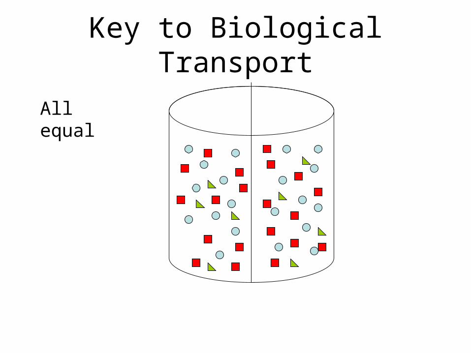
Key to Biological Transport
All equal
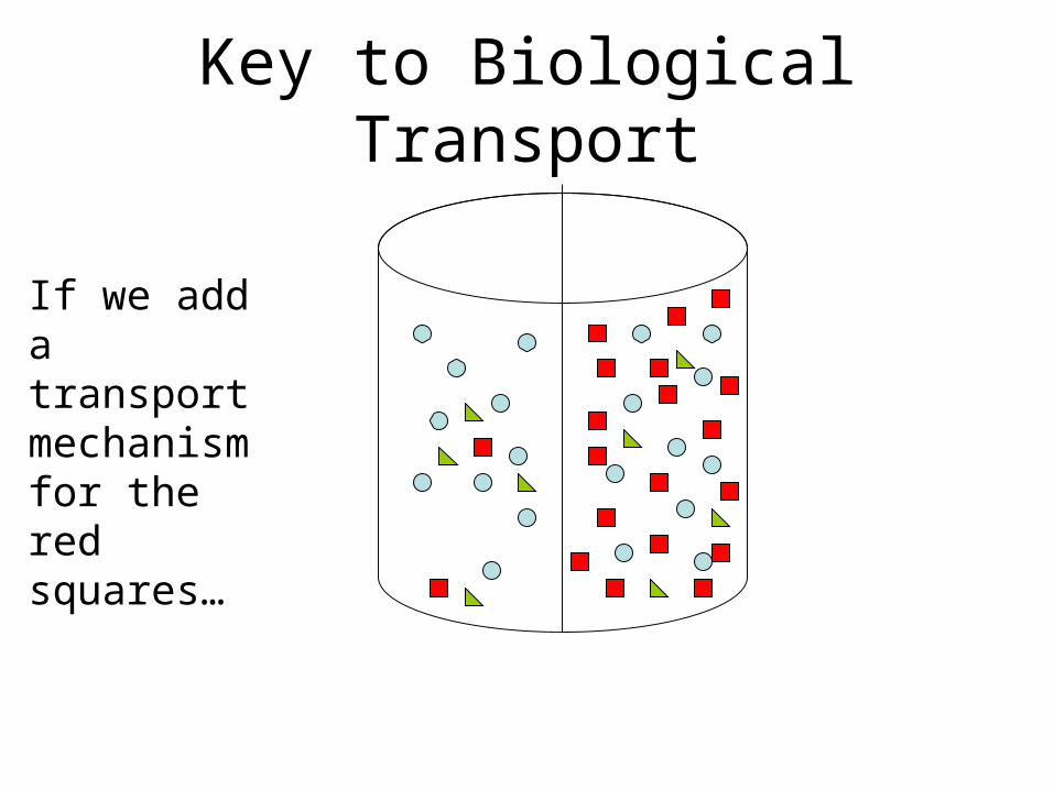
Key to Biological Transport
If we add a transport mechanism for the red squares…
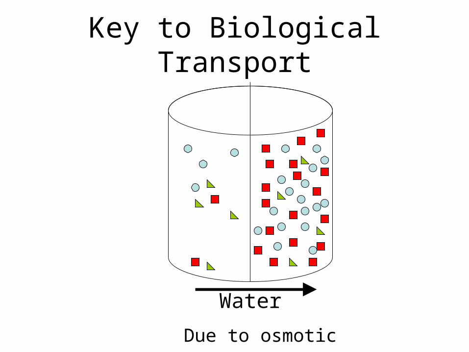
Key to Biological Transport
Water
Due to osmotic pressure
Key to Biological Transport
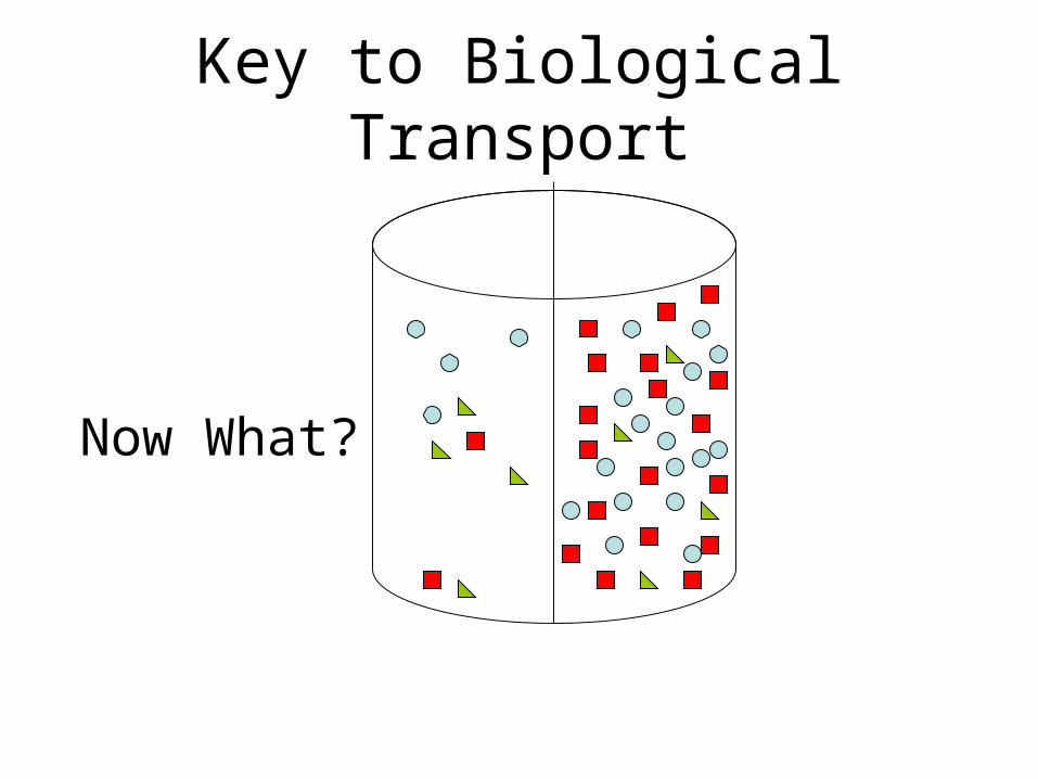
Now What?
Key to Biological Transport
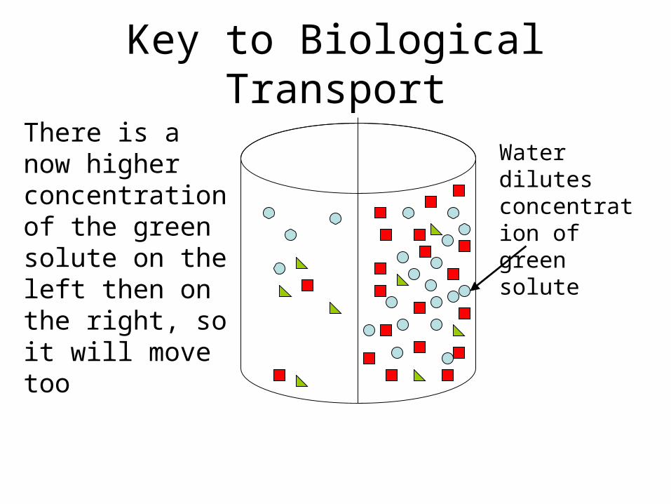
There is a now higher concentration of the green solute on the left then on the right, so it will move too
Water dilutes concentration of green solute
Key to Biological Transport
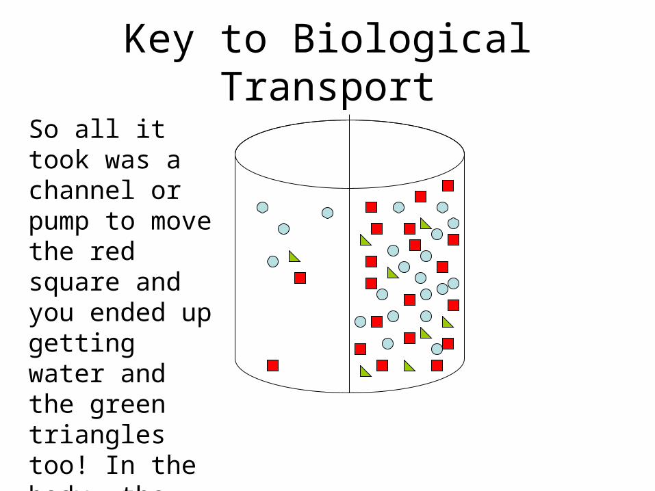
So all it took was a channel or pump to move the red square and you ended up getting water and the green triangles too! In the body, the red square is?
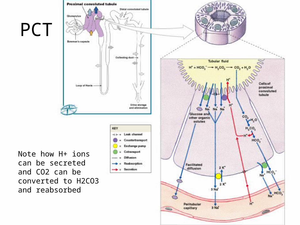
PCT
Figure 26–12 (Navigator)
Note how H+ ions can be secreted and CO2 can be converted to H2CO3 and reabsorbed
The Loop of Henle
• Reabsorbs about 25% of water and 20- 25% of sodium and chloride ions through countercurrent multiplication:– exchange that occurs between 2 parallel
segments of loop of Henle: • the thin, descending limb• the thick, ascending limb
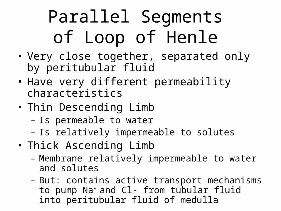
Parallel Segments of Loop of Henle
• Very close together, separated only by peritubular fluid
• Have very different permeability characteristics• Thin Descending Limb
– Is permeable to water– Is relatively impermeable to solutes
• Thick Ascending Limb – Membrane relatively impermeable to water and
solutes– But: contains active transport mechanisms to pump
Na+ and Cl- from tubular fluid into peritubular fluid of medulla
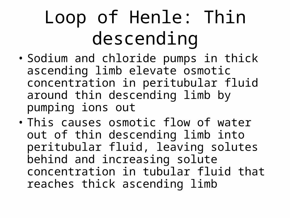
Loop of Henle: Thin descending
• Sodium and chloride pumps in thick ascending limb elevate osmotic concentration in peritubular fluid around thin descending limb by pumping ions out
• This causes osmotic flow of water out of thin descending limb into peritubular fluid, leaving solutes behind and increasing solute concentration in tubular fluid that reaches thick ascending limb
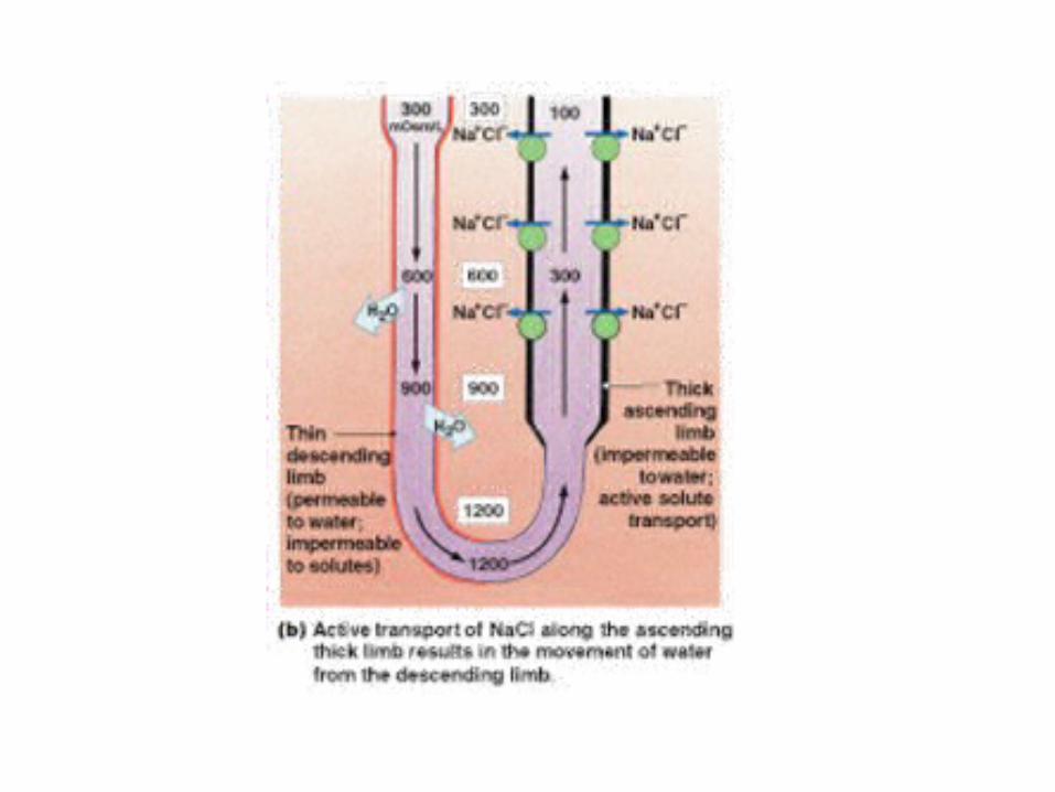
Loop of Henle: Thick Ascending
• Now the solution that arrives at the thick ascending limb is highly concentrated (lost a lot of water in descending limb via osmosis)
• This Accelerates Na+ and Cl- transport into peritubular fluid of medulla
• It’s positive feedback
Positive Feedback
• Solute pumping at ascending limb increases solute concentration in descending limb which accelerates solute pumping in ascending limb… etc
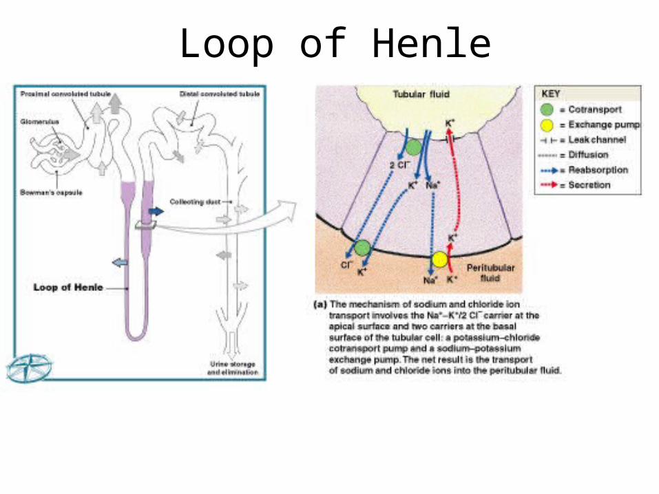
Figure 26–13a (Navigator)
Loop of Henle
Countercurrent Multiplication and Concentration of Urine
Figure 26–13b, c
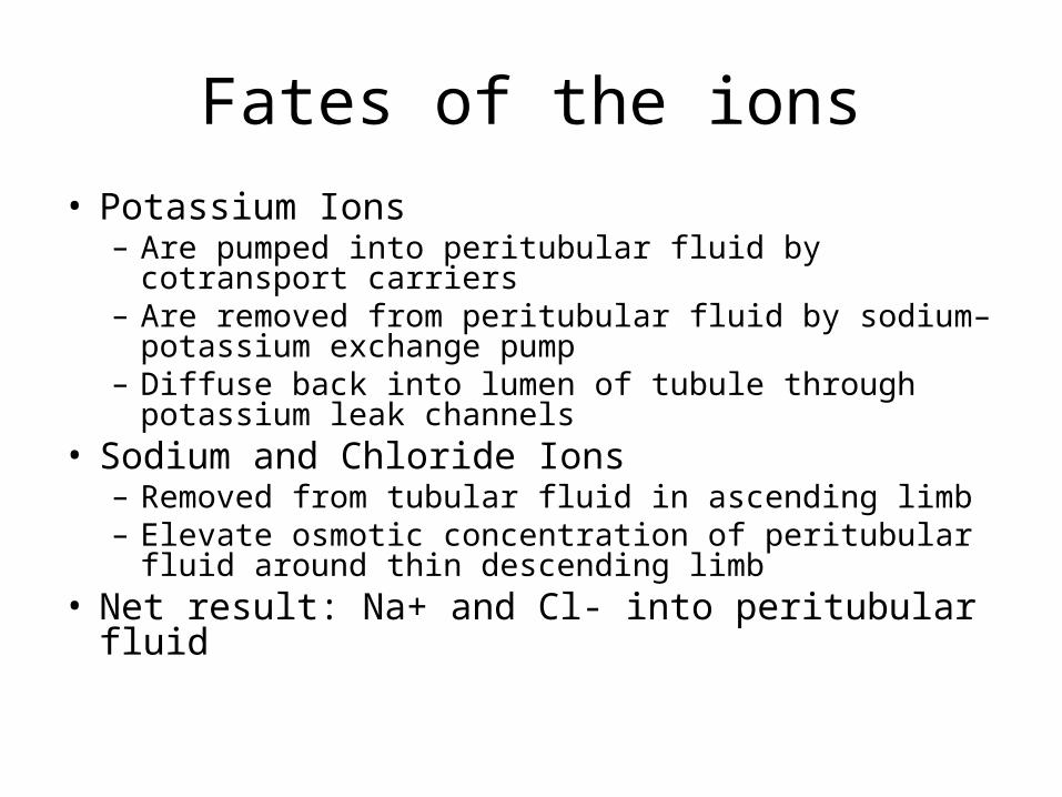
Fates of the ions
• Potassium Ions – Are pumped into peritubular fluid by cotransport
carriers– Are removed from peritubular fluid by sodium–
potassium exchange pump– Diffuse back into lumen of tubule through potassium
leak channels• Sodium and Chloride Ions
– Removed from tubular fluid in ascending limb– Elevate osmotic concentration of peritubular fluid
around thin descending limb• Net result: Na+ and Cl- into peritubular fluid

Regional Differences
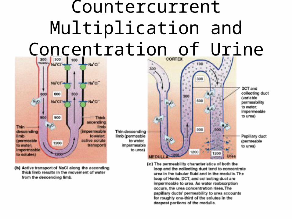
• Rate of ion transport across thick ascending limb is proportional to ion’s concentration in tubular fluid
• More Na+ and Cl- are pumped into medulla at the start of the thick ascending limb than near cortex
• Regional difference in ion transport rate causes a concentration gradient within medulla: high concentration in deep medulla, low in superficial medulla
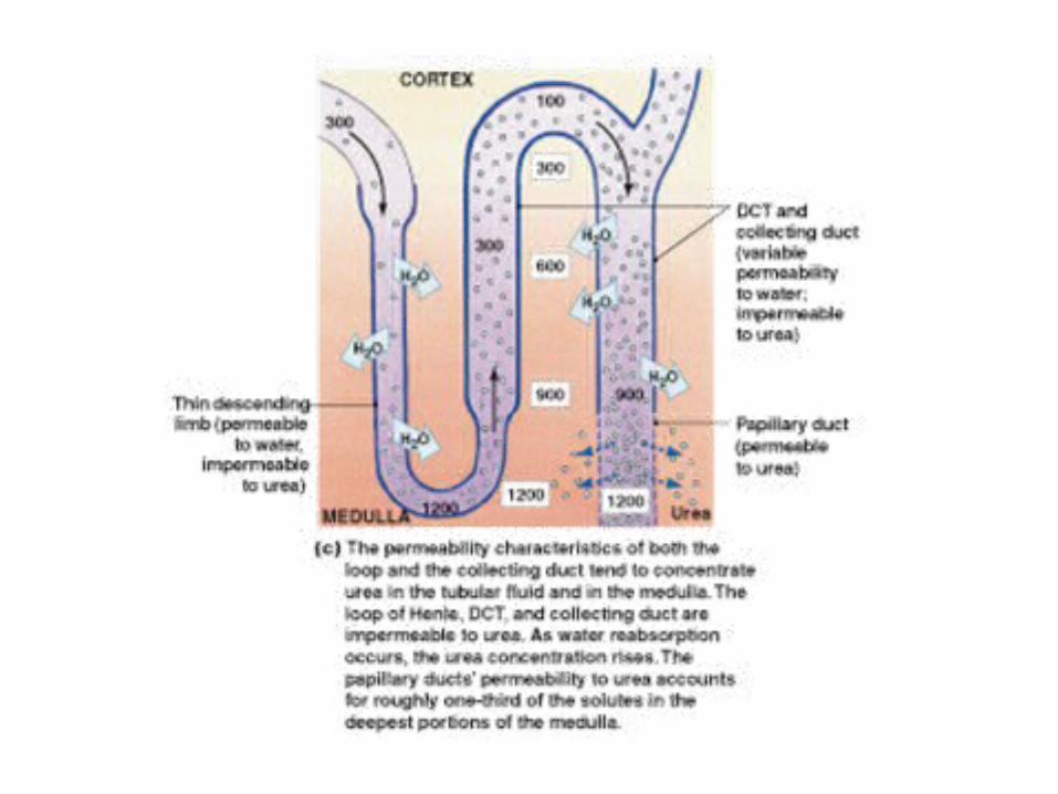
• Normal maximum solute concentration of peritubular fluid near turn of loop of Henle in deepest medulla is 1200 mOsm/L (important)
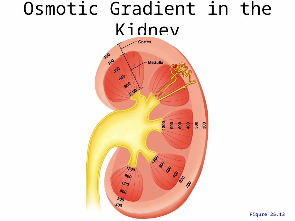
Osmotic Gradient in the Kidney
Figure 25.13
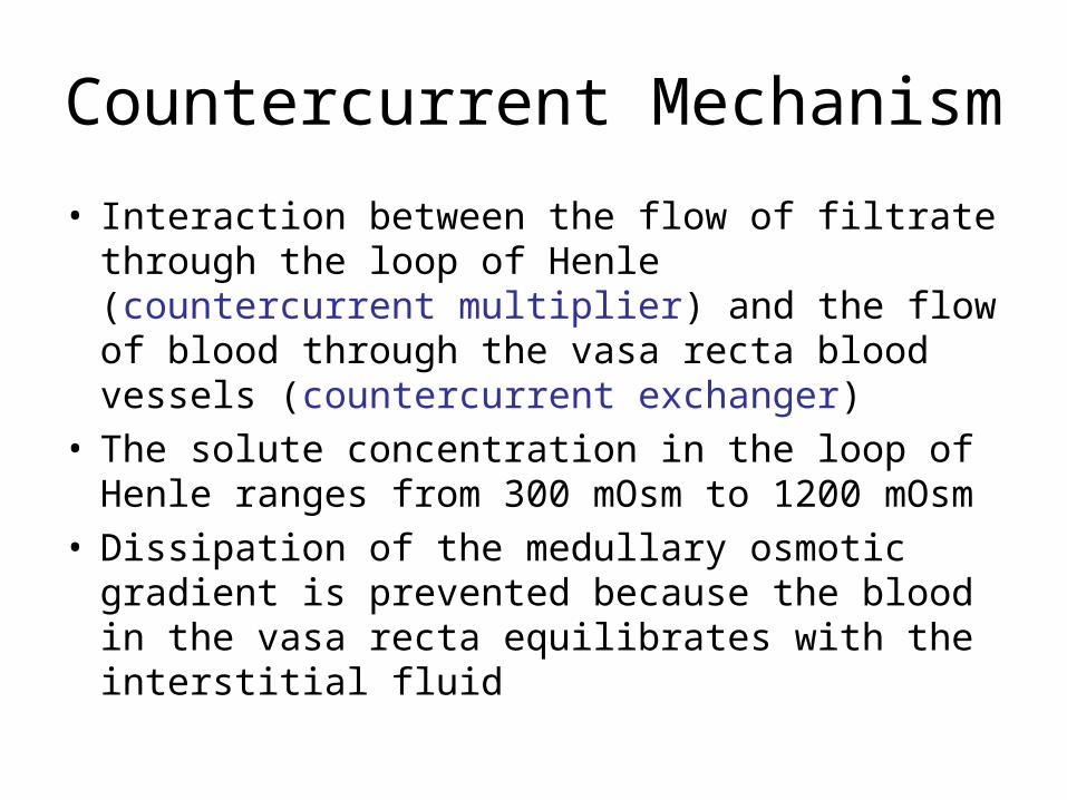
Countercurrent Mechanism
• Interaction between the flow of filtrate through the loop of Henle (countercurrent multiplier) and the flow of blood through the vasa recta blood vessels (countercurrent exchanger)
• The solute concentration in the loop of Henle ranges from 300 mOsm to 1200 mOsm
• Dissipation of the medullary osmotic gradient is prevented because the blood in the vasa recta equilibrates with the interstitial fluid
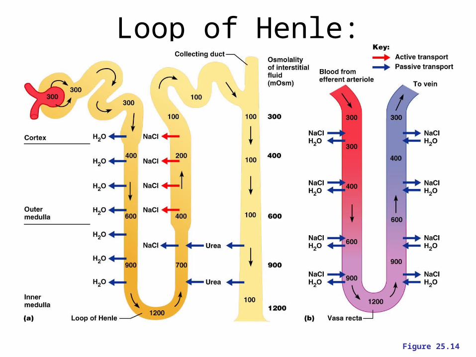
Loop of Henle: Countercurrent Mechanism
Figure 25.14
Tubular Fluid at DCT
• Arrives with osmotic concentration of 100 mOsm/L:– 1/3 concentration of peritubular fluid of renal
cortex because of ion reabsorption in ascending limb
The Concentration Gradient of the Medulla
• 2/3 (or 750 mOsm/L) is from Na+ and Cl- pumped out of ascending limb
• Remainder is from urea• Tubular fluid reaching papillary duct
contains 450 mOsm/L urea (just from loss of water in tubular fluid)
• Papillary ducts are permeable to urea, concentration in medulla averages 450 mOsm/L
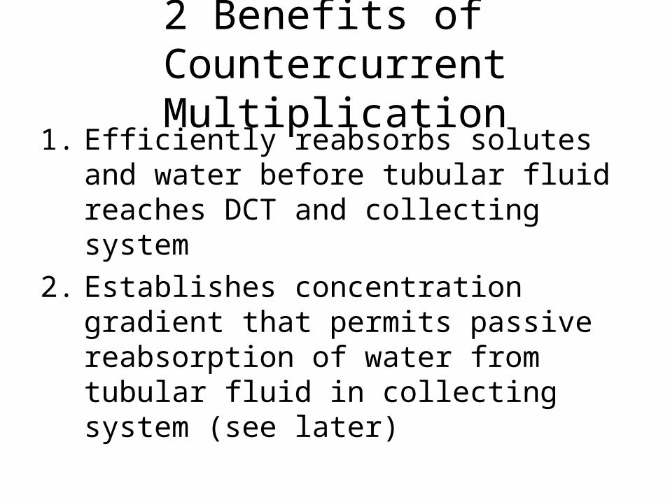
2 Benefits of Countercurrent Multiplication
1. Efficiently reabsorbs solutes and water before tubular fluid reaches DCT and collecting system
2. Establishes concentration gradient that permits passive reabsorption of water from tubular fluid in collecting system (see later)
Changes in Tubular Fluids
• Composition and volume of tubular fluid change from capsular space to distal convoluted tubule– Only 15–20% of initial filtrate volume reaches
DCT– Concentrations of electrolytes and organic
wastes in arriving tubular fluid no longer resemble blood plasma (most organic nutrients gone, most electrolytes gone. Urea and other wastes remain)
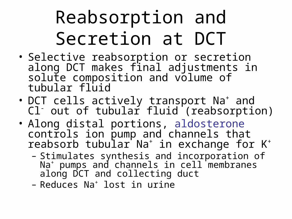
Reabsorption and Secretion at DCT
• Selective reabsorption or secretion along DCT makes final adjustments in solute composition and volume of tubular fluid
• DCT cells actively transport Na+ and Cl- out of tubular fluid (reabsorption)
• Along distal portions, aldosterone controls ion pump and channels that reabsorb tubular Na+ in exchange for K+
– Stimulates synthesis and incorporation of Na+ pumps and channels in cell membranes along DCT and collecting duct
– Reduces Na+ lost in urine
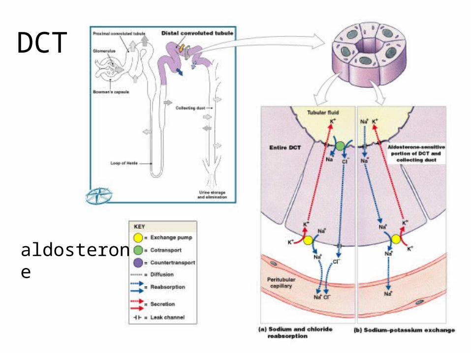
DCT
Figure 26–14a, b (Navigator)
aldosterone
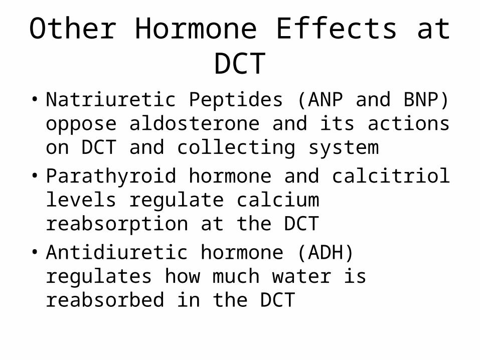
Other Hormone Effects at DCT
• Natriuretic Peptides (ANP and BNP) oppose aldosterone and its actions on DCT and collecting system
• Parathyroid hormone and calcitriol levels regulate calcium reabsorption at the DCT
• Antidiuretic hormone (ADH) regulates how much water is reabsorbed in the DCT
Secretion at the DCT
• Blood entering peritubular capillaries still contains some undesirable substances that did not cross filtration membrane at glomerulus
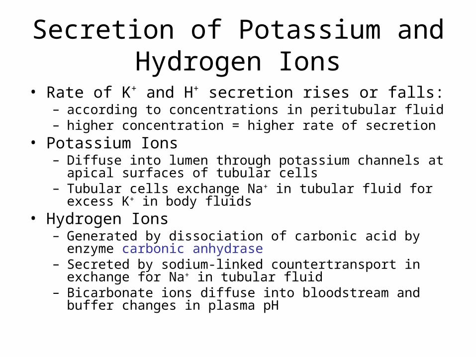
Secretion of Potassium and Hydrogen Ions
• Rate of K+ and H+ secretion rises or falls:– according to concentrations in peritubular fluid– higher concentration = higher rate of secretion
• Potassium Ions – Diffuse into lumen through potassium channels at apical
surfaces of tubular cells– Tubular cells exchange Na+ in tubular fluid for excess K+ in body
fluids• Hydrogen Ions
– Generated by dissociation of carbonic acid by enzyme carbonic anhydrase
– Secreted by sodium-linked countertransport in exchange for Na+ in tubular fluid
– Bicarbonate ions diffuse into bloodstream and buffer changes in plasma pH
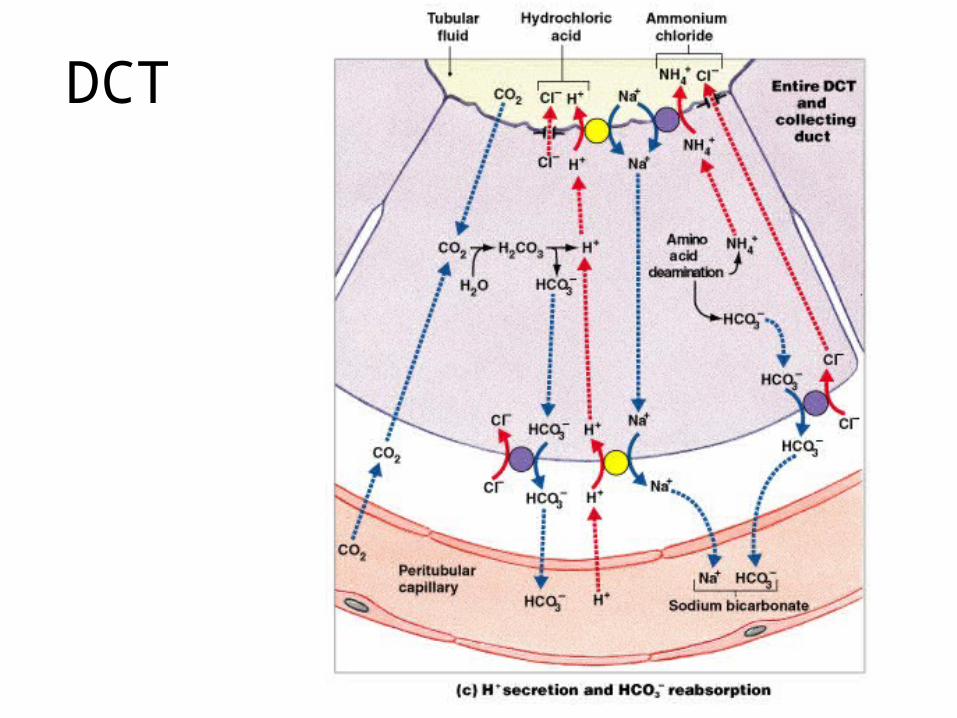
DCT
Control of Blood pH
• By H+ removal and bicarbonate production at kidneys is important to homeostasis
• Acidosis– Lactic acidosis:
• develops after exhaustive muscle activity
– Ketoacidosis:• develops in starvation or diabetes mellitus
• Alkalosis
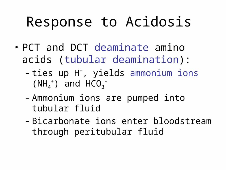
Response to Acidosis
• PCT and DCT deaminate amino acids (tubular deamination): – ties up H+, yields ammonium ions (NH4
+) and HCO3
-
– Ammonium ions are pumped into tubular fluid– Bicarbonate ions enter bloodstream through
peritubular fluid
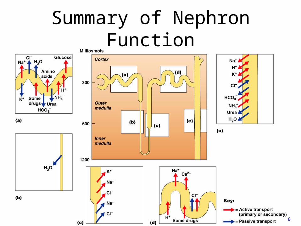
Summary of Nephron Function
Figure 25.16
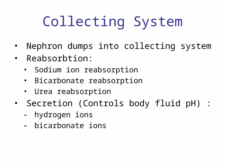
Collecting System
• Nephron dumps into collecting system• Reabsorbtion:• Sodium ion reabsorption• Bicarbonate reabsorption• Urea reabsorption
• Secretion (Controls body fluid pH) :– hydrogen ions– bicarbonate ions
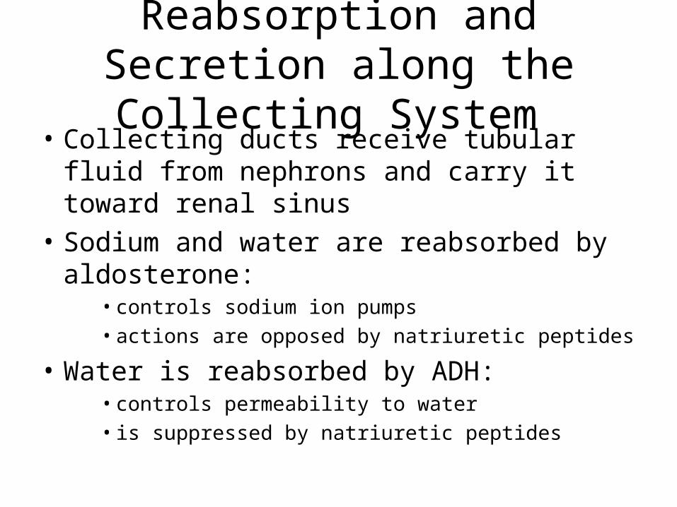
Reabsorption and Secretion along the Collecting System
• Collecting ducts receive tubular fluid from nephrons and carry it toward renal sinus
• Sodium and water are reabsorbed by aldosterone:
• controls sodium ion pumps • actions are opposed by natriuretic peptides
• Water is reabsorbed by ADH:• controls permeability to water• is suppressed by natriuretic peptides
Urea Reabsorption
• As mentioned, urea is highly concentrated in the papillary duct (at the end of the collecting duct) and the cells are permeable to it. Thus urea diffuses out to the peritubular fluid of the medulla, adding to the medullary concentration gradient and allowing production of concentrated urine
Collecting System pH regulation
• Low pH:– Carrier proteins:
• pump H+ into tubular fluid• reabsorb bicarbonate ions
• Hi pH:– Collecting system:
• secretes bicarbonate ions• pumps H+ into peritubular fluid
KEY CONCEPT
• Reabsorption involves diffusion, osmosis, channel-mediated diffusion, and active transport
• Many processes are independently regulated by local or hormonal mechanisms
• The primary mechanism governing water reabsorption is “water follows salt”
• Secretion is a selective, carrier mediated process
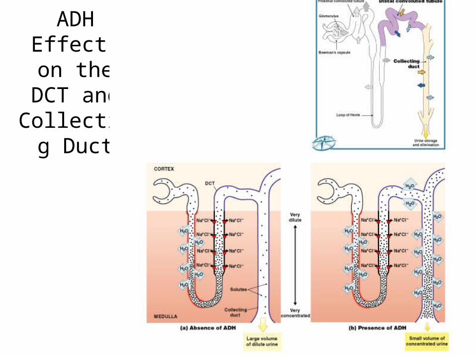
ADH Effects on the
DCT and Collecting
Duct
Figure 26–15 (Navigator)
Urine Volume and Osmotic Concentration
• Are regulated through control of water reabsorption
• Water is reabsorbed by osmosis in: – proximal convoluted tubule– descending limb of loop of Henle
• Occurs when osmotic concentration of peritubular fluid exceeds that of tubular fluid. Ions leave, then water follows.
• In DCT and collecting system, water absorption is controlled by hormones
Water Reabsorption
• 1–2% of water in original filtrate is recovered during sodium ion reabsorption in distal convoluted tubule and collecting system (in addition to the 60 + 25 pecent in PCT and loop)
• Obligatory water reabsorption is reabsorption based on osmosis, can’t be prevented– Recovers 85% of original filtrate
• Facultative Water Reabsorption controls volume of water reabsorbed along DCT and collecting system (15% of filtrate volume) – segments are relatively impermeable to water except
in presence of ADH
ADH
• Hormone causes special water channels to appea in apical cell membranes
• Increases rate of osmotic water movement• Higher levels of ADH increases:
– number of water channels– water permeability of DCT and collecting system
• Water can move by itself into interstitial fluid around the collecting duct because the concentration there is 1200 mOsm/L!
Concentration Gradient of the Medulla
• 2/3 (or 750 mOsm/L) is from Na+ and Cl- pumped out of ascending limb
• Remainder is from urea• Tubular fluid reaching papillary duct contains
450 mOsm/L urea (just from loss of water in tubular fluid)
• Papillary ducts are permeable to urea, concentration of urea in medulla averages 450 mOsm/L. This adds to the concetration generated by ion pumping in the loop of Henle to create a 1200mOsm/L concentration gradient
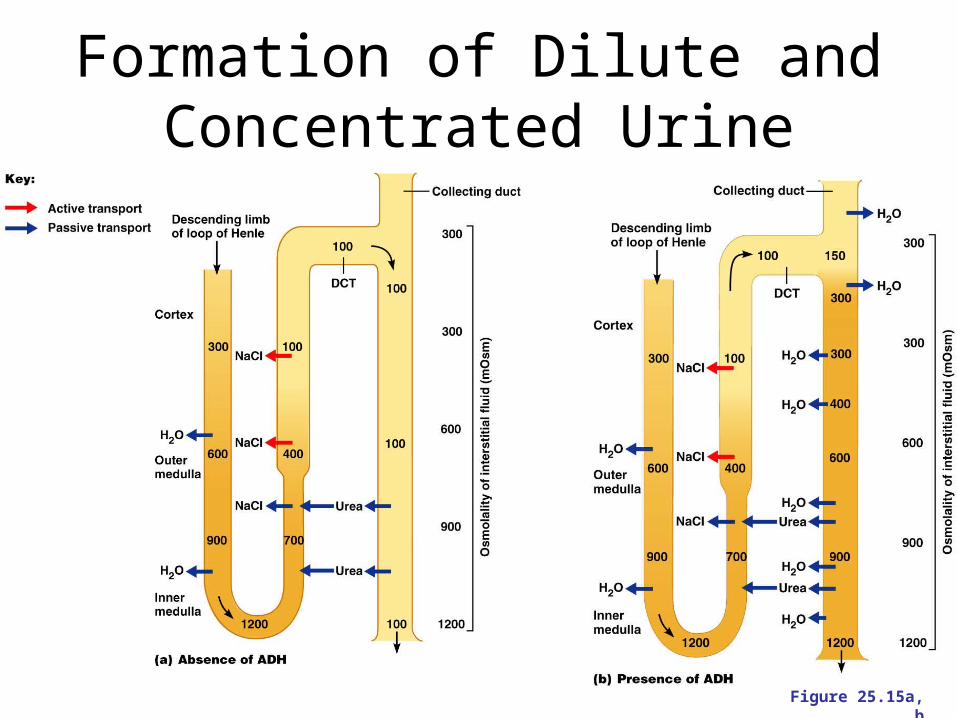
Formation of Dilute and Concentrated Urine
Figure 25.15a, b
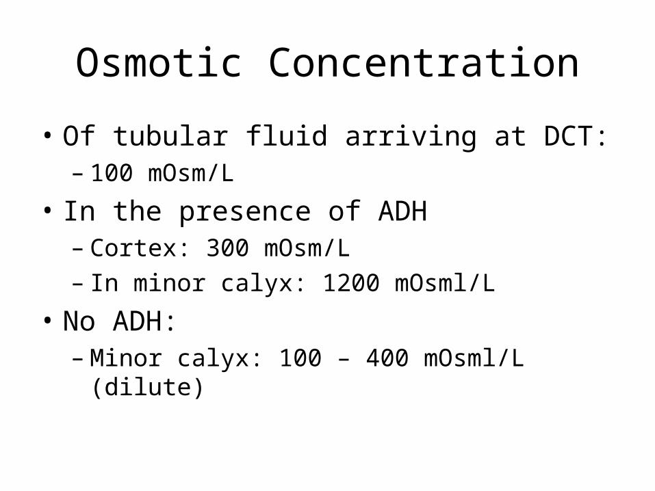
Osmotic Concentration
• Of tubular fluid arriving at DCT:– 100 mOsm/L
• In the presence of ADH – Cortex: 300 mOsm/L– In minor calyx: 1200 mOsml/L
• No ADH:– Minor calyx: 100 – 400 mOsml/L (dilute)
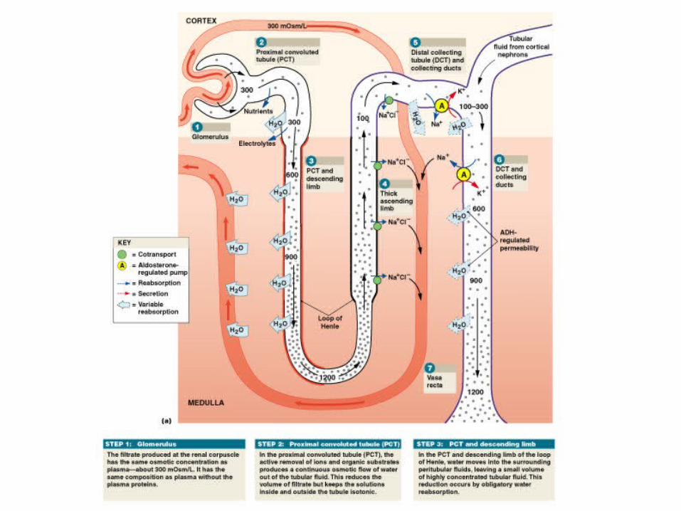
Figure 26–16a
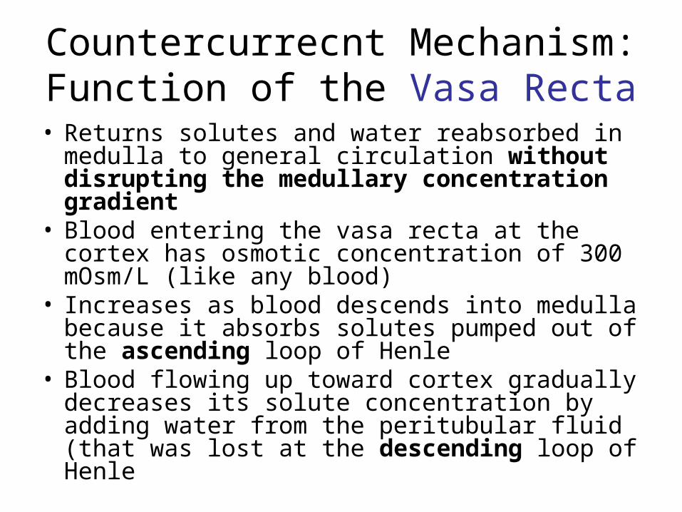
Countercurrecnt Mechanism: Function of the Vasa Recta
• Returns solutes and water reabsorbed in medulla to general circulation without disrupting the medullary concentration gradient
• Blood entering the vasa recta at the cortex has osmotic concentration of 300 mOsm/L (like any blood)
• Increases as blood descends into medulla because it absorbs solutes pumped out of the ascending loop of Henle
• Blood flowing up toward cortex gradually decreases its solute concentration by adding water from the peritubular fluid (that was lost at the descending loop of Henle
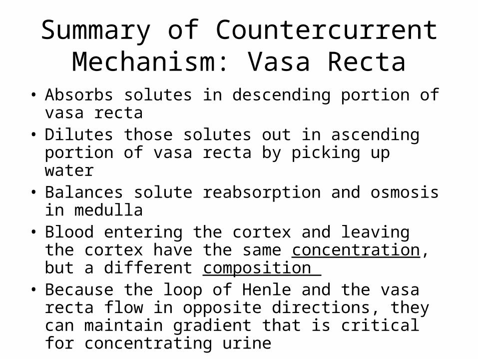
Summary of Countercurrent Mechanism: Vasa Recta
• Absorbs solutes in descending portion of vasa recta
• Dilutes those solutes out in ascending portion of vasa recta by picking up water
• Balances solute reabsorption and osmosis in medulla
• Blood entering the cortex and leaving the cortex have the same concentration, but a different composition
• Because the loop of Henle and the vasa recta flow in opposite directions, they can maintain gradient that is critical for concentrating urine
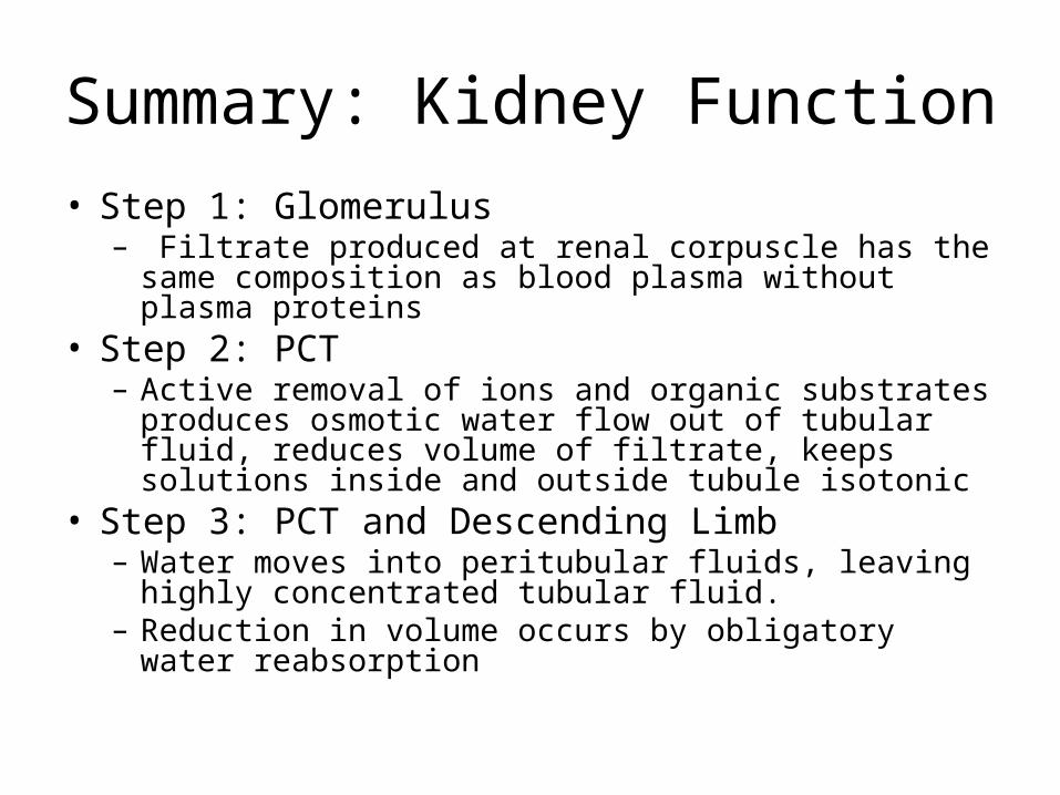
Summary: Kidney Function
• Step 1: Glomerulus– Filtrate produced at renal corpuscle has the same
composition as blood plasma without plasma proteins• Step 2: PCT
– Active removal of ions and organic substrates produces osmotic water flow out of tubular fluid, reduces volume of filtrate, keeps solutions inside and outside tubule isotonic
• Step 3: PCT and Descending Limb– Water moves into peritubular fluids, leaving highly
concentrated tubular fluid. – Reduction in volume occurs by obligatory water
reabsorption
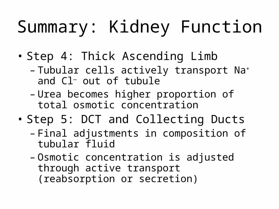
Summary: Kidney Function
• Step 4: Thick Ascending Limb – Tubular cells actively transport Na+ and Cl—
out of tubule– Urea becomes higher proportion of total
osmotic concentration
• Step 5: DCT and Collecting Ducts – Final adjustments in composition of tubular
fluid – Osmotic concentration is adjusted through
active transport (reabsorption or secretion)
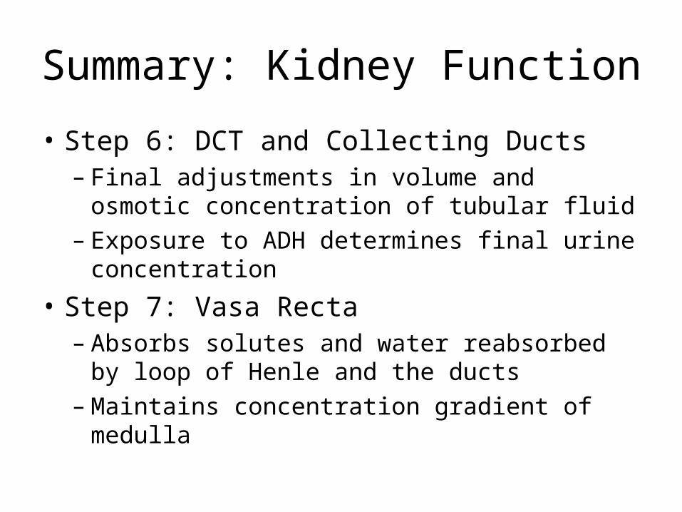
Summary: Kidney Function
• Step 6: DCT and Collecting Ducts– Final adjustments in volume and osmotic
concentration of tubular fluid– Exposure to ADH determines final urine
concentration
• Step 7: Vasa Recta – Absorbs solutes and water reabsorbed by
loop of Henle and the ducts– Maintains concentration gradient of medulla
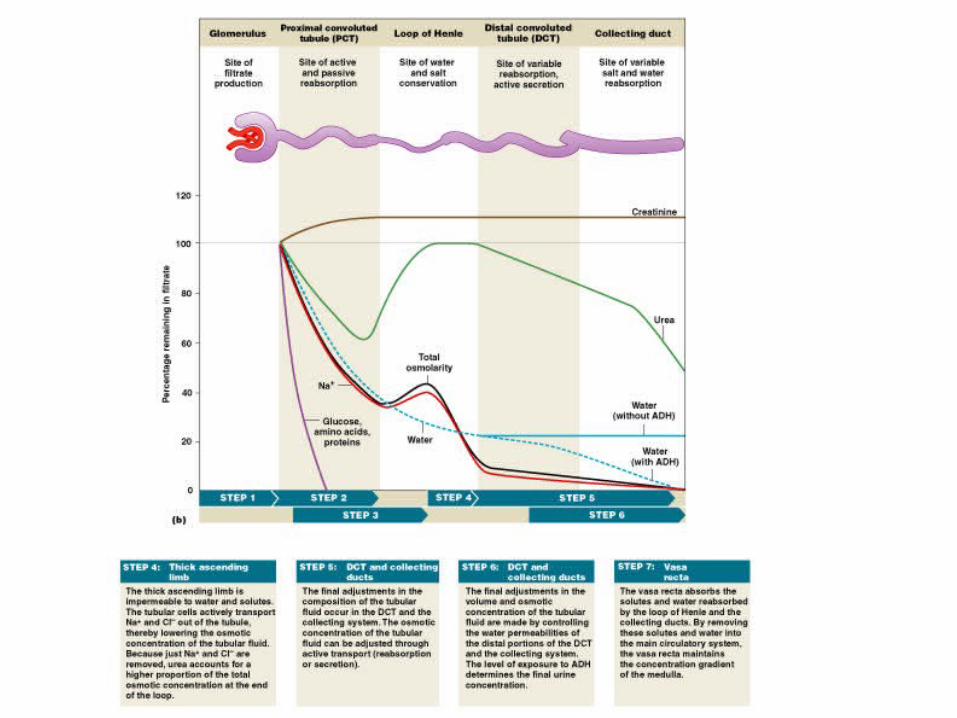
Figure 26–16b
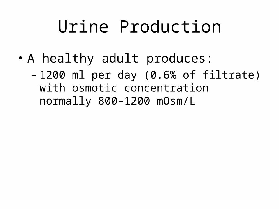
Urine Production
• A healthy adult produces:– 1200 ml per day (0.6% of filtrate) with osmotic
concentration normally 800–1200 mOsm/L
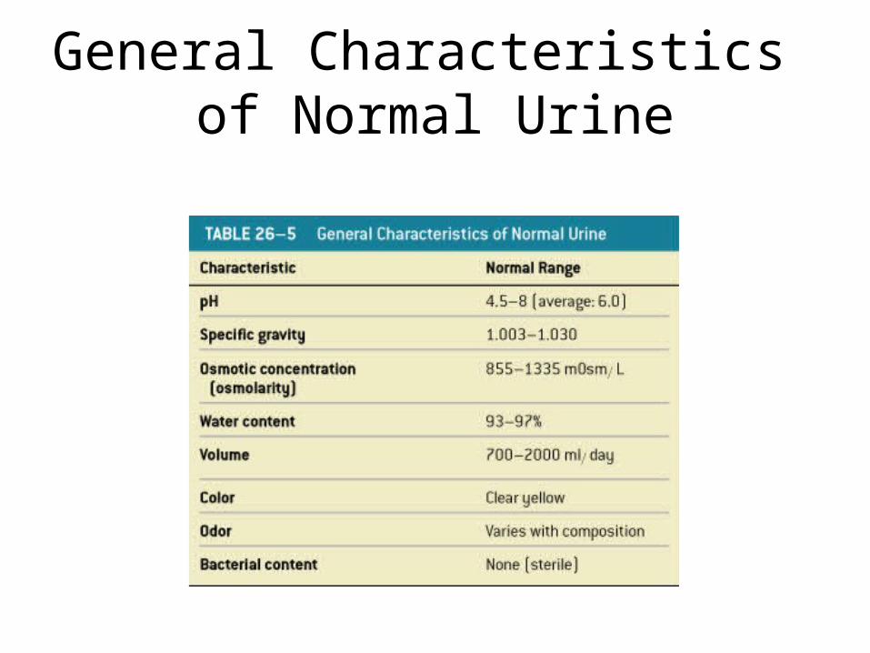
General Characteristics of Normal Urine
Table 26–5
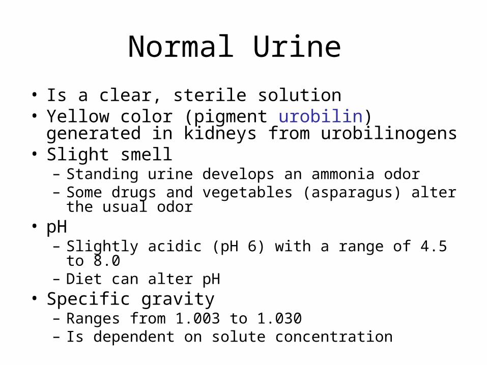
Normal Urine
• Is a clear, sterile solution• Yellow color (pigment urobilin) generated in
kidneys from urobilinogens • Slight smell
– Standing urine develops an ammonia odor– Some drugs and vegetables (asparagus) alter the
usual odor• pH
– Slightly acidic (pH 6) with a range of 4.5 to 8.0– Diet can alter pH
• Specific gravity– Ranges from 1.003 to 1.030 – Is dependent on solute concentration
Chemical Composition of Urine• Urine is 95% water and 5% solutes• Nitrogenous wastes: urea, uric acid, and
creatinine• Other normal solutes include:
– Sodium, potassium, phosphate, and sulfate ions
– Calcium, magnesium, and bicarbonate ions
• Abnormally high concentrations of any urinary constituents may indicate pathology
Urination
• Peristaltic Contractions– Begin at renal pelvis– Sweep along ureter– Force urine toward urinary bladder– Every 30 seconds or so
Urethral Sphincters
• Internal urethral sphincter– Found at the neck of the Urinary Bladder
surrounding urethral opening– Smooth muscle
• External urethral sphincter in both sexes: – is a circular band of skeletal muscle where
urethra passes through urogenital diaphragm
External Urethral Sphincter
• Is under voluntary control
• Has resting muscle tone
• Voluntarily relaxation permits micturition
The Micturition Reflex
• Coordinates the process of urination• Stretch Receptor Stimulation Increases
with urinary volume, causes urge to urinate
• Both sphincters (internal and external) are closed until voluntary opening of the external triggers the internal to open too.
• Volume > 500 ml triggers involuntary micturition reflex