the ultrastructure of contact lens induced changes
TRANSCRIPT

A C T A O P H T H A L M O L O G I C A 62 (1984) 320-333
Department of Ophthalmology (Head: Salnw Vannas), University of Hehnki, Finhnd
Cornea and Contact Lens Research Unit (Head: Barry Collin), School of Optmet?, Uniuersily of New South Wales, Sydney, Australia
TH E U LTRASTRUCTU RE OF CONTACT LENS INDUCED CHANGES
ANITTI VANNAS, BRIEN A. HOLDEN and JUKKA MAKlTlE
The endothelium of 15 human corneas was studied with specular and electron microscopy after exposure to a thick, low water content, soft contact lens (SCL). Five control corneas (no lens wear) were studied using the same methods. SCL wear produced obvious changes in endothelial morphology in 12 of the 15 eyes. With specular microscopy, the changes consisted of an apparent increase in separation of cells and development of areas of loss of membrane reflectivity (blebs). When viewed with electron microscopy, the changes in the same corneas consisted of oedema in the nuclear area of the cells and bulging of the posterior endothelial surface, in some cases over an area of several cells. In 4 cases, the cellular oedema was marked showing both intracellular and intercellular vacuoles. It was concluded that the transient endothelial changes seen with specular microscopy following SCL lens wear were produced by alterations in the contour of the posterior endothelial surface resulting from disturbance to the endothelial environment.
Key words: corneal endothelium - ultrastructure - contact lens - blebs - nuclear capping.
Soft contact lens (SCL) wear can induce transient changes in the appearance of the human corneal endothelium (Zantos & Holden 1977). The changes consist of an apparent increase in separation of cells and the development of black non- reflecting areas (blebs) covering up to 3 or 4 cells. This phenomenon has been
Received on May 2nd, 1983.
320

Vannas et al. Contact lens induced endothelial changes
confirmed by subsequent work using both non-contact and contact specular microscopes of different designs (Vannas et al. 1979; Sherrard & Buckley 1981; Kamiya 1982). The response has been found to have the following characteristics: it is extremely rapid, occurring within 2 min; peaks within 30-40 min; falls to a steady level after 1-2 h and disappears on lens removal (Holden & Zantos 198 1 ; Vannas et al. 198 1). Similar changes have been reported with hard contact lens wear (Barr & Schoessler 1979).
The reversibility of morphological alterations to the endothelium is an important consideration, and though no cell loss appears to occur with SCL induced endothelial response (Vannas et al. 198 I ) , the nature of these changes at a cellular level is not fully understood.
The aim of the present study was to correlate the specular microscopic appearance of SCL induced changes in the endothelium with changes in ultrastructure of the endothelial cells seen with scanning electron microscopy (SEM) and transmission electron microscopy (TEM).
Materials and Methods
The endothelium of 20 corneas was studied. Fourteen of the eyes were to be enucleated (Group 1) mainly because of choroidal or conjunctival melanoma (Table 1). Six corneas were obtained from 3 beating-heart braindeath cadavers (Group 2).
Fifteen of these eyes (12 from Group 1 and 3 from Group 2) wore a thick SCL (0.3 mm thick, 32% water content, 8.0 mm base curve). Five corneas (2 from group 1 and 3 from group 2) served as controls.
Three eyes from the Group 1 patients were tested one day prior to enucleation, where they showed typical bleb responses i.e., black, non-reflecting areas within minutes of lens insertion, the changes reaching a maximum in 20-40 min and disappearring after lens removal (Fig. 1).
On the day of enucleation, the endothelium of the 12 eyes to be tested was photographed before, and again, after approximately 30 min of SCL wear. The latter photographs were taken immediately after compression of the optic nerve and enucleation.
In the Group 2 patients, the SCL was worn on one eye of the braindeath beating-heart cadavers for 20-30 min, while the other cornea served as a control. These corneas were excised with a 12 mm rotor trephine.
Five of the corneas (3 test and 2 controls) were fixed prior to excision to prevent possible surgical artefacts. The remaining corneas were fixed immediately after specular microscopic photography using either 2.5% glutaraldehyde or 1 %
32 1 Acta ophthal. 62. 2 21

Vannas et al. Contact lens induced endothelial changes
Table I. Clinical Data of the patients.
No. I Sex I Age I Diagnosis
Group 1
1 2 3 4 5 6 7 9
10 1 1 12 13 14
M F M M M F M M F F F F F
65 46 49 60 38 63 48 58 33
1 1
48 58
M M M M M1 M M M M Rb Rb M M
Group 2 15 M 33 C, SAH 16 M 58 C, SAH 17 M 43 C, SAH
Abbreviations : M = choroidal malignant melanoma, M1 = conjunctival malignant melanoma, Rb = retino- blastoma, C = brain-death beating-heart cadaver, SAH = subarachnoid haemorrhage.
osmium tetroxide in 0.1 M phosphate buffer (pH 7.25) for 3 h. Different primary fixatives were used because glutaraldehyde may not preserve the posterior contour of endothelial cells (Hodson 1968). The glutaraldehyde fixed specimens were post-fixed in osmium tetroxide. The corneal buttons were bisected for TEM and SEM. Specimens were dehydrated and embedded in Epon. Cut specimens were stained with uranylacetate and lead citrate. For scanning electron microscopy the corneal buttons were critical point dried with C02. Dried tissue was fixed with silver conductive paint and coated with gold in a Blazers BA3 evaporator.
The specular microscope used in this study has been described elsewhere (Vannas et al. 1981). The angle between observation and illumination system was kept constant at 50 degrees.
322
Vannas et al. Contact lens induced endothelial changes
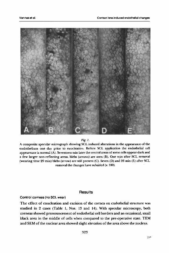
Fig. I. A composite specular micrograph showing SCL induced alterations in the appearance of the endothelium one day prior to enucleation. Before SCL application the endothelial cell appearnace is normal (A). Seventeen min later the central areas of some cells appear dark and a few larger non-reflecting areas, blebs (arrows) are seen (B). One min after SCL removal (wearing time 29 min) blebs (arrow) are still present (C). Seven (D) and 26 min (E) after SCL
removal the changes have subsided (x 100).
Results Control corneas (no SCL wear)
The effect of enucleation and excision of the cornea on endothelial structure was studied in 2 cases (Table 1, Nos. 13 and 14). With specular microscopy, both corneas showed pronouncement of endothelial cell borders and an occasional, small black area in the middle of cells when compared to the pre-operative state. TEM and SEM of the nuclear area showed slight elevation of the area above the nucleus.
323

Vannas et al. Contact lens induced endothelial changes
In the control corneas removed from the beating-heart dadavers, the endothelial cells appeared flat with TEM, and contained normal mitochondria and endo- plasmic reticulum. Regular tight apical junctions were observed between cells, and the intercellular spaces were not dilated. In SEM these endothelial cells appeared flat and hexagonal, but there was slight prominence of the nuclear area (Fig. 2). There were no ruptured cell membranes.
Corneas following SCL wear
Individual variation was observed in the SCL induced endothelial response. Three Group 1 eyes presented specular and electron microscopic findings that were similar to those seen in the control eyes.
Eight eyes showed, at enucleation, more pronounced changes than seen in control eyes. In specular microscopy endothelial cells presented dark central areas and larger non reflecting areas (Fig. 3). In TEM and SEM these eyes demonstrated
Fig. 2. SEM of a Group 2 control eye (no SCL wear). The endothelial cells appear intact (x 900).
324

Vannas et al. Contact lens induced endothelial changes
Fig. 3. Specular micrographs before (A) and immediately after enucleation and SCL wear (B). Considerable variation in cell size and shape is seen in a 65-year-old patient. After SCL wear
many endothelial cells present a dark central area and a few blebs are seen (arrows) (x 100).
distension of the cellular surface especially in the nuclear region (Fig. 4 and 5) , that in some instances covered an area of 2-3 cells (Fig. 6). The nuclear shape was rounded and the chromatin dispersed. These eyes did not show disorganization or swelling of either the mitochondira or the endoplasmic reticulum of the endothelial cells.
Extensive changes were seen in the remaining 4 corneas, one of an enucleated eye and all 3 of the braindeath beating-heart cadavers. Black non-reflecting areas covering several cells were observed, In semi-thin sections the cellular surface was
325

Vannas et al. Contact lens induced endothelial changes
uneven, and many endothelial cells contained clear vacuoles. Thin sections from the same areas contained oedematous cells with intracellular vacuoles and oedematous interdigitating marginal processes (Fig. 7). In SEM the number of inter-cellular pores was high (Fig. 8). and in some areas the posterior cell surface was disrupted showing distended nuclear elements and swollen mitochondria.
Discussion
An important consideration in this paper is the effect of the experimental procedures. Dissection and handling of the cornea can result in cell lesions, but such lesions usually have a striate appearance. The changes observed in this study did not show such a pattern. Cellular oedema can also be induced by inadequate fixation and increased post-mortem time. In our work corneas were fixed quickly after enucleation, and in 5 cases before corneal excision.
Fig. 4. In a Group 1 eye following SCL wear at surgery the nucleus occupies a large space and the posterior cell membrane is distorted. Cellular junctions and the intercellular space appear
normal (x 7700).
326

Vannas et al. Contact lens induced endothelial changes
Fig. 5. Prominent and elevated nuclei are shown in SEM after SCL wear. Cellular junctions appear
normal (x 1500).
The enucleation procedure caused accentuation of the cell borders and small dark spots in the central area of the cell with specular microscopy. SEM and TEM revealed slight projection of the nuclear area in these control eyes.
Considerable individual variation occurred in response to SCL wear. Three eyes showed no significant changes. Eight eyes showed obvious effects of SCL wear, with good correlation between changes in the specular microscopic appearance of the endothelium and the alterations seen in endothelial cell ultrastructure. The small, black areas seen in the centre of many cells with specular microscopy were caused by elevation of the nuclear area. Large blebs correlated in appearance and distribution with distension of the posterior border of the endothelium over an area involving several cells (see Fig. 5). The black areas (blebs) therefore, are apparently caused by alteration to the angle of the cell membrane needed for specular reflection (Laing et al. 1979).
Our finding of occasional ruptures of cell membranes and cellular connections is difficult to correlate with the transient and reversible nature of the specular microscopic changes seen in in vivo studies (Zantos & Holden 1977; Holden &
327

Vannas et al. Contact lens induced endothelial changes
Zantos 198 1 ; Vannas et al. 198 1). Though previous studies did not find endothelial cell loss following SCL induced blebs, it could be argued that the cell loss might be too subtle to detect with conventional cell-counting methods. This is considered unlikely as repeated experiments in subjects showing massive bleb response do not show demonstrable changes in cell density.
A possible explanation for the cell rupture seen in this study is that the SCL effects on the cornea make the endothelial cells more susceptible to damage. In the case of the 3 beating-heart braindeath cadavers, the aqueous oxygen partial
Fig. 6. Following SCL wear in SEM, the cellular surface and nuclear region are distended over area
of several cells (x 1200).
328

Vannas et al. Contact lens induced endothelial changes
pressure was undoubtedly low, and in the one enucleatd eye showing cell rupture, surgical trauma may have been the cause. In the in vivo cornea, recovery of the endothelium from the influence of the SCL could be completed without such severe stress.
Oedema of the nuclear area is known to occur under such diverse influences as perfusion in different media (McCarey et al. 1973; Edelhauser et at. 1975; Conneringet al. 1979), increased postmortem time (Hoefle et al. 1970; Davies et al. 1976) and increased intraocular pressure (Melamed et al. 1980). UV-radiation has also been described to cause localized impressions of the posterior membrane resulting in defects in the specular reflection (Ringvold et al. 1982). Therefore, oedema of the nuclear area with a change in cell contour could reflect a general response of a cell to a change in environment.
It' is interesting to speculate on the aetiology of the SCL induced response. Atmospheric hypoxia can produce transient endothelial changes similar in appearance and pattern to those described in this study (Holden & Zantos 1981).
Fig. 7. Two endothelial cells with oedematous cytoplasm following 20 min SCL wear. The nucleus
on the left is swollen while on the right pyknotic (x 9500).
329

Vannas et at. Contact lens induced endothelial changes
Fig. 8. SEM of enucleated eye showing pronounced changes following SCL wear. Posterior endothelial cell membrane is uneven, and the nuclei are prominent. Interdigitations between
cells have opened and many pores are present (x 1850).
The thick SCL used in this study would reduce atmospheric oxygen availability to cornea to less than 1 % (Hill 1977).
Controversy exists, however, whether reduction in atmospheric oxygen availability effects the oxygen supply to the endothelium. In the view of some authors (Kwan et al. 1972; Mishima 1972; Fatt et al. 1974) the endothelial oxygen supply is independant of the atmosphere, whereas other studies (Hamano 1976; Barr et al. 1977; Wolbarsht et al. 1981) strongly suggest that lowering the atmospheric oxygen tension causes a significant drop in aqueous and endothelial pOz. Regardless of whether contact lenses produce endothelial hypoxia, it is clear that factors other than hypoxia are also involved in the bleb response. For instance, unilateral cataract surgery also considerably reduces the bleb response in the aphakic eye (Holden et al. 1982).
The nature of the endothelial response, cellular oedema with changes in the orientation of the posterior endothelial border usually in a relatively small number of cells, is also interesting. Perhaps disturbance to the endothelial environment
330

Vannas et al. Contact lens induced endothelial changes
Fig. 9. High power SEM showing ruptured cell membranes, oedematous nuclei and swollen
mitochondria (x 5000).
produces a cellular reaction involving contractile cytoplasmic filaments (Kaye et al. 1974; Edelhauser et al. 1976; Goldminz et al. 1979; Hull 8c Staehelin 1979; Stern et al. 198 1). This results in changes in the spatial organization of endothelial cells, seen as non-reflecting areas in specular microscopy, nuclear capping and changes in membrane orientation in SEM and TEM.
Acknowledgments
Supported by Silmasaatio (Dr. Vannas) and the Optometric Vision Research Foundation Grant 7803 (Dr. Holden).
33 1

Vannas et al. Contact lens induced endothelial changes
References
Barr R E, Hennessey M & Murphy V G (1977): Diffusion of oxygen at the endothelial surface of the rabbit cornea. J Physiol270: 1-8.
Barr J T & Schoessler J P (1979): Measurement of endothelial cell density and quantification of endothelial mosaic change. Int Contact Lens 16: 65-68.
Davies P D, Kirkham J B & Villanueva S (1976): Surface ultrastructure of human donor corneal endothelium. Trans Ophthalmol Soc UK 96: 96- 106.
Edelhauser H F, Van Horn D L, Hyndiuk R A & Schultz R 0 (1975): Intraocular imgating solutions. Their effect on the corneal endothelium. Arch Ophthalmol93 : 648-657.
Edelhauser H F, Van Horn D L, Miller P T , Pederson H J (1976): Effect of thioloxidation of glutathione with diamide on corneal endothelial function, junctional complexes, and microfilaments. J Cell Biol68: 567-578.
Fatt I, Freeman R D & Lin S (1974): Oxygen tension distributions in the cornea: A re-examination. Exp Eye Res 18: 357-365.
Goldminz D, Vlodavsky I, Jonhson L K ; Gospodarowicz D (1979): Contact inhibition and the regulation of endocytosis in the corneal endothelium: Correlation with a restricted surface receptor lateral mobility and the appearance of a fibronectin meshwork. Exp Eye Res 29:
Gonnering R, Edelhauser H F, Van Horn D L & Durant W (1979): The pH tolerance of rabbit and human corneal epithelium. Invest Ophthalmol Vis Sci 18: 373-389.
Hamano H (1976): Fundamental information of contact lens wear on the eye. J Jpn Contact LensSoc 18: 1-18.
Hill R M (1977): Oxygen permeable contact lenses: How convinced is the cornea? Int Contact Lens 14: 34-35.
Hodson S (1968): Inadequacy of aldehyde fixatives in preserving the ultrastructure of corneal endothelium in rabbit and monkey. Exp Eye Res 7: 22 1-224.
Hoefle F B, Maurice D M & Sibley R C (1970): Human corneal donor material. A method of examination before keratoplasty. Arch Ophthalmol84: 74 1-744.
Holden R A, Polse K A, Fonn D & Mertz G W (1982): Effects of cataract surgery on corneal function. Invest Ophthalmol Vis Sci 22: 343-350.
Holden B A & Zantos S G (198 1): Corneal endothelium: Transient changes with atmospheric anoxia. In: The Cornea in Health and Disease. R Soc Med Int Congr Symp Ser 40: 79-83. Academic Press Inc, London.
Hull B E & Staehlin L A (1979): The terminal web. A reevaluation of its structure and function. J Cell Biol81: 67-82.
Kaye G I, Fenoglio C M, Hoefle F B & Fischbarg J (1974): Studies on the cornea. Physiologic and the morphologic effects of cytochalasin B on endothelium of rabbit corneas perfused in vitro. J Cell Biol61: 537-543.
Kwan M, Niinikoski J & Hunt T K (1972): In vivo measurement of oxygen tension in the cornea, aqueous humor, and anterior lens of open eye. Invest Ophthalmol 11 : 108- 114.
Laing R A, Sandstrom M M & Leibowitz H M (1979): Clinical specular microscopy. I. Optical principles. Arch Ophthalmol97: 1714- 1719.
McCarey B E, Edelhauser H F & Van Horn D L (1973): Functional and structural changes in the corneal endothelium during in vitro perfusion. Invest Ophthalmol 12: 410-417.
33 1-35 1.
332

Vannas et at. Contact lens induced endothelial changes
Melamed S, Ben-Sira I & Ben-Shad Y (1980): Corneal endothelial changes under induced intraocular pressure elevation : A scanning and transmission electron microscopic study in rabbits. Br J Ophthalmol64: 164- 169.
Mishima S (1972): Corneal physiology under contact lenses. In: Gasset A R & Kaufman H E (eds). Soft Contact Lens, p 19. Mosby Co.
Ringvold A, Davanger M & Olsen E (1982: Changes of the corneal epithelium after ultraviolet radiation. Acta Ophthalmol (Copenh) 60: 4 1-53.
Sherrard E S & Buckley R J (1981): Contact clinical specular microscopy of the corneal endothelium: optical modification to the applanating objective cone. Invest Ophthalmol VisSci20: 816-820.
Stern M E, Edelhauser H F, Pederson H J & Staatz W D (1981): Effects of ionophores X537A and A23 187 and calcium-free medium on corneal endothelial morphology. Invest Ophthalmol Vis Sci 20: 497-508.
Vannas A, Holden B A, Makitie J, Ruusuvaara P & ODonnell J J (1979): Specular microscopy and ultrastructure of endothelial blebs. Invest Ophthalmol Vis Sci 18: (ARVOSuppl): 143.
Vannas A, Makitie J, Sulonen J, Ahonen R & Jarvinen E (1981): Contact lens induced transient changes in corneal endothelium. Acta Ophthalmol (Copenh) 59: 552-559.
Wolbarsht M L, Stefansson E & Landers M B (1981): A polymethylmethacrylate corneal contact lens decreases the oxygen tension in the anterior chamber of the cat. Invest Ophthalmol Vis Sci 20 (ARVO Suppl): 24 1.
Zantos S G & Holden B A (1977): Transient endothelial changes soon after wearing soft contact lenses. Am J Optom Physiol Optics 54: 856-857.
Author's address: Dr. Antti Vannas, Department of Ophthalmology, University of Helsinki, Haartmanink, 4C, SF-00290 Helsinki 29, Finland.
333