the role of low endothelial shear stress in the conversion
TRANSCRIPT

C
The role of low endothelial shea
r stress in the conversion ofatherosclerotic lesions from stable to unstable plaqueKonstantinos C. Koskinasa, Yiannis S. Chatzizisisa,b, Aaron B. Bakerb,Elazer R. Edelmana,b, Peter H. Stonea and Charles L. Feldmana
aCardiovascular Division, Brigham and Women’sHospital, Harvard Medical School, Boston andbHarvard-MIT Division of Health Sciences &Technology, Massachusetts Institute of Technology,Cambridge, Massachusetts, USA
Correspondence to Peter H. Stone, MD, CardiovascularDivision, Brigham and Women’s Hospital, HarvardMedical School, 75 Francis Street, Boston, MA 02115,USATel: +1 857 307 1963; fax: +1 857 307 1955;e-mail: [email protected]
Current Opinion in Cardiology 2009,24:580–590
Purpose of review
Local hemodynamic factors are major determinants of the natural history of individual
atherosclerotic plaque progression in coronary arteries. The purpose of this review is to
summarize the role of low endothelial shear stress (ESS) in the transition of early, stable
plaques to high-risk atherosclerotic lesions.
Recent findings
Low ESS regulates multiple pathways within the atherosclerotic lesion, resulting in
intense vascular inflammation, progressive lipid accumulation, and formation and
expansion of a necrotic core. Upregulation of matrix-degrading proteases promotes
thinning of the fibrous cap, severe internal elastic lamina fragmentation, and extracellular
matrix remodeling. In the setting of plaque-induced changes of the local ESS, coronary
regions persistently exposed to very low ESS develop excessive expansive remodeling,
which further exacerbates the proinflammatory low ESS stimulus. Recent studies
suggest that the effect of recognized cardioprotective medications may be mediated by
attenuation of the proinflammatory effect of the low ESS environment in which a plaque
develops.
Summary
Low ESS determines the severity of vascular inflammation, the status of the extracellular
matrix, and the nature of wall remodeling, all of which synergistically promote the
transition of stable lesions to thin cap fibroatheromata that may rupture with subsequent
formation of an occlusive thrombus and result in an acute coronary syndrome.
Keywords
coronary atherosclerosis, extracellular matrix-degrading enzymes, inflammation,
remodeling, shear stress, unstable plaque
Curr Opin Cardiol 24:580–590� 2009 Wolters Kluwer Health | Lippincott Williams & Wilkins0268-4705
Introduction
Atherosclerosis is a systemic disease with heterogeneous
manifestations. Lesions with various morphologies typi-
cally coexist in the coronary arteries of affected individ-
uals (Fig. 1) [1]. Early lesions may remain quiescent for
long periods, evolve toward flow-limiting, fibrocalcific
plaques that become clinically evident as stable angina
or develop a more inflamed phenotype. Only few among
the many inflamed plaques develop a particularly
unstable phenotype that renders them prone to rupture
and consequently trigger an acute coronary event [2].
Fibrocalcific stable plaques may also evolve from sub-
clinical plaque rupture and subsequent healing and fibro-
sis [3]. Increasing interest has focused on the mechanisms
by which a subpopulation of early lesions transition to
vulnerable, rupture-prone plaques. Early in-vivo identi-
fication of plaques destined to become vulnerable would
be of enormous clinical value, as it would provide the
opyright © Lippincott Williams & Wilkins. Unautho
0268-4705 � 2009 Wolters Kluwer Health | Lippincott Williams & Wilkins
rationale for intensive systemic pharmacological treat-
ment and possibly selective, prophylactic local interven-
tions to avert future coronary events.
Despite the exposure of the entire coronary artery system
to identical systemic risk factors, the distribution of
atherosclerotic plaques [4], and particularly of high-risk
plaques [5�,6�], is highly focal. Local hemodynamic fac-
tors related to disturbed flow patterns are responsible for
the nonrandom susceptibility to atherosclerosis. Local
flow properties exhibit remarkable heterogeneity over
short distances, even in adjacent arterial regions, corre-
sponding to the heterogeneity in the spatial distribution
of atherosclerotic plaque [7,8]. Endothelial shear stress
(ESS) is the tangential force per unit area exerted on the
endothelial surface of the arterial wall by flowing blood.
Local low ESS, in particular, determines proatherogenic
endothelial cell morphologic and functional character-
istics that induce local plaque formation [9]. Spatial
rized reproduction of this article is prohibited.
DOI:10.1097/HCO.0b013e328331630b

C
Endothelial shear stress and unstable atherosclerotic plaque Koskinas et al. 581
Figure 1 Atherosclerotic plaque heterogeneity within a human
coronary artery
(a). Cross section of a human coronary artery just distal to a bifurcation.The atherosclerotic plaque to the left (circumflex branch) is fibrotic andpartly calcified, whereas the plaque to the right (marginal branch) is lipid-rich with a nonoccluding thrombus superimposed. (b) Higher magnifi-cation of the plaque–thrombus interface reveals that the fibrous cap overthe lipid-rich core is extremely thin, inflamed, and ruptured with a realdefect in the cap. Trichrome stain, staining collagen blue and thrombusred. Reproduced with permission from [1].
Figure 2 Role of low endothelial shear stress in fibrous cap
attenuation and the formation of thin cap fibroatheromata
Digital photomicrographs of oil red O-stained (a and c) and CD45-stained (b and d) fibroatheroma from a porcine coronary artery, with athin fibrous cap inflamed at its shoulders (small black arrowheads). (cand d) Magnifications of the black box in a and b, respectively. Thenecrotic core is extended into the media through the disrupted IEL (a andb; large black arrowheads). (e) Association of minimum cap thicknesswith the magnitude of baseline ESS; dashed lines represent 95% CI for
gradients of ESS in geometrically irregular regions, as
well as temporal shear stress gradients, have also been
implicated in atherogenesis. Arterial regions of naturally
occurring low ESS, such as branch points, bifurcations,
and inner surfaces of curvatures, are the regions primarily
involved in atherogenesis and plaque progression [7,10–
12]. Accumulating evidence now suggests that low ESS is
a critical determinant not only of plaque formation and
growth, but also of the transition of a developing plaque
to a rupture-prone phenotype [13��].
The purpose of this review is to summarize the mech-
anisms linking low ESS with the transition of developing
plaques to unstable, rupture-prone atheromata.
the regression line. The arteries were snap frozen and not pressure-fixedimmediately after harvesting; the actual lumen dimensions can thereforenot be accurately assessed because of tissue shrinkage at �808C. A,adventitia; C, calcification; ESS, endothelial shear stress; F, fibrous cap;IEL, internal elastic lamina; L, lumen; M, media; N, necrotic core.Reproduced with permission from [13��].Morphologic features of unstable plaquesPlaque rupture with superimposed thrombosis is the
predominant cause of acute coronary syndromes (ACSs)
opyright © Lippincott Williams & Wilkins. Unauth
and sudden coronary death and may contribute to the
progression of stable, fibrocalcific plaque. The thin-cap
fibroatheroma (TCFA) is the typical precursor lesion of
rupture-mediated thrombosis, accounting for about
two-thirds of ACS. The remaining coronary events are
attributed to plaque erosion or calcified nodules [14].
TCFAs are characterized by a thin fibrous cap, measuring
less than 65 mm, overlying a large necrotic core. The
fibrous cap is intensely inflamed particularly at its
shoulders (Fig. 2a–d). TCFAs contain less collagen
and fewer vascular smooth muscle cells (VSMCs) and
are more often located within expansively remodeled
orized reproduction of this article is prohibited.

C
582 Ischemic heart disease
arterial segments, compared with lesions with a stable
phenotype [14].
Endothelial shear stress as a determinant ofplaque formation and localizationExtensive in-vitro work has elucidated the molecular basis
of the flow-dependent, region-specific susceptibility to
atherosclerosis. Shear stress is sensed by endothelial cell
mechanoreceptors [15]. A complex system of mechano-
transduction activates signaling pathways that modulate
gene expression profiles [16] and ultimately evokes dis-
tinct endothelial cell phenotypes [17]. The transcription
factor Krupell-like factor-2 (KLF-2) orchestrates multiple
shear-responsive atheroprotective genes under favorable
flow conditions [18,19]. Atheroprotective flow also upre-
gulates NF-E2-related factor-2 (Nrf2)-dependent anti-
oxidant genes [20]. The mitogen-activated protein kinase
phosphatase (MKP)-1, a negative regulator of p38 and
c-Jun NH(2)-terminal kinase (JNK), is a critical mediator
of the anti-inflammatory effects of physiologic values of
ESS [21�]. In contrast, low ESS mutes the effect of KLF-2
and induces the transcriptional regulator nuclear factor-
kappa B (NF-kB), which promotes proinflammatory gene
and protein expression [22,23��]. Overall, the low ESS-
induced endothelial cell changes convert biomechanical
forces to biochemical responses, enhancing the formation
of an early atherosclerotic lesion.
Figure 3 Association of the magnitude of low endothelial shear
stress with the severity of high-risk plaque characteristics
2.0
1.5* *
*
Role of low endothelial shear stress in thedestabilization of progressing plaquesThe extent of the maladaptive local inflammatory
response to the subendothelial accumulation of apolipo-
protein B-containing lipoproteins critically influences the
subsequent differentiation of an early lesion. In the
setting of local low ESS, the predominance of inflam-
mation, cell death, and extracellular matrix (ECM) degra-
dation over ECM synthesis and fibroproliferation largely
determines the transition of a subpopulation of early
lesions to rupture-prone plaques.
n=
18
n=
18
n=
18
n=
61
n=
61
n=
61
n=
32
n=
32
n=
32
n=
31
n=
31
n=
31
1.0
0.5
0.0Very low(<0.5)
Low(0.5–1.0)
Baseline ESS (Pa)
Moderate(1.0–1.5)
High(>1.5)
Association of ESS magnitude at baseline (week 23) with the severity ofhigh-risk plaque characteristics at follow-up (week 30), in a serial studyutilizing a diabetic, hyperlipidemic model of atherosclerosis. ESS, endo-thelial shear stress; , intima-to-media ratio; , lipid content; , inflam-mation.
�P<0.05 for each characteristic in very low ESS vs. the respective
characteristic in moderate or high ESS; P¼0.13 for inflammation in verylow ESS vs. low ESS. Reproduced with permission from [13��].
Upregulation of local vascular inflammationLow ESS exerts a key role in the ongoing recruitment of
circulating inflammatory cells into the vessel wall, where
they differentiate to potent sources of proinflammatory
mediators [24]. NF-kb induces the expression of adhesion
molecules [intracellular adhesion molecule (ICAM)-1,
vascular cell adhesion molecule (VCAM)-1, E-selectin],
chemoattractant chemokines, such as monocyte chemoat-
tractant protein (MCP)-1, and proinflammatory cytokines
[tumor necrosis factor (TNF)-a, interleukin (IL)-1,
and interferon (IFN)-g] [7,22,25]. Adhesion molecules
facilitate the adhesion of circulating leukocytes to the
endothelial surface, whereas MCP-1 promotes their trans-
opyright © Lippincott Williams & Wilkins. Unautho
migration into the intima. In-vivo experimental studies
have confirmed that low ESS indeed fosters an inflamed
plaque phenotype, consistent with the abundance of in-
vitro evidence. In a mouse carotid artery model of induced
flow pattern variations, regions exposed to low ESS devel-
oped vulnerable lesions, with increased expression of
VCAM-1, ICAM-1, IL-6 [26], and chemokines, predomi-
nantly fraktalkine [27]. Our group recently demonstrated
that the extent of inflammatory cell infiltration is related
in a dose-dependent manner to the magnitude of the
preceding low ESS, utilizing a well established diabetic
hyperlipidemic swine model of atherosclerosis (Fig. 3)
[13��]. This natural history study clearly indicated that
highly inflamed TCFAs exclusively develop in sites of
preceding low ESS.
Subendothelial lipid retention and expansionof the necrotic coreNecrotic core expansion is a key factor of lesion desta-
bilization [14]. Plaque progression rate is determined by
the dose-responsive, synergistic effect of systemic hyper-
lipidemia and local low ESS [28��]. Low ESS colocalizes
with elevated luminal surface low-density lipoprotein
(LDL) cholesterol concentration, thereby locally expos-
ing the endothelium to maximal lipoprotein levels [29�].
Increased permeability of endothelial cells to lipopro-
teins [30], widening of intercellular junctions [7,31], and
the upregulation of LDL receptors [32] potentiate intra-
cellular lipid influx in fibroatheromata developing in a
low ESS milieu. Furthermore, low ESS-induced proin-
rized reproduction of this article is prohibited.
C
Endothelial shear stress and unstable atherosclerotic plaque Koskinas et al. 583
flammatory cytokines, oxidative stress, and activation of
the Fas ligand promote macrophage apoptosis. When
inflammation prevails in the intima, the apoptotic process
is not properly coupled with phagocytic clearance (effer-
ocytosis); as a result, accumulating necrotic debris con-
tributes to the expansion of the necrotic core [33��].
Intimal neovascularization is also critical in promoting
plaque progression and instability [34��]. Neovessels may
serve as conduits for the extravasation of inflammatory
cells and erythrocyte membrane-derived cholesterol
and proinflammatory interleukins into the core [35�,36�].
Low ESS may contribute to plaque neovascularization by
promoting intimal thickening with resulting local hypoxia,
a potent angiogenic stimulus [37], and by upregulating the
expression of the angiogenic factor vascular endothelial
growth factor (VEGF) [26].
Extracellular matrix degradationThe status of the ECM is regulated by the balance
between macromolecule synthesis and enzymatic break-
down. Collagenases degrade the major plaque-stabilizing
structural protein, that is, interstitial collagen. Elastases
break down elastin fibers, facilitating the migration of
macrophages and VSMCs and promoting arterial remo-
deling [38]. Recent work has shown that the activation of
JNK, a regulator of flow-responsive inflammatory gene
expression, is modulated by ECM remodeling [39�],
suggesting a positive feedback pathway by which inflam-
mation-driven matrix remodeling further augments the
endothelium-dependent vascular inflammation. In-vitro
studies have associated low ESS with the upregulation of
matrix metalloproteinases (MMPs) [40] and cathepsins
[41]. Mohler et al. [42��] determined time-dependent
patterns in the expression of genes implicated in porcine
coronary plaque progression and found that MMP-9 was
remarkably upregulated at advanced stages of plaque
evolution. Our group further showed a lesion-specific,
ESS-related variability in the expression of ECM cata-
bolizing enzymes. Exposure to very low ESS induced the
activity of MMP-2, 9, 12 and cathepsins K, L, S relative to
their endogenous inhibitors, ultimately resulting in the
formation of TCFAs [43��]. Minimum cap thickness in
these TCFAs was related to the magnitude of baseline
ESS (Fig. 2e). These in-vivo investigations clearly demon-
strated that low ESS-induced ECM degradation promotes
matrix remodeling and thinning of the fibrous cap, both
critical steps in rendering a plaque rupture-prone.
Heparan sulfate proteoglycansRecent evidence suggests a role of low ESS in the enzy-
matic regulation of matrix glycosaminoglycans. Proteo-
glycans can alter subendothelial lipid deposition and
retention. Although proteoglycans with predominantly
chondroitin and dermatan sulfate chains display increased
opyright © Lippincott Williams & Wilkins. Unauth
affinity to atherogenic lipoproteins [44], heparan sulfate
proteoglycans are considered antiatherogenic due to their
potential to inhibit LDL binding [45] and monocyte
adhesion [46]. Heparanase-mediated removal of heparan
sulfate chains from ECM proteins facilitates proteolytic
digestion by MMPs [47]. We recently found that hepar-
anase was upregulated in coronary segments that were
exposed to low ESS and evolved to TCFAs; notably,
heparanase colocalized with inflammatory cells, lipid
deposition, and MMP expression (Fig. 4) [48��]. Overall,
heparanase may be a powerful regulator of plaque pro-
gression and may act in concert with MMPs in the degra-
dation of the ECM under low ESS conditions.
Smooth muscle cell migration and apoptosisInflammatory mediators including IFN-g, FasL, TNF-a,
and reactive oxygen species can activate caspases and elicit
mitochondrial dysfunction and apoptosis of VSMCs [49],
resulting in a likely decrease in the number of smooth
muscle cells (SMCs) in TCFAs [14]. Low ESS promotes
VSMC migration from the media to the intima through
upregulation of the SMC mitogens platelet-derived
growth factor (PDGF)-A and PDGF-B [50], endothelin-
1 [51], and VEGF [26]. However, low ESS also induces
VSMC apoptosis, mediated by the downregulation of
protein-Rho-GDP dissociation inhibitor alpha (Rho-
GDIa), a modulator of Rho family signal transduction
[52��]. The apoptotic death of this major cellular source
for the renewal of the fibrous cap’s collagen may represent
a further mechanism of plaque destabilization. Further-
more, VSMCs exert an antiapoptotic effect on macro-
phages and monocytes [53], such that VSMC apoptosis
may indirectly facilitate apoptosis of inflammatory cells
and thus cause an increase in the size of the necrotic core.
Effect of low endothelial shear stress onarterial remodelingThe arterial wall dynamically responds to plaque for-
mation. The nature of the wall’s remodeling, regulated
by systemic, genetic, and local hemodynamic factors, can
range from constrictive to compensatory expansive to
excessive expansive [54]. Expansively remodeled coron-
ary plaques are associated with increased inflammation
[55] and unstable clinical presentation [56]. Interestingly,
Okura et al. [57��] reported that the remodeling pattern of
ACS culprit lesions might be a better predictor of long-
term (3-year) clinical outcome than the presence of
plaque rupture. A dynamic interplay between the local
ESS and the vascular remodeling is a critical determinant
of the natural history of an individual lesion [2]. We
recently showed that regions culminating in high-risk
excessive expansive remodeling had been exposed to
very low ESS throughout their natural history, utilizing
serial, in-vivo vascular profiling of porcine coronary
orized reproduction of this article is prohibited.

C
584 Ischemic heart disease
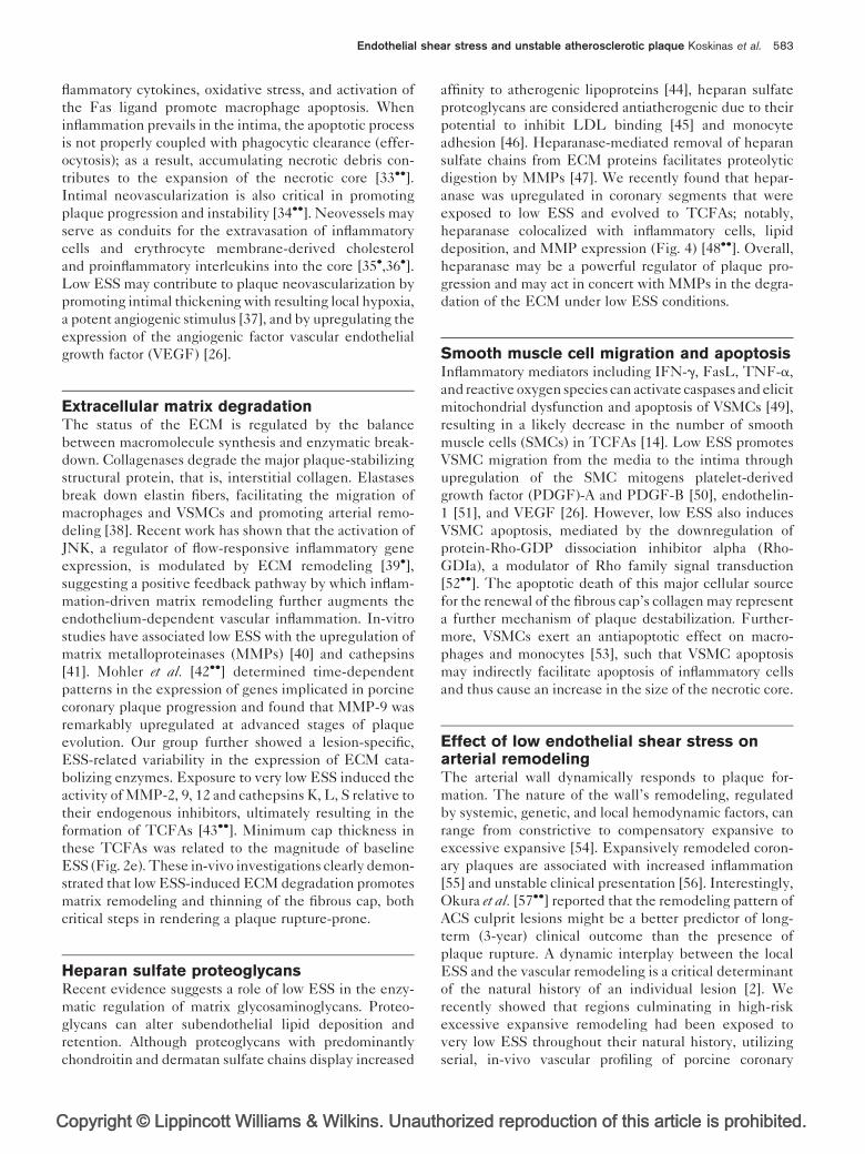
Figure 4 Colocalization of heparanase with lipids and inflammatory cells in a coronary region of low endothelial shear stress
Figure from a porcine coronary artery section, showing a thin cap fibroatheroma that developed in a coronary region of preceding low ESS. Note thatimmunostaining for heparanase (HPA, a) colocalizes with staining oil-red-O (lipids, b), and immunostaining with CD45 (inflammatory cells, c). Stainingwith picrosirius red (PR, d) indicates collagen and reveals the thin fibrous cap overlying the necrotic core.
arteries at five consecutive time points [58��] (Fig. 5). In
the setting of plaque-induced changes of the local geo-
metry and thereby of local flow waveforms, only a small
subpopulation of developing atheromata is located in a
persistently low ESS milieu, related to the magnitude of
low ESS and, consequently, the magnitude of intense
inflammation. The elaboration of matrix-degrading pro-
teases, with subsequent elastolysis in the internal elastic
laminae and the media beneath the plaque, is critical
in promoting aneurysm-like expansion of the highly
inflamed wall and turning the ostensibly protective func-
tion of compensatory remodeling into a detrimental local
environment [13��]. Local excessive expansive remodel-
ing contributes to further lowering of the low ESS, thus
reinforcing the vicious cycle of the intense proinflamma-
tory stimulus and ultimately promoting the evolution of
an early atheroma to a TCFA.
Thrombogenicity of the necrotic coreLow ESS augments the thrombogenicity of the necrotic
core and thus the extent of thrombosis in the event of
acute plaque disruption. Low ESS-induced macrophage
accumulation and SMC apoptosis are sources of tissue
factor, a potent procoagulant factor [59]. Although ather-
opyright © Lippincott Williams & Wilkins. Unautho
oprotective flow-activated KLF-2 induces thrombo-
modulin and endothelial nitric oxide synthase expression
and reduces plasminogen activator inhibitor-1 and tissue
factor expression, muting of KLF-2 shifts the balance
toward a prothrombotic state [60].
Plaque-induced changes of local endothelialshear stressThe magnitude, directionality, and spatial distribution of
local shear stress all change in response to the changes of
local arterial geometry induced by a growing plaque.
Thus, a developing plaque itself can modify the local
ESS milieu in specific parts of and adjacent to the lesion.
Lumen narrowing because of a stenotic plaque results in
increased flow velocity at the throat of the plaque, low
ESS in the upstream region, and disturbed flow in the
form of directionally oscillatory ESS in the downstream
shoulder of the plaque [61]. The composition of an
individual plaque displays considerable spatial hetero-
geneity. The downstream region contains significantly
more smooth muscle cells, whereas the upstream portion
is more inflamed, containing a high number of macro-
phages [62] and expressing higher gelatinolytic activity
[63]. Cheng et al. [26] showed in a mouse model that
rized reproduction of this article is prohibited.

C
Endothelial shear stress and unstable atherosclerotic plaque Koskinas et al. 585
Figure 5 Effect of persistently low endothelial shear stress in the formation of expansively remodeled atherosclerotic plaque
Representative example of a serially profiled porcine coronary artery. (a) Two-dimensional maps show the ESS distribution along the artery length at fiveconsecutive time points of in-vivo vascular profiling at weeks 4, 11, 16, 23, and 36 after the induction of diabetes and hyperlipidemia. In each map, thehorizontal axis represents the artery circumference (8) and the vertical axis the artery length (mm). The red rectangle includes a proximal segment whichis peristently exposed to low ESS, throughout its natural history. (b) Two-dimensional maps showing the plaque thickness, external elastic lamina radius,and lumen radius distribution along the artery length at final week 36; in each map, the horizontal axis denotes the artery circumference (8) and thevertical axis the artery length (mm). Red rectangles include the same proximal segment, as in subpart a. This arterial segment displays maximal plaquethickness and also significant expansion of the vessel wall, as indicated by the orange–red color in the corresponding maps. Note that even the lumenexhibits maximal expansion, despite the formation of significant plaque, indicating excessive expansive remodeling, that is, an exaggerated, aneurysm-like form of arterial remodeling that not only preserves normal lumen dimensions, but also actually causes lumen increase under the effect of sufficientlylow ESS. ESS, endothelial shear stress.
regions exposed to low ESS upstream of a perivascular
shear stress modifier exhibited the most profound devel-
opment of highly inflamed, vulnerable carotid plaques,
whereas stable lesions formed in the downstream vortices
of lowered/oscillatory ESS. Overall, the plaque-induced
changes of local ESS seem to exert a differential effect in
distinct portions of a stenotic lesion and critically affect
the longitudinal distribution of plaque morphology; a
self-perpetuating local environment conducive to further
plaque growth is established downstream of the lesion,
whereas a vulnerable, rupture-prone phenotype develops
in the low-ESS upstream shoulder (Fig. 6).
Plaque rupture represents the most devastating compli-
cation of atherosclerotic disease. Frank plaque rupture
may be related to low ESS-mediated inflammation and
opyright © Lippincott Williams & Wilkins. Unauth
matrix degradation culminating in severe plaque fragility
and severe proclivity to rupture from simple daily hemo-
dynamic stresses [64]. It has also been postulated that
localized high shear stress may actually trigger fibrous cap
rupture [65–67]. Because the values of wall shear stress
are markedly lower than the values of blood pressure-
induced tensile stress in the plaque cap, it is unlikely that
high wall shear stress contributes significantly to the
direct mechanical failure of the cap. However, high
ESS may induce pathobiologic responses within the
plaque that also exacerbate plaque fragility, as suggested
by the reported association of regions with high ESS with
high strain, a presumed surrogate marker of vulnerable
plaque composition [68,69]. Further, high ESS may be
implicated in local endothelial erosion, increased platelet
adhesion, and induction of acute coronary thrombosis [7].
orized reproduction of this article is prohibited.

Copyright © Lippincott Williams & Wilkins. Unauthorized reproduction of this article is prohibited.
586 Ischemic heart disease
Figure 6 Association between longitudinal atherosclerotic plaque morphology and spatial distribution of local endothelial shear
stress
(a) Top: histologic appearance of a human carotid artery plaque stained with Elastic–van Gieson. Horizontal arrow indicates the direction of blood flow.Box on the left indicates proximal (upstream) shoulder of plaque; box on the right represents the distal (downstream) shoulder. Middle: boxed area ofproximal shoulder stained with anti-CD68 (macrophages; MF) and anti-a-actin (smooth muscle cell, SMC). Bottom: boxed area of distal shoulderstained with anti-CD68 (macrophages; MF) and anti-a-actin (smooth muscle cell, SMC). Note the abundance of macrophages in the upstreamshoulder and of smooth muscle cells in the downstream shoulder, respectively. Modified from [62]. (b) Differential spatial distribution of ESS along alumen-protruding plaque. Arrows represent velocity vectors. The upstream shoulder is exposed to low ESS. Local ESS is elevated in the throat, andlow/oscillatory in the downstream shoulder of the developing plaque. These local ESS conditions promote the formation of a vulnerable, rupture-proneplaque phenotype, indicated by the red rectangle, upstream of the lesion, and additional growth, indicated by the dashed line, downstream of theplaque. ESS, endothelial shear stress; NC, necrotic core.
C
Endothelial shear stress and unstable atherosclerotic plaque Koskinas et al. 587
Effect of antiatherosclerotic medications onthe proinflammatory effect of low endothelialshear stressThe plaque-stabilizing effect of recognized antiathero-
sclerotic drugs may, at least in part, be mediated by the
attenuation of the proinflammatory low-ESS environment
of high-risk plaques, indirectly emphasizing the critical
role of low ESS in plaque destabilization. We have shown
that lifetime administration of valsartan in a diabetic,
hyperlipidemic swine model, alone or in combination with
simvastatin, attenuated the proinflammatory effect of local
low ESS. These medications reduced the expression of
MMP-9, which is actively involved in the ECM degra-
dation, as well as the MMP/tissue inhibitor of metallopro-
teinases (TIMP) ratio, thereby shifting the ECM balance
toward less degradation and limiting the severity of expan-
sive remodeling. The beneficial effect of valsartan and
simvastatin in reducing the severity of inflammation and
stabilizing high-risk plaque characteristics in regions of
low ESS was independent of a blood pressure-lowering
and lipid-lowering effect [70��]. Statins, in particular, are
potent promoters of the atheroprotective regulator KLF-2
and up-regulate several of its downstream transcriptional
targets [71]. Statins may thereby exert their well described
nonlipid-lowering vasculoprotective effects by counter-
balancing the proatherogenic effect of low ESS on the
KLF-2-regulated genes cassette.
opyright © Lippincott Williams & Wilkins. Unauth
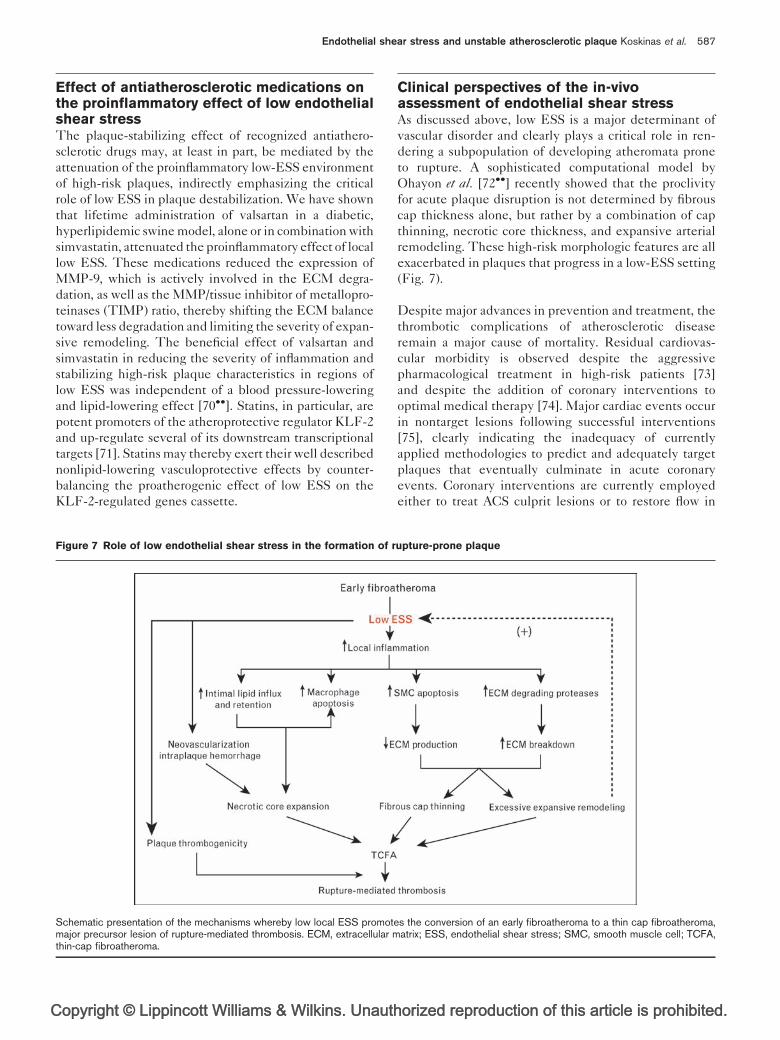
Figure 7 Role of low endothelial shear stress in the formation of r
Schematic presentation of the mechanisms whereby low local ESS promotemajor precursor lesion of rupture-mediated thrombosis. ECM, extracellular mthin-cap fibroatheroma.
Clinical perspectives of the in-vivoassessment of endothelial shear stressAs discussed above, low ESS is a major determinant of
vascular disorder and clearly plays a critical role in ren-
dering a subpopulation of developing atheromata prone
to rupture. A sophisticated computational model by
Ohayon et al. [72��] recently showed that the proclivity
for acute plaque disruption is not determined by fibrous
cap thickness alone, but rather by a combination of cap
thinning, necrotic core thickness, and expansive arterial
remodeling. These high-risk morphologic features are all
exacerbated in plaques that progress in a low-ESS setting
(Fig. 7).
Despite major advances in prevention and treatment, the
thrombotic complications of atherosclerotic disease
remain a major cause of mortality. Residual cardiovas-
cular morbidity is observed despite the aggressive
pharmacological treatment in high-risk patients [73]
and despite the addition of coronary interventions to
optimal medical therapy [74]. Major cardiac events occur
in nontarget lesions following successful interventions
[75], clearly indicating the inadequacy of currently
applied methodologies to predict and adequately target
plaques that eventually culminate in acute coronary
events. Coronary interventions are currently employed
either to treat ACS culprit lesions or to restore flow in
orized reproduction of this article is prohibited.
upture-prone plaque
s the conversion of an early fibroatheroma to a thin cap fibroatheroma,atrix; ESS, endothelial shear stress; SMC, smooth muscle cell; TCFA,

C
588 Ischemic heart disease
obstructive, flow-limiting plaques, thereby ignoring a large
proportion of minimally stenotic TCFAs, potential pre-
cursors of ACS. Although several imaging techniques have
been proposed to assess morphologic or functional charac-
teristics of rupture-prone plaques before they rupture, no
widely accepted diagnostic method to prospectively
identify such high-risk plaques is available at present.
Knowledge of local flow patterns and identification of
arterial regions with naturally occurring low ESS cannot
currently prompt interventions in native anatomy to pre-
vent atherosclerosis or atherosclerotic sequelae. However,
in-vivo identification of coronary regions of low ESS and
expansive remodeling, utilizing catheterization-based [10]
or noninvasive techniques of vascular profiling [76��], may
be predictive of future high-risk plaque formation and can
be used to risk-stratify individual lesions that have, or are
likely to acquire, characteristics of vulnerability [77��].
The early characterization of a coronary region most likely
to progress to a rupture-prone phenotype may enable
primary prevention at the level of individual atherosclero-
tic plaque and thereby provide the rationale for focused
systemic or local treatments to avert future coronary
events. Established systemic approaches and novel thera-
peutic targets may be employed to impede or even reverse
the progression towards vulnerable plaques and stabilize
high-risk plaque characteristics. Furthermore, regional
therapy of high-risk plaque in the form of highly selective,
prophylactic coronary interventions may be justified to
‘eradicate’ plaques destined to become vulnerable. The
association of drug-eluting stents [78��,79��], drug-coated
balloon catheters [80��], and, more recently, bioabsorbable
everolimus-eluting stents [81] with a lower risk of coronary
in-stent restenosis or stent thrombosis may render pre-
emptive stenting of high-risk plaques plausible. A large-
scale clinical natural history study (the PREDICTION
trial) is currently underway to investigate whether coronary
segments with low ESS and expansive remodeling are the
regions that result in rapid progression of atherosclerosis
and eventually in plaque rupture. This study may validate
the predictive value of vascular profiling for the accurate
risk stratification of early coronary plaques and thereby
potentially change the paradigm for management of
patients with coronary artery disease.
ConclusionLow ESS regulates multiple pathways that synergistically
induce plaque destabilization. Low ESS upregulates local
inflammation, promotes reduced synthesis and increased
catabolism of ECM macromolecules, and elicits exces-
sive expansive wall remodeling, a high-risk remodeling
pattern that further exacerbates the adverse low-ESS
stimulus. Early lesions persistently exposed to low ESS
may thereby progress towards highly inflamed TCFAs.
The in-vivo measurement of ESS may be used for the
opyright © Lippincott Williams & Wilkins. Unautho
early identification of lesions that are likely to acquire a
rupture-prone phenotype and trigger coronary thrombo-
sis. Early identification of high-risk plaques before they
become vulnerable may set the stage for focused systemic
treatments or prophylactic local interventions to avert
future coronary events.
AcknowledgementsThis work was supported by grants from Boston Scientific, Inc., theNovartis Pharmaceuticals, Inc., the George D. Behrakis ResearchFellowship, the Onassis Foundation, the Hellenic Harvard Foundation,the Hellenic Atherosclerosis Society, the AG Leventis Foundation, thePropondis Foundation, and the National Institutes of Health R01 GM49039 (to E.R.E.).
The authors thank Prof. George D. Giannoglou for his encouragementand support.
References and recommended readingPapers of particular interest, published within the annual period of review, havebeen highlighted as:� of special interest�� of outstanding interest
Additional references related to this topic can also be found in the CurrentWorld Literature section in this issue (p. 620).
1 Falk E. Pathogenesis of atherosclerosis. J Am Coll Cardiol 2006; 47:C7–C12.
2 Chatzizisis YS, Coskun AU, Jonas M, et al. Role of endothelial shear stress in thenatural history of coronary atherosclerosis and vascular remodeling: molecular,cellular and vascular behavior. J Am Coll Cardiol 2007; 49:2379–2393.
3 Burke AP, Kolodgie FD, Farb A, et al. Healed plaque ruptures and suddencoronary death: evidence that subclinical rupture has a role in plaqueprogression. Circulation 2001; 103:934–940.
4 Asakura T, Karino T. Flow patterns and spatial distribution of atheroscleroticlesions in human coronary arteries. Circ Res 1990; 66:1045–1066.
5
�Tanaka A, Imanishi T, Kitabata H, et al. Distribution and frequency of thin-capped fibroatheromas and ruptured plaques in the entire culprit coronaryartery in patients with acute coronary syndrome as determined by opticalcoherence tomography. Am J Cardiol 2008; 102:975–979.
This clinical, optical coherence tomography study demonstrated in vivo theproximal distribution of TCFAs.
6
�Hong MK, Mintz GS, Lee CW, et al. A three-vessel virtual histology intravas-cular ultrasound analysis of frequency and distribution of thin-cap fibroather-omas in patients with acute coronary syndrome or stable angina pectoris. Am JCardiol 2008; 101:568–572.
This three-vessel human study demonstrated the proximal localization of TCFAs,identified by virtual histology.
7 Malek AM, Alper SL, Izumo S. Hemodynamic shear stress and its role inatherosclerosis. JAMA 1999; 282:2035–2042.
8 Richter Y, Edelman ER. Cardiology is flow. Circulation 2006; 113:2679–2682.
9 Gimbrone MA Jr, Topper JN, Nagel T, et al. Endothelial dysfunction, hemo-dynamic forces, and atherogenesis. Ann N Y Acad Sci 2000; 902:230–239.
10 Stone PH, Coskun AU, Kinlay S, et al. Effect of endothelial shear stress on theprogression of coronary artery disease, vascular remodeling, and in-stentrestenosis in humans: in vivo 6-month follow-up study. Circulation 2003;108:438–444.
11 Stone PH, Coskun AU, Kinlay S, et al. Regions of low endothelial shear stressare sites where coronary plaque progress and vascular remodeling occurs inhumans: an in-vivo serial study. Eur Heart J 2007; 28:705–710.
12 Giannoglou GD, Soulis JV, Farmakis TM, et al. Haemodynamic factors and theimportant role of local low static pressure in coronary wall thickening. Int JCardiol 2002; 86:27–40.
13
��Chatzizisis YS, Jonas M, Coskun AU, et al. Prediction of the localization of highrisk coronary atherosclerotic plaques on the basis of low endothelial shearstress: an intravascular ultrasound and histopathology natural history study.Circulation 2008; 117:993–1002.
This intravascular ultrasound and histopathology study in porcine coronary arteriesshowed that the magnitude of low ESS determines the localization and hetero-geneity of atherosclerotic lesions and predicts the development of TCFAs.
rized reproduction of this article is prohibited.

C
Endothelial shear stress and unstable atherosclerotic plaque Koskinas et al. 589
14 Virmani R, Burke AP, Farb A, Kolodgie FD. Pathology of the vulnerable plaque.J Am Coll Cardiol 2006; 47:C13–C18.
15 Lehoux S, Castier Y, Tedgui A. Molecular mechanisms of the vascularresponses to haemodynamic forces. J Intern Med 2006; 259:381–392.
16 Garcıa-Cardena G, Comander JI, Blackman BR, et al. Mechanosensitiveendothelial gene expression profiles: scripts for the role of hemodynamicsin atherogenesis? Ann N Y Acad Sci 2001; 947:1–6.
17 Mott RE, Brian P, Helmke BP. Mapping the dynamics of shear stress-inducedstructural changes in endothelial cells. Am J Physiol Cell Physiol 2007;293:C1616–C1626.
18 Dekker RJ, Boon RA, Rondaij MG, et al. KLF2 provokes a gene expressionpattern that establishes functional quiescent differentiation of the endothe-lium. Blood 2006; 107:4354–4363.
19 Parmar KM, Larman HB, Dai G, et al. Integration of flow-dependent endothelialphenotypes by Kruppel-like factor 2. J Clin Invest 2006; 116:49–58.
20 Dai G, Vaughn S, Zhang Y, et al. Biomechanical forces in atherosclerosis-resistant vascular regions regulate endothelial redox balance via phosphoi-nositol 3-kinase/Akt-dependent activation of Nrf2. Circ Res 2007; 101:723–733.
21
�Zakkar M, Chaudhury H, Sandvik G, et al. Increased endothelial mitogen-activated protein kinase phosphatase-1 expression suppresses proinflamma-tory activation at sites that are resistant to atherosclerosis. Circ Res 2008;103:726–732.
The in-vitro investigation showed that MAP kinase phosphatase (MKP)-1 isrequired for the anti-inflammatory effects of atheroprotective shear stress.
22 Orr AW, Sanders JM, Bevard M, et al. The subendothelial extracellular matrixmodulates NF-kappaB activation by flow: a potential role in atherosclerosis.J Cell Biol 2005; 169:191–202.
23
��Gareus R, Kotsaki E, Xanthoules S, et al. Endothelial cell-specific NF-kappaBinhibition protects mice from atherosclerosis. Cell Metab 2008; 8:372–383.
This experimental study confirmed in vivo that NF-kB regulates proinflammatorygene expression and promotes atherosclerosis.
24 Cicha I, Goppely-Struebe M, Yilmaz A, et al. Endothelial dysfunction andmonocyte recruitment in cells exposed to nonuniform shear stress. ClinHemorheol Microcirc 2008; 39:113–119.
25 Matharu NM, McGettrick HM, Salmon M, et al. Inflammatory responses ofendothelial cells experiencing reduction in flow after conditioning by shearstress. J Cell Physiol 2008; 216:732–741.
26 Cheng C, Tempel D, van Haperen R, et al. Atherosclerotic lesion size andvulnerability are determined by patterns of fluid shear stress. Circulation2006; 113:2744–2753.
27 Cheng C, Tempel D, van Haperen R, et al. Shear stress-induced changes inatherosclerotic plaque composition are modulated by chemokines. J ClinInvest 2007; 117:616–626.
28
��Chatzizisis YS, Koskinas K, Jonas M, et al. Differential atherosclerotic vascularresponse to local endothelial shear stress based on the severity of hyperli-pidemia [abstract]. Arterioscler Thromb Vasc Biol 2009; 29:e118.
This serial, in-vivo intravascular ultrasound investigation in swine coronary arteriesshowed the synergistic effect of systemic hypercholesterolemia and local low ESSin determining the natural history of individual plaque.
29
�Soulis JV, Giannoglou GD, Papaioannou V, et al. Low-density lipoproteinconcentration in the normal left coronary artery tree. BioMed Eng Online2008; 7:26.
This computational study showed that the concentration of LDL at the luminalsurface is higher in regions of low shear stress.
30 Himburg HA, Grzybowski DM, Hazel AL, et al. Spatial comparison betweenwall shear stress measures and porcine arterial endothelial permeability. Am JPhysiol Heart Circ Physiol 2004; 286:H1916–H1922.
31 Chien S. Molecular and mechanical bases of focal lipid accumulation inarterial wall. Prog Biophys Mol Biol 2003; 83:131–151.
32 Liu Y, Chen BP, Lu M, et al. Shear stress activation of SREBP1 in endothelialcells is mediated by integrins. Arterioscler Thromb Vasc Biol 2002; 22:76–81.
33
��Seimon T, Tabas I. Mechanisms and consequences of macrophage apoptosisin atherosclerosis. J Lipid Res 2009; 50:S382–S387.
This comprehensive review summarizes current knowledge on the differential roleof apoptotic macrophages in early vs. advanced atherosclerotic lesions and theunderlying mechanisms of macrophage death during atherosclerosis.
34
��Sluimer JC, Kolodgie FD, Bijnens AP, et al. Thin-walled microvessels in humancoronary atherosclerotic plaques show incomplete endothelial junctionsrelevance of compromised structural integrity for intraplaque microvascularleakage. J Am Coll Cardiol 2009; 53:1517–1527.
This human histopathology study revealed increased microvessel density andcompromised structural integrity of the microvascular endothelium in advancedhuman atherosclerotic plaques.
opyright © Lippincott Williams & Wilkins. Unauth
35
�Giannoglou GD, Koskinas KC, Tziakas D, et al. Total cholesterol content oferythrocyte membranes and coronary atherosclerosis. An intravascular ultra-sound pilot study. Angiology 2009 [Epub ahead of print]
This human IVUS study showed for the first time in vivo that erythrocyte membrane-derived cholesterol is associated with coronary plaque burden.
36
�Tziakas DN, Chalikias GK, Tentes IK, et al. Interleukin-8 is increased in themembrane of circulating erythrocytes in patients with acute coronary syn-drome. Eur Heart J 2008; 29:2713–2722.
This human study showed that IL-8 contained in erythrocyte membranes maycontribute to coronary plaque instability.
37 Kolodgie FD, Narula J, Yuan C, et al. Elimination of neoangiogenesis forplaque stabilization: is there a role for local drug therapy? J Am Coll Cardiol2007; 49:2093–2101.
38 Libby P. The molecular mechanisms of the thrombotic complications ofatherosclerosis. J Intern Med 2008; 263:517–527.
39
�Hahn C, Orr AW, Sanders JM, et al. The subendothelial extracellular matrixmodulates JNK activation by flow. Circ Res 2009; 104:995–1003.
This experimental study identified JNK as a matrix-specific, flow-activated inflam-matory regulator.
40 Sun HW, Li CJ, Chen HQ, et al. Involvement of integrins, MAPK, and NF-kappaB in regulation of the shear stress-induced MMP-9 expression inendothelial cells. Biochem Biophys Res Commun 2007; 353:152–158.
41 Platt MO, Ankeny RF, Shi GP, et al. Expression of cathepsin K is regulated byshear stress in cultured endothelial cells and is increased in endothelium inhuman atherosclerosis. Am J Physiol Heart Circ Physiol 2007; 292:H1479–H1486.
42
��Mohler ER 3rd, Sarov-Blat L, Shi Y, et al. Site-specific atherogenic geneexpression correlates with subsequent variable lesion development in cor-onary and peripheral vasculature. Arterioscler Thromb Vasc Biol 2008;28:850–855.
This in-vivo study investigated swine coronary and peripheral arteries and asso-ciated temporal and spatial variations of gene expression with variable plaquedevelopment and progression.
43
��Chatzizisis YS, Baker A, Beigel R, et al. Low endothelial shear stressupregulates extracellular matrix degrading enzymes and promotes the forma-tion of thin cap fibroatheromas in the coronary arteries [abstract]. ArteriosclerThromb Vasc Biol 2008; 28:e44.
This in-vivo intravascular ultrasound (IVUS) and histopathology investigation ofporcine coronary arteries revealed the low shear stress-mediated upregulation ofmatrix-degrading enzymes as a critical step in the formation of thin cap fibroather-omata.
44 Wight TN, Merrilees MJ. Proteoglycans in atherosclerosis and restenosis: keyroles for versican. Circ Res 2004; 94:1158–1167.
45 Pillarisetti S, Paka L, Obunike JC, et al. Subendothelial retention of lipoprotein(a). Evidence that reduced heparan sulfate promotes lipoprotein binding tosubendothelial matrix. J Clin Invest 1997; 100:867–874.
46 Sivaram P, Obunike JC, Goldberg IJ. Lysolecithin-induced alteration of sub-endothelial heparan sulfate proteoglycans increases monocyte binding tomatrix. J Biol Chem 1995; 270:29760–29765.
47 Munesue S, Yoshitomi Y, Kusano Y, et al. A novel function of syndecan-2,suppression of matrix metalloproteinase-2 activation, which causes suppres-sion of metastasis. J Biol Chem 2007; 282:28164–28174.
48
��Baker AB, Chatzizisis YS, Beigel R, et al. Heparanase expression in thedevelopment of thin cap fibroatheromas (TCFAs): effects of plaque stage,endothelial shear stress, and pharmacologic interventions [abstract]. Arter-ioscler Thromb Vasc Biol 2008; 28:e48.
This in-vivo experimental study of swine coronary arteries showed that low shearstress upregulates heparanase and modifies the economy of extracellular matrixand the natural history of individual plaques.
49 Geng YJ, Libby P. Progression of atheroma: a struggle between death andprocreation. Arterioscler Thromb Vasc Biol 2002; 22:1370–1380.
50 Palumbo R, Gaetano C, Antonini A, et al. Different effects of high and lowshear stress on platelet-derived growth factor isoform release by endothelialcells: consequences for smooth muscle cell migration. Arterioscler ThrombVasc Biol 2002; 22:405–411.
51 Ziegler T, Bouzourene K, Harrison VJ, et al. Influence of oscillatory andunidirectional flow environments on the expression of endothelin and nitricoxide synthase in cultured endothelial cells. Arterioscler Thromb Vasc Biol1998; 18:686–692.
52
��Qi YX, Qu MJ, Long DK, et al. Rho-GDP dissociation inhibitor alpha down-regulated by low shear stress promotes vascular smooth muscle cell migrationand apoptosis: a proteomic analysis. Cardiovasc Resc 2008; 80:114–122.
This ex-vivo experimental study showed that low shear stress induces smoothmuscle cell migration and apoptosis by downregulating Rho-GDIa.
orized reproduction of this article is prohibited.

C
590 Ischemic heart disease
53 Cai Q, Lanting L, Natarajan R. Interaction of monocytes with vascular smoothmuscle cells regulates monocyte survival and differentiation through distinctpathways. Arterioscler Thromb Vasc Biol 2004; 24:2263–2270.
54 Feldman CL, Coskun AU, Yeghiazarians Y, et al. Remodeling characteristicsof minimally diseased coronary arteries are consistent along the length of theartery. Am J Cardiol 2006; 97:13–16.
55 Varnava AM, Mills PG, Davies MJ. Relationship between coronary arteryremodeling and plaque vulnerability. Circulation 2002; 105:939–943.
56 Nakamura M, Nishikawa H, Mukai S, et al. Impact of coronary artery remodel-ing on clinical presentation of coronary artery disease: an intravascularultrasound study. J Am Coll Cardiol 2001; 37:63–69.
57
��Okura H, Kobayashi Y, Sumitsuji S, et al. Effect of culprit-lesion remodelingversus plaque rupture on three-year outcome in patients with acute coronarysyndrome. Am J Cardiol 2009; 103:791–795.
This human IVUS study in patients with ACSs showed that culprit lesion positiveremodeling was a stronger predictor of long-term clinical outcome than plaquerupture.
58
��Koskinas K, Chatzizisis YS, Coskun AU, et al. High-risk coronary plaquesdevelop in a persistently low endothelial shear stress environment: A long-term, serial natural history IVUS study [abstract]. Arterioscler Thromb VascBiol 2009; 29:e76.
This in-vivo, natural history IVUS study on swine coronary arteries showed thatregions of persistently low shear stress develop high-risk excessive expansiveremodeling.
59 Tedgui A, Mallat Z. Apoptosis as a determinant of atherothrombosis. ThrombHaemost 2001; 86:420–426.
60 Lin Z, Kumar A, SenBanerjee S, et al. Kruppel-like factor 2 (KLF2) regulatesendothelial thrombotic function. Circ Res 2005; 96:e48–e57.
61 Davies PF. Hemodynamic shear stress and the endothelium in cardiovascularpathophysiology. Nat Clin Pract Cardiovasc Med 2009; 6:16–26.
62 Dirksen MT, van der Wal AC, van den Berg FM, et al. Distribution ofinflammatory cells in atherosclerotic plaques relates to the direction of flow.Circulation 1998; 98:2000–2003.
63 Segers D, Helderman F, Cheng C, et al. Gelatinolytic activity in atheroscleroticplaques is highly localized and is associated with both macrophages andsmooth muscle cells in vivo. Circulation 2007; 115:609–616.
64 Muller JE, Tofler GH, Stone PH. Circadian variation and triggers of onset ofacute cardiovascular disease. Circulation 1989; 79:733–743.
65 Fukumoto Y, Hiro T, Fujii T, et al. Localized elevation of shear stress is relatedto coronary plaque rupture: a 3-dimensional intravascular ultrasound studywith in-vivo color mapping of shear stress distribution. J Am Coll Cardiol2008; 51:645–650.
66 Gijsen FJ, Wentzel JJ, Thury A, et al. Strain distribution over plaques in humancoronary arteries relates to shear stress. Am J Physiol Heart Circ Physiol2008; 295:H1608–H1614.
67 Groen HC, Gijsen FJ, van der Lugt A, et al. Plaque rupture in the carotid arteryis localized at the high shear stress region: a case report. Stroke 2007;38:2379–2381.
68 Slager CJ, Wentzel JJ, Gijsen FJ, et al. The role of shear stress in thegeneration of rupture-prone vulnerable plaques. Nat Clin Pract CardiovascMed 2005; 2:401–407.
69 Schaar JA, Regar E, Mastik F, et al. Incidence of high-strain patterns in humancoronary arteries: assessment with three-dimensional intravascular palpogra-phy and correlation with clinical presentation. Circulation 2004; 109:2716–2719.
opyright © Lippincott Williams & Wilkins. Unautho
70
��Chatzizisis YS, Jonas M, Beigel R, et al. Attenuation of inflammation and expan-sive remodeling by valsartan alone or in combination with simvastatin in high-riskcoronary atherosclerotic plaques. Atherosclerosis 2009; 203:387–394.
In this in-vivo experimental study valsartan, alone or combined with simvastatin,attenuated the proinflammatory effect of low shear stress and reduced high-riskplaque characteristics.
71 Parmar KM, Nambudiri V, Dai G, et al. Statins exert endothelial athero-protective effects via the KLF2 transcription factor. J Biol Chem 2005;280:26714–26719.
72
��Ohayon J, Finet G, Gharib AM, et al. Necrotic core thickness and positive arterialremodeling index: emergent biomechanical factors for evaluating the risk ofplaque rupture. Am J Physiol Heart Circ Physiol 2008; 295:H717–H727.
This computational study showed that the risk of acute plaque disruption isdetermined by the combination of fibrous cap thickness, necrotic core thickness,and arterial remodeling.
73 Cannon CP, Braunwald E, McCabe CH, et al. Intensive versus moderate lipidlowering with statins after acute coronary syndromes. N Engl J Med 2004;350:1495–1504.
74 Boden WE, O’Rourke RA, Teo KK, et al. Optimal medical therapy with or withoutPCI for stable coronary disease. N Engl J Med 2007; 356:1503–1516.
75 Cutlip DE, Chhabra AG, Baim DS, et al. Beyond restenosis: five-year clinicaloutcomes from second-generation coronary stent trials. Circulation 2004;110:1226–1230.
76
��Rybicki FJ, Melchionna S, Mitsouras D, et al. Prediction of coronary arteryplaque progression and potential rupture from 320-detector row prospec-tively ECG-gated single heart beat CT angiography: Lattice Boltzmannevaluation of endothelial shear stress. Int J Cardiovasc Imaging 2009. [Epubahead of print]
This human study employed computed tomography angiography for the noninva-sive measurement of coronary ESS.
77
��Koskinas K, Coskun AU, Chatzizisis YS, et al. In-vivo identification of extremehigh-risk coronary plaque based on assessment of low endothelial shearstress and wall remodeling; an IVUS study [abstract]. Arterioscler ThrombVasc Biol 2009; 29:e44.
This natural history IVUS study proposed the in-vivo assessment of low ESS andarterial remodeling for the risk stratification of coronary atherosclerotic plaques.
78
��Stone GW, Lansky AJ, Pocock SJ, et al. Paclitaxel-eluting stents versus bare-metal stents in acute myocardial infarction. N Engl J Med 2009; 360:1946–1959.
This human study showed that implantation of paclitaxel-eluti stents was asso-ciated with reduced in-stent restenosis compared with bare-metal stents inpatients with acute myocardial infarction.
79
��Malenka DJ, Kaplan AV, Lucas FL, et al. Outcomes following coronary stentingin the era of bare-metal vs the era of drug-eluting stents. JAMA 2008;299:2868–2876.
This observational study showed that the use of drug-eluting stents was asso-ciated with a decline in the need for repeat revascularization, compared with bare-metal stents.
80
��Unverdorben M, Vallbracht C, Cremers B, et al. Paclitaxel-coated ballooncatheter versus paclitaxel-coated stent for the treatment of coronary in-stentrestenosis. Circulation 2009; 119:2986–2994.
This clinical study demonstrated a comparable efficacy and tolerability of paclitax-el-coated balloon catheter and paclitaxel-eluting stent for the inhibition of coronaryin-stent restenosis.
81 Serruys PW, Ormiston JA, Onuma Y, et al. A bioabsorbable everolimus-elutingcoronary stent system (ABSORB): 2-year outcomes and results from multipleimaging methods. Lancet 2009; 373:897–910.
rized reproduction of this article is prohibited.