vascular lab as a diagnostic tool for pad · peripheral artery disease bifurcation turbulence shear...
TRANSCRIPT
Vascular Lab as a diagnostic
tool for PAD
Richard F. Neville, MDAssociate Director, INOVA Heart and Vascular Institute
Vice-Chairman, Department of Surgery
Director of Vascular Services
Clinical Professor, George Washington University
Adjunct Professor, Biomedical Engineering, GMU
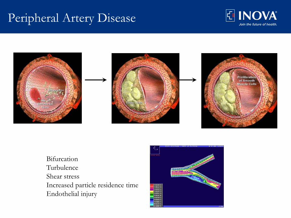
Peripheral Artery Disease
Bifurcation
Turbulence
Shear stress
Increased particle residence time
Endothelial injury

3
Peripheral Artery Disease
Asymptomatic Disease
66%
Symptomatic Disease
34%
Hiatt WR. N Engl J Med. 2001;344:1608-1621

Prevalence of PAD
• Over 9 million Americans have PAD
• One million new Medicare patients annually
• 30% over 70 years old have PAD
• Without proper treatment, are likely to die in 5
years
• Under-diagnosis of PAD
– Patients – “part of getting old”
– Physicians – “don’t ask – don’t tell policy”
• Diabetes mellitus is exploding……..350 million
worldwide
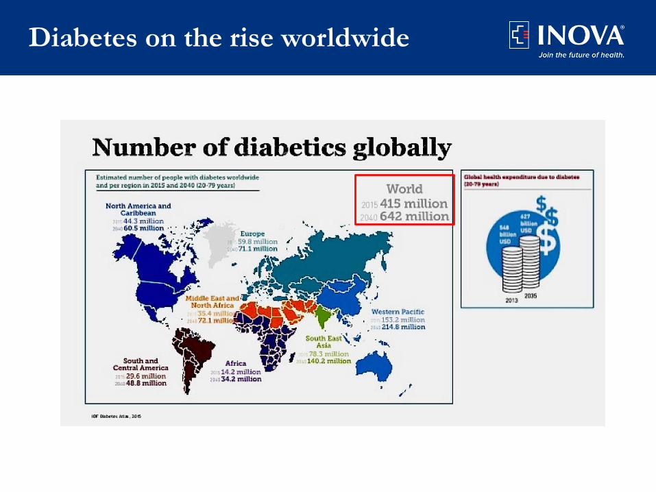
$627 Billion
Diabetes on the rise worldwide

Increasing problem worldwide
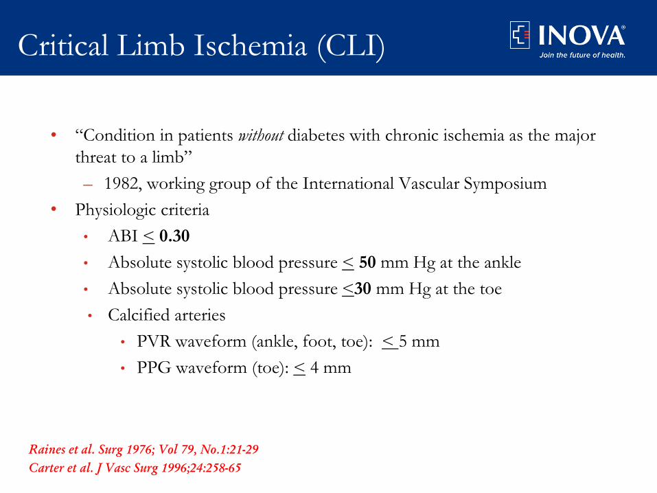
Critical Limb Ischemia (CLI)
• “Condition in patients without diabetes with chronic ischemia as the major
threat to a limb”
– 1982, working group of the International Vascular Symposium
• Physiologic criteria
• ABI < 0.30
• Absolute systolic blood pressure < 50 mm Hg at the ankle
• Absolute systolic blood pressure <30 mm Hg at the toe
• Calcified arteries
• PVR waveform (ankle, foot, toe): < 5 mm
• PPG waveform (toe): < 4 mm
Raines et al. Surg 1976; Vol 79, No.1:21-29
Carter et al. J Vasc Surg 1996;24:258-65

Definition of CLI
• Rutherford classification (1986, 1997)
– CLI under categories 4-6
• TASC II (2008)
– Chronic ischemic rest pain, ulcers or gangrene due
to objectively proven PAD
– AHA/ACC adopted TASC II definition in 2016
• Society of Vascular Surgery (2014)
– Classification based on the wound, degree of
ischemia, and foot infection (WIfI)
Gerhard-Herman MD, et al. J Am Coll Cardiol. 2016;doi:10.1016/j.jacc.2016.11.007.
Hardman RL, et al. Semin Intervent Radiol. 2014;doi:10.1055/s-0034-1393976.
Mills JL, et al. J Vasc Surg. 2014;doi:10.1016/j.jvs.2013.08.003
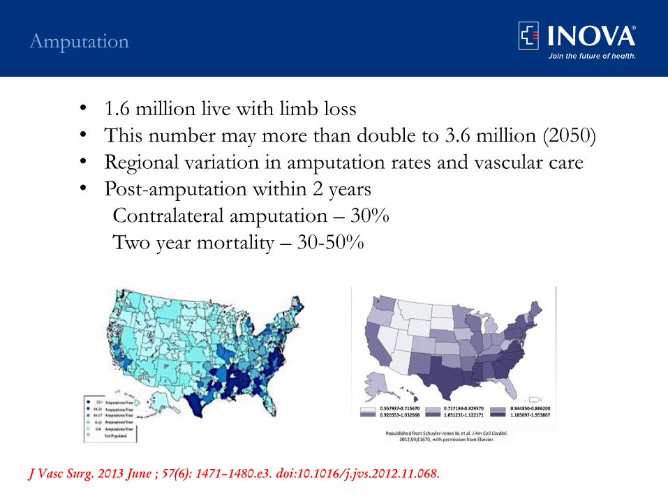
Amputation
J Vasc Surg. 2013 June ; 57(6): 1471–1480.e3. doi:10.1016/j.jvs.2012.11.068.
• 1.6 million live with limb loss
• This number may more than double to 3.6 million (2050)
• Regional variation in amputation rates and vascular care
• Post-amputation within 2 years
Contralateral amputation – 30%
Two year mortality – 30-50%

Mortality
TASC II. (2007). JOURNAL OF VASCULAR SURGERY
Volume 45, Number 1, Supplement S
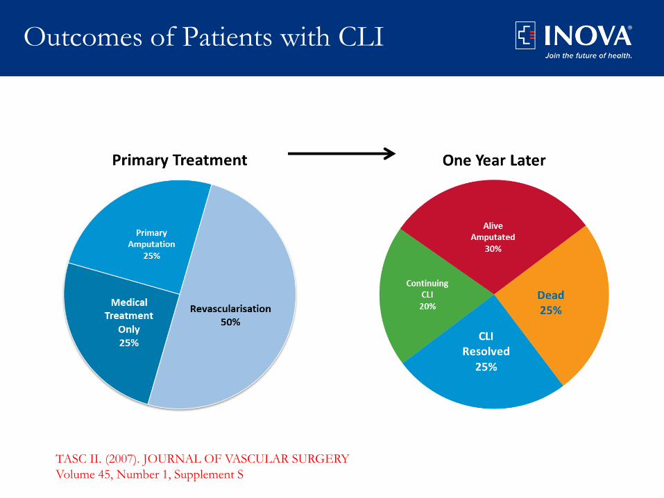
Outcomes of Patients with CLI

Diagnostic assessment
• History
– Risk factors
– Symptoms at presentation
• Physical Exam
• Noninvasive vascular testing
• Arterial imaging
– Arteriography
– MRA
– CTA

Non-invasive Vascular Lab
• ABI
• Segmental waveforms
• Segmental pressures
• Pulse Volume Recordings
• Digital pressures
• Duplex imaging
• Tissue perfusion
• TcO2
• Skin perfusion pressure
• Photoplethysmography
• Hyperspectral imaging
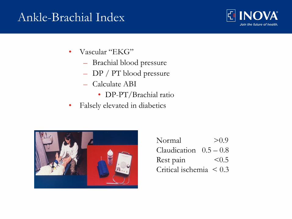
Ankle-Brachial Index
• Vascular “EKG”
– Brachial blood pressure
– DP / PT blood pressure
– Calculate ABI
• DP-PT/Brachial ratio
• Falsely elevated in diabetics
Normal >0.9
Claudication 0.5 – 0.8
Rest pain <0.5
Critical ischemia < 0.3
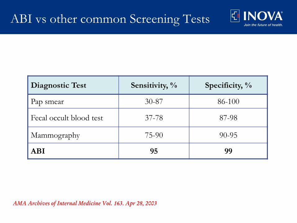
ABI vs other common Screening Tests
Diagnostic Test Sensitivity, % Specificity, %
Pap smear 30-87 86-100
Fecal occult blood test 37-78 87-98
Mammography 75-90 90-95
ABI 95 99
AMA Archives of Internal Medicine Vol. 163. Apr 28, 2003

Segmental pressures/waveforms
Arm
Pressures
For ABI
Ankle
Pressures
for ABI
Waveforms
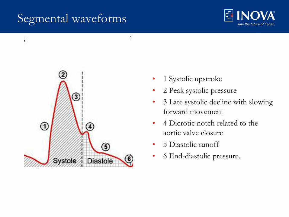
Segmental waveforms
• 1 Systolic upstroke
• 2 Peak systolic pressure
• 3 Late systolic decline with slowing
forward movement
• 4 Dicrotic notch related to the
aortic valve closure
• 5 Diastolic runoff
• 6 End-diastolic pressure.
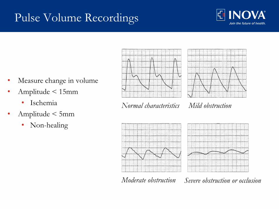
Pulse Volume Recordings
• Measure change in volume
• Amplitude < 15mm
• Ischemia
• Amplitude < 5mm
• Non-healing
Normal characteristics Mild obstruction
Moderate obstruction Severe obstruction or occlusion

Photoplethysmography: Toe pressures
• Reflection of light in microcirculation
• Toe pressures
– Toe Brachial index (> 0.4)
• Non-healing < 30-40 mmHg
Toe Cuffs Here
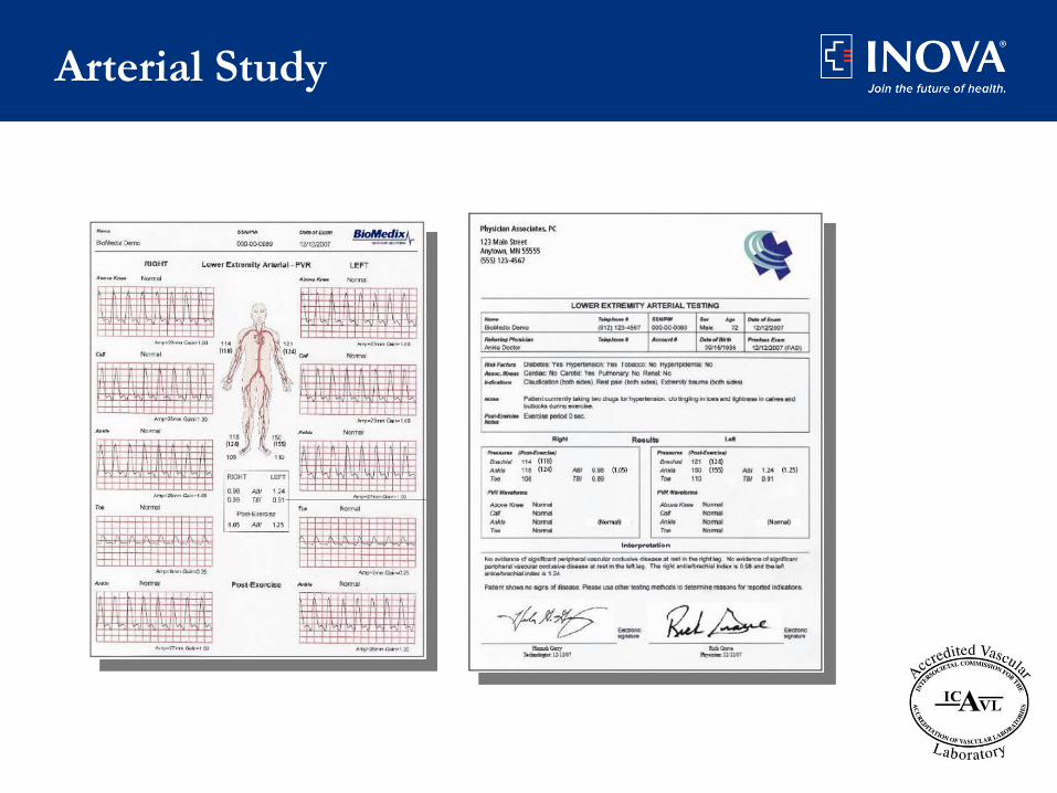
Arterial Study
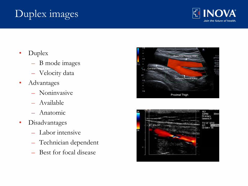
Duplex images
• Duplex
– B mode images
– Velocity data
• Advantages
– Noninvasive
– Available
– Anatomic
• Disadvantages
– Labor intensive
– Technician dependent
– Best for focal disease
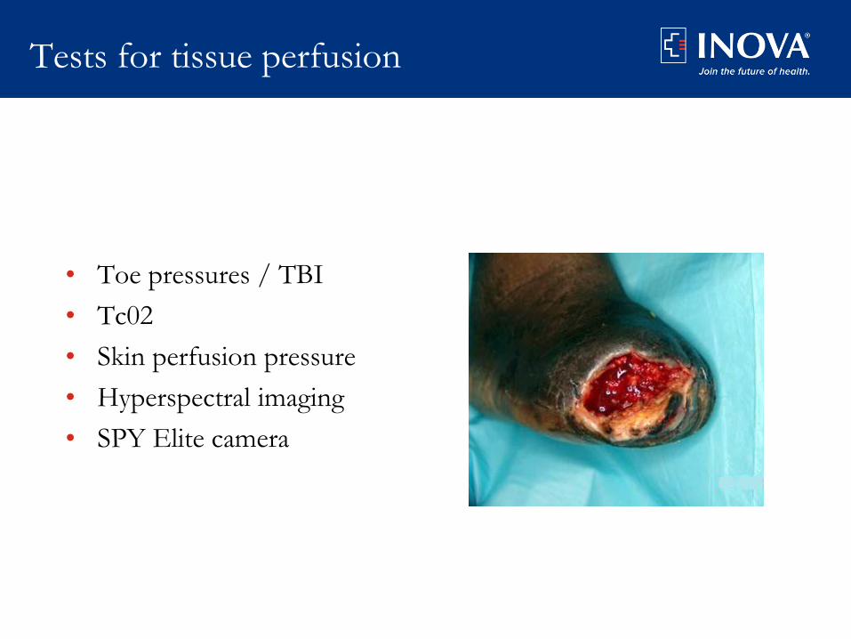
Tests for tissue perfusion
• Toe pressures / TBI
• Tc02
• Skin perfusion pressure
• Hyperspectral imaging
• SPY Elite camera

Transcutaneous Oxygen
• Healing potential and tissue perfusion
• Values measured at foot and chest wall
• Non-healing
– Value < 20-30 mmHg
– Chest – foot index < 0.4
• TcO2 peaks two to four weeks after revascularization
Caselli A, et al Diabetic Med 2005;22(4):460-465.

Skin perfusion pressure (SPP)
• Laser Doppler technology
– Laser signal below skin surface (1.5mm)
– Measures blood flow to capillary/tissue
• Pressure cuff occludes capillary flow
• System performs a controlled release of pressure
• Laser uses the Doppler “shift effect” of capillary flow return
• Automatically calculates the Skin Perfusion Pressure - the
pressure at which blood flow first returns to the capillaries
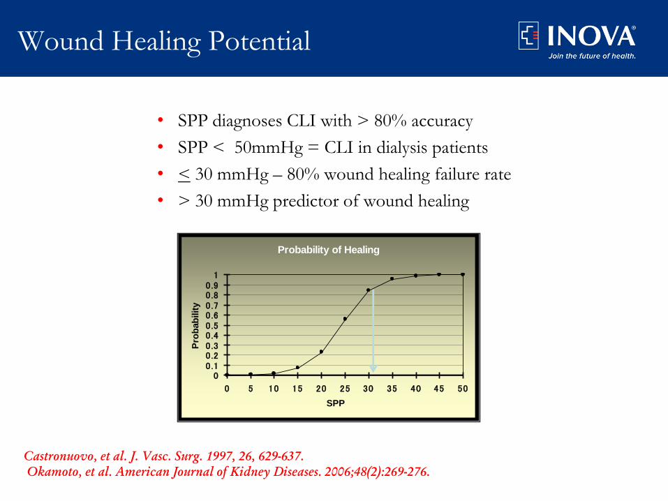
• SPP diagnoses CLI with > 80% accuracy
• SPP < 50mmHg = CLI in dialysis patients
• < 30 mmHg – 80% wound healing failure rate
• > 30 mmHg predictor of wound healing
Probability of Healing
00.10 .20 .30 .40 .50 .60 .70 .80 .91
0 5 10 15 20 25 30 35 40 45 50
SPP
Pro
bab
ilit
y
Castronuovo, et al. J. Vasc. Surg. 1997, 26, 629-637.
Okamoto, et al. American Journal of Kidney Diseases. 2006;48(2):269-276.
Wound Healing Potential
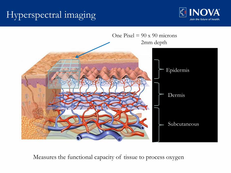
Hyperspectral imaging
One Pixel = 90 x 90 microns
2mm depth
Measures the functional capacity of tissue to process oxygen
Epidermis
Dermis
Subcutaneous
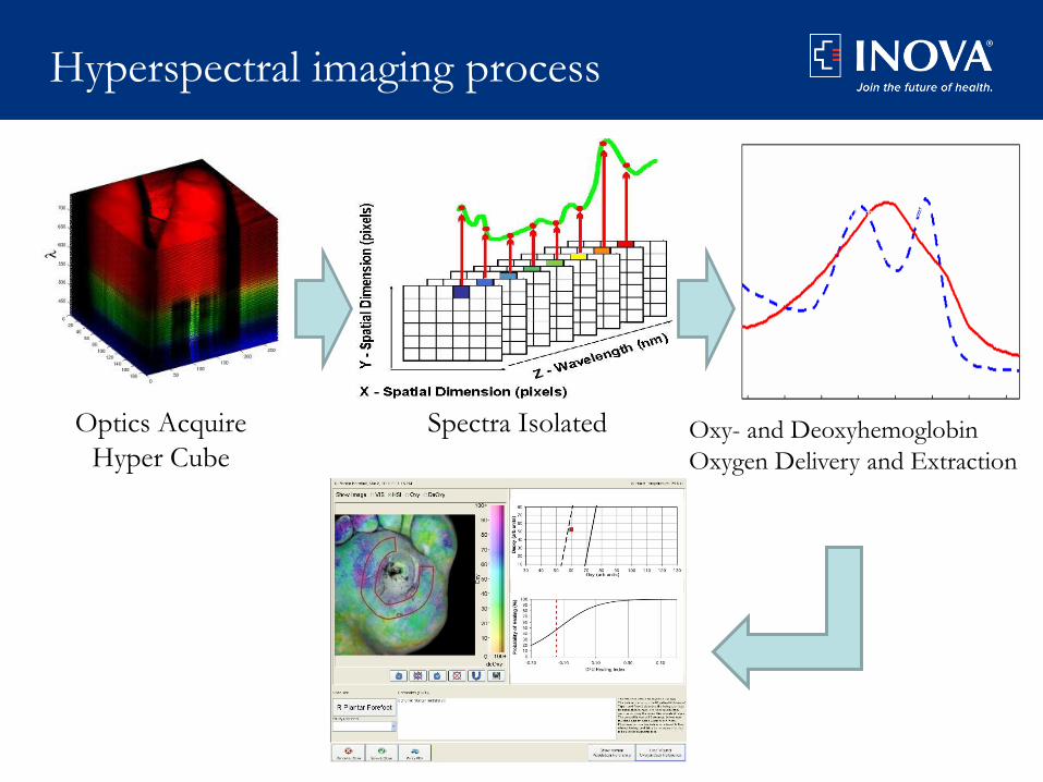
Hyperspectral imaging process
Optics Acquire
Hyper CubeOxy- and Deoxyhemoglobin
Oxygen Delivery and Extraction
540nm 577nm
555nm
Spectra Isolated

Hyperspectral analysis
• Oxyhemoglobin low
• DeOxy low
• O2 Sat low relative to the
normal population values
• Results consistent with
decreased perfusion
Value Observed Normal
Mean
Std. Dev.
Oxy 19 38 18
DeOxy 28 40 12
O2 Sat 40 48 10
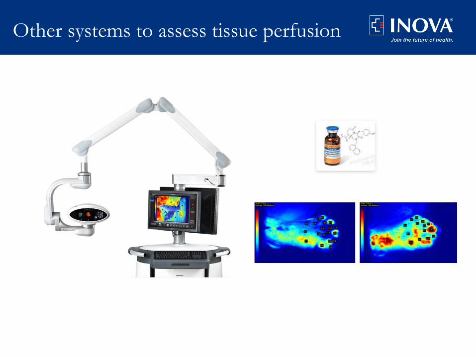
Other systems to assess tissue perfusion
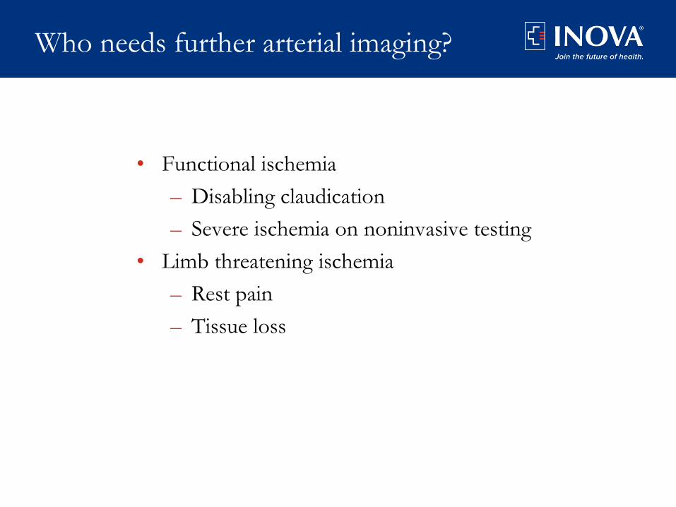
Who needs further arterial imaging?
• Functional ischemia
– Disabling claudication
– Severe ischemia on noninvasive testing
• Limb threatening ischemia
– Rest pain
– Tissue loss
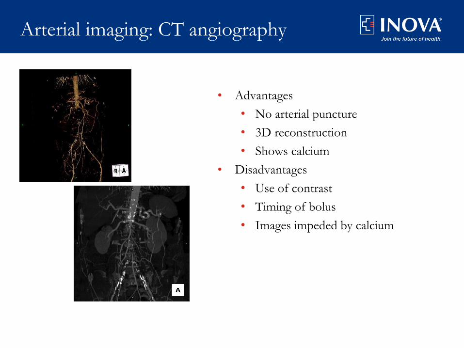
Arterial imaging: CT angiography
• Advantages
• No arterial puncture
• 3D reconstruction
• Shows calcium
• Disadvantages
• Use of contrast
• Timing of bolus
• Images impeded by calcium

Arterial imaging: MR angiography
• Flow dependent
• Poor distal image quality
• Overestimate stenosis
• Time to acquire images
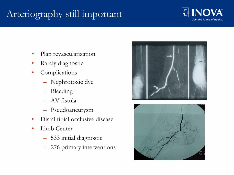
Arteriography still important
• Plan revascularization
• Rarely diagnostic
• Complications
– Nephrotoxic dye
– Bleeding
– AV fistula
– Pseudoaneurysm
• Distal tibial occlusive disease
• Limb Center
– 533 initial diagnostic
– 276 primary interventions
Role of the Non-invasive Vascular Lab
• Role of Vascular lab
– Screening
– Determine need for further imaging and intervention
– Determine success of therapy
• Medical
• Endovascular
• Surgical
– Follow up revascularization procedures
• Vascular lab in a CLI practice
– Is there adequate perfusion for a wound to heal?
– Has the intervention provided adequate perfusion for healing?
– Is perfusion maintained or is a re-intervention required?

Thank you