the role of autophagy in parkinson’s disease
TRANSCRIPT

The Role of Autophagy in Parkinson’s Disease
Melinda A. Lynch-Day, Kai Mao, Ke Wang, Mantong Zhao, and Daniel J. Klionsky
University of Michigan, Life Sciences Institute, Ann Arbor, Michigan 48109
Correspondence: [email protected]
Great progress has been made toward understanding the pathogenesis of Parkinson’s disease(PD) during the past two decades, mainly as a consequence of the discovery of specific genemutations contributing to the onset of PD. Recently, dysregulation of the autophagy pathwayhas been observed in the brains of PD patients and in animal models of PD, indicating theemerging role of autophagy in this disease. Indeed, autophagy is increasingly implicatedin a number of pathophysiologies, including various neurodegenerative diseases. Thisarticle will lead you through the connection between autophagy and PD by introducingthe concept and physiological function of autophagy, and the proteins related to autosomaldominant and autosomal recessive PD, particularlya-synuclein and PINK1-PARKIN, as theypertain to autophagy.
There seem to be various causes of Parkin-son’s disease (PD), yet the pathogenesis of
this disease appears to be converging on com-mon themes—oxidative stress, mitochondrialdysfunction, and protein aggregation—all ofwhich are tightly linked to autophagy, a highlyconserved cellular homeostatic process essentialfor bulk degradation of cytoplasmic contents.In particular, the recent identification of auto-somal dominant and autosomal recessive muta-tions in familial PD has revealed the involve-ment of the corresponding gene products inautophagy. Although autophagy has commonlybeen regarded as an adaptive response to nu-trient deprivation, increasing evidence indicatesthat basal, constitutive autophagy is essentialfor neuronal survival and that its dysregulationleads to neurodegeneration.
AUTOPHAGY
Main Types of Autophagy
Autophagy is an evolutionarily conserved ca-tabolic process that mediates the degradationof long-lived proteins and dysfunctional orsuperfluous organelles in eukaryotic cells. Au-tophagy is induced by various adverse condi-tions such as limited nutrients, low oxygen lev-els, and decreased energy supply, and its actionresults in the release of degradation products,especially amino acids, back into the cytoplasmto be used in essential biosynthetic pathways.
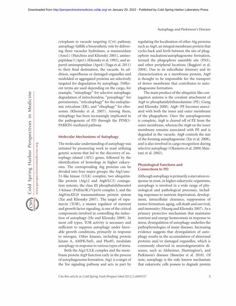
According to the different pathways bywhich cargo is delivered to the lysosome or va-cuole, autophagy can be divided into threemain types (Fig. 1): macroautophagy, microau-tophagy, and chaperone-mediated autophagy
Editor: Serge Przedborski
Additional Perspectives on Parkinson’s Disease available at www.perspectivesinmedicine.org
Copyright # 2012 Cold Spring Harbor Laboratory Press; all rights reserved; doi: 10.1101/cshperspect.a009357
Cite this article as Cold Spring Harb Perspect Med 2012;2:a009357
1
ww
w.p
ersp
ecti
vesi
nm
edic
ine.
org
on January 20, 2022 - Published by Cold Spring Harbor Laboratory Presshttp://perspectivesinmedicine.cshlp.org/Downloaded from

(CMA). CMA involves direct translocation ofunfolded proteins across the lysosome mem-brane. Chaperone proteins mediate this processby binding to cytosolic substrates that enter thelysosome through interaction with a receptor/channel on the lysosomal membrane (Majeskiand Dice 2004). Microautophagy describes theprocess of direct uptake of cytoplasmic materialsat the lysosome surface by invagination of thelysosome membrane. After vesicles containingthe cytosolic substrates pinch off into the lyso-somal lumen, they are rapidly degraded (Kunzet al. 2004). In contrast, during macroautophagy,portions of the cytoplasm are engulfed by a dou-ble-membrane phagophore that expands into acytosolic vesicle called an autophagosome; thecompleted autophagosome is targeted to thelysosome in mammalian cells or the vacuole inyeast (Klionsky 2005). The outer membrane ofthe autophagosome subsequently fuses with thelysosomal/vacuolar membrane, allowing hydro-
lases access to the inner autophagosome mem-brane and its cargo, which is degraded and re-cycled. In contrast to the ubiquitin-26S pro-teasome system, macroautophagy can mediatenonselective and bulk degradation of cytoplas-mic contents, including entire organelles (Shin-tani et al. 2002; Kanki et al. 2009). Among thethree main types of autophagy, macroautophagyis the best characterized process and will here-after be referred to as autophagy.
Selective Autophagy
In some cases, autophagy displays substratespecificity, even though autophagy is often con-sidered to be a nonselective pathway for thedegradation of bulk cytoplasmic components.Indeed, the unique feature of the autophagyprocess where the initial sequestering compart-ment expands into an autophagosome allowsfor flexible cargo selection. For example, in the
Autolysosome
Lysosomalhydrolase
Permease
Unfoldedsubstrate protein
hsc70
Peroxisome
Microautophagy(micropexophagy)
Macroautophagy
Chaperone-mediatedautophagy
Lysosome
Autophagosome
PhagophoreMitochondrion
LAMP-2A
Figure 1. Schematic model of the three main types of autophagy. The modes of autophagy differ depending onthe nature of the substrate and the site of sequestration. In chaperone-mediated autophagy, the substrates con-tain a KFERQ-consensus motif, are unfolded by HSC70 chaperones, and translocate directly across the lysosomemembrane via interaction with a LAMP-2A oligomer. There are various types of microautophagy-like processesincluding micropexophagy and micromitophagy, the selective degradation of peroxisomes and mitochondria,respectively. Again, sequestration occurs at the lysosome-limiting membrane, but the substrates do not have tobe unfolded. Macroautophagy uses a double-membrane phagophore to sequester the cargo. Essentially any cy-toplasmic component can be enwrapped by a phagophore, which expands into an autophagosome. Fusion withthe lysosome allows the cargo to be degraded, and the resulting macromolecules are released into the cytosolthrough permeases, allowing them to be reused for anabolic processes.
M.A. Lynch-Day et al.
2 Cite this article as Cold Spring Harb Perspect Med 2012;2:a009357
ww
w.p
ersp
ecti
vesi
nm
edic
ine.
org
on January 20, 2022 - Published by Cold Spring Harbor Laboratory Presshttp://perspectivesinmedicine.cshlp.org/Downloaded from
cytoplasm to vacuole targeting (Cvt) pathway,autophagy fulfills a biosynthetic role by deliver-ing three vacuolar hydrolases, a-mannosidase(Ams1) (Hutchins and Klionsky 2001), amino-peptidase I (Ape1) (Klionsky et al. 1992), and as-partyl aminopeptidase (Ape4) (Yuga et al. 2011)to their final destination, the vacuole. In ad-dition, superfluous or damaged organelles andmisfolded or aggregated proteins are selectivelytargeted for degradation by autophagy. Differ-ent terms are used depending on the cargo, forexample, “mitophagy” for selective autophagicdegradation of mitochondria, “pexophagy” forperoxisomes, “reticulophagy” for the endoplas-mic reticulum (ER), and “ribophagy” for ribo-somes (Klionsky et al. 2007). Among them,mitophagy has been increasingly implicated inthe pathogenesis of PD through the PINK1-PARKIN-mediated pathway.
Molecular Mechanisms of Autophagy
The molecular understanding of autophagy wasinitiated by pioneering work in yeast utilizinggenetic screens that led to the discovery of au-tophagy-related (ATG) genes, followed by theidentification of homologs in higher eukary-otes. The corresponding Atg proteins can bedivided into four major groups: the Atg1/unc-51-like kinase (ULK) complex, two ubiquitin-like protein (Atg12 and Atg8/LC3) conjuga-tion systems, the class III phosphatidylinositol3-kinase (PtdIns3K)/Vps34 complex I, and theAtg9/mATG9 transmembrane protein system(Xie and Klionsky 2007). The target of rapa-mycin (TOR), a master regulator of nutrientand growth factor signaling, is one of the criticalcomponents involved in controlling the induc-tion of autophagy (He and Klionsky 2009). Inmost cell types, TOR activity is necessary andsufficient to suppress autophagy under favor-able growth conditions, primarily in responseto nitrogen. Other kinases, including proteinkinase A, AMPK/Snf1, and Pho85, modulateautophagy in response to various types of stress.
Both the Atg1/ULK complex and the mem-brane protein Atg9 function early in the processof autophagosome formation. Atg1 is a target ofthe Tor signaling pathway and acts in part by
regulating the localization of other Atg proteinssuch as Atg9, an integral membrane protein thatcycles back and forth between the site of phag-ophore nucleation/autophagosome formation,termed the phagophore assembly site (PAS),and other peripheral locations (Reggiori et al.2004). Due to its subcellular itinerary and itscharacterization as a membrane protein, Atg9is thought to be responsible for the transportof donor membrane that contributes to auto-phagosome formation.
The main product of the ubiquitin-like con-jugation systems is the covalent attachment ofAtg8 to phosphatidylethanolamine (PE) (Gengand Klionsky 2008). Atg8–PE becomes associ-ated with both the inner and outer membraneof the phagophore. Once the autophagosomeis complete, Atg8 is cleaved off of PE from theouter membrane, whereas the Atg8 on the innermembrane remains associated with PE and isdegraded in the vacuole. Atg8 controls the sizeof the forming autophagosome (Xie et al. 2008),and is also involved in cargo recognition duringselective autophagy (Okamoto et al. 2009; Shin-tani et al. 2002).
Physiological Functions andConnections to PD
Although autophagy is primarily a starvation re-sponse in yeast, in higher eukaryotic organisms,autophagy is involved in a wide range of phy-siological and pathological processes, includ-ing responses to nutrient deprivation, develop-ment, intracellular clearance, suppression oftumor formation, aging, cell death and survival,and immunity (Huang and Klionsky 2007). As aprimary protective mechanism that maintainsnutrient and energy homeostasis in response tostress, dysregulation of autophagy underlies thepathophysiologies of many diseases. Increasingevidence suggests that dysregulation of auto-phagy results in the accumulation of abnormalproteins and/or damaged organelles, which iscommonly observed in neurodegenerative di-seases, such as Alzheimer, Huntington’s, andParkinson’s diseases (Banerjee et al. 2010). Ofnote, autophagy is the only known mechanismthat eukaryotic cells possess to degrade protein
Autophagy and Parkinson’s Disease
Cite this article as Cold Spring Harb Perspect Med 2012;2:a009357 3
ww
w.p
ersp
ecti
vesi
nm
edic
ine.
org
on January 20, 2022 - Published by Cold Spring Harbor Laboratory Presshttp://perspectivesinmedicine.cshlp.org/Downloaded from

aggregates and damaged organelles that cannot beprocessed by the proteasome. Recent studies fromtransgenic mice, animal, and cell models of PDsuggest the involvement of proteins geneticallylinked to autosomal dominant PD, particularlya-synuclein and LRRK2, in the autophagy path-way (Bandyopadhyay and Cuervo 2007; Alegre-Abarrategui and Wade-Martins 2009). In addi-tion, proteins related to recessive PD, such asPINK1 and PARKIN, have an important role inthe process of mitophagy.
EARLY DISCOVERIES: a-SYNUCLEINAND AUTOPHAGY
a-Synuclein and PD
a-Synuclein was found to localize to the presy-naptic terminals in the central nervous systemand is involved in vesicular release (Claytonand George 1998, 1999; Spencer et al. 2009). Itis a natively unfolded protein, but can be foundin several aberrant conformational states in-cluding an oligomer, a protofibril, and an amy-loid fibril (Martinez-Vicente et al. 2008). a-Syn-uclein was identified as a component of Lewybodies, cytosolic inclusions that are a patholog-ical trait of PD (Spillantini et al. 1997, 1998).Studies of familial cases of autophagy revealtwo separate autosomal dominant mutationsin the a-synuclein gene: A53T and A30P (Poly-meropoulos et al. 1997). In addition to thepoint mutations, several posttranslational mod-ifications such as phosphorylation, ubiquitina-tion, nitration, oxidation, and dopamine-de-pendent adduct formation also create toxicforms of the protein (Martinez-Vicente et al.2008).
Chaperone-Mediated Autophagy
a-Synuclein in its native form is degraded bychaperone-mediated autophagy (Cuervo et al.2004). The protein contains a 15 amino acidsequence that consists of imperfect yet overlap-ping variations of the KFERQ CMA recogni-tion motif. The chaperone protein HSC70 rec-ognizes the pentapeptide sequence motif andbinds to a-synuclein. a-Synuclein then bindsto the lysosomal-associated membrane proteintype 2A (LAMP-2A) at the lysosomal membrane.
This CMA receptor with the aid of a lysosomallumenal HSC70 transports a-synuclein intothe lysosome where it is degraded by proteases.Mutant forms of the protein prevent its degra-dation by the CMA pathway resulting in toxicaggregation in the cytoplasm as was seen incell culture and postmortem tissues (Martinez-Vicente et al. 2008). Autophagy can partiallycompensate for the lack of CMA-mediated deg-radation, but may cause autophagic cell deathunder stress conditions. Cells expressing mutanta-synuclein can be characterized by an increasein cell death, accumulation of autophagosomes,and a loss of ability to store catecholamine alongwith a failure to release dopamine (Stefanis et al.2001).
All mutant forms of a-synuclein vary in thedegree to which they hamper the lysosomal/CMA degradation pathway and thus have differ-ent levels of toxicity. In cell culture studies, theA53T and A30P mutants of the protein bindmore strongly to the LAMP-2A receptor than thewild-type form, but fail to be transported acrossthe lysosomal membrane (Martinez-Vicente etal. 2008). The mutants act as receptor inhibitors,preventing other CMA targets from binding.This leads to a complete block in CMA resultingin a higher degree of toxicity. Overexpression ofthe wild-type protein is matched by an increasein expression of the CMA receptor proteinLAMP-2A, but high levels of protein expressionlead to the formation of oligomeric forms thatcannot be degraded by CMA (Vogiatzi et al.2008; Xilouri et al. 2009). This results in atoxicitylevel that is lower than the familial point mutantforms. Similar intermediate levels of toxicity areseen in certain posttranslationally modified ver-sions of a-synuclein as was illustrated in cell andpost-mortem tissues. Phosphorylated, ubiquiti-nated, nitrated, and oxidized forms are less sus-ceptible to CMA degradation than the nonmodi-fied protein, but they do not block CMA in itsentirety. It is thought that these modificationspromote higher-order oligomers that cannot bebroken down and degraded. This is reflected inthe observation of all of thesemodifications in cy-tosolic aggregates.
Modification of the protein with dopaminegives a phenotype that more closely resembles
M.A. Lynch-Day et al.
4 Cite this article as Cold Spring Harb Perspect Med 2012;2:a009357
ww
w.p
ersp
ecti
vesi
nm
edic
ine.
org
on January 20, 2022 - Published by Cold Spring Harbor Laboratory Presshttp://perspectivesinmedicine.cshlp.org/Downloaded from

the point mutations. a-Synulcein can be modi-fied with oxidized dopamine through a nonco-valent interaction. Dopaminea-synuclein (DA-a-syn) inhibits not only its own degradation,but it also blocks CMA activity in general. Thisdefect can be seen in a variety of experimentalconditions including isolated lysosomes, dopa-minergic cell lines, and visceral motor neurons.The CMA defect results from DA-a-syn expres-sion because it is not seen in a-synuclein dele-tion cells, it is not observed with the dopamineinsensitive form of a-synuclein, and it is repro-duced with isolated lysosomes when presentedwith DA-a-syn. The complete blockage of CMAcreates a high level of toxicity (Martinez-Vicenteet al. 2008). What is interesting about the DA-a-syn modification is that it is the dopaminergicsubstantia nigra and the norepinephrine-releasinglocus coeruleus neurons that are killed first in theprogression of PD. Both of these types of neuronscontain cytosolic dopamine and produce neuro-melanin (a product of dopamine modifications).The toxic DA-a-syn form of the protein may ex-plain why those two types of neurons are particu-larly sensitive.
The blockage of CMA activity with mutantforms of a-synuclein not only results in the di-rect buildup of toxicity in the neuron throughthe formation of aggregates, but it also preventsthe protective activityof the protein myocyte en-hancer factor 2D (MEF2D). MEF2D,atranscrip-tion factor, is an important player in neuronalsurvival. Patients with PD show an increase ofthis protein in brain neurons, and a genetic poly-morphism of a related protein (MEF2A) hasbeen linked to Alzheimer disease. CMA-depen-dent degradation regulates MEF2D activity.MEF2D is continuously shuttled to the cytosolfrom the nucleus where it interacts with hsc70.In cells, when CMA is inhibited, an inactiveform of the protein accumulates in the cytosoland the amount of protein in the nucleus drops.This inactive form can no longer bind DNA.Wild-type and mutant forms of a-synucleinprevent binding between HSC70 and MEF2D(Yangetal.2009).Thissuggeststhatnotonlydoesa-synuclein promote neuronal death throughthe formation of aggregates, but it also promotescell death by inhibiting cell survival proteins.
a-Synuclein and Autophagy
As noted previously, inhibition of CMA byaberrant a-synuclein leads to an increase in au-tophagy. This appears to be a compensatory re-sponse, but rather than leading to cell survival,the induction of autophagy can be detrimentalcausing autophagic cell death. Blocking autoph-agy by knocking down the autophagy proteinAtg5 in cells expressing the A53T a-synucleinmutant can rescue the cell from toxicity-in-duced cell death (Xilouri et al. 2009). However,autophagy-induced neuronal death is not al-ways the outcome. One study suggests that thesignaling pathway for activation of autophagymay be important as to whether or not autoph-agy will be protective or detrimental. Autophagyis mainly initiated through the mTOR signal-ing pathway either directly or indirectly throughthe autophagy protein Atg1. An additional sig-naling pathway for initiation of autophagy isthe Vps34-Beclin 1 complex. It is this secondarysignaling pathway that appears to promote cellsurvival. For example, a reduction in a-syn-uclein accumulation is seen when Beclin 1 isoverexpressed. In addition, Beclin 1 overexpres-sion decreases cell death and increases autoph-agy activity observed through enhanced lysoso-mal degradation (Spencer et al. 2009).
Not only does aberrant a-synuclein inhibitCMA, but it also inhibits autophagy throughRAB1A and omegasome formation as seen inboth cell and mouse models (Winslow et al.2010). RAB1A is a GTPase involved in the earlysecretory pathway, specifically ER-to-Golgi trans-port. The early secretory pathway is importantfor autophagy, and inhibition of the secretorypathway blocks autophagy. Furthermore, RABproteins can play a role in autophagy indepen-dent of the secretory pathway, as seen with Ypt1in yeast (Lynch-Day et al. 2010) and RAB1A inmammalian cells (Huang et al. 2011). a-Syn-uclein overexpression blocks autophagosome for-mation, inhibits secretion, and increases Golgifragmentation. Overexpression of RAB1A rescuesthis defect. The block in autophagy due toa-syn-uclein overexpression occurs early in the pathway,before autophagosome formation, suggesting aneffect on ATG9, which is the only transmembrane
Autophagy and Parkinson’s Disease
Cite this article as Cold Spring Harb Perspect Med 2012;2:a009357 5
ww
w.p
ersp
ecti
vesi
nm
edic
ine.
org
on January 20, 2022 - Published by Cold Spring Harbor Laboratory Presshttp://perspectivesinmedicine.cshlp.org/Downloaded from

protein required for autophagy. It is thought thatATG9 is responsible for the transport of mem-brane to the site of autophagosome formationand thus acts early in the process. ATG9 normallyforms puncta at a perinuclear location (the site ofautophagosome formation) and at the trans-Golgi network in mammalian cells. Whena-syn-uclein is overexpressed, ATG9 is mislocalized andis diffuse throughout the cytoplasm of the cell.The same phenotype is seen with the knock-down of RAB1A. One preautophagosome struc-ture that branches off of the ER is the omegasome,which generates an autophagosome, at least un-der some circumstances. Omegasome formationis reduced in cells that overexpress a-synucleinand in cells that have reduced RAB1A proteinlevels. a-Synuclein blocks autophagy by inhibit-ing the activity of RAB1A, which results in themislocalization of ATG9 and inhibition of auto-phagosome formation (Winslow et al. 2010).
a-Synuclein and Mitophagy
More recent studies of a-synuclein in PD haveshown a relationship between its aberrant ex-pression and mitophagy. Mitochondrial dys-function is another characteristic of PD and willbe described in more detail later in this article.However, a connection has been made betweenthe activation of autophagy by aberrant a-syn-uclein expression and mitochondrial dysfunc-tion. In cells expressing the A53T a-synucleinmutant, there is an observed increase in colocal-ization between autophagosomes and normal,polarized mitochondria. In addition, there is adecrease in the number and length of mitochon-dria in these cells. Similar results are seen whenwild-type a-synuclein is overexpressed; how-ever, the phenotype is not as severe. The increasein mitochondria clearance in these cell lines isdependent on mitochondrial fragmentation andon the protein PARKIN (Choubey et al. 2011).PARKIN, currently another large area of focusfor autophagy and PD research, will be discussedin the next section of this article. However, therole of a-synuclein in the promotion of mito-phagy of polarized mitochondria suggests thatthere may be a connection between it and PAR-KIN in the promotion of the disease.
RECENT STUDIES: CONTROVERSIESABOUND
In this section, we will discuss new yet contro-versial areas of research with regard to PD andautophagy. Recent studies have focused on thehypothesis of mitochondrial dysfunction as acause of the disease. These studies have resultedin some interesting data, but to date there is noclear indication as to whether mitochondrialdysfunction is a cause of Parkinson or is rathercorrelated with the progression of the disease.
The Hypothesis: MitochondriaDysfunction in PD
Mitochondria are essential organelles that pro-vide .90% of the energy in all eukaryotic cellsthrough oxidative phosphorylation (McBrideet al. 2006). Mitochondria are also involved invarious other processes such as calcium ho-meostasis (Celsi et al. 2009) and regulation ofapoptosis (Keeble and Gilmore 2007). However,mitochondria are also the major source of cellu-lar reactive oxygen species (ROS). Normal levelsof ROS can be tolerated because of cellular anti-oxidants, whereas in pathological situations ofmitochondrial respiratory defect, dramatic ROSproduction exceeds the capability of antioxidantprotection and causes severe damage to a widerange of cellular components including mito-chondria. Accumulation of this damage is re-lated to aging, cancer, and recently to neurode-generative diseases such as PD (Wallace 2005).
PD is characterized primarily by the selectiveloss of dopaminergic neurons in the substantianigra pars compacta leading to a dopamine def-icit in the striatum. Recent evidence suggeststhat mitochondria dysfunction may play a rolein the pathogenesis of both sporadic PD and fa-milial Parkinsonism. One current model sug-gests that mitochondrial dysfunction resultsfrom damage to complex I of the mitochon-drial electron transport chain (Schuler andCasida 2001). Indeed, some studies have showncomplex I activities to be significantly reducedin post-mortem substantia nigra of PD patients(Schapira et al. 1989; Schapira 1993). There areseveral lines of evidence that suggest that in-
M.A. Lynch-Day et al.
6 Cite this article as Cold Spring Harb Perspect Med 2012;2:a009357
ww
w.p
ersp
ecti
vesi
nm
edic
ine.
org
on January 20, 2022 - Published by Cold Spring Harbor Laboratory Presshttp://perspectivesinmedicine.cshlp.org/Downloaded from

creased oxidative damage and ATP depletion maycause dopaminergic neuronal cell death, but thehypothesis linking PD and complex I deficiencyis still highly debatable, and the question of cau-sationversus correlation remainsto be answered.
Familial variants of PD account for up to10% of all cases (Gasser 2009). In familial PD,several genes have been linked to autosomal re-cessive (PARK2, PARK6, PARK7) or dominant(LRRK2) Parkinson (Hardy et al. 2009). Thesegenes have been linked to mitochondrial func-tion and several very recent studies have demon-strated that the corresponding gene productsare involved in the selective removal of damagedmitochondria through autophagy (Narendra etal. 2008, 2010b). Thus, these proteins may pro-vide a link between mitophagy and PD.
Mitophagy: Autophagic MitochondrialRemoval
As discussed above, autophagy can be highlyspecific. During autophagy, the phagophoregradually expands and engulfs a portion of thecytoplasm, or specific cargos, to form the dou-ble-membrane autophagosome (Nair and Kli-onsky 2005). The diameter of a typical autopha-gosome is approximately 500 nm (Xie et al.2008); however, the mechanism of autophago-some formation, involving the sequential ex-pansion of the phagophore, provides autophagywith the capacity to sequester essentially any cel-lular components, including entire organelles,and deliver them into the lysosome for degra-dation.
Pioneering studies in yeast have demon-strated that autophagic degradation of mito-chondria, mitophagy, can be a highly selectiveand tightly regulated process (Kanki et al. 2009;Mao et al. 2011). In yeast cells, mitophagy fitswith the common model of a receptor-adaptorsystem for the selective degradation of a specificcargo by autophagy; a tag on the cargo is recog-nized by a receptor and/or adapter, which linksthe cargo with the autophagy machinery via in-teraction with Atg8 (Shintani et al. 2002; Wangand Klionsky 2011). In the case of mitophagy,yeast genetic screens discovered a mitochondrialouter membrane resident protein, Atg32, which
functions as the receptor for the sequestration ofmitochondria into an autophagosome (Kankiet al. 2009; Okamoto et al. 2009). During mi-tophagy, Atg32 is recognized by an adaptor pro-tein, Atg11, which is proposed to play a role inmediating cargo recognition and transport tothe phagophore assembly site (PAS), the nucleat-ing structure for generation of the phagophore(Yorimitsu and Klionsky 2005). Mitochondrialfragments containing Atg32 are then enwrappedby the expanding phagophore, ultimately beingincorporated into an autophagosome. The de-tailed mechanism of this process is still understudy.
In higher eukaryotes, autophagy also plays acritical role in degrading mitochondria. In fact,mitochondria were first detected inside an auto-phagosome in the 1950s (Eskelinen et al. 2011);however, a molecular understanding of this pro-cess is occurring only now. Studies suggest thatthe selective removal of mitochondria, espe-cially damaged mitochondria, is part of animportant homeostatic pathway for organellequality control. Since mitochondria functionis compromised in some PD models, a defectin mitochondria quality control may play a crit-ical role in the pathogenesis of PD.
Mitophagy and PD
As mentioned above, several genes related to PDhave been recently reported to participate inthe removal of damaged mitochondria throughautophagy. The PARK2 gene has been reportedto be mutated in nearly 50% of autosomal reces-sive, and 10%–15% of sporadic early-onset PD.PARKIN, the gene product of PARK2 is a pri-marily cytosolic ubiquitin E3 ligase that con-tains a ubiquitin-like domain, two RING fingerdomains, and a conserved region between theRING domains (Schapira 2008). PARKIN hasbeen previously reported to function in thecytosol, in the ER, on mitochondrial targets,and at the plasma membrane; however, no clearevidence had linked PARKIN function to thepathogenesis of PD. Recent studies from RichardYoule’s group and others, however, have pro-vided a model for PARKIN’s role in eliminatingimpaired mitochondria (Narendra et al. 2008):
Autophagy and Parkinson’s Disease
Cite this article as Cold Spring Harb Perspect Med 2012;2:a009357 7
ww
w.p
ersp
ecti
vesi
nm
edic
ine.
org
on January 20, 2022 - Published by Cold Spring Harbor Laboratory Presshttp://perspectivesinmedicine.cshlp.org/Downloaded from

PARKIN is specifically recruited to damaged mi-tochondria and promotes their autophagic deg-radation (Gasser 2009). At steady state, PARKINis cytosolic. However, treatment of PARKIN-overexpessing cells with the mitochondrial un-coupler carbonyl cyanide m-chlorophenylhy-drazone (CCCP) leads to a rapid and significantrelocation of PARKIN to mitochondria, fol-lowed by substantial mitochondria loss fromthe treated cells. The loss of mitochondria is de-pendent on the expression of PARKIN and thepresence of autophagic proteins, demonstratingthat degradation of mitochondria is throughautophagy. Extensive mitochondria fragmenta-tion is observed following CCCP treatment, ina PARKIN-independent manner. Microscopystudies show that PARKIN is selectively re-cruited to mitochondria fragments that havedecreased or no membrane potential, suggestinga role for PARKIN in distinguishing betweenhealthy and damaged mitochondria. Furtherobservation shows that these PARKIN-markedmitochondrial fragments are LC3 (a mamma-lian homolog of yeast Atg8) positive, furtherdemonstrating that clearance of damaged mi-tochondria occurs through autophagy. Over-expressed PARKIN is also recruited to mito-chondria upon an increase in complex one-dependent ROS, which follows treatment withthe herbicide paraquat, a toxin frequently usedto induce a PD phenotype in some animal andcell culture models (Terzioglu and Galter 2008).
Mitochondrial Targets of PARKIN
The translocation of PARKIN to mitochondriais an indispensable step in PARKIN-dependentmitophagy. Therefore, the identification of mi-tochondrial targets of PARKIN is significant forelucidating the underlying mechanism of thiscellular activity. Although the mitochondrialvoltage-dependent anion channel 1 (VDAC1)was reported to be ubquitinated by PARKIN inHeLa cells, this ubquitination does not seem tobe required for mitochondrial clustering or mi-tophagy (Geisler et al. 2010; Narendra et al.2010a,b). The other putative mitochondrial tar-gets of PARKIN include the mitochondrial fu-sion proteins MFN1 and MFN2 (Gegg et al.
2010; Ziviani et al. 2010). After translocation tomitochondria, PARKIN ubquitinates MFN1/2causing their degradation, which facilitates mi-tochondrial fission; normal fission may be neces-sary forefficient mitophagy. However, if MFN1/2are the only substrates of PARKIN, the lattermight play a role in facilitating, but not activatingmitophagy; it is thought that mitochondrial fis-sion is required, but not sufficient to initiate mi-tophagy. Therefore, to determine the real role ofPARKIN in mitophagy, some other specific sub-strates of PARKIN, if they exist, have to be iden-tified. Along these lines, a recent study fromDavid Chan’s group suggests that PARKIN acti-vates the ubiquitin-proteasome system, which re-sults in the ubiquitination of a large number ofmitochondrial proteins (Chan et al. 2011).
The Role of p62 in PARKIN-DependentMitophagy
p62 connects ubiquitinated proteins to LC3 forautophagic degradation (Pankiv et al. 2007). Asaccumulation of p62 is strikingly elevated whenautophagy is blocked, it is widely used as anautophagy marker. The loss of mitochondri-al membrane potential promotes the accumu-lation of p62 on clustered mitochondria in aPARKIN-dependent manner. Whether p62 is re-quired for mitophagy, however, is controversialand further studies are needed to determine itsrole (Geisler et al. 2010; Narendra et al. 2010a).
PINK1
PARKIN interacts with another PD-related pro-tein, PTEN-induced kinase 1 (PINK1), a mito-chondrial membrane-anchored kinase. In Dro-sophila melanogaster, the phenotype resultingfrom the loss of PINK1 is rescued on overexpres-sion of PARKIN; however, loss of PARKIN is notrescued by the overexpression of PINK1 (Clarket al. 2006; Yang et al. 2006), suggesting thatPINK1 acts upstream of PARKIN. Subsequentstudies show that PINK1 plays a role in the re-cruitment of PARKIN (Geisler et al. 2010; Mat-suda et al. 2010; Narendra et al. 2010b; Vives-Bauza et al. 2010). Expression of PINK1 on in-dividual mitochondria is regulated by voltage-
M.A. Lynch-Day et al.
8 Cite this article as Cold Spring Harb Perspect Med 2012;2:a009357
ww
w.p
ersp
ecti
vesi
nm
edic
ine.
org
on January 20, 2022 - Published by Cold Spring Harbor Laboratory Presshttp://perspectivesinmedicine.cshlp.org/Downloaded from

dependent proteolysis; thus, low levels of PINK1are maintained on healthy, polarized mitochon-dria. In steady-state cells, PINK1 is importedinto the mitochondrial inner membrane in amembrane potential-dependent manner. Whenimported into the inner membrane, the mi-tochondrial inner membrane rhomboid prote-ase presenilin-associated rhomboidlike protein(PARL) mediates the cleavage of PINK1 (Jin etal. 2010). Upon mitochondria depolarization,PINK1 import into the inner membrane is im-paired, leading to a rapid PINK1 accumulationon the outer membrane of damaged mitochon-dria. PINK1 accumulation on mitochondria isboth necessary and sufficient for PARKIN re-cruitment to mitochondria. How recruitedPARKIN on damaged mitochondria can pro-mote their degradation is still under extensiveinvestigation. An intriguing possibility is thatPARKIN may mediate the ubiquitination of cer-tain substrates on mitochondria, and the ubiq-uitinated substrates may serve as a recognitiontarget for p62/SQSTM1, a ubiquitin-bindingprotein that interacts with LC3 and is proposedto play a role in cargo recruitment to the phag-ophore (Gegg et al. 2010; Geisler et al. 2010).Importantly, several follow-up studies show thatdisease-associated PARK2 and PARK6 muta-tions result in defective mitophagy, thereby im-plicating mitophagy defects in the developmentof PD (Lee et al. 2010; Matsuda et al. 2010; Na-rendra et al. 2010b).
Functions of Different Isoforms of PINK1
PINK1 has at least two isoforms: a full-lengthform and an N-terminally truncated form (Bei-lina et al. 2005; Silvestri et al. 2005). PINK1 cleav-age is mediated by the mitochondrial proteaserhomboid-7/PARL in flies and mammalian cells(Whitworth et al. 2008; Deas et al. 2010; Jin et al.2010). However, which is the functional iso-form ofPINK1 remains unclear.Early workindi-cated the cleaved PINK1 might be the functionalform, as the expression of a cytoplasmic, cleavedPINK1 is sufficient to protect neurons from mi-tochondrial stress by MPTP (1-methyl-4-phe-nyl-1,2,3,6-tetrahydropyridine) (Haque et al.2008). In contrast, recent work suggests that full-
length PINK1 is the only functional form. Full-length PINK1 is rapidlydegraded in normal con-ditions, but accumulates in dysfunctional mito-chondriatoactivatemitophagywhenmitochon-dria lose their membrane potential (Matsudaet al. 2010; Narendra et al. 2010a,b). These latterresults imply that truncated PINK1 is an inter-mediate product destined for degradation. Asdifferent isoforms of PINK1 are related to differ-ent cellular locations and functions and mightrespond to different stresses, further studies arestill needed to elucidate this issue.
Protective Function of PINK1 in DifferentAnimal Models
Although the significant role of PINK1 in neu-ron protection is clear, an apparent differenceof displayed phenotypes is observed between flyand mouse models when PINK1 is depleted. InPink1 mutant Drosophila, the obvious pheno-types, including loss of dopaminergic neurons,reduced life span, mitochondrial impairment,and mobility abnormalities, are strikingly sim-ilar to the PD pathology in humans (Clark et al.2006; Park et al. 2006). However, dopaminergicneurons remain normal in the park62/2 mouse,which implies an even more complicated mech-anism of PINK1 function in PD (Kitada et al.2007).
Other PD-Related Genes and Autophagy
Although 95% of PD cases are sporadic, iden-tification of genes responsible for monogenicforms has improved our knowledge of this neu-rodegenerative disease. In addition to SNCA(encoding a-synuclein), PARK2, and PARK6,two other monogenic PD-related genes, encod-ing the leucine-rich repeat kinase 2 (LRRK2)and DJ-1, also play a role in autophagy or mito-chondrial dynamics.
Mutation of the gene encoding LRRK2 is re-sponsible for an autosomal dominant form ofPD. LRRK2 is mainly localized in membranemicrodomains, multivesicular bodies, and auto-phagic vesicles. Mutation or depletion of LRRK2results in autophagy impairment and the ac-cumulation of the autophagy marker proteins
Autophagy and Parkinson’s Disease
Cite this article as Cold Spring Harb Perspect Med 2012;2:a009357 9
ww
w.p
ersp
ecti
vesi
nm
edic
ine.
org
on January 20, 2022 - Published by Cold Spring Harbor Laboratory Presshttp://perspectivesinmedicine.cshlp.org/Downloaded from

LC3 and p62 (Alegre-Abarrategui et al. 2009;Tong et al. 2010). In contrast, DJ-1 was identifiedas mediating autosomal recessive PD. Recentstudies also made a link between autophagy andDJ-1, as depletion of DJ-1 in both human neuro-blastoma cells and Drosophila results in mito-chondrial dysfunction and impaired autophagy(Hao et al. 2010; Thomas et al. 2010).
These findings imply that mitochondriaand autophagy might play a significant role oreven be the convergence points for differentmonogenic PD-related mutations that give riseto similar symptoms. An alternative possibilityis that these PD-related gene products mightfunction together. However, to date, only PINK1and PARKIN have been shown to geneticallyand physically interact, especially in modulatingneuron protection, mitochondrial function, andmitophagy. Thus, even though many studieshave begun to uncover the connections amongPINK1, PARKIN, and mitochondria, severalcontroversies remain to be resolved.
BASIC SCIENCE RESEARCH ANDCLINICAL TREATMENT
Based on the significant roles of mitochondriaand autophagy in PD, maintaining and stabiliz-ing mitochondrial function or promoting thedegradation of damaged mitochondria mightbenefit the protection of dopaminergic neurons.Data on the possible connection between defectsin mitophagy and PD suggest that modulationof autophagy might be one avenue for treatingsome types of this disease. However, autophagyis described as a double-edged sword, becauseboth reduced and excessive autophagy can bedetrimental; therefore, simply upregulating au-tophagy is not a practical course of action, andthe application of autophagy-inducing drugsmust be undertaken with extreme caution.
CONCLUDING REMARKS
The turnover of proteins has been the focusof attention across neurodegenerative diseases,given that many, if not all, of these diseasesshow characteristic protein aggregation as partof their cellular pathology. There have been tre-
mendous advances in our understanding of thecauses of PD. Novel genes causing familial PDhave been discovered, and have been shown tobe involved in the autophagy pathway, one ofthe major proteolytic systems that maintaincellular protein homeostasis. Because autoph-agy is part of the cell’s homeostatic machinery,maintaining a proper level of autophagy is im-portant for minimizing abnormal protein ag-gregates and for facilitating organelle turnover.Discovery of therapeutic agents that boost au-tophagic activity or that directly maintain mi-tochondrial homeostasis, could potentially re-duce neuronal loss and slow down diseaseprogression. A better understanding of the reg-ulatory mechanism of autophagy in the patho-genesis of PD will enable the identification ofpossible methods for clinical intervention.
ACKNOWLEDGMENTS
This work was supported by NIH Grant GM5-3396 to D.J.K.
REFERENCES
Alegre-Abarrategui J, Wade-Martins R. 2009. Parkinson dis-ease, LRRK2 and the endocytic-autophagic pathway. Au-tophagy 5: 1208–1210.
Alegre-Abarrategui J, Christian H, Lufino MM, Mutihac R,Venda LL, Ansorge O, Wade-Martins R. 2009. LRRK2regulates autophagic activity and localizes to specificmembrane microdomains in a novel human genomic re-porter cellular model. Hum Mol Genet 18: 4022–4034.
Bandyopadhyay U, Cuervo AM. 2007. Chaperone-mediatedautophagy in aging and neurodegeneration: Lessons froma-synuclein. Exp Gerontol 42: 120–128.
Banerjee R, Beal MF, Thomas B. 2010. Autophagy in neuro-degenerative disorders: Pathogenic roles and therapeuticimplications. Trends Neurosci 33: 541–549.
Beilina A, Van Der Brug M, Ahmad R, Kesavapany S, MillerDW, Petsko GA, Cookson MR. 2005. Mutations inPTEN-induced putative kinase 1 associated with reces-sive parkinsonism have differential effects on proteinstability. Proc Natl Acad Sci 102: 5703–5708.
Brewer GJ, Wallimann TW. 2000. Protective effect of the en-ergy precursor creatine against toxicity of glutamate andb-amyloid in rat hippocampal neurons. J Neurochem 74:1968–1978.
Celsi F, Pizzo P, Brini M, Leo S, Fotino C, Pinton P, RizzutoR. 2009. Mitochondria, calcium and cell death: A deadlytriad in neurodegeneration. Biochim Biophys Acta 1787:335–344.
Chan NC, Salazar AM, Pham AH, Sweredoski MJ, KolawaNJ, Graham RL, Hess S, Chan DC. 2011. Broad activation
M.A. Lynch-Day et al.
10 Cite this article as Cold Spring Harb Perspect Med 2012;2:a009357
ww
w.p
ersp
ecti
vesi
nm
edic
ine.
org
on January 20, 2022 - Published by Cold Spring Harbor Laboratory Presshttp://perspectivesinmedicine.cshlp.org/Downloaded from

of the ubiquitin-proteasome system by Parkin is criticalfor mitophagy. Hum Mol Genet 20: 1726–1737.
Choubey V, Safiulina D, Vaarmann A, Cagalinec M, WareskiP, Kuum M, Zharkovsky A, Kaasik A. 2011. Mutant A53Ta-synuclein induces neuronal death by increasing mito-chondrial autophagy. J Biol Chem 286: 10814–10824.
Clark IE, Dodson MW, Jiang C, Cao JH, Huh JR, Seol JH,Yoo SJ, Hay BA, Guo M. 2006. Drosophila pink1 is re-quired for mitochondrial function and interacts geneti-cally with parkin. Nature 441: 1162–1166.
Clayton DF, George JM. 1998. The synucleins: A family ofproteins involved in synaptic function, plasticity, neuro-degeneration and disease. Trends Neurosci 21: 249–254.
Clayton DF, George JM. 1999. Synucleins in synaptic plasti-city and neurodegenerative disorders. J Neurosci Res 58:120–129.
Cuervo AM, Stefanis L, Fredenburg R, Lansbury PT, SulzerD. 2004. Impaired degradation of mutant a-synucleinby chaperone-mediated autophagy. Science 305: 1292–1295.
Deas E, Plun-Favreau H, Gandhi S, Desmond H, Kjaer S,Loh SH, Renton AE, Harvey RJ, Whitworth AJ, MartinsLM, et al. 2010. PINK1 cleavage at position A103 bythe mitochondrial protease PARL. Hum Mol Genet 20:867–879.
Eskelinen E-L, Reggiori F, Baba M, Kovacs AL, Seglen PO.2011. Seeing is believing. The impact of electron micro-scopy on autophagy research. Autophagy 7: 935–956.
Ferrante RJ, Andreassen OA, Jenkins BG, Dedeoglu A,Kuemmerle S, Kubilus JK, Kaddurah-Daouk R, HerschSM, Beal MF. 2000. Neuroprotective effects of creatinein a transgenic mouse model of Huntington’s disease. JNeurosci 20: 4389–4397.
Gasser T. 2009. Mendelian forms of Parkinson’s disease.Biochim Biophys Acta 1792: 587–596.
Gegg ME, Cooper JM, Chau KY, Rojo M, Schapira AH,Taanman JW. 2010. Mitofusin 1 and mitofusin 2 areubiquitinated in a PINK1/parkin-dependent mannerupon induction of mitophagy. Hum Mol Genet 19:4861–4870.
Geisler S, Holmstrom KM, Skujat D, Fiesel FC, RothfussOC, Kahle PJ, Springer W. 2010. PINK1/Parkin-mediated mitophagy is dependent on VDAC1 and p62/SQSTM1. Nat Cell Biol 12: 119–131.
Hao LY, Giasson BI, Bonini NM. 2010. DJ-1 is critical for mi-tochondrial function and rescues PINK1 loss of function.Proc Natl Acad Sci 107: 9747–9752.
Haque ME, Thomas KJ, D’Souza C, Callaghan S, Kitada T,Slack RS, Fraser P, Cookson MR, Tandon A, Park DS.2008. Cytoplasmic Pink1 activity protects neurons fromdopaminergic neurotoxin MPTP. Proc Natl Acad Sci105: 1716–1721.
Hardy J, Lewis P, Revesz T, Lees A, Paisan-Ruiz C. 2009. Thegenetics of Parkinson’s syndromes: A critical review. CurrOpin Genet Dev 19: 254–265.
Huang J, Klionsky DJ. 2007. Autophagy and human disease.Cell Cycle 6: 1837–1849.
Huang J, Birmingham CL, Shahnazari S, Shiu J, Zheng YT,Smith AC, Campellone KG, Heo WD, Gruenheid S,Meyer T, et al. 2011. Antibacterial autophagy occurs at
PI(3)P-enriched domains of the endoplasmic reticulumand requires Rab1 GTPase. Autophagy 7: 17–26.
Hutchins MU, Klionsky DJ. 2001. Vacuolar localization ofoligomeric a-mannosidase requires the cytoplasm to va-cuole targeting and autophagy pathway components inSaccharomyces cerevisiae. J Biol Chem 276: 20491–20498.
Jin SM, Lazarou M, Wang C, Kane LA, Narendra DP, YouleRJ. 2010. Mitochondrial membrane potential regulatesPINK1 import and proteolytic destabilization by PARL.J Cell Biol 191: 933–942.
Kanki T, Wang K, Cao Y, Baba M, Klionsky DJ. 2009. Atg32 isa mitochondrial protein that confers selectivity duringmitophagy. Dev Cell 17: 98–109.
Keeble JA, Gilmore AP. 2007. Apoptosis commitment—Translating survival signals into decisions on mitochon-dria. Cell Res 17: 976–984.
Kitada T, Pisani A, Porter DR, Yamaguchi H, Tscherter A,Martella G, Bonsi P, Zhang C, Pothos EN, Shen J. 2007.Impaired dopamine release and synaptic plasticity inthe striatum of PINK1-deficient mice. Proc Natl AcadSci 104: 11441–11446.
Klionsky DJ. 2005. The molecular machinery of autophagy:Unanswered questions. J Cell Sci 118: 7–18.
Klionsky DJ, Cueva R, Yaver DS. 1992. Aminopeptidase Iof Saccharomyces cerevisiae is localized to the vacuole in-dependent of the secretory pathway. J Cell Biol 119:287–299.
Klionsky DJ, Cuervo AM, Dunn WA Jr, Levine B, van derKlei I, Seglen PO. 2007. How shall I eat thee? Autophagy3: 413–416.
Kunz JB, Schwarz H, Mayer A. 2004. Determination of foursequential stages during microautophagy in vitro. J BiolChem 279: 9987–9996.
Lambert AJ, Brand MD. 2004. Inhibitors of the quinone-binding site allow rapid superoxide production from mi-tochondrial NADH:ubiquinone oxidoreductase (com-plex I). J Biol Chem 279: 39414–39420.
Lee JY, Nagano Y, Taylor JP, Lim KL, Yao TP. 2010. Disease-causing mutations in parkin impair mitochondrial ubiq-uitination, aggregation, and HDAC6-dependent mitoph-agy. J Cell Biol 189: 671–679.
Littarru GP, Tiano L. 2007. Bioenergetic and antioxidantproperties of coenzyme Q10: Recent developments. MolBiotechnol 37: 31–37.
Lynch-Day MA, Bhandari D, Menon S, Huang J, Cai H, Bar-tholomew CR, Brumell JH, Ferro-Novick S, Klionsky DJ.2010. Trs85 directs a Ypt1 GEF, TRAPPIII, to the phago-phore to promote autophagy. Proc Natl Acad Sci 107:7811–7816.
Majeski AE, Dice JF. 2004. Mechanisms of chaperone-mediated autophagy. Int J Biochem Cell Biol 36: 2435–2444.
Mao K, Wang K, Zhao M, Xu T, Klionsky DJ. 2011. TwoMAPK signaling pathways are required for mitophagyin Saccharomyces cerevisiae. J Cell Biol 193: 755–767.
Martinez-Vicente M, Talloczy Z, Kaushik S, Massey AC,Mazzulli J, Mosharov EV, Hodara R, Fredenburg R, WuDC, Follenzi A, et al. 2008. Dopamine-modified a-synu-clein blocks chaperone-mediated autophagy. J Clin Invest118: 777–788.
Autophagy and Parkinson’s Disease
Cite this article as Cold Spring Harb Perspect Med 2012;2:a009357 11
ww
w.p
ersp
ecti
vesi
nm
edic
ine.
org
on January 20, 2022 - Published by Cold Spring Harbor Laboratory Presshttp://perspectivesinmedicine.cshlp.org/Downloaded from

Matsuda N, Sato S, Shiba K, Okatsu K, Saisho K, GautierCA, Sou YS, Saiki S, Kawajiri S, Sato F, et al. 2010.PINK1 stabilized by mitochondrial depolarization re-cruits Parkin to damaged mitochondria and activates la-tent Parkin for mitophagy. J Cell Biol 189: 211–221.
McBride HM, Neuspiel M, Wasiak S. 2006. Mitochondria:More than just a powerhouse. Curr Biol 16: R551–R560.
Naderi J, Somayajulu-Nitu M, Mukerji A, Sharda P, SikorskaM, Borowy-Borowski H, Antonsson B, Pandey S. 2006.Water-soluble formulation of Coenzyme Q10 inhibitsBax-induced destabilization of mitochondria in mam-malian cells. Apoptosis 11: 1359–1369.
Nair U, Klionsky DJ. 2005. Molecular mechanisms and reg-ulation of specific and nonspecific autophagy pathwaysin yeast. J Biol Chem 280: 41785–41788.
Narendra D, Tanaka A, Suen DF, Youle RJ. 2008. Parkin is re-cruited selectively to impaired mitochondria and pro-motes their autophagy. J Cell Biol 183: 795–803.
Narendra D, Kane LA, Hauser DN, Fearnley IM, Youle RJ.2010a. p62/SQSTM1 is required for Parkin-induced mi-tochondrial clustering but not mitophagy; VDAC1 is dis-pensable for both. Autophagy 6: 1090–1106.
Narendra DP, Jin SM, Tanaka A, Suen DF, Gautier CA, ShenJ, Cookson MR, Youle RJ. 2010b. PINK1 is selectivelystabilized on impaired mitochondria to activate Parkin.PLoS Biol 8: e1000298.
Okamoto K, Kondo-Okamoto N, Ohsumi Y. 2009. Mito-chondria-anchored receptor Atg32 mediates degradationof mitochondria via selective autophagy. Dev Cell 17:87–97.
Pankiv S, Clausen TH, Lamark T, Brech A, Bruun JA, OutzenH, Overvatn A, Bjorkoy G, Johansen T. 2007. p62/SQ-STM1 binds directly to Atg8/LC3 to facilitate degrada-tion of ubiquitinated protein aggregates by autophagy. JBiol Chem 282: 24131–24145.
Papucci L, Schiavone N, Witort E, Donnini M, Lapucci A,Tempestini A, Formigli L, Zecchi-Orlandini S, OrlandiniG, Carella G, et al. 2003. Coenzyme q10 prevents apopto-sis by inhibiting mitochondrial depolarization inde-pendently of its free radical scavenging property. J BiolChem 278: 28220–28228.
Park J, Lee SB, Lee S, Kim Y, Song S, Kim S, Bae E, Kim J,Shong M, Kim J-M, et al. 2006. Mitochondrial dysfunc-tion in Drosophila PINK1 mutants is complemented byparkin. Nature 441: 1157–1161.
Polymeropoulos MH, Lavedan C, Leroy E, Ide SE, Dehejia A,Dutra A, Pike B, Root H, Rubenstein J, Boyer R, et al. 1997.Mutation in the a-synuclein gene identified in familieswith Parkinson’s disease. Science 276: 2045–2047.
Schapira AH. 1993. Mitochondrial complex I deficiency inParkinson’s disease. Adv Neurol 60: 288–291.
Schapira AH. 2008. Mitochondria in the aetiology andpathogenesis of Parkinson’s disease. Lancet Neurol 7:97–109.
Schapira AH, Cooper JM, Dexter D, Jenner P, Clark JB,Marsden CD. 1989. Mitochondrial complex I deficiencyin Parkinson’s disease. Lancet 1: 1269.
Schuler F, Casida JE. 2001. Functional coupling of PSSTandND1 subunits in NADH:ubiquinone oxidoreductase es-tablished by photoaffinity labeling. Biochim Biophys Acta1506: 79–87.
Shintani T, Huang W-P, Stromhaug PE, Klionsky DJ. 2002.Mechanism of cargo selection in the cytoplasm to va-cuole targeting pathway. Dev Cell 3: 825–837.
Silvestri L, Caputo V, Bellacchio E, Atorino L, DallapiccolaB, Valente EM, Casari G. 2005. Mitochondrial importand enzymatic activity of PINK1 mutants associated torecessive parkinsonism. Hum Mol Genet 14: 3477–3492.
Spencer B, Potkar R, Trejo M, Rockenstein E, Patrick C,Gindi R, Adame A, Wyss-Coray T, Masliah E. 2009. Beclin1 gene transfer activates autophagy and ameliorates theneurodegenerative pathology in a-synuclein models ofParkinson’s and Lewy body diseases. J Neurosci 29:13578–13588.
Spillantini MG, Schmidt ML, Lee VM-Y, Trojanowski JQ,Jakes R, Goedert M. 1997. a-Synuclein in Lewy bodies.Nature 388: 839–840.
Spillantini MG, Crowther RA, Jakes R, Hasegawa M, Goe-dert M. 1998. a-Synuclein in filamentous inclusions ofLewy bodies from Parkinson’s disease and dementiawith Lewy bodies. Proc Natl Acad Sci 95: 6469–6473.
Stefanis L, Larsen KE, Rideout HJ, Sulzer D, Greene LA.2001. Expression of A53T mutant but not wild-typea-synuclein in PC12 cells induces alterations of theubiquitin-dependent degradation system, loss of dopa-mine release, and autophagic cell death. J Neurosci 21:9549–9560.
Szeto HH. 2008. Development of mitochondria-targetedaromatic-cationic peptides for neurodegenerative dis-eases. Ann NY Acad Sci 1147: 112–121.
Terzioglu M, Galter D. 2008. Parkinson’s disease: Geneticversus toxin-induced rodent models. FEBS J 275: 1384–1391.
Thomas KJ, McCoy MK, Blackinton J, Beilina A, van derBrug M, Sandebring A, Miller D, Maric D, Cedazo-Minguez A, Cookson MR. 2010. DJ-1 acts in parallel tothe PINK1/parkin pathway to control mitochondrialfunction and autophagy. Hum Mol Genet 20: 40–50.
Tong Y, Yamaguchi H, Giaime E, Boyle S, Kopan R, KelleherRJ III, Shen J. 2010. Loss of leucine-rich repeat kinase 2causes impairment of protein degradation pathways, ac-cumulation of a-synuclein, and apoptotic cell death inaged mice. Proc Natl Acad Sci 107: 9879–9884.
Vives-Bauza C, Zhou C, Huang Y, Cui M, de Vries RL, Kim J,May J, Tocilescu MA, Liu W, Ko HS, et al. 2010. PINK1-dependent recruitment of Parkin to mitochondria in mi-tophagy. Proc Natl Acad Sci 107: 378–383.
Vogiatzi T, Xilouri M, Vekrellis K, Stefanis L. 2008. Wild typea-synuclein is degraded by chaperone-mediated autoph-agy and macroautophagy in neuronal cells. J Biol Chem283: 23542–23556.
Wallace DC. 2005. A mitochondrial paradigm of metabolicand degenerative diseases, aging, and cancer: A dawn forevolutionary medicine. Annu Rev Genet 39: 359–407.
Wang K, Klionsky DJ. 2011. Mitochondria removal by au-tophagy. Autophagy 7: 297–300.
Whitworth AJ, Lee JR, Ho VM, Flick R, Chowdhury R,McQuibban GA. 2008. Rhomboid-7 and HtrA2/Omiact in a common pathway with the Parkinson’s diseasefactors Pink1 and Parkin. Dis Model Mech 1: 168–174.
Winslow AR, Chen C-W, Corrochano S, Acevedo-ArozenaA, Gordon DE, Peden AA, Lichtenberg M, Menzies FM,
M.A. Lynch-Day et al.
12 Cite this article as Cold Spring Harb Perspect Med 2012;2:a009357
ww
w.p
ersp
ecti
vesi
nm
edic
ine.
org
on January 20, 2022 - Published by Cold Spring Harbor Laboratory Presshttp://perspectivesinmedicine.cshlp.org/Downloaded from

Ravikumar B, Imarisio S, et al. 2010.a-Synuclein impairsmacroautophagy: Implications for Parkinson’s disease. JCell Biol 190: 1023–1037.
Xie Z, Nair U, Klionsky DJ. 2008. Atg8 controls phagophoreexpansion during autophagosome formation. Mol BiolCell 19: 3290–3298.
Xilouri M, Vogiatzi T, Vekrellis K, Park D, Stefanis L. 2009.Abberant a-synuclein confers toxicity to neurons inpart through inhibition of chaperone-mediated autoph-agy. PLoS One 4: e5515.
Yang Y, Gehrke S, Imai Y, Huang Z, Ouyang Y, Wang J-W,Yang L, Beal MF, Vogel H, Lu B. 2006. Mitochondrialpathology and muscle and dopaminergic neurondegeneration caused by inactivation of DrosophilaPink1 is rescued by Parkin. Proc Natl Acad Sci 103:10793–10798.
Yang Q, She H, Gearing M, Colla E, Lee M, Shacka JJ, Mao Z.2009. Regulation of neuronal survival factor MEF2D
by chaperone-mediated autophagy. Science 323: 124–127.
Yorimitsu T, Klionsky DJ. 2005. Atg11 links cargo to thevesicle-forming machinery in the cytoplasm to vacuoletargeting pathway. Mol Biol Cell 16: 1593–1605.
Yuga M, Gomi K, Klionsky DJ, Shintani T. 2011. Aspartylaminopeptidase is imported from the cytoplasm to thevacuole by selective autophagy in Saccharomyces cerevi-siae. J Biol Chem 286: 13704–13713.
Zhao K, Zhao GM, Wu D, Soong Y, Birk AV, Schiller PW,Szeto HH. 2004. Cell-permeable peptide antioxidantstargeted to inner mitochondrial membrane inhibit mito-chondrial swelling, oxidative cell death, and reperfusioninjury. J Biol Chem 279: 34682–34690.
Ziviani E, Tao RN, Whitworth AJ. 2010. Drosophila parkinrequires PINK1 for mitochondrial translocation andubiquitinates mitofusin. Proc Natl Acad Sci 107: 5018–5023.
Autophagy and Parkinson’s Disease
Cite this article as Cold Spring Harb Perspect Med 2012;2:a009357 13
ww
w.p
ersp
ecti
vesi
nm
edic
ine.
org
on January 20, 2022 - Published by Cold Spring Harbor Laboratory Presshttp://perspectivesinmedicine.cshlp.org/Downloaded from

February 15, 20122012; doi: 10.1101/cshperspect.a009357 originally published onlineCold Spring Harb Perspect Med
Melinda A. Lynch-Day, Kai Mao, Ke Wang, Mantong Zhao and Daniel J. Klionsky The Role of Autophagy in Parkinson's Disease
Subject Collection Parkinson's Disease
Functional Neuroanatomy of the Basal Ganglia
ObesoJosé L. Lanciego, Natasha Luquin and José A. Dysfunction in Parkinson's Disease
as a Model to Study MitochondrialDrosophila
Ming Guo
GeneticsAnimal Models of Parkinson's Disease: Vertebrate
DawsonYunjong Lee, Valina L. Dawson and Ted M. Pathways
and DJ-1 and Oxidative Stress and Mitochondrial Parkinsonism Due to Mutations in PINK1, Parkin,
Mark R. CooksonInnate Inflammation in Parkinson's Disease
V. Hugh PerryProgrammed Cell Death in Parkinson's Disease
Katerina Venderova and David S. Park
NeuropathologyParkinson's Disease and Parkinsonism:
Dennis W. DicksonDiseaseGenomics and Bioinformatics of Parkinson's
al.Sonja W. Scholz, Tim Mhyre, Habtom Ressom, et
Parkinson's DiseasePhysiological Phenotype and Vulnerability in
Sanchez, et al.D. James Surmeier, Jaime N. Guzman, Javier
DiseaseMotor Control Abnormalities in Parkinson's
CortésPietro Mazzoni, Britne Shabbott and Juan Camilo
ManagementFeatures, Diagnosis, and Principles of Clinical Approach to Parkinson's Disease:
João Massano and Kailash P. Bhatia
Parkinson's Disease: Gene Therapies
Patrick AebischerPhilippe G. Coune, Bernard L. Schneider and
The Role of Autophagy in Parkinson's DiseaseMelinda A. Lynch-Day, Kai Mao, Ke Wang, et al.
Functional Neuroimaging in Parkinson's Disease
EidelbergMartin Niethammer, Andrew Feigin and David
Parkinson's DiseaseDisruption of Protein Quality Control in
PetrucelliCasey Cook, Caroline Stetler and Leonard
Key QuestionsLeucine-Rich Repeat Kinase 2 for Beginners: Six
Lauren R. Kett and William T. Dauer
http://perspectivesinmedicine.cshlp.org/cgi/collection/ For additional articles in this collection, see
Copyright © 2012 Cold Spring Harbor Laboratory Press; all rights reserved
on January 20, 2022 - Published by Cold Spring Harbor Laboratory Presshttp://perspectivesinmedicine.cshlp.org/Downloaded from