the origin of the hydrochloric acid in the gastric...
TRANSCRIPT

56
The Origin of the Hydrochloric Acid in the Gastric Tubules ABy M abel P urefoy F itzGerald.
(Communicated by Prof. A. B. Macallum, F.R.S. Received June 4, 1910.)
(From the Biochemical Laboratory of the University of Toronto.)
[Plates 7—9.]
In 1823 William Proutf brought forward the view that the acid normally existing in the stomach was free hydrochloric acid, or to quote his own words, “ free or, at least, unsaturated muriatic acid.” This opinion was based on the analyses made by him of the gastric contents of the rabbit and of other animals, and of the fluid ejected from the human stomach in severe cases of dyspepsia. He said further : “ With respect to the nature of this acid, very various opinions have been entertained. Some of the older chemists seem to have considered it as an acid sui generis; by others it was supposed to be the phosphoric, the acetic, the lactic, etc. No less various have been the opinions respecting its origin and use, some supposing that it is derived from the stomach itself, and is essential to the digestive process, others that it is derived from the food, or is a result of fermentation, etc.; in short, there seems to be no physiological subject so imperfectly understood, or concerning which there has been such a variety of opinions.”
These words written in retrospection by the first exponent of the free hydrochloric acid theory, when read in the twentieth century, have the significance also of a prognostication, for during the past eighty-seven years interminable discussion has ensued between the advocates of the mineral and organic acid theories respectively, and in spite of the efforts of the physiologist, biologist, and bio-chemist in their several fields, uncertainty still exists on many and similar points. This is true in particular of the structure or structures of the gastric mucosa directly concerned with the formation and secretion of the hydrochloric acid, as well as of the existence even of hydrochloric acid in a demonstrable form within the gland tubules.
Bickel,* one of the most recent writers on the subject, calls attention to
* Part of this research was carried out during the tenure of a Travelling Fellowship of the Rockefeller Institute for Medical Research, New York, 1908.
t W. Prout, ‘ Phil. Trans.,’ 1824, p. 45.I A. Bickel, “ Magen und Magensaft,” article in ‘ Handb. d. Biochemie d. Menschen u.
Tiere.’ Carl Oppenheimer, 1909, vol. 3, Part 1, p. 72.
on July 7, 2018http://rspb.royalsocietypublishing.org/Downloaded from
Origin of the Hydrochloric Acid in the Gastric Tubules. 57
this uncertainty, and says an answer is still required to the following questions:—“ Wird die Salzsaure in den Belegzellen oder iiberhaupt in Zellen der Magenschleimhaut selbst. gebildet, oder scheiden die Zellen nur das Ausgangsmaterial ab, aus dem die Salzsaure im Ausfuhrungsgang der Driisen oder an der Oberflache der Schleimhaut entsteht ? ”
The investigation, of which the present communication is the outcome, was undertaken (1908-9) with the hope of advancing, by means of micro- chemistry, the knowledge requisite to the solution of such questions. Before entering into details, an account must be given of some of the work of earlier investigators, which in the limits of the present paper must be confined mainly to researches definitely directed towards tracing the acid to its seat of formation by micro-chemical methods. Respecting the nature of the acid, the brief statement must suffice, that the tendency of recent researches has been to show that organic acids are only adventitiously present, and the view held to-day is that the hydrochloric acid is not entirely free.
In 1846 Frerichs* held the opinion that the gastric juice was secreted in an acid condition, and the free acid present was not of external origin. He pointed out that the acid nature of the secretion could be proved, even if the fluid found in the stomach was neutral in reaction, for, if in such cases the mucosa were scraped, the contents of the deepest part of the glands (“ Labdriisen ”) would be found to be acid in reaction. He also states that this fact is easily proved in the stomach (“ Yormagen ”) of a bird, such as the goose, for in the deepest part of the glands a material is found which colours litmus red.
Claude Bernard (1850)f was the first to apply micro-chemistry to determine the precise seat of formation of the acid of the gastric juice. In his well- known Prussian blue experiment the hypothesis was that, since the presence of an acid was necessary to the formation of Prussian blue from the interaction of lactate of iron and potassium ferrocyanide, solutions of such salts, owing to the alkalinity of the blood, could be introduced into the circulation of an animal without fear of their interaction, and that a precipitation of Prussian blue would occur when the two salts came into contact with an acid fluid or tissue of the body; therefore, if acid were formed in any of the glandules of the gastric mucosa its presence would thus be revealed.
He injected consecutively into the jugular vein of a semi-fasting rabbit a solution of lactate of iron and a solution of potassium ferrocyanide, both solutions being tepid. Three-quarters of an hour later the animal was killed,
* 1' ■ Th. Frerichs, ‘ Wagner’s Handworterb. d. Physiol.,’ vol. 3, Part 1, p. 780.t C. Bernard, ‘Le§ons sur les Propri6tes physiologiques et les Alterations patliologiques
des Liquides de 1’Organisme,’ Paris, 1859.
on July 7, 2018http://rspb.royalsocietypublishing.org/Downloaded from
58
and the tissues immediately examined. No blue colour was visible macro- scopically until the stomach was opened. A deposit of Prussian blue was then found on the surface of the mucosa, more particularly in the region corresponding to the lesser curvature of the organ. Microscopical examination, however, furnished no evidence of the presence of a similar deposit in the gland tubules. Bernard therefore concluded that the gastric secretion' did not acquire its acid properties until it reached the superficial region of the mucosa and had mixed with other fluids of the stomach.
That both the salts had reached the tissues, and were in a condition to allow' their interaction to form Prussian blue in a suitable medium, was proved by their presence in the urine, and the instantaneous occurrence of the Prussian blue reaction on the addition to it of either hydrochloric or sulphuric acid. Other tissues of the injected rabbit were placed in an acid bath (H2SO4) ; as a result Prussian blue was found in the lymphatic glands of the upper part of the neck and in the orifices of the glands of the pharynx.
Bernard was further strengthened in his belief that the acid was formed externally to the glands and on the surface of the mucosa, by the negative results obtained in later experiments* ; also, by the fact that after washing the mucosa with water, or injecting it with a continuous current of water charged with carbonate of soda, the acid reaction reappeared on the surface at the end of a certain interval of time depending on the method of procedure. This led him to the conclusion that the acidity was due to a fermentation of the mucus, and as proof he pointed out that if the non-acid mucus were removed and isolated for a short time it would become acid, a result excluding any possibility of action by the so-called acid glands. At a still later datef he regarded the acid as a remote product of the stomach secretion, holding the view that the glands secreted a liquid which split up into an acid fluid and another undetermined product, and that the appearance of the acid on the surface of the mucosa after death, occurring even after the stomach had been immersed in a dilute solution of sodium carbonate, was due to a continuation of secretion, the secretory processes continuing after death until putrefaction causes the impairment of an organ. He also mentions that the acid thus found was soluble in alcohol.
Observations on the reaction of the gastric mucosa of birds were made by Bracked in 1859. Experimenting with a living pigeon, he found that the reaction of the surface of the mucosa of both the oesophagus and gizzard was alkaline, but that this was markedly acid in the region of the stomach glands..
* ‘ Gaz. Heb. de Med.,’ 1873, p. 691 ; discussion on paper by.Lepine.t C. Bernard, ‘Gazette Medicale,’ Paris, 1877 (‘Soc. de Biol.'),pp. 224 and 261.J Briicke, ‘Sitz. K. Akad. Wiss./ Vienna, vol. 37, 1859.
Miss M. P. FitzGerald. Origin of the [June 4,
on July 7, 2018http://rspb.royalsocietypublishing.org/Downloaded from

59'
After quickly detaching the glands from the external coats of a portion of the stomach wall, isolated immediately after death, and pressing these between blue litmus paper, he found the reaction to be, as a rule, neutral, or, at most, faintly acid, even during full digestive activity. Nevertheless, he was of the opinion that the acid was formed in the glands, and he explains the lack of the acid reaction by supposing (1) the acid to be secreted in very small quantities, and to be rapidly and completely expelled after formation from the glandular lumina; and (2), by the neutralisation of the acid if it diffused out into the surrounding alkaline lymph.
In the stomach (“ Vormagen ”) of the hen, which is composed of many multilobular glands—the secretion of each lobe being discharged into a central cavity and thence into a common excretory canal in communication with the stomach—the aggregation of the secretion rendered it sometimes possible to demonstrate an acid reaction to litmus paper.
In rabbits, he also found the reaction of the tubules to be neutral to litmus paper, although the secretion of the surface of the mucosa was intensely acid. Briicke also held the view that when hydrochloric acid is formed in the cells, an alkaline fluid of equal concentration must also be formed which passes into the lymph and blood of the gastric mucosa; thus the isolated glandular tissue should not give an acid reaction with litmus paper.
The recognition of the existence of two kinds of cells in the fundus glands (“ Labdriisen ”), the description of which was afforded by Heidenhain* and Eollettf in quick succession, together with the suggestion made by Heidenhain of their functional difference, i.e.of the formation of pepsin by the chief cells (Eollett’s adelomorph cells), and of the acid of the gastric juice by the parietal cells (Eollett’s delomorph cells), gave renewed impetus to the investigation of the seat of the acid formation; and many micro-chemical methods were subsequently employed to determine a possible connection between the secretion of acid and the parietal cells in particular.
Eollett (1871),+ hoping to establish such a connection between different regions or certain cells of the gastric glands (“ Labdrusen ”), repeated the experiment of Claude Bernard, but used sodium ferrocyanide instead of the potassium salt, in conjunction with lactate of iron. He was unable to obtain uniform results, and although many rabbits were injected, in only two cases did he obtain a light blue tinge on the surface of the mucosa, which appeared in the transitional region of the P o r t i o P y l o r i c a .
1910.] Hydrochloric Acid in the Gastric Tubules.
* Heidenhain, ‘ Arch. Mikros. Anat.,’ 1870.+ A. Rollett, * Centralbl. Med. Wiss.,’ 1870, pp. 21, 22.+ A. Rollett, ‘ Untersuch. a. d. Inst. f. Physiol, u. Histol., Graz,’ 1871, Part 2, p. 192.
on July 7, 2018http://rspb.royalsocietypublishing.org/Downloaded from

60
Lepine (1872)* also repeated the injection experiment of Claude Bernard, and injected a dog with a solution of potassium ferrocyanide and a solution of lactate of iron by the left carotid artery, thus causing the animal’s death. Like Claude Bernard, he found a deposit of Prussian blue on the surface of the gastric mucosa, hut after fixing the organ in Muller’s fluid, in which Prussian blue, being insoluble, is unaffected, he obtained no evidence of its occurrence in the cells.
This method failing, he altered the technique in a variety of ways, hoping by one or other process to prove the point in question. Thus, he macerated the stomach of an uninjected animal (dog) in full secretion in a solution of potassium ferrocyanide and lactate of iron; or, after death, macerated the stomach of a dog, previously injected with a solution of potassium ferrocyanide, in one of lactate of iron. Both methods gave negative results.
Later, Lepine prepared a solution in which sulphate of iron and potassium ferrocyanide were present in the proportions and manner necessary for the formation of Prussian blue, and neutralised this with dilute caustic potash (litmus paper as indicator), thus causing the blue colour to disappear and the solution to become a dirty yellow. A drop of dilute hydrochloric acid added to several cubic centimetres of the fluid was sufficient to bring back the blue colour. In this solution he placed fresh sections of the stomach of normal dogs, killed by section of the medulla, one to three hours after the ingestion of a meal given after a fast of 24 hours. The contents of the stomach and the surface of the mucosa were in each case found to be acid to litmus. In some instances, before the meal, a dilute solution of bicarbonate of soda was injected into the stomach. Lepine argued that if acid were present in the glands, even if very dilute, the blue colour would appear in the sections when placed in the neutral fluid. The tissues were left in the solution for varying lengths of time, but a blue colour did not appear.
A portion of the mucosa detached from the outer coats of the stomach was also arranged as a dialyser, with an alcoholic solution of sulphate or lactate of iron on the one side, and an aqueous solution of potassium ferrocyanide on the other. Sometimes the dialysing solutions were varied, an aqueous solution of the iron salt being used, and a glycerine solution of potassium ferrocyanide. By this method a blue colour was invariably seen on the free surface after a certain number of hours, the degree varying with the method of procedure. The microscopical examination of the tissue, fixed in absolute alcohol, furnished no evidence of blue colour in the cells, but a slight deposit
* R. Lepine, ‘Comptes Rendus/ 1872, vol. 24, p. 221 ; and ‘Gaz. Heb. de Med.,’ Paris, 1873, p. 691.
Miss M. P. FitzGerald. Origin of the [June 4,
on July 7, 2018http://rspb.royalsocietypublishing.org/Downloaded from

61
of Prussian blue was occasionally observed in a rift running parallel to a gland, and perpendicular to the surface, regarded by Lepine as, in all probability, a lymphatic channel. He explained this as due to the lymphatic space readily permitting the osmosis of the two fluids, and as iron solutions are always slightly acid (a solution of sulphate and lactate of iron always giving an acid reaction), the conditions sufficed for the formation of Prussian blue.
The gastric mucosae of dogs, killed by section of the medulla during full digestion, were also placed in contact with a litmus solution, but in these cases an acid reaction was not obtained.
From these varied experiments Lepine concluded that the cells of the gastric glands did not possess an acid reaction, but he also stated that the failure to demonstrate acid did not prevent one kind of cell possessing the function of preparing the acid, if not of completely elaborating it. In this connection he refers to the work of Ebstein and Grutzner,* in which the view is expressed that probably the secretion elaborated by the parietal cells is rich in chlorides of the alkalies, this, by an unknown process, together with the inactive pepsin from the chief cells, becoming acid first on the free surface of the gastric mucosa.
In a discussion that followed the reading of the report of Lepine’s earlier experiments (p. 60),f Yulpian did not regard the results as decisive, and Rabuteau recommended the substitution of sodium ferrocyanide for the potassium salt, because of the rapidly fatal action of the latter on cells, which possibly prevented their coloration. Ranvier, however, stated that by slow injection of small doses an enormous quantity of the potassium salt could be introduced into rabbits without causing their death. In his own experiments he observed that the diffusion of the salt was not the same in all the tissues and fluids, and thought that Lepine’s negative results were due to this inequality of diffusion. He found the salt present in great quantity in the connective tissue and lymph, but not in the muscles. He also found that the elimination of the salt was very rapid, that it made its appearance very rapidly in the urine, and was present in great quantity, the urine at the same time being increased in amount.
In an abstract of Lepine’s work, Maly£ makes the criticism that the alkaline solution used was not free from objection, since it would contain the colloidal ferric hydrate, and therefore, owing to the lack of diffusion of one of the salts, Prussian blue could not be deposited within the cell if acid were present.
* W. Ebstein and Grutzner, ‘Pfliiger’s Archiv,’ 1874, vol. 8, p. 150.t ‘Comptes Eendus,’ 1872, vol. 24, p. 221.X R. Maly, ‘ Jahresb. d. Thier. Chem.,’ 1873, p. 174.
1910.] Hydrochloric Acid in the Gastric Tubides.
on July 7, 2018http://rspb.royalsocietypublishing.org/Downloaded from

Various micro-chemical reactions were employed by Edinger in 1882.* Hoping to solve the question by only colouring alkaline tissue, this investigator injected into a rabbit a colourless solution of phenolphthalein, which became of a deep red colour in the presence of alkali, but as the results obtained were lacking in clearness this method was abandoned.
More promising but indefinite results were obtained by the use of a neutral concentrated solution of sodium alizarin. From this solution, purple-red in ■colour, is precipitated on the addition of a drop of acid the golden-yellow colloidal alizarin, insoluble in water. The precipitation was brought about by all acids, but of the salts investigated by him Edinger found that it was caused only by potassium and sodium phosphate. He considered this solution suitable for the enquiry on account of (1) its extreme sensitiveness to acid (0'0007 HC1, Lieberkiihn); and (2) the colloidal nature of the precipitate.
He accordingly injected intravenously a solution of the above-named salt into rabbits and dogs, and as a result found the gastric mucosa flecked with a yellow colour. These orange-yellow areas corresponded to gland groups, and were separated one from another by broad red-violet zones. In some places the yellow areas were confluent. On making sections of the mucosa corresponding to one of these yellow areas, the colour could be traced into the deeper part of the tissue, in many cases throughout the length of the gland, in others through the upper third or to the middle of the gland tubule, but unfortunately in thin sections the colour was too faint for positive information of its precise situation to be obtained; therefore, knowledge of the function or reaction of the cells of the gastric glands was not furthered. He considered these results proved that the acid reaction of the gland tissue is spontaneous, but is not evident in all the glands at the same time, the red areas corresponding to resting glands. The reaction disappeared quickly or entirely in hunger (dogs). Owing to the rapid disappearance of the acid reaction from dying tissue, the animals were killed immediately after the injection if they had not already succumbed. The application of acid or alkali to the red or yellow spots caused an immediate alteration to yellow or red respectively, and this test could be repeated many times.
In the rabbit the golden islands, separated by red-violet zones, occurred in the fundus, once in the anterior region of the lesser curvature, and in the region of the greater curvature, where they were particularly large, owing to confluence. The pyloric region was of a dirty yellow or red-yellow
62 Miss M. P. FitzGerald. Origin of the [June 4,
•’Wfa
* L. Edinger, ‘Pfluger’s Archiv,’ 1882, vol. 29.
on July 7, 2018http://rspb.royalsocietypublishing.org/Downloaded from

63
colour. Edinger mentions that his results confirm those of Lieberkiihn,* who also observed the yellow colour of the stomach following the use of alizarin.
Tropseolin was another reagent used by Edinger. This yellow dye, known as Tropseolin 00 , is unaffected by organic acids, but is exceedingly sensitive to mineral acids, the smallest trace of the latter being sufficient to cause the colour to change to carmine red. I t was brought into medical technique for determining the presence of free hydrochloric acid in the gastric juice by Yon den Velden,f who used it in the form of a crystalline salt of potassium and not in the brown commercial state. He pointed out that in the presence of much peptone, or of a small quantity of albumen, the colour change does not occur on the addition of hydrochloric acid.
Edinger also points out that in the presence of strongly saline fluids the reaction does not occur. He applied the reagent by injecting a solution into the large artery of the stomach of fasting dogs killed just before injection. He found no trace of red colour in the gastric mucosa, and obtained a yellow colour everywhere. Previous to this, Edingerf had employed tropseolin in an investigation of the stomach of the frog with negative results.
Trinkler§ also employed tropseolin. After carefully washing the stomach of the dog, he placed the teased glands in a solution of the dye and made his observations under the microscope. He found both the parietal and the chief cells yellow, the tint of a neutral solution of tropseolin, and therefore concluded that neither kind of cell held or formed free acid. He was further strengthened in his view by the negative results obtained with litmus.
StintzingH endeavoured to determine the' acid-forming element by means of Congo red, which changes in colour from deep red to blue in the presence of a trace of hydrochloric acid, and forms a firm compound in either alcoholic or aqueous solution. He applied the red solution to the gastric mucosa of the following animals:—Frog, mouse, rat, guinea-pig, and rabbit, and in each case within certain cells of the fundus glands he detected blue granules, sometimes small, sometimes the size of a cell nucleus. Although not venturing to affirm that these results proved the existence of acid within these cells, which could be classed as parietal cells, he held the opinion that they furnished some supporting evidence of the formation of acid in the parietal cells.
* Lieberkuhn, ‘Sitzungsb. d. Gesell. z. Beford. d. ges. Naturwiss. zu Marburg,’ 1874. Original paper unobtainable for reference.—M. P. F. G.
t Yon den Yelden, ‘Deutsch. Archiv Klin. Med./ 1879, vol. 23 ; 1880, vol. 27.+ L. Edinger, ‘ Archiv Mikros. Anat.,’ 1879, vol. 17.§ N. Trinkler, ‘Archiv Mikros. Anat.,’ 1885, vol. 24.|| Stintzing, ‘Munchener Med. Wochenschr.,’ 1889, p. 793.
1910.] Hydrochloric Acid in Gastric Tubules.
on July 7, 2018http://rspb.royalsocietypublishing.org/Downloaded from

64
The Prussian blue method was again resorted to by Sehrwald in 1889 * He criticised the experiment of Claude Bernard, and stated that in it the chemical premise was wrong, as the lactate of iron and potassium ferro- cyanide had been employed for the production of Prussian blue in the presence of an acid ;and such a result could not follow, since the iron in the lactate acts as a divalent metal, and therefore the salt belongs to the ferrous oxide compounds, which will not form Prussian blue with potassium ferrocyanide either in the presence or in the absence of an acid, a ferric salt being necessary for such a- formation. He also says that if Claude Bernard obtained Prussian blue in the stomach,' notwithstanding his chemical error, this would be due to the inherent tendency of ferrous salts to oxidise and therefore change to ferric salts, which with potassium ferrocyanide will form Prussian blue in an acid solution. Through this tendency, which would naturally persist in the body, the blue colour could appear in the acid parietal cells.
Sehrwald, therefore, used lactate of iron together with potassium ferricyanide, which, in an acid solution, forms Turnbull’s blue (Prussian blue). To avoid the possibility of the reaction being prevented by one or other of the salts undergoing change in the body, he abandoned the injection method, and placed small sections of a fresh stomach, first, into a solution of lactate of iron for one day, and thence, after washing quickly, into a solution of potassium ferricyanide.
As Lepine before him, Sehrwald had observed the acid reaction of different iron salts in solution, including the lactate, and in such cases, without the further addition of an acid, that the formation of Prussian blue would occur when solutions of an iron salt and potassium ferricyanide were brought together. Taking this fact into consideration, he argued that if the cells were alkaline the acidity of the reagent would be neutralised in part and the cells would be uncoloured, and that if this alkalinity exceeded a certain degree the cells must remain so, but if acid, they would become intensely blue in colour.
As the result of his experiment, he found that the parietal cells had become deep blue in colour, while the chief cells were absolutely colourless. He therefore concluded that the parietal cells were less alkaline than the chief cells—the alkaline reaction being demonstrated by their colourless condition—and that the intensity of the blue colour in the parietal cells pointed to their being neutral if not acid in reaction, and strongly supported the view of their forming the acid.
Sehrwald also observed a blue colour on the surface of the mucosa, and in * Selirwald, ‘Munchener Med. Wochenschr./ 1889, p. 177.
Miss M. P. FitzGerald. Origin of the [June 4,
on July 7, 2018http://rspb.royalsocietypublishing.org/Downloaded from

65
the outer border of the chief cells in the necks of the glands; this he interpreted as due to early post-mortem changes permitting the imbibition of acid from the stomach contents. Dark blue masses were also found filling the lumina of the neck region of the glands in certain places; these he regarded as acid ingredients of the stomach and masses of mucus. By the same method Sehrwald found that the connective tissue and the walls of the blood-vessels became blue, and very frequently the blood corpuscles also, enabling even small capillaries to be traced. The production of colour in such corpuscles he regarded as due to the small quantity of plasma or lymph in their immediate neighbourhood being insufficient to neutralise the acid of the iron salt, and thus, when taken up by the cells, coloration would be rendered possible.
This occurrence might also be explained by the supposition that, as regards the acid of the iron salt, the absorptive power of certain cellular elements— the connective tissue elements, the parietal cells, and certain of the red corpuscles included—is so great that, although they were previously alkaline in reaction, as a result of this absorption they take on an acid character. This, together with the fact of the experiment being based on the assumption that the acid reaction of the parietal cells would persist throughout the day the sections were allowed to remain in the solution of lactate of iron before being placed in one of potassium ferricyanide, and that the acid fluid, if present, would not diffuse from, or the alkaline fluids of surrounding elements would not diffuse into the parietal cells and neutralise any acid present, makes it evident .that the results obtained cannot be regarded as decisive.
A criticism similar to the above was passed by Friinkel in 1891,* and he further states that if sections of a stomach, hardened for some time in alcohol, are placed in a solution of Prussian blue, the same effect is produced that Sehrwald regarded as evidence of the formation of acid by the parietal cells.
Sehrwaldt also made experiments in vitro with anilin black, for the purpose of deciding whether the differential staining of the parietal cells, observed by Griitzner when using this dye, was due to their acid reaction. The latter observer* had found by treating sections of the stomach fixed in alcohol with 1-per-cent, aqueous solution of anilin black, and subsequently with bichromate of potassium, that the parietal cells became violet-black in colour, while the chief cells were of a dirty grey hue with blackish nuclei. He did not interpret this as due to an acid reaction of the parietal cells,
* S. Frankel, * Pfluger’s Archiv,’ 1891, vol. 48.t Sehrwald, ibid.J Griitzner, ‘ Pfluger’s Archiv,’ 1879, vol. 20.
VOL. LXXXIII.— B.
1910.] Hydrochloric Acid in the Gastric Tubules.
F
on July 7, 2018http://rspb.royalsocietypublishing.org/Downloaded from

66
but Heidenhain had previously suggested this as possibly responsible for such cells being stained by anilin blue.*
Sehrwald found that if hydrochloric acid was added to a dilute solution of anilin black, which is of a blue colour in transmitted light, the blue was unchanged, but that, on the addition of alkali, the colour changed to a dirty pale violet, with no resemblance to that of the original solution. On the addition of potassium bichromate, the blue and pale violet colour disappeared respectively from the acid and alkaline solutions, but a striking difference was observable in the intensity and character of the colour then attained, the acid solution being grey blue, and the alkaline pale grey; with highly concentrated solutions, the colour difference obtained was similar to that shown by Griitzner to prevail in the parietal and chief cells of preparations of the gastric mucosa stained by anilin black. Sehrwald therefore concluded that the darkly-stained parietal cells, described by Griitzner, were richer in acid, and the pale grey chief cells more alkaline, and that these results furnish evidence of acid formation in the parietal cells.
Frankelf repeated the alizarin experiment of Edinger on the dog, and fully confirmed the results obtained by the earlier investigator, but could not determine in which cellular element the acid occurred. He also points out that an objection can be raised to the use of sodium alizarin for the end in view, since the reaction may be brought about by acid resulting from dissociation of the neutral salt.
Frankel therefore turned his attention to another reagent. Following the method employed by Dreserj to demonstrate the presence of acid in muscle during activity, he injected into the jugular vein of a dog a 5-per-cent, solution of acid fuchsin, previously decolourised with either caustic soda or with carbonate of soda, the latter being usually employed. The amount injected varied from 50 to 100 c.cms., according to the size of the animal. Prior to injection, the gastric juice of the dog to be experimented upon was tested with phloroglucin and vanillin and with Congo paper. The dogs used were in full digestion. As result, the mucosa, macroscopically, was seen to be of a beautiful red colour, the pylorus region included, while the oesophagus and duodenum were uncoloured. From the microscopical examination of teased portions of the mucosa placed in a drop of distilled water, he found both the parietal and chief cells, readily distinguishable one from another, intensely red, the colour of both kinds being of ecpial intensity.
Miss M. P. FitzGerald. Origin of the [June 4,
* Heidenhain, ‘ Hanclbucli d. Physiol.,.Hermann,’ 1883, vol. 5, p. 150. + S. Frankel, ibid.+ H. Dreser, ‘Centralb. f. Physiol.,’ 1887, vol. I.
on July 7, 2018http://rspb.royalsocietypublishing.org/Downloaded from

671910.] H ydrochloric A c id i n the G astric Tub ales.♦
The cylindrical epithelium and the interstitial tissue were, on the other hand, uncoloured.
In repeating the experiment with the rabbit, the results obtained were, in the main, the same; but in this animal, owing to the glands not being uniformly active, the mucosa was flecked with red instead of being uniformly coloured, while the intervening tissue was of the normal hue. The pyloric region was also uncoloured, except in one instance, when faintly acid spots were observed. In the microscopical examination of teased glands token from uncoloured areas, no red colour whatever was seen, nor was the red colour peculiar to one kind of cell in the borderland between a coloured and an uncoloured area.
In testing the reagent used, Frankel found that all acids would produce the red colour, also acid phosphates, and acid ammonium tartrate, but that neutral salts would not. He regarded the results obtained as proving the acid reaction of the gastric mucosa and the formation of the acid in the gland cells, but did not feel justified in saying that this formation could be ascribed to one or other kind of cell exclusively. He considered the view held by others that only one kind of cell produced the free acid, was by no means established.
Oppel* and Gmelinf also hold the opinion that an inter-relation between the parietal cells and hydrochloric acid is not yet proven. Gmelin, in the course of the experiments upon which he based this opinion, stained the parietal cells of the puppy with Congo red and obtained a brown colour.
During the course of the years in whiph the experiments previously mentioned were performed, indirect evidence in support of Heidenhain’s theory of the acid-forming function of the parietal cells was supplied from time to time by experimental work of another kind, a complete account of which is outside the scope of the present paper.
Of special interest in reference to the hydrochloric acid originating from the chlorides of the organism are the observations of Mary Greenwood made in 1884-54 She found, when treating the gastric glands of the pig with silver nitrate, that the parietal cells stained readily and deeply with the reagent on exposure to light, the reducing portion of the cell apparently being the interstitial substance and not the network of the cytoplasm.
She further found that the cells of the gastric glands of the frog, which secrete an acid fluid, gave a colour reaction with the silver compound, while those of the oesophagus, which secrete an alkaline fluid, did not. She
* Oppel, ‘Lehrbuch der vergleich. Mikrosk. Anat. I. Theil,’ “ Der Magen,” 1896, p. 253.t Gmelin, ‘ Archiv fur d. ges. Physiol.,’ 1902, vol. 90, p. 103.+ M- Greenwood, ‘ Journ. Physiol.,’ 1884-1885, vol. 5, p. 195.
on July 7, 2018http://rspb.royalsocietypublishing.org/Downloaded from

68 Miss M. P. FitzGerald. The Origin of the [June 4,
therefore regarded the reaction as indicating the presence of chlorides in the parietal cells, and associated this with their acid-forming function.
Though giving support to the presence of chlorides within the parietal cells, the results cannot be regarded as proving the point, for Maeallum* has shown that besides the chlorides other silver salts, such as the phosphate, carbonate, etc., produce a coloured reduction compound under the action of light, and that it is only in the presence of free nitric acid, which prevents the formation of these other coloured reduction compounds, that the reaction of silver nitrate can be relied upon for the detection of chlorides in animal and vegetable tissues, and in this case also taurine and creatine must be absent, since they give a reaction similar to that of the chloride in an acid solution.
Before accepting the conclusion of Mary Greenwood, or attributing the reaction to the presence of hydrochloric acid in the cells, it would, therefore, be a matter of especial importance to eliminate the possibility of the reaction being due either to the presence of phosphate of sodium, the diacid form of which is supposed by Malyt to be the effective agent in bringing about the production of hydrochloric acid from sodium chloride, or to other organic compounds of physiological origin, which will also in the absence of free nitric acid produce a dark reduction compound with silver nitrate under the influence of light.
This has been accomplished by Macallum,| who at the same time proved by the use of his silver nitrate—nitric acid method that in comparison with the chief cells and adjacent tissues chlorides were present in abundance in the parietal cells of the gastric glands of the rabbit and guinea-pig, the reaction being so pronounced as to readily demonstrate the occurrence of these cells along the course of the tubules.
From this necessarily condensed account of even a portion of the work done it will be seen that hitherto certainty has not been attained in reference to the structure elaborating the hydrochloric acid of the gastric secretion, although the view that its elaboration was in some way connected with the parietal cells has been maintained. Neither has it hitherto been proved that the hydrochloric acid exists in a demonstrable form in the secretion of the glands before this reaches the free surface of the mucosa. Therefore, in spite of all endeavour during the intervening years, the outlook of Bickel in 1908 (see p. 50) closely resembles that of Prout in 1823.
Notwithstanding so many failures, the opinion was held hy Prof. A. B. Macallum that micro-chemistry would still reveal the seat of formation
* A. B. Macallum, ‘ Boy. Soc. Proc.,’ 1905, B, vol. 76. t B, Maly, ‘Zeitsch. f. Phys. Chem.,’ 1878 vol. 1, p. 174.
XA. B. Macallum, ‘Ergebn. der Physiol ’ 1908, vol. 7, p. 628.
on July 7, 2018http://rspb.royalsocietypublishing.org/Downloaded from

69
of the hydrochloric acid, and by some modification of technique or change of salt the Prussian blue reaction in particular might still be of service in the solution of the problem.
This opinion has received its justification, and results of a positive nature have been obtained by the substitution of the double citrate of iron and ammonia for the lactate of iron as used by Bernard in conjunction with potassium ferrocyanide.
For the purpose to be accomplished, and the methods to be employed, the double citrate of iron and ammonia was found to be a salt free from objectionable qualities inherent in the lactate and other salts of iron previously employed in Claude Bernard’s method. In its officinal preparation, and to meet the requirements of the pharmacopoeia, the salt is generally made by dissolving freshly precipitated ferric hydrate in citric acid. Ammonia is added until the neutral point is reached, and the solution is then evaporated to the consistency of a syrup. In this condition it is spread out in thin layers on porcelain plates, and completely dried. The resulting scale-like residue is not uniform in its composition, but as a rule does not contain less than 18 per cent, of iron, and may contain considerably more. The salt gives either a very feeble reaction to blue litmus paper or no reaction at all. The reaction of the sample used in the experiments was very faintly alkaline to red litmus paper ; and in aqueous solution the salt possessed a sweetish taste and very faintly astringent properties.
If an aqueous solution of the double citrate is added to one of potassium ferrocyanide a formation of Prussian blue does not occur, nor will it have developed at the end of 24 hours. But if hydrochloric acid is added to the mixture, sufficient in quantity to make it of 0‘03645 per cent, strength, which is a concentration less than that existing in the gastric juice, the Prussian blue reaction begins to develop and becomes more marked at the end of two hours, the colour of the mixture at the outset being greenish. If the acid added corresponds to 0 0729 per cent, strength, the formation of Prussian blue begins very quickly, and if it is of greater strength, 0T per cent., the reaction is both immediate and intense.
The addition of sodium dihydrogen phosphate, or of disodium hydrogen phosphate, however concentrated, to a mixture of the double citrate and potassium ferrocyanide in solution will not produce Prussian blue, even during the course of 24 hours. Neither will carbon dioxide produce the reaction, even when present in such a mixture under considerable tension.*
* The addition of the corresponding phosphates of potassium, or of mono-sodium carbonate, also gives a negative result. A series of experiments showed further that a Prussian blue reaction developed only after the ammonium ferric citrate and potassium
1910.] Hydrochloric Acid in the Gastric Tubules.
on July 7, 2018http://rspb.royalsocietypublishing.org/Downloaded from

70
Prussian blue is not formed by the addition of a solution of the double citrate to one of potassium ferricyanide, even when acid is present, showing that the iron of the compound is all in the ferric state.
It is thus seen that the double citrate presents none of the disadvantages pertaining to the use of other iron salts for subcutaneous or intravascular injection, either alone or in conjunction with a solution of ferrocyanide of potassium, and, moreover, when it gives in solution with the latter a distinct Prussian blue reaction, the additional presence of an acid, i.e. of extraneous origin, can be regarded as certain, whereas with other iron salts such a certainty cannot exist, as an acid may arise from the dissociation of such salts. For example, in a solution of ferric chloride, hydrochloric acid, arising from the dissociation of the salt, is present in sufficient quantity to cause an immediate formation of Prussian blue on the addition of potassium ferrocyanide to the solution. Owing to the acid thus inherent, a similar formation occurs with the acetate, lactate, phosphate, sulphate (ferrous and ferric), nitrate, and iodide of iron, as well as with the ferrous and ferric ammonium sulphates, and, consequently, all such compounds are more or less unsuitable for the purpose and methods of an investigation such as the present.
The preparation of the double citrate used in these experiments was found by analysis to contain 25-7 per cent, of iron. Therefore, to ensure a solution of it and of the potassium ferrocyanide being so balanced that, on addition of a sufficient quantity of acid, all the iron of the citrate and all the Fe(CN)o atom groups would go out of solution as Prussian blue, it would be necessary to have present in solution five parts of the ferrocyanide to approximately every three parts by weight of the double citrate dissolved.
In this investigation, however, the solution used contained 1*5 per cent, of potassium ferrocyanide and 2'25 per cent, of ammonium ferric citrate, proportions which did not give a balanced solution as designated above. For the purpose of intravenous or'subcutaneous injection, such balanced solutions are better than unbalanced ones, only, if the rate of diffusion of the two salts is the same through membranes, vascular walls, and cellular structures, which has not been ascertained to be the case.
ferrocyanide solutions had been standing in contact for from 36—48 hours with the following :—
(1) Potassium dihydrogen phosphate.(2) Potassium dihydrogen phosphate and carbon dioxide.(3) Carbon dioxide.(4) Potassium dihydrogen phosphate and mono-sodium carbonate.
The reaction was very slight in the tubes containing the latter compounds.
Miss M. P. FitzGerald. Origin of the [June 4,
on July 7, 2018http://rspb.royalsocietypublishing.org/Downloaded from
71
It is likewise not known that either salt diffuses through all the tissues of the body with a uniform velocity, the observations of Eanvier previously mentioned, p. 61, point to the contrary in the case of potassium ferrocyanide, so that a solution which in a test-tube is “ balanced ” may not be so in the gastric mucosa. Therefore it is better for the object in view to have an excess of the double citrate, so that in the gastric tubules a complete disposal of all the ferrocyanide can he effected, and thus enable the gland-cells engaged in the formation of hydrochloric acid to demonstrate the presence of the acid in such structures.
The animals selected for the investigation were the dog, the rabbit, and the guinea-pig. Their physiological condition as regards digestive activity varied from one of semi-starvation to one of full digestion.
The injection fluid was freshly prepared at the time of injection by taking equal parts of a 1'5-per-cent, aqueous solution of potassium ferrocyanide and of a 2'25-per-cent, aqueous solution of ammonium ferric citrate.
In one instance sodium ferrocyanide was substituted for the potassium salt, but no appreciable difference in results was observed.
The total amount of fluid injected, by two or more injections given at varying intervals, varied from 10-11 to 45 c. eras, in rabbits: from 16 to 22 c. eras, in guinea-pigs; and from 42 to 55 c. eras, in dogs.
The subcutaneous method was employed as a rule, and was found to give the best results. The flank was the region selected for injection in the dog and the rabbit, and the abdominal region in the guinea-pig. In one case (rabbit) the injections were given intravenously (lateral branch of the posterior auricular veiu), and in another (rabbit) an intravenous injection was supplemented by two given by the subcutaneous method.
In many instances, but not in all, toxic effects were produced during life by the solution injected, and these varied somewhat in the different animals. By a process of exclusion, the toxic effect could be attributed to the potassium ferrocyanide, as experiments made by Prof. A. B. Macallum showed that large doses of the double citrate could be injected into rabbits without causing any physiological disturbance.
The toxic effects were manifested by refusal of food, by discomfort, restlessness and agitation, or by lethargy. Inflammatory conditions supervened at the site of inoculation in the guinea-pig, and much gas was found in the stomach and intestines. Diuresis was observed in the guinea-pig, and there were indications of this also in the rabbit, but in the dog, diminution in quantity, or suppression, of urine ensued. In the first dog experimented upon vomiting occurred once, but apart from this no marked disturbance was observed. In the second dog lethargy was a
1910. ] Hydrochloric Acid in the Gastric Tubules.
on July 7, 2018http://rspb.royalsocietypublishing.org/Downloaded from
72
pronounced symptom, and within three hours from the beginning of the experiment this had approached the comatose condition. The amount of the solution (ammonium ferric citrate and potassium ferrocyanide) injected in this case was 42 c. cms., two injections being given, with an interval of two hours between the first and the second. The weight of the dog was 12 lbs.
A general toxic effect was usually observed in rabbits which had received a large amount of the injection fluid during a short space of time, i.e. 40 to 50 c. cms. within the space of seven hours. Large amounts given over a long period were less likely to produce disturbance, and toxic effects were absent when small doses were given in a short space of time, i.e. 10 to 17 c. cms. in five hours.
These observations are in close agreement with some of those of Combemale and Dubiquet.* These investigators administered by the stomach an aqueous solution of potassium ferrocyanide to dogs and guinea- pigs, the doses varying from 008 gramme to 2 grammes per kilo, of body weight. They found that in the dog the repeated use of the salt caused an irritation of the alimentary canal, manifested by diarrhoea and vomiting, the latter symptom being invariably present when the dose attained or exceeded 80 centigrammes per kilo, of body weight, whatever the concentration of the fluid, and that suppression or diminution in quantity of urine sometimes ensued, but diuresis never, while in the guinea-pig diuresis was produced even by small doses, and the digestive tract was, as a rule, unaffected. They found further that the temperature, the circulatory, respiratory, and the nervous systems were not affected in any constant manner either in the dog or the guinea-pig.
These observers did not regard potassium ferrocyanide as toxic. They were also of the opinion that the ferrocyanide was transformed in the body to ferricyanide of potassium, and that in the guinea-pig it was eliminated in the urine entirely in this changed form, provided the dose did not exceed 45 centigrammes per kilo, of body weight, but that with a greater amount the power to transform the salt was lost, and ferrocyanide was then present in the urine. They held that the condensation of the ferrocyanide molecule took place probably in the blood, and, since ferricyanide of potassium did not cause diuresis in guinea-pigs, that the occurrence of this was due to the two atoms of potassium thus set free, forming salts of known diuretic action. They found the ferricyanide present in the urine as early as 45 minutes after the ingestion of the salt and in the third micturition, and in another case the urine gave the ferricyanide
* Combemale et Dubiquet, ‘Compt. Rend. Soc. Biol.,’ 1890, vol. 42.
Miss M. P. FitzGerald. Origin the [June 4,
on July 7, 2018http://rspb.royalsocietypublishing.org/Downloaded from

73
reaction as late as 20 hours and in the eighth micturition after ingestion of the ferrocyanide of potassium.
A change, such as this recorded by Combemale and Dubiquet, in the potassium salt would not prohibit the occurrence of the Prussian blue reaction with certain of the iron salts, but would be absolutely prohibitive to any such formation with ammonium ferric citrate, for if a solution of this salt is mixed with one of potassium ferricyanide, the colour of the fluid remains brown, and becomes olive brown on the addition of hydrochloric acid.
On the addition of hydrochloric acid to the urine of the animals personally investigated (dogs, rabbits, and guinea-pigs), the Prussian blue reaction occurred in almost every case, thus proving the presence of unchanged potassium ferrocyanide, as well as that of the iron salt.
In cases of exception to this rule, proof of both the salts having existed in the body in an unchanged condition was furnished either by the presence of Prussian blue on the surface of the gastric mucosa, or by the occurrence of the reaction in one or other of the tissues treated after removal from the body with dilute hydrochloric acid, and tests were not made to ascertain which salt was lacking in the urine. The absolute exception, that is, entire disability to obtain the Prussian blue reaction in the urine, occurred in a dog and in a rabbit in which suppression and retention of urine were respectively observed. In the case of the dog (see Table, Dog 1), a very small quantity of urine was voided shortly before death, 3 | hours after the beginning of the experiment, and the bladder was found to be empty at the post-mortem examination, while in the case of the rabbit (Rabbit 3) no urine, as far as known, was voided during the 25f hours of the experiment and the bladder was found enormously distended, but the bladder tissue in this case became slightly blue in the hydrochloric acid alcohol fixing solution. For the amount of solution injected see Table, Dog 1 and Rabbit 3. In the case of Rabbit 2, the Prussian blue reaction was obtained during the first day of the experiment, after 17 c. cms. of the injection fluid had been administered; but it could not be obtained in the urine of the bladder at the time of death, 30 hours after the first and 2\ hours after the last injection, when the total quantity of fluid injected had amounted to 37 c. cms.
The earliest time that the Prussian blue reaction was obtained in the voided urine was 1^ hours after the beginning of the experiment (Rabbit 6). A few minutes previous to this micturition, the rabbit had received the third injection of 11 c. cms., two others of the same amount having been given during the first hour of the experiment. The reaction at this time was faint, but was more marked in the urine voided three-quarters of an hour later.
1910.] Hydrochloric Acid in the Gastric Tubules.
on July 7, 2018http://rspb.royalsocietypublishing.org/Downloaded from

74
The reaction was generally obtained in the urine, either voided during life or taken from the bladder after death, about 3 hours after the beginning of the experiment in each kind of animal.
Taking Rabbit 7, in which case sodium ferrocyanide had been substituted for the potassium salt, the amount of the injection fluid given prior to the obtaining of the Prussian blue reaction in the voided urine was 30 c. cms. The weight of the animal being 193 kilo. (4J lbs.), this would prove the presence of unchanged sodium ferrocyanide in a dose representing 23'3 centigrammes of sodium ferrocyanide per kilo, of body weight. The reaction was also obtained in the urine of the bladder after death, the total amount of the injection fluid received being 40 c. cms., or a dose equal to 31 centigrammes per kilo, of body weight.
Dog 2, whose weight was 5‘45 kilos. (12 lbs.), received 42 c. cms. of the potassium ferrocyanide and ammonium ferric citrate solution; the Prussian blue reaction was obtained in the urine of the bladder at death 3 | hours from the beginning of the experiment, so that evidence of untransformed potassium ferrocyanide was obtained in this animal after a dose equivalent to 11’5 centigrammes per kilo, of body weight.
Dating from the time of the first injection, the experiments varied in duration from 3 to 30 hours, and the animals were killed at times varying from three quarters of an hour to two hours after receiving the last injection. Death was caused in dogs by pithing, in rabbits by an anaesthetic (chloroform ; in one case ether preceded chloroform), and in guinea-pigs by shock (blow over medulla). In every case, the stomach was removed and opened at the earliest possible moment after death, the mucosa exposed, and the surface of this, and of the contents, examined for a deposit of Prussian blue. The organ was then freed from the contents and placed in absolute alcohol for fixation. When sufficiently hard, thin vertical sections were cut by hand.
With the exception of the spontaneous occurrence of the Prussian blue reaction in the skin at the site of inoculation in one or two instances, the tissue of the stomach alone exhibited a blue colour at the post-mortem examinations. Evidence of the presence of Prussian blue on the surface of the gastric mucosa was obtained in each kind of animal; and, in the rabbit, Prussian blue was also demonstrated in the lumina of the tubules, and in the canaliculi branching off from these into the parietal cells.
A more complete examination of the stomach contents was made after their removal from the organ; extracts were made of these in distilled water, also of the contents of different regions of the intestines in the majority of cases, and the filtered fluid tested for the presence of iron salts and potassium ferrocyanide. In certain cases the bile was examined, but always
Miss M. P. FitzGerald. Origin of the [June 4,
on July 7, 2018http://rspb.royalsocietypublishing.org/Downloaded from

with negative results. An arbitrary division of the contents of the stomach was made, and a portion taken from each of these, viz., from the contents of (1) fundus proper, (2) the intermediate region corresponding to the region of the oesophageal opening and the lesser curvature immediately adjacent, and (3) the pyloric region.
The production of the Prussian blue reaction was used as the general test, and the presence or absence of any one of the three necessary factors determined by adding, to a portion of the filtrate, one or both of the others as required. In many cases, in addition to this, haunatoxylin was employed as a test for iron. In the application of the test, a minute quantity of a 1-per-cent, aqueous solution of the reagent was added to portions of the filtrates previously rendered neutral to litmus paper. As shown by Macallum,* dilute solutions of haeinatoxylin in pure water give a blue-black reaction in the presence of “ unmasked” iron, but not in the presence of “ masked” iron, under which category the ferrocyanide and ferricyanide fall.
The presence of iron was not demonstrated by the hsematoxylin method in the filtrates of the gastric contents of the dog, nor could the Prussian blue reaction be obtained in these.
In those filtrates pertaining to the rabbit and guinea-pig in which it was tound possible to produce a Prussian blue reaction, it was generally observed that a sufficiency of iron salt already existed, but that potassium ferrocyanide had to be supplied or augmented, and, in many eases, the hydrochloric acid also.
In the guinea-pig, the reactions were obtained in one instance only (Guinea- pig 3); in this it was found that, although sufficient acid existed for the production of the Prussian blue reaction in the filtrates of the different portions of the contents removed in the first place—the filtrate of the contents of the intermediate region being the most acid—both salts had to be added; and it was found necessary to make a filtrate of the “ total ” remainder of the contents to obtain sufficient iron for the spontaneous occurrence of the reaction on the addition of potassium ferrocyanide, as well as for the demonstration of iron by the hsematoxylin method. In Guinea-pig 2, sufficient acid was present in the filtrate of the fundus and of che “ total ” contents, but the salts were lacking; both the acid and the salts were lacking in the filtrates of the contents of the intermediate and pyloric regions.
In the rabbit, the Prussian blue reaction was rarely obtainable in the filtrate of the contents of the fundus region proper, but in the case of babbit i, in which the solution injected had contained sodium, ferrocyanide
* A. B. Macallum, ‘ Journ. Physiol.,’ 1897, vol. 22, p. 92.
1910.] Hydrochloric Acid in the Gastric Tubules. 75
on July 7, 2018http://rspb.royalsocietypublishing.org/Downloaded from

76
instead of the potassium salt, the reaction was produced in the filtrate of the contents of this region to a marked degree. A similar result was obtained in Babbit 3. On the other hand, the reaction was usually obtained in the filtrate of the contents removed from the intermediate region; or, in other words, in the filtrate of the contents contiguous or adjacent to that part of the mucosa to which the Prussian blue deposit generally adhered. Occasionally, a very faint reaction was obtained in the filtrate of the contents of the pyloric region. In Babbit 6, the hsematoxylin reaction was obtained in the filtrate pertaining to the intermediate region, and in that of the upper part of the pyloric region; but not in the filtrate of the contents removed from the superior half of the organ in this region. It is of interest to note that in the control (uninjected) rabbit, the Prussian blue reaction was also obtained in the filtrate of the contents of both the intermediate and pyloric regions, on the addition of hydrochloric acid and potassium ferrocyanide; but the reaction was slight in comparison to that obtained in the injected animal. This appears to indicate an excretion of iron by the cells of the gastric mucosa, since only a small quantity of iron could have been derived from the food (carrots). In the analysis of carrots given by Bottger,* the ash represents l -03 per cent, of the total material, and of this ash, iron (Fe20.3) represents P01 per cent. In certain instances, notably in Eabbit 3, the Prussian blue reaction was obtained in the filtrate of the contents of the upper portion of the duodenum.
As before stated, a deposit of Prussian blue was observed adhering to the surface of the mucosa and in the gastric contents of each species of animal experimented upon, being present, frequently to a marked degree, in the majority of the rabbits, in each of the three guinea-pigs, and, in a minor degree, in one of two dogs employed.
The earliest observed occurrence of the deposit on the surface of the mucosa was three hours after the first injection ; but as a minimum period of three hours, dating from the first injection, was allowed to elapse before any of the animals were killed, no information can be supplied from the present series of experiments as to the earliest appearance of the deposit. I t will, however, be recalled that Claude Bernard observed a superficial deposit three-quarters of an hour after the injection of the lactate of iron and potassium ferrocyanide.
A statement in reference to the earliest observed excretion by the kidneys of the two salts injected in solution will be found on p. 73.
In the rabbit and the guinea-pig the Prussian blue thus observed on opening the stomach was invariably situated in a comparatively speaking
* Rottger, ‘ Nahrungsmittel-Chemie,’ 3rd ed., p. 344.
Miss M. P. FitzGerald. Origin of the [June 4,
on July 7, 2018http://rspb.royalsocietypublishing.org/Downloaded from
77
circumscribed area in the oesophageal region, close to the opening of the oesophagus, and in the region of the lesser curvature adjacent to this.* In the guinea-pig the deposit of Prussian blue was found to occur further to the left of the oesophagus, i.e.further towards the fundus, than in the rabbit; but in neither of these animals was it detected in the fundus region proper, nor in the superior half of the organ.
In the dog the deposit was also found in the inferior half of the organ, and in a region slightly to the left of the oesophagus in the direction of the fundus, but further evidence would be required before definite statement could be made as to the usual situation of the deposit in this animal.
A case of special interest, in which there was an extension of the area wherein the Prussian blue deposit usually occurred, was that of a rabbit (Eabbit 5, see Table) which had received a large amount of the ammonium ferric citrate and potassium ferrocyanide solution in a series of five injections during a comparatively short space of time (50 c. eras, within 6^ hours, injected in quantities of 10 to 11 c. cms.). The animal was killed 6-| hours after the last injection. The injection solution had exerted a toxic influence, manifested by quietude and refusal of food (carrots) offered during the experiment.
The Prussian blue reaction occurred with such intensity that on the removal of the stomach from the body the deposit on the mucosa was visible through the stomach wall, appearing as a diffuse green coloration from the outer surface. A diagram (fig. 1) from a drawing made at the autopsy is given to shpw the area over which the colour was visible through the wall of the unopened stomach.
Indications of colour similarly transmitted through the same region of the stomach wall were obtained in other instances in a minor degree.
On opening the stomach the mucosa presented a remarkable appearance (see Plate 7, fig. 1), for in the region indicated a total area of approximately 11 or 12 sq. cms. was covered with a thick deposit of Prussian blue of great intensity of colour. The deposit was greatest in amount in the region of the oesophageal opening and in the adjacent region of the lesser curvature. The sharpness of the demarcation of the deposit in a line with the base of the oesophagus was a noteworthy feature. In a vertical direction the deposit covered about one-half of the stomach wall. In addition to the deposit in
* The portions of the gastric mucosa designated in this communication as the cesophageal region and the adjacent region of the lesser curvature correspond to the inferior half of the median part of the greater curvature and to a portion of the inferior half of the smaller curvature in the arbitrary divisions of the rabbit’s stomach given by Langley. (J. N. Langley and H. Sewall, ‘ Journ. Physiol.,’ 1879-1880, vol. 2.)
1910.] Hydrochloric Acid in the Gastric Tubules.
on July 7, 2018http://rspb.royalsocietypublishing.org/Downloaded from

78 Miss M. P. FitzGerald. The Origin the [June 4.
F ig. 1 . — Diagram of Unopened Stomach of Rabbit. (Rabbit 5.) § size, ventral aspect. Crosses (x ) are placed in region in which occurred the diffuse green coloration transmitted from the deposit of Prussian blue within stomach.
Broken line indicates outer limit of visible colour ; A.P., Antrum pylori.
the usual situation in the region before named, a further deposit, slightly separated from the main deposit, covering an area of about 2 sq. cms., was found in the pyloric region beneath the antrum, extending to within 6 mms. of the pyloric opening. This was the solitary instance of an occurrence of a deposit of Prussian blue in the pyloric region; whether due to the intensity of the reaction and mechanical transference, or to a cause inherent in the mucosa of this region, can only be ascertained by further experiments. In another rabbit, in which the mucosa was tested with litmus paper, an acid reaction was obtained in the pyloric region, although less marked than in the other regions of the stomach.
Ebstein* found that the surface of the gastric mucosa of the rabbit in the pyloric region was acid in reaction, and it will be remembered that this was also observed by Edingerf when experimenting with sodium alizarin (see p. 62), and by Frankel also, on one occasion, when using a decolorised solution of acid fuchsin (see p. 67).+
A diagram is given of the dorsal wall of the opened stomach of the rabbit (Pabbit 5) to illustrate the region in which the Prussian blue was usually found, its maximum observed extent, as well as the additional and unusual area in the pyloric region. It is copied from a drawing of the stomach in which the areas covered by the deposit were carefully depicted, immediately after the mucosa was exposed.
The contents of the stomach (Rabbit 5) contiguous to the areas of the mucosa over which the Prussian blue was deposited were impregnated with
* ‘Arcbiv Mikros. Anat./ 1870, vol. 6.t Ed in get-, ibid.| Franks!, ibid.
on July 7, 2018http://rspb.royalsocietypublishing.org/Downloaded from

1910.] Hydrochl ori c. A c i d m the 179
p JG 2._Diagram of Dorsal Wall of Opened Stomach of Babbit (Babbit 5), illustratingregion in which Prussian blue deposit usually occurred, maximum intensity of occurrence of reaction, and unusual area covered by deposit in pyloric region. | size.
the blue compound, and this coloration extended for a considerable depth beneath the contiguous layer in the region of the oesophageal opening and of the lesser curvature adjacent to it. On the other hand, the contents of the fundus and of the superior half of the organ, as well as the greater portion of the contents of the pyloric region, were of the yellow colour normally found with a carrot diet, and exhibited no trace of the presence of Prussian blue.
On testing the filtrates obtained by extracting portions of the contents removed from (1) the fundus region; (2) the region of the oesophageal opening and of the lesser curvature adjacent to i t ; and (3) the pyloric region, proof was not obtained of the presence of either the iron salt or potassium ferrocyanide in the filtrate of the fundus contents, but the Prussian blue reaction occurred to a marked degree in the filtrate of region (2), and to a slight degree in that of region (3), on the addition of a small quantity of 0'5 per cent, hydrochloric acid and of one drop of a 1'5-per-cent, solution of potassium ferrocyanide, bearing witness to the presence of an iron salt, and, therefore, to the salt in question, or possibly to more than one of the factors necessary to the reaction, being mixed more particularly with the contents of region (2).
The contents of the intestines, similarly extracted, and the bile were likewise tested, but with negative results.
On the addition of 0'5 per cent, hydrochloric acid the Prussian blue reaction was obtained in the urine voided four hours after the first injection. Further interesting points observed in the same rabbit (5) will be dealt with later.
In some instances the examination of sections of the tissue underlying the superficial deposit of Prussian blue furnished no evidence of the presence of
on July 7, 2018http://rspb.royalsocietypublishing.org/Downloaded from

80
the blue compound. In others, it was observed only in the upper portion of the crypts of the glands; on the surface of the gland cells, or in structures external to the gland tubules; but in one of the rabbits proof of the most decisive character was obtained, both of the presence of the deposit of Prussian blue in the central lumina of the gland tubules and of the occurrence of the reaction within the parietal cells.
This rabbit (Rabbit 2, see Table) received eight injections by the subcutaneous method, during the space of 30 hours. The total amount.of the solution of the two salts injected was 32 c. cms., and this was given in quantities varying from 4 to 6 c. cms.; 23 c. cms. were given during the first 12 hours of the experiment, the five injections being given at intervals of 1 | to I f hours. After receiving this amount (23 c. cms.) the rabbit was allowed to rest for about 17 hours. During the subsequent seven hours three injections of 4 to 5 c. cms. of the fluid were given at intervals of 2 or 2 | hours.
The animal partook heartily of carrots supplied during the experiment. No toxic effect due to the injection fluid was observed. The earliest evidence of the excretion of the two salts contained in the injection fluid was obtained in the urine voided about six hours after the first injection. The rabbit was killed 30 hours after the first and 2 | hours after the last injection had been administered.
In this instance, only a slight deposit of Prussian blue was found adhering to the surface of the gastric mucosa and in the contents directly in contact with it, and this deposit, greenish in colour, was situated in a small area close to the oesophageal opening, and in the region adjacent, corresponding to the border of the lesser curvature.
In addition to the superficial deposit, the examination of vertical sections, with the low power of the microscope, revealed a deposit of Prussian blue in some of the crypts, and at isolated points in the upper two-thirds of the central lumina of certain of the gland tubules. (See Plate 7, fig. 2.) Occasionally the deposit extended continuously, through more than a third of the gland to the surface, but more frequently it occurred in isolated lengths, and was as a rule confined to that third of the tubule adjoining the crypt. The deposit was never found in the lower third of the tubule. It is thus seen that the reaction occurred in the neck of the gland, in which region the parietal cells predominate. By further examination with the high power, the crypts appeared to be lined and filled with a material composed of wavy blue strands, frequently of considerable thickness. In these, deeply coloured varicosities occurred at irregular intervals, which were regarded as being probably agglomerations of the Prussian blue and mucus.
Miss M. P. FitzGerald. Origin [June 4,
on July 7, 2018http://rspb.royalsocietypublishing.org/Downloaded from
81
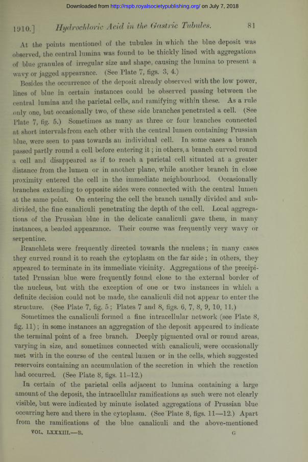
At the points mentioned of the tubules in which the blue deposit was observed, the central luinina was found to be thickly lined with aggregations of blue granules of irregular size and shape, causing the lumina to present a wavy or jagged appearance. (See Plate 7, figs. 3, 4.)
Besides the occurrence of the deposit already observed with the low power, lines of blue in certain instances could be observed passing between the central lumina and the parietal cells, and ramifying within these. As a rule only one, but occasionally two, of these side branches penetrated a cell. (See Plate 7, fig. 5.) Sometimes as many as three or four branches connected at short intervals from each other with the central lumen containing Prussian blue, were seen to pass towards an individual cell. In some cases a branch passed partly round a cell before entering i t ; in others, a branch curved round a cell and disappeared as if to reach a parietal cell situated at a greater distance from the lumen or in another plane, while another branch in close proximity entered the cell in the immediate neighbourhood. Occasionally branches extending to opposite sides were connected with the central lumen at the same point. On entering the cell the branch usually divided and subdivided, the fine canaliculi penetrating the depth of the cell. Local aggregations of the Prussian blue in the delicate canaliculi gave them, in many instances, a beaded appearance. Their course was frequently very wavy or serpentine.
Branchlets were frequently directed towards the nucleus; in many cases they curved round it to reach the cytoplasm on the far side; in others, they appeared to terminate in its immediate vicinity. Aggregations of the precipitated Prussian blue were frequently found close to the external border of the nucleus, but with the exception of one or two instances in which a definite decision could not be made, the canaliculi did not appear to enter the structure. (See Plate 7, fig. 5 ; Plates 7 and 8, figs. 6, 7, 8, 9, 10, 11.)
Sometimes the canaliculi formed a fine intracellular network (see Ilate 8, fig. 11); in some instances an aggregation of the deposit appeared to indicate the terminal point of a free branch. Deeply pigmented oval or round areas, varying in size, and sometimes connected with canaliculi, were occasionally met with in the course of the central lumen or in the cells, which suggested reservoirs containing an accumulation of the secretion in which the reaction had occurred. (See Plate 8, figs. 11-12.)
In certain of the parietal cells adjacent to lumina containing a large amount of the deposit, the intracellular ramifications as such were not clearly visible, but were indicated by minute isolated aggregations of Prussian blue occurring here and there in the cytoplasm. (See Plate 8, figs. 11—12.) Apart from the ramifications of the blue canaliculi and the above-mentioned
VOL. l x x x iii .— B.
1910.1 Hydrochloric Acid in Gastric
G
on July 7, 2018http://rspb.royalsocietypublishing.org/Downloaded from
82
indications of these, the cytoplasm was, as a rule, free from colour, but sometimes, under the most favourable daylight illumination, it appeared to be tinged with a faint, almost imperceptible, blue. In some eases it was uncertain whether this was due to a scattering of the light reflected from the material in the eanaliculi.
In view of the fact previously stated (p. 69), that the presence of a free acid is a necessary factor for the production of Prussian blue in vitro, from the interaction of solutions of ammonium ferric citrate and potassium ferro- cyanide, the occurrence of the Prussian blue reaction in the canaliculi of the parietal cells of an animal injected with a solution of these two salts affords conclusive evidence of the presence of free acid within these structures.
The evidence of the occurrence of the reaction was obtained in a very limited portion of the mucosa. The examination of a large number of sections cut from the same part of the mucosa as those which furnished the evidence of Prussian blue within the parietal cells and the lumina of the tubules afforded no proof whatever of the presence of the blue compound in the glandular structures.
This indicates the regional nature of the secretory activity, and suggests the possibility that only a very few of the gastric tubules or parietal cells are functionally active at any one time. The amount of the superficial deposit, frequently found in cases in which direct evidence of the occurrence of the reaction in the underlying tissue was not forthcoming, appears also to indicate that the Prussian blue, when formed, is removed from the tubules with great rapidity. The rapid removal of the Prussian blue, together with the variation in the functioning time of the several gland tubules, or of certain cells within these, may be largely responsible for the difficulty encountered in obtaining evidence of a positive character within the glandular structures, and for the lack of success experienced by earlier investigators.
Factors, the nature of which is unknown, are also present, which under certain conditions, equally unknown, influence the secretion of the acid in such a manner that it is no longer directed entirely towards the free surface, and consequently the Prussian blue may be found in situations other than those hitherto named.
Proof of this was obtained in two rabbits (Eabbits 5 and 6, see Table). The remarkable quantity of Prussian blue found in the case of Eabbit 5 adhering to the surface of the gastric mucosa at the end of an experiment of 6-g- hours’ duration has already been referred to. (For experimental data see pp. 77—79.)
In Eabbit 6, which was ’killed 3 hours after receiving the first, and between three-quarters of an hour and one hour after the last injection, the
Miss M. P. FitzGerald. Origin [June 4,
on July 7, 2018http://rspb.royalsocietypublishing.org/Downloaded from

83
superficial deposit of Prussian blue was very slight. The animal had received 43 to 45 c. cms. of the injection solution by four subcutaneous injections of about 11 c. cms. each. One hour had elapsed between the first and second injections, and the remainder had been given at intervals of half-an-hour. A toxic effect was not observed. Food had been given prior to the beginning of the experiment.
The experiments with these two rabbits (Eabbits 5 and 6) although differing in some respects, both in minor details in the method of procedure and in certain of the results obtained, possess essential features in common, namely, the subcutaneous administration of a large quantity of the injection solution during the space of a few hours, and the similarity of the situation in which the Prussian blue was ultimately found in the gastric mucosa. Instead of occurring within the parietal cells, the Prussian blue was found as a granular deposit scattered on their surface remote from the lumen. The cells frequently presented the appearance of being partly covered with a delicate pale blue film, punctuated with dark blue. (See Plate 8, fig. 14.) The deposit only occurred on certain of the cells.
In many instances the surface of the cells was clear and the deposit was found in isolated areas, evidently lymphatic spaces, between the cells and close to the interstitial tissue adjacent to the external border of the tubule. In such areas, also, the dark blue granules appeared to be set in a pale blue ground substance. (See Plate 8, fig. 15.)
Certain of the intertubular lymphatic channels, as well as other lymphatic vessels adjacent to the gland tubules, were found to be lined with a deposit of Prussian blue and frequently to contain large aggregations of the blue compound. The course of the lymphatic vessels could be readily followed from their blue content. (See Plate 8, fig. 13 and fig. 16.) On the other hand, the central lumina of the gland tubules contained no Prussian blue.
Wandering cells found between the tubules, and the lymph cells in the course of the larger channels, were frequently observed to be deeply impregnated with Prussian blue, and large phagocytic cells studded with blue granules were found in the submucous coat (Rabbit 5). (See Plate 8, fig. 17.) In certain of the preparations Prussian blue was also found in the contents of the blood vessels. (See Plate 8, fig. 16.)
It is therefore evident that, under certain conditions the acid, instead of being wholly discharged into the canaliculi and thence into the central lumina of the gland tubules, may also be discharged in the inverse direction. Whether the occurrence of the Prussian blue in the lymphatic and bloodvessels thus reveals a normal tendency in the direction of the secretion or part of the secretion of the parietal cells, or whether the occurrence is
1910.] Hydrochloric Acid in the Gastric Tubules.
on July 7, 2018http://rspb.royalsocietypublishing.org/Downloaded from

84
due to an abnormal condition of the cell membrane brought about by one or more of the salts contained in the injection solution, can only be ascertained by further investigation.
In reference to this point, Carnot and Lelievre* in a recent paper, brought forward the view that the disposition of the parietal cells indicates a double physiological role, and they suggest that these cells give origin to both a digestive and an “ internal ” secretion. They show that the situation of the greater number of the parietal cells in the necks of the gland tubules adjoining the crypts facilitates the ready discharge of their secretion directed towards the surface, but that in addition to this the cells are in contact with the peri-tubular vascular network. They point out that this reciprocal relationship is best seen in the body of the gland tubule, where the chief cells exceed the parietal cells in number, and that in longitudinal sections, particularly in those of the stomach in which the vessels have been injected with carmine, the blood-vessels are seen to be surrounded on all sides by parietal cells, just as if the capillary constituted the central lumen of a gland. The parietal cells which thus surround a vessel belong to different gland tubules, and their intra-cellular canaliculi, demonstrated in this instance both by the Golgi-Cajal method and by iron haematoxylin, accordingly diverge toward three or four independent acini (dog).
These investigators further point out that this arrangement of the parietal cells in relation to the blood-vessels is very similar to that existing between the hepatic cells and the blood-vessels, and hold that it indicates an imbrication of two glandular systems, the one orientated in relation to the excretory canal, and the other in relation to the vessels. They also find it difficult to accept the idea that this concentric arrangement of the parietal cells in relation to the capillaries could have for its sole object the withdrawal from the blood of secretory products, and therefore they advance the view that the surface of contact indicates that a secretion, indiscernable histologically, is discharged in an inverse direction to that going forth from the cell to the central lumen of the tubule. They further state that evidence in support of the view of an elaboration of an internal secretion by the gastric glands, and attributable probably to the parietal cells, had already been obtained from physiological experiments in process of execution at the time of publication.
If such be the case it can be understood in the event of an upsetting of the unknown mechanism governing the direction of the digestive secretion that hydrochloric acid might then be discharged in the same direction as the internal secretion, and thus be found to be present in the lymph and blood-
* P. Carnot et A. Lelievre, ‘Compt. Rend. Soe. Biol.,’ 1909, vol. 66, No. 3.
Miss M. P. FitzGerald. Origin of the [June 4,
on July 7, 2018http://rspb.royalsocietypublishing.org/Downloaded from

85
vessels. The richness of the blood supply in the region of the mucosa in which the parietal cells predominate has been demonstrated in the rabbit by Bohm and David off.*
With reference to the spontaneous occurrence of the Prussian blue reaction in the skin previously mentioned, this was observed in four instances (dog, rabbit, guinea-pigs) in the muscles of the skin at the site of inoculation. The microscopical examination showed that the deposit of Prussian blue was situated in the inner structure of the muscle fibre itself. Further particulars are withheld, as the results obtained may appear in a future paper. Doubtless the explanation is that the reaction was here brought about by the presence of an organic acid, probably sarcolactic acid, resulting from injury to the fibre. Dreser,f when demonstrating the acidity of the muscles during activity by means of a decolourised solution of acid fuchsin, found the red reaction in the lymph spaces between the muscle fibres, but not in the fibres themselves.
Before passing to experiments made with other reagents, brief mention is here made of the occurrence of mitosis in the cells of the gastric glands, observed in one or two instances in the rabbit, notably in Rabbit 2. Mitotic figures in various stages were remarkably prevalent in the cells lining the crypts, and were present also in the cells of the upper part of the necks of the glands, and occasionally in some of the parietal cells in the lower portion of the tubules.
Among other observers this phenomenon has previously been observed by Sachsj: in the surface epithelium of the gastric mucosa in man, and in the surface epithelium and in the cells of the crypts in the dog in pathological conditions; by Bizzozero§ in the cells of the crypts; and by Bensleyl in the cells of the crypts and in the chief cells of the necks of mammalian gastric glands.
A few experiments were made to see if a result approaching that recorded by Sehrwald^T could be obtained by placing portions of the fresh gastric mucosa of the rabbit and of the guinea-pig into an aqueous solution of 2'25 per cent, ammonium ferric citrate for 18 to 24 hours, and later, after teasing, into one of 1'5-per-cent. potassium ferrocyanide. In some cases the tissues were * * * §
* Bohm and Davidoff, ‘ Lelirb. d. Histol. d. Menschen,’ 1895.t H. Dreser, ‘ Centralbl. f. Physiol.,’ 1887, vol. I.f A. Sachs, ‘Archiv f. exp. Path, und Pharmakologie,’ 1886, vol. 22, pp. 155-173;
1887, vol. 24, p. 109.§ Bizzozero, ‘Anat. Anz.,’ 1888, vol. 3, No. 26, p. 781 ; ‘Archiv f. Mikros. Anat.,’ 1893,
vol. 42.|| R. Bensley, ‘Quart. Journ. Micr. Sci.,’ 1899, vol. 41.
IT Sehrwald, ‘ Miinchener Med. Wochenschr.,’ 1889, p. 177.
1910.] Hydrochloric Acid in the Gastric Tubules.
on July 7, 2018http://rspb.royalsocietypublishing.org/Downloaded from

washed rapidly in distilled water after their removal from the solution of the double citrate and before being placed in the potassium ferrocyanide solution. The results obtained by this method, as might be anticipated, were quite of an indeterminate nature. In the case of one guinea-pig, a diffuse greenish coloration was observed here and there over the gland tubules, and, in another, a peculiar brown crystalline precipitate was observed in the parietal cells.
86 Miss M. P. FitzGerald. Origin of the [June 4,
Table giving Results Obtained by Injecting a Solution containing Equal Parts of an Aqueot Solution of 2‘25-Per-Cent. Ammonium Ferric Citrate and of 1‘5-Per-Cent. Potassiu; Ferrocyanide.
Animal.Mode and
No. of injections.
Amountinjected.
No. of hours since first Condition. Toxic
Prussian blue reaction—spontaneous.
D itto, on additions 0*5 per cent. HCl
and last injections.
effect.Gastric mucosa. Skin. Urine. i Varioui
j tissues,"
c. cms. First.35£
Last. Surface. Interior.Babbit
(1)Intra
venous, 1 Subcuta
neous, 2
10—11 2 Food given. 4-
p
+ ,
Babbit(2)
Subcutaneous, 8
37 30 n Food given. 4- 4- J 4-(1st day)
(2nd day)
+ |
•
Babbit(3)
Subcutaneous, 5
31—32 25| n Starved 24 hrs.(1st day)
+(2nd day)
# 4- f(Betention of urine)
+ , |
Babbit(4)
Intravenous, 2 17 4i 2f Sem i-fasting. (Starved
36 hrs. Food. Starved 24 hrs.)
.4- +
Babbit(5)
Subcutaneous, 5
50 6$ 1 No food eaten during expt.
+ + 4- + + |
Babbit(6)
Subcutaneous, 4
4 3 -4 5 3 i - i Food before exp."
4- . 4- .4- + I
Babbit (7) (sod. ferrocyanide)
Subcutaneous, 4
40 5* l Semi-fasting. Food given after fast of 20 hrs. and during expt. Eaten
well until after 4tli injection.
+ 4- + ■
G-uinea- Pig (1)
Subcutaneous, 2
22 3} i} i Food given, + 4- 4- j j j 4 - 1
Guinea- pig (2)
Subcutaneous, 2
- 16 Si 3 ' . . . 4-(Polyuria)
+ 4- — ? + .1
Guinea- pig (3)
Subcutaneous, 2
19 si . . . 4-(Polyuria)
+ 4- 4- 4- :«■;
D og (1)... Subcutaneous, 2
55 S i u Starved 48 hrs. Food given 1 hr. before expt.
+(Vomit-
in g )
+ m
D og (2)... Subcutaneous, 2 ;
42 31 +(Coma
tose)
+ 4- 4- 4* S
-------J;
+ means positive result. means negative result.
* After their removal from the body, and subsequent exposure to th e action of dilute hydrochloric acid, the Prussian blue react® developed, at various tim es, in th e following tissues :—Bladder, kidney, liver, small and large intestines, ovary, fallopian tubes, uterf
.gina, and lym ph gland of axillary region (dog). The kidney became quite blue in m any cases, and under the microscope * miferous tubules presented a very striking appearance.
va,urin
In view of the proof obtained of formed acid existing in the canaliculi of the parietal cells, it was a matter both of interest and of importance to repeat the experiments of Mary Greenwood* and of MacallumJ referring to the
* M. Greenwood, ibid,t A. B. Macallum, ‘Ergebn. d. Physiol.,’ 1908, vol. 7, p. 628.
on July 7, 2018http://rspb.royalsocietypublishing.org/Downloaded from

presence of chlorides in the cytoplasm of the parietal cells, and, if possible, thereby to increase collateral evidence of the elaboration of the hydrochloric acid of the gastric juice within these cellular elements of the gastric mucosa.
As before stated (p. 68), owing to the fact that, on exposure to sunlight, the phosphate and the carbonate of silver form coloured reduction compounds similar in colour to that of the chloride, the results obtained by Mary Greenwood by the use of silver nitrate furnished only a partial proof of the presence of an excess of chlorides in the parietal cells as compared with the chief cells, but definite proof of this was afforded by Macallum, who eliminated the error which could arise in the interpretation of the results obtained by the silver nitrate method as used by the earlier investigator, by the addition of free nitric acid to the silver nitrate solution, which permits the formation of the coloured reduction compound from chlorides, but prohibits a similar formation from phosphates and carbonates.
Macallum* also pointed out that, in the presence of free nitric acid, organic substances, with the exception of taurine and creatine, will not give the colour reaction similar to that of the sub-chloride of silver, and that proteids and albuminoids, when freed from all traces of haloids, do not give any compound of silver that will reduce under the action of light.
In the present series of experiments, the methods used were similar to those employed for the determination of the distribution of the chlorides in nerve-cells and fibres by Macallum and Menten,f and of the phosphates in muscle by Maud Menten.J For the detection of salts, such as chlorides, phosphates, and carbonates, giving a coloured reduction compound of silver on exposure to sunlight, a deci-normal solution of silver nitrate was used; and, for demonstrating the chlorides alone, a deci-normal solution of silver nitrate containing 1*5 per cent, free nitric acid. The silver solution was in each case made with pure water.
Investigations were carried out on the fresh gastric mucosa of the rabbit and of the guinea-pig, the animals having been killed beforehand by the methods described previously (p. 74) for the two species named. In each case portions of the fresh stomach, washed quickly in distilled water, were rapidly cut into sections, and the mucosa detached from the outer layers of the stomach wall placed in vessels containing the respective solutions. The tissues were then carefully teased with goose quills, to permit the reagent to penetrate. Penetration does not take place readily, and, although the presence of the free nitric acid aids the diffusion of the nitrate in the
1910.] Hydrochloric Acid in Gastric Tubules. 87
* A. B. Macallum, ‘Roy. Soc. Proc.,’ 1905, B, vol. 76, p. 217.+ A. B. Macallum and Maud L. Menten, ‘ Roy. Soc. Proc.,’ 1906, B, vol. 77, p. 165. I Maud L. Menten, ‘Canadian Inst. Trans.,’ 1908-1909, vol. 8, p. 414.
on July 7, 2018http://rspb.royalsocietypublishing.org/Downloaded from

88
solution which contains the acid, careful teasing is necessary, whether nitric acid is present in the solution or not. After allowing the respective solutions to act upon the tissues for a certain length of time, the vessels containing them were then exposed to sunlight, or to as bright a light as was available, for several days; and when the maximum reaction had been obtained, the tissues were mounted in 50-per-cent, glycerine ; or, after being exposed to light for some time, portions of the tissues were mounted in 50-per-cent, glycerine, to which a few drops of the reagent were added, and again exposed to sunlight in this medium.
The characteristic reaction was obtained in the tissues acted upon by each of the solutions, and drawings were made of specimens mounted in 50-per-cent, glycerine. Fig. 18 shows the appearance of a tubule treated with the silver nitrate solution without acid, and tigs. 19 to 22 the appearance of tubules after treatment with silver nitrate solution containing free nitric acid.
It will be seen that in both cases the parietal cells are differentiated from the chief cells, but since the “ reduced ” phosphate and carbonate compounds of silver are brown in colour, and as the sub-chloride, although violet, reddish violet, or bluish violet when in mass, may also be reddish brown, as shown by Macallum,* when it occurs in thin layers or deposits, the shade apparently depending on the existence of the compound in a finely divided state or otherwise, it is clear that it would be impossible to make a definite statement as to the presence of an excess of chlorides in the parietal cells in the tubule depicted in fig. 18, because the brown colour could also be attributed to the precipitation of the “ reduced ” phosphate or carbonate. Such a statement can, however, safely be made with regard to the reaction shown in figs. 19 to 22, for in these cases, owing to the differentiating action of the free nitric acid present in the silver nitrate solution, the reaction can only be due to the precipitation of the sub-chloride of silver.
The intensity of the reaction obtained in the parietal cells of both the rabbit and the guinea-pig, and the marked manner in which the parietal cells stand out in situ in the tubules of the rabbit shown in figs. 19, 20, due to the chlorides being more abundantly present in these cells than in the chief cells, are results which fully corroborate those obtained by Macallum,f when using the same method and the same animals, and furnish evidence of the source of the hydrochloric acid in the parietal cells, and also give support to the view that this is elaborated in the cytoplasm of these cells.
With the hope of establishing the presence of the acid in the cells by more than one method, some experiments were also made with Iropseolin 0 0
* A. B. Macallum, ‘Roy. Soc. Proc.,’ 1905, B, vol. 76, p. 217. t A. B. Macallum, ‘Ergebn. d. Physiol.,’ 1908, vol. 7, p. 628.
Miss M. P. FitzGerald. Origin of the [June 4,
on July 7, 2018http://rspb.royalsocietypublishing.org/Downloaded from

89
(rf. p. 63), but the results obtained were of an indefinite nature, although indicating the presence of acid. The rabbit and the guinea-pig were the animals used. In the experiment with the rabbit, the animal was killed by chloroform, and portions of the gastric mucosa (fresh) were teased in a saturated aqueous solution of tropmolin, or in a saturated glycerine solution ('25 or 50 per cent.). A red colour was observed in the region of the mucosa containing the peptic glands.
With the guinea-pig, the tropseolin was injected subcutaneously as a saturated solution made up in either distilled water or in Ringer’s solution. The amount injected varied from 18 to 22 c. cms., and was administered by two injections in the abdominal region, at intervals varying from 1 to 1| hours, or from 2 to 2£ hours. The duration of the experiment varied from 4 to 5 | hours. The reagent, both in aqueous solution and when dissolved in Ringer’s fluid, exerted a decidedly toxic influence, and its presence in the body appeared to cause the animals considerable discomfort. The contents of the stomach were in a very watery condition, and much gas was present both in the stomach and intestines. Inflammatory conditions supervened at the site of inoculation. The urine on some occasions gave the characteristic rose-coloured reaction on the addition of concentrated hydrochloric acid, but not on the addition of a 05 per cent, solution. Death was produced by shock. On opening the stomach a rose-coloured area was observed on the surface of the mucosa near the oesophageal opening and in the region corresponding to the border of the lesser curvature. In the guinea-pig injected with the reagent dissolved in Ringer’s fluid, a rose colour was also observed in the pyloric region. I t extended across the mucosa of the superior half of the organ in this region, and was visible also on the surface of the mucosa of the duodenum adjacent to the orifice of the pylorus. Portions of the gastric mucosa were teased and examined in 50 per cent, glycerine without delay, but owing to the general distribution and diffuseness of the rose colour in the gland tubules, no positive information as to the occurrence of the acid within any one type of cell was obtained by this method.
The drawings accompanying the paper were made with the aid of an Abbe camera lucida (Zeiss or Leitz) with a Zeiss microscope, compensating ocular No. 4 (Zeiss) or No. 2 (Leitz), and Leitz objectives No. 3 or one-twelfth oil immersion. The drawing paper was laid flat on the table or on a board placed on a level with the stage of the microscope. The drawings illustrating the results obtained in the Prussian blue experiments were mostly made from unstained preparations. In some cases, illustrations of which have not been included, the sections were stained with a 0‘5-per-cent. aqueous solution of
v o l . l x x x iii .— B.
1910 . ] Hydrochloric Acid in the Gastric Tubules.
H
on July 7, 2018http://rspb.royalsocietypublishing.org/Downloaded from

90 Miss M. P. FitzGerald. The Origin of the [June 4
neutral red. Previous to immersion in the staining fluid, the sections were carefully washed in distilled water to remove the’ alcohol used for fixation, to prevent the yellow coloration which would otherwise have resulted on the application of the dye. With the exception of the nuclei staining a pink colour of moderate intensity, the parietal cells remain uncoloured by this method; they are, therefore, well differentiated from the chief cells, which are stained crimson. In a vertical section of that part of the gastric mucosa in which the parietal cells are most numerous, the neck region of the gland tubules appears as a clear band, as the surface epithelium and the cells of the crypts are also stained crimson.
From the results obtained in the gastric mucosa of the rabbit previouslyinjected with a solution containing equal parts of an aqueous solution of2’25-per-cent. ammonium ferric citrate and of 1’5-per-cent. potassium ferro-cyanide, it may be said in conclusion that, the occurrence of Prussian blue,not only in the lumina of the gland tubules, but also in the canaliculi in theparietal cells, makes it certain that the hydrochloric acid is, at least, alreadyformed and free in the secretion as it appears in the canaliculi. Whetherthe acid, or, more correctly speaking, whether its hydrogen ions occur in thecytoplasm of the cells still remains an open question, although the very faintblue occasionally observed in a parietal cell seems to postulate an affirmative.It is, moreover, to be noted that if the acid is formed and set free in thecanaliculi, then the cytoplasm lining the latter must constitute a membrane
+ + —* —
permeable to the II and Cl ions, but not to Na, Iv, PO4, or CO3. If that were entirely the case, no free acid should occur in the lymphatics besides the tubules. The acid here can only be derived from the cytoplasm of the parietal cells, and if the latter have absorbed it from their canaliculi, then it would be difficult to explain why the cytoplasm does not neutralise the acid, for the removal of the hydrogen ions from the parietal cells must make their cytoplasm distinctly alkaline. If, to overcome this difficulty, we suppose that the cytoplasm is so constituted as to maintain the ionisation of the hydrogen and the chlorine in the presence of alkalies, we attribute a property to it that could be also responsible for the formation of the acid in the first place in the cytoplasm.
The occurrence of free acid in the canaliculi, and also, in certain cases, in the lymphatic vessels between the gastric tubules, is, therefore, best explained by supposing that the cytoplasm of the parietal cells forms in itself the free- acid, and, under ordinary conditions, that the free acid so formed diffuses into the canaliculi, but, under certain conditions, the character of which is unknown, the cell loses the sense of direction, so to speak, and, as a
on July 7, 2018http://rspb.royalsocietypublishing.org/Downloaded from

consequence, the free acid diffuses in the opposite direction, that is, out, into the inter-tubular tissue.
The source of the hydrochloric acid in the parietal cells is, without doubt, the chlorides, present, as already shown, in greater abundance in them than in the chief cells or other adjacent tissue elements. The results of observations with Macallum’s cobalt sodium hexanitrite method for demonstrating potassium* have not yet shown that the latter element occurs more abundantly in the parietal than in the chief cells, consequently the chlorine demonstrable in the parietal cells must be associated chiefly, if not entirely, with sodium.
Eosemannf made analysis of the gastric juice of the dog, and, calculating from his results, it was found that in the ash the proportion of the potassium to the sodium was 1: 1*23 and 1: 2T7, whereas in the blood plasma the proportion is 1: 0'0685. Rosemann consequently concluded that the acid arises not through simple diffusion out of the blood, but from the bodies of the cells concerned. As the amounts of these elements in the gastric juice are very small (K = 0,03077-(H)4328, and Na = 0,01979-0,02502), and, further, as the percentage of potassium in the juice does not greatly exceed that of potassium in the blood plasma ( = 0*025 per cent.), it is possible that, though the parietal cells take up the two alkaline chlorides from the blood plasma and lymph, yet in the formation of the acid they utilise only the sodium chloride, the sodium being retained to pass back into the blood and lymph. If potassium chloride constituted a source of the chlorine of the acid in the gastric juice, the potassium in the latter would have, as in the case of sodium, undergone relatively a great diminution in amount.
In conclusion, I desire to express my grateful thanks to Prof. A. B. Macallum, at whose suggestion this work was undertaken, for his supervision and for affording me the facilities for carrying out the research in the Bio- Chemical Laboratory of the University of Toronto, and, in particular, I wish to acknowledge my indebtedness to him for his valuable help in the chemical matters connected with the investigation, and with the physico-chemical interpretation of the results.
1910.] Hydrochloric Acid in the Gastric Tubules. 91
* A. B. Macallum, ‘ Journ. Physiol.,’ 1905, vol. 32, p. 95. t Rosemann, loc. cit., pp. 522—523.
on July 7, 2018http://rspb.royalsocietypublishing.org/Downloaded from

92 Miss M. P. FitzGerald. The Origin of the [June 4.
EXPLANATION OF PLATES.
Figs. 1-17 represent results obtained in the gastric mucosae of rabbits injectedsubcutaneously with a solution containing equal parts of an aqueous solution of 2'25 percent, ammonium ferric citrate and L'5 per cent, potassium ferrocyanide.
P late 7.
Fig. 1.—Opened stomach of Rabbit 5 fixed in absolute alcohol. Natural size. Drawing shows deposit of Prussian blue adhering to surface of mucosa of dorsal wall and on that of unremoved portion of ventral wall (pyloric region). Maximum reaction as observed on surface.—Note that Prussian blue is distributed in region of oesophageal opening and in adjacent region of lesser curvature, and that in this instance deposit also occurs in pyloric region.
Fig. 2.—Vertical section of oesophageal region of stomach fixed in absolute alcohol, showing deposit of Prussian blue in central lumina of gland tubules, in crypts, and on surface of mucosa. (Rabbit 2, 30 hours after the first injection.) X 96.
Fig. 3.—Gland tubules of gastric mucosa in which lumina are lined with granular deposit of Prussian blue. The irregularly occurring aggregations of deposit cause lumina to present a wavy, jagged appearance. Two side branches can be traced passing to a cell; within other cells small aggregations of deposit are seen. Xl 100.
Fig. 4.—Gastric tubule showing deposit of Prussian blue in central lumen, in side branch, and in eanaliculi resulting from its bifurcation, x 1100. (Rabbit 2.)
Fig. 5.—Parietal cell penetrated by two side branches connected with lumen and containing Prussian blue. xllOO. (Rabbit 2.)
Figs. 6, 7, 8, 9.—Parietal cells, the canaliculi within which contain Prussian blue. Note larger side channel, ramification of fine canaliculi in the cytoplasm of cells, their wavy course, frequent proximity to nucleus, and beaded appearance due to isolated aggregations of deposit within. Figs. 6-9, x 1100. (Rabbit 2.)
P late 8.
Fig. 10.—Parietal cells with Prussian blue in canaliculi. X720. (Rabbit 2.)Fig. 11.—Gland tubules cut tangentially. Along the course of one of the central lumina
and of a side channel, lined with Prussian blue, deeply stained oval enlargements occur, suggesting reservoirs containing accumulation of secretion. In one of the parietal cells canaliculi are seen in form of a network, x 1100. (Rabbit 2.)
Fig. 12.—Gland tubule. Prussian blue seen to be irregularly deposited along course of central lumen and to be present in side branch. An oval area suggesting a reservoir containing accumulation of secretion is seen, and small isolated aggregations of deposit within cells, x 1100. (Rabbit 2.)
Fig. 13._Intertubular lymph channel containing Prussian blue, x 920. (Rabbit 6.)Fig. 14._Granular deposit of Prussian blue in a film of blue on surface of cells and in
adjacent lymph space, x 920. (Rabbit 5.)Fig. 15._Intercellular lymph space containing granular deposit of Prussian blue in a
pale blue ground substance. X 640. (Rabbit 5.)Fig. 16.—Transverse section of gastric mucosa. Prussian blue is seen in an inter-
glandular lymph vessel, and mixed with contents of blood vessel. x 640. (Rabbit 6.)
Fig. 17.-—Phagocytes in sub-mucous coat of stomach, which have taken up the granular deposit of Prussian blue, x 800. (Rabbit 5.)
on July 7, 2018http://rspb.royalsocietypublishing.org/Downloaded from

R oy.Soc. P toc.jB.VoI. 83. P la te 7.F i t z Ger
8West* Kewma.n life}*.
on July 7, 2018http://rspb.royalsocietypublishing.org/Downloaded from
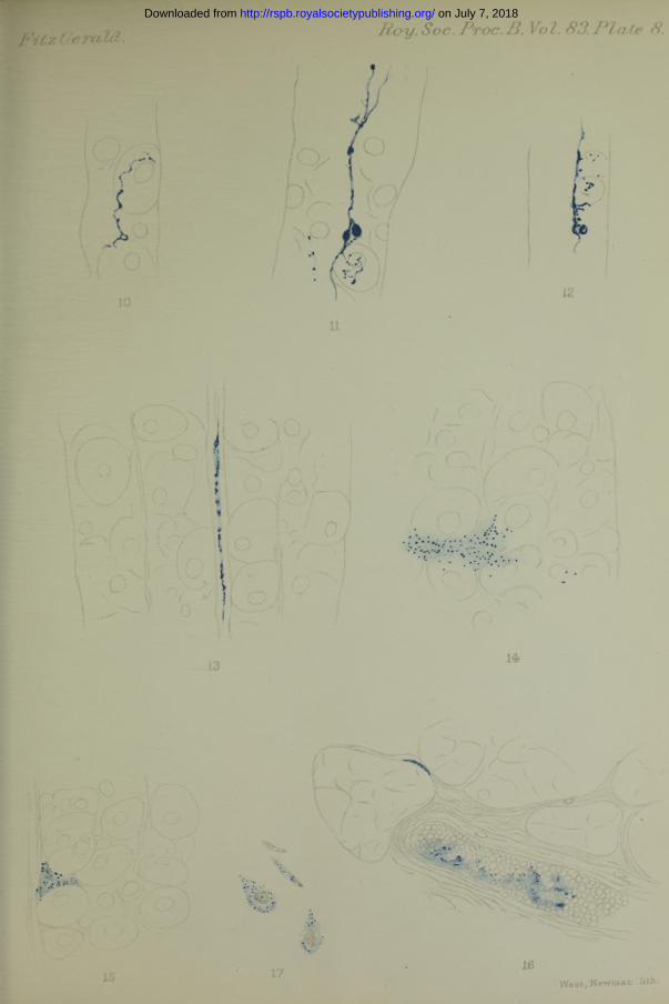
Roy.Soc. Ptoc.B. Vot. 83.P la te 8. on July 7, 2018http://rspb.royalsocietypublishing.org/Downloaded from

Roy. Soc. Proc. B, ool. 83, PI. 9.
Grout, sc. & imp*
on July 7, 2018http://rspb.royalsocietypublishing.org/Downloaded from

Hydrochloric Acid in Gastric Tubules. 93
P late 9.I
Figs. 18-22 represent results obtained by action of silver nitrate on teased gastricglands of rabbit and guinea-pig.Fig. 18.—Portion of the gastric gland of a rabbit exposed’to the action of N/10 AgNO,
for two days. The parietal cells are differentiated from the chief cells by their brown colour, but in this instance the brown reaction may be due to presence of phosphates, carbonates, or chlorides, or to all three of these “ reduced ” compounds of ilver. x 690.
Fig. 19.—Portion of teased gastric gland tubule from oesophageal region of normal rabbit, treated when fresh with N/10 AgN03 + 1*5 per cent. H N 03. Parietal cells are differentiated from chief cells by the intensity of their brown coloration. The reaction in this case is due to precipitation of “ reduced ” chloride of silver ; its occurrence in such marked degree demonstrates the presence of greater abundance of chlorides in these cells than in the chief cells, x 700.
Fig. 20.—Base of gastric tubule from oesophageal region of normal rabbit, treated when fresh with N/10 AgN03 + 1 ‘5 per cent. PIN03. Excess of chlorides again demonstrated in parietal cells by their brown coloration. (See explanation of fig. 19.) X690.
Fig. 21.—Teased preparation of gastric gland tubules treated with N/10 AgN03 + 1*5 per cent. HN03. Abundance of chlorides in parietal cells is demonstrated by their reddish-brown colour. X840. Guinea-pig.
Fig. 22.—Teased preparation of gastric gland tubule from fundus region of normal guinea-pig, treated With N/10 AgN03 + l ‘5 per cent. H N 03, showing presence of chlorides in deposit in lumen and in intercellular and intracellular channels, x 640.
VOL. LXXXIII.— B. I
on July 7, 2018http://rspb.royalsocietypublishing.org/Downloaded from