the interactions of outer membrane proteins with the
TRANSCRIPT

The interactions of outer membrane
proteins with the periplasmic chaperone Skp of E.coli and with LPS
Dissertation zur Erlangung des akademischen Grades des Doktors der Naturwissenschaften
an der Universität Konstanz Mathematisch – Naturwissenschaftliche Sektion
Fachbereich Biologie
Vorgelegt von Jian Qu
Tag der mündlichen Prüfung: 20.06.2007
Referent 1: PD Dr. Jörg H. Kleinschmidt Referent 2: Prof. Dr. Winfried Boos

Table of contents
1 General Introduction
1.1 Biomembranes…...…..…………………………………………….…. 001
1.2 Membrane lipids………………………………………………….…... 003
1.3 Lipopolysaccharides (LPS)...........……………………………….….... 005
1.4 Membrane proteins…………..…………………………………….…. 006
1.4.1 Outer membrane protein A (OmpA)………………………... 011
1.4.1.1 Structure……………………………………….….. 011
1.4.1.2 Functions……………………………………….…. 012
1.4.1.3 Refolding……………………………………….…. 012
1.4.2 OmpG………………………………………………………. 015
1.4.3 NalP……………………………………………………….… 015
1.4.4 YaeT…………………………………………………….….. 015
1.4.5 FomA…………………………………………………….…. 016
1.4.6 hVDAC1…………………………………………………… 016
1.4.7 FhuA…………………………………………………….…. 017
1.5 Molecular Chaperones………………………………………………... 017
1.5.1 Periplasmic chaperones…………………………………….. 019
1.5.1.1 Skp…………………………………………….….. 020
1.6 Methods…………………………………………………………….… 022
1.6.1 Fluorescence spectroscopy…………………………………. 022
1.6.2 Fluorescence Quenching………………………………….… 024
1.6.3 Site-directed Spin Labeling (SDSL)………………………... 024
1.6.4 S-Methylation…………………………………………….… 025
1.6.5 ATR-FTIR……………………………………………….…. 026
1.6.6 CD spectroscopy...………………………………………….. 026
2 The periplasmic chaperone Skp of E. coli forms 1:1 complexes with outer membrane
proteins via hydrophobic and electrostatic interactions
2.1 Abstract…………………………………………………………….…. 028
2.2 Introduction…………………………………………………………… 029
2.3 Materials and methods…………………………………………….….. 032
i
2.4 Results………………………………………………………………… 036
2.5 Discussion………………………………………………………….…. 049
3 The interaction of OmpA with LPS and the periplasmic chaperone Skp of Escherichia coli
studied by site-directed mutagenesis
3.1 Abstract………………………………………………………….……. 055
3.2 Introduction…………………………………………………………… 056
3.3 Materials and methods………………………………………….…….. 058
3.4 Results………………………………………………………………… 064
3.5 Discussion…………………………………………………….………. 082
4 Orientation of β-barrel proteins OmpA and FhuA in lipid membranes. Chainlength
dependence from infrared dichroism
4.1 Abstract……………………………………………………………….. 086
4.2 Introduction…………………………………………………………… 087
4.3 Theoretical background………………………………………………. 089
4.4 Materials and methods………………………………………………... 091
4.5 Results………………………………………………………………… 095
4.6 Discussion…………………………………………….………………. 105
Summary………………………………………………………………………… 109
Zusammenfassung………………………………………………………………. 114
References………………………………………………………………………. 119
List of publications……………………………………………………………… 133
Acknowledgements……………………………………………………………… 134
ii

Abbreviations
2-ME Beta Mercaptoethanol
ATR attenuated total reflection
BR bacteriorhodopsin
bromphenol
blue 3',3'',5',5''-tetrabromophenolsulfonephthalein
BSA bovine serum albumin
CAPS 3-(Cyclohexylamino)-1-propanesulfonic acid
CD circular dichroism
CMC critical micelle concentration
DOPC 1,2-dioleoyl-sn-glycero-3-phosphocholine
DTNB 5,5´-dithiobis(2-nitrobenzoic acid)
EDTA ethylenediaminetetraacetic acid
Eq. equation
E. coli Escherichia coli
ESR electron spin resonance
FhuA ferric hydroxamate uptake protein A from Escherichia coli
FomA major outer membrane protein A from Fusobacterium nucleatum
FTIR Fourier transform infrared spectroscopy
HEPES 4-(2-hydroxyethyl) piperazine-1-ethanesulfonic acid
hVDAC1 human voltage-dependent anion-selective channel protein isoform 1
IMPs integral membrane proteins
IPTG isopropyl-1-thio-β-D-galactopyranoside
kDa kilo Dalton
λ wavelength
LDAO N-lauroyl-N,N-dimethylammonium-N-oxide
LPS lipopolysaccharide
LUVs large unilamellar vesicles
Mcps million counts per second
mdeg millidegrees
MTSSL (1-oxyl-2,2,5,5-tetramethylpyrroline-3-methyl) methanethiosulfonate
NalP autotransporter from Neisseria meningitides
iii

OmpA outer membrane protein A from Escherichia coli
OmpG outer membrane protein G
OMPs outer membrane proteins
PFD prefoldin
pI isoelectric point
PPIases peptidyl-proly cis/trans isomerases
SDS sodium dodecyl sulfate
SDSL site-directed spin labeling
SDS-PAGE SDS-polyacrylamide gel electrophoresis
Skp Seventeenth Kilodalton Protein
SurA the survival factor A
SUVs small unilamellar vesicles
TCEP Tris(2-carboxyethyl)phosphine hydrochloride
Tris tris(hydroxymethyl)aminomethane
TM transmembrane
TMD-OmpA transmembrane domain of OmpA, i.e. amino acid residues 0-176
WT wildtype
YaeT homologue of Omp85 in E.coli
iv

Notes on contributions:
Hereby I declare that all the experiments in this thesis are carried out by me and all the
chapters of the thesis are written by me under the direct supervision of PD Dr. Jörg
Kleinschmidt. The exceptions to the above are:
Plasmid pSkp was provided by Dr. Susanne Behrens; LPS was provided by Dr. Otto Holst;
plasmid pET28OmpGm2 was provided by Dr. Christoph Mayer; plasmid pPU320 was
provided by Prof. Dr. Peter van Ulsen; Plasmid pET15_EcOMP85 was purchased from
Trenzyme GmbH; E.coli B strain PC2889 with plasmid pET-10953 was provided by Prof.
Dr. H. B. Jensen; all the single Trp/single Cys plasmids were constructed by Paula Bulieris.
In chapter 4, my contributions are construction of the plasmid pET22bB1, isolation and
purification of TMD-OmpA and WT-OmpA, and refolding of TMD-OmpA and WT-OmpA
into LDAO micelles.
Jian Qu
Konstanz, 06.2007
v

Interaction of OMPs with Skp and LPS
1 General introduction
1.1 Biomembranes
Since cells were first observed under a microscope, it is known that they have very
definite boundaries: biological membranes or biomembranes. All biomembranes are based
on a similar lipid bilayer structure and membrane proteins are embedded within or at the
surface of the bilayers. The basic function of the biomembranes is to separate the lumen on
the inside from the outside of the cells. Preserving the integrity of the cell and of its
organelles as well as facilitating specific transport of solutes into the cell and between the
different cell organelles inside the cell is very essential for cell function and a major purpose
of biomembranes. At the same time many other important functions are performed by the
biomembranes, such as barrier, signaling, energy conversion, recognition [Lehninger et al.
2005].
The Gram-negative cell envelope (Figure 1.1) has been studied extensively. It
contains three layers: an outer membrane (OM), a periplasmic space and a plasma membrane
[Beveridge 1999]. A plasma membrane, cell membrane, or cytoplasmic membrane is a
selectively permeable lipid bilayer coated and embedded by proteins. The fluid mosaic model
for cell membrane (Figure 1.2) presented by Singer and Nicholson in 1972 is generally
accepted now [Singer et al. 1972]. Most plasma membrane lipids are phospholipids, which
spontaneously self-assemble to a bilayer. Nonpolar tails of the phospholipids are hydrophobic
and form the hydrophobic core of the bilayer; polar heads are hydrophilic and form the
apolar/polar interface at the membrane surfaces to face extracellular and intracellular fluids.
There are two major membrane proteins: peripheral proteins and integral proteins. The
peripheral proteins are bound to the surface of the bilayer. The integral proteins, also called
transmembrane proteins, have a hydrophobic region spanning in the bilayer and hydrophilic
regions that stick out on both sides of the bilayer.
The periplasmic space or simply periplasm is the space between the cytoplasmic
membrane and the outer membrane. The composition of the periplasm differs strongly from
that of the surrounding medium. Peptidoglycan is a major component, which plays roles of
supporting and sieving. The periplasm contains many important solutes, such as
monosaccharides, oligosaccharides, amino acids, peptides, proteins, soluble biosynthetic
1

Interaction of OMPs with Skp and LPS
Figure 1.1 The gram-negative cell envelope The Gram-negative cells envelop is constituted of an outer membrane, a periplasmic space and a plasma
membrane (see text for details).
Figure 1.2 Fluid mosaic model for cell membranes In this model, the plasma membrane is a phospholipid bilayer, in which protein molecules are partially or
wholly embedded. Embedded proteins are scattered throughout the membrane in an irregular pattern, which
varies among membranes.
precursors of the peptidoglycan and other small molecules, and also degrading and
detoxifying enzymes [Seltmann et al. 2002]. The periplasm is involved in various
biochemical functions including nutrient acquisition, synthesis of peptidoglycan, electron
transport, protein folding and alteration of substances toxic to the cell. Some important
2

Interaction of OMPs with Skp and LPS
proteins are distributed in this space, i.e. enzymes, chaperones [Costerton et al. 1974; Koch
1998; Behrens 2003].
In addition to proteins and phospholipids, the outer membrane contains
lipopolysaccharide (LPS). LPS is important to bacteria since it provides a permeability
barrier to hydrophobic substances. While phospholipids form the periplasmic leaflet of the
lipid bilayer of the outer membrane, LPS is exclusively found in the outer leaflet. The inner
leaflet of the OM is composed of ~70 to 80% phosphatidylethanolamine and ~20 to 30%
phosphatidylglycerol and diphosphatidylglycerol. Porins are integral membrane proteins that
form a pore through which molecules can diffuse. Unlike channel structures of transport
proteins of cytoplasmic membranes, porins have a larger channel opening that allows either
unspecific or specific passive diffusion, i.e. porins do not require energy or a transmembrane
potential for transport.
1.2 Membrane lipids Lipid bilayers are the major architectural feature of the biomembrane. Lipids are
water-insoluble cellular components of diverse structure that can be extracted by nonpolar
solvents. Most membrane lipids are amphipathic: one end is hydrophobic and nonpolar, the
other hydrophilic and polar. The two major classes of gram-negative bacterial membrane
lipids are phospholipids and glycolipids (Figure 1.3) [Seltmann et al. 2002].
Figure 1.3 Some common types of membrane lipids All the lipid types shown here have either glycerol or sphingosine as the backbone (pink screen), to which are
attached one or more long chain alkyl groups (yellow) and a polar head group (blue). In triacylglycerols,
glycerophospholipids, galactolipids, and sulfolipids, fatty acids are ester-linked to hydroxyl groups of the
backbone. Sphingolipids contain a single fatty acid in amide linkage to the sphingosine backbone. In
phospholipids the polar head group is joined through a phosphodiester, whereas glycolipids have a direct
glycosidic linkage between the head-group sugar and the backbone glycerol. [Lehninger et al. 2005]
3

Interaction of OMPs with Skp and LPS
Glycerophospholipids, also called phosphoglycerides, are the most abundant class of
lipids in most membranes. Phosphoglycerides have three essential parts: a backbone of
glycerol, two long-chain fatty acids esterified to carbons 1 and 2 of the glycerol, and a polar
head group attached to a phosphate esterified to carbon 3 of the glycerol (Figure 1.4).
Phosphoglycerides are classified by their polar head group. Phosphoglycerides ester-linked to
ethanolamine, choline, serine, glycerol and inositol are called phosphatidylethanolamines
(PE), phosphatidylcholines (PC), phosphatidylserines (PS), phosphatidylglycerols (PG) and
phosphatidylinositols (PI). Mostly, phosphoglycerides contain a saturated fatty acid at carbon
1 and an unsaturated fatty acid at carbon 2. Natural fatty acids are carboxylic acids with
hydrocarbon chains ranging from 4 to 36 carbons long.
Figure 1.4 Glycerophospholipids The common glycerophospholipids are diacylglycerols linked to head-group alcohols through a phosphodiester
bond. Each derivative is named for the head-group alcohol
(X), with the prefix “phosphatidyl-.” An atom model of PC is shown (right).
Sphingolipids are derived from sphingosine, an amino alcohol with a long
hydrocarbon chain, and contain a long-chain fatty acid attached to the sphingosine amino
4

Interaction of OMPs with Skp and LPS
group. There are three main types of sphingolipids: ceramides, sphingomyelins, and
glycosphingolipids. Sphingolipids play very important roles in the cells as second messengers
and endogenous modulators of cell regulation [Hannun et al. 1989].
Glycoglycerolipids are the glycolipids containing one or more glycerol residue.
Glycoglycerolipids are classified to neutral glycoglycerolipids (e.g., monogalactosyl
diacylglycerol (MGDG)) and acidic glycoglycerolipids (e.g. Diacylglyceryl-α-D-
glucuronide).
Lipids in bacteria perform various functions. For example, they serve as structural
elements, protective components, biologically active materials, and energy sources [Seltmann
et al. 2002].
1.3 Lipopolysaccharides
LPS is a major component of the outer membrane of Gram-negative bacteria,
contributing greatly to the structural integrity of the bacteria and protecting the membrane
from certain kinds of chemical attacks.
LPS, wild-type or smooth-type(S), consists of three regions: (i) the O-specific region,
also called O-chain, which is a long-chain polysaccharide consisting of 20-40 repeating units
containing 2-8 sugars; (ii) the oligosaccharide core region, composes of up to 15
monosaccharides; and (iii) lipid A, consists of two glucosamine units with attached long
chain fatty acids, and normally containing one phosphate group on each carbohydrate [Caroff
et al. 2003] (Figure 1.5).
LPS is an endotoxin, and induces a strong response from normal animal immune
systems. LPS has been historically described as heat-stable, non-proteinaceous, endotoxic
microbial cell wall component consisting of highly variable (O-chain) as well as highly
conserved segments (lipid A). Lipid A, which is inserted into the outer membrane, is the
conserved region between bacterial species. It assists in either the development and/or
maintenance of a component or structure that is essential for survival of the bacterium. The
O-specific chain is the variable region, which represents the segment that is not essential for
the bacterium, allowing for evolutionary variation without catastrophic consequences [Dixon
et al. 2005]. Changes within these regions can either result in simple modifications of LPS,
such as minor alteration in the length of the segment, or can have dramatic effects, like
5

Interaction of OMPs with Skp and LPS
changing the overall chemical configuration, composition, or attached charged groups, which
can affect overall structure [Dixon et al. 2005].
Figure 1.5 Schematic structure of LPS R, SR, and S indicate the structures of Rough-type (lacking O-chain), Semi-Rough type (with only one O-chain
subunit) and Smooth-type LPS, respectively.
Several periplasmic proteins and LPS have been demonstrated to interact with OMPs
in the periplasm and initial studies suggested that LPS is required for efficient assembly of
OMPs such as monomeric OmpA [Schweizer et al. 1978; Freudl et al. 1986] and trimeric
PhoE [de Cock et al. 1996; de Cock et al. 1999a] into OMs. Further evidence for a role of
LPS came from genetic studies. In rfa mutants, the σE-dependent stress response was
activated. The htrM (rfaD) gene product was shown to encode an ADP-L-glycero-D-
mannoheptose-6-epimerase, an enzyme required for the biosynthesis of an LPS precursor
[Pegues et al. 1990; Raina et al. 1991]. Lack of the core heptose moiety in htrM mutants led
to an altered LPS [Missiakas et al. 1996]. In such mutants, the assembly of certain OMPs was
affected, caused by the absence of proper LPS [Nikaido et al. 1985; Schnaitman et al. 1993].
Also, the rate of OMP synthesis was decreased [Ried et al. 1990].
1.4 Membrane proteins
Proteins, from the Greek πρώτα, meaning ‘of primary importance’, are present in and
vital to every living cell. Proteins are polymers of amino acids, with each amino acid residue
joined to its neighbor by a peptide bond. The sequence of amino acids in this chain is termed
their primary structure. Within the chain, different peptide bonds are often connected via
6

Interaction of OMPs with Skp and LPS
hydrogen bonds between their amide hydrogens and their carbonyl oxygens. The different
patterns of hydrogen bonding between peptide groups are termed secondary structure. α-
helix, β-sheet, and random coil are most often observed types of secondary structure. The
various elements of secondary structure are folded into a unique three-dimensional spatial
arrangement, the protein tertiary structure. A fourth level of structure is observed for proteins
composed of more than one polypeptide chain. The three-dimensional arrangement of these
chains that are called subunits is termed quaternary structure (Figure 1.6).
(i) Primary structure: also called covalent
structure, the linear arrangement of amino
acids. (ii) Secondary structure: local
ordered structure stabilized by hydrogen
bond. The most common secondary
structure elements are α-helix and β-sheet.
(iii) Tertiary structure: the overall three-
dimensional structure of the polypeptide;
the spatial relationship of the secondary
structures to one another. (iv) Quaternary
structure: the combination of two or more
polypeptide chains to form a multi-subunit
complex [Lehninger et al. 2005].
Figure 1.6 The four levels of
protein structure
Membrane proteins can be classified into two broad categories—integral and
peripheral—based on the nature of the membrane-protein interactions. Integral membrane
proteins, also called transmembrane proteins, span a phospholipid bilayer and compose of
7

Interaction of OMPs with Skp and LPS
three domains. The cytosolic and exoplasmic domains have hydrophilic exterior surfaces that
interact with the aqueous solutions and resemble other water-soluble proteins in their amino
acid composition and structure. The transmembrane domain consists of one or more α-helices
or of multiple β-strands and contains many hydrophobic amino acids whose side chains
protrude outward and interact with the hydrocarbon core of the lipid bilayer. Peripheral
membrane proteins are usually bound to the membrane indirectly by interactions with integral
membrane proteins or directly by interactions with lipid polar head groups. Peripheral
membrane proteins are localized either at the cytosolic or at the exoplasmic surface of the
membrane [Lodish 2003].
Membrane proteins provide a variety of major cell functions such as transport,
enzymatic activity, signal transduction, intercellular joining, endocytosis, cell-cell
recognition, or ECM attachment [Campbell et al. 2003].
Two classes of integral membrane proteins are currently known that are characterized
by the structure of their transmembrane domain: α-helical proteins and β-barrel proteins
(Figure 1.7).
α-helical proteins are the major class of the transmembrane proteins. α-helical
proteins contain one or more α-helices and the proteins with seven α-helices are the most
common types for example, bacteriorhodopsin [Pebay-Peyroula et al. 1997]. Monomeric and
oligomeric α-helix bundle membrane proteins are known.
To date, β-barrel membrane proteins are found only in outer membranes of Gram-
negative bacteria, mitochondria and chloroplasts, and cell wall of Gram-positive bacteria. In
these β-barrel membrane proteins, antiparallel β-strands span the membrane and form a barrel
like structure. The H-bonding pattern is not evenly spaced with respect to the amino acid
sequence. H-bonds span between amino acids on separate β-strands, which may be quite
distant from each other in the sequence, in contrast to α-helices where the peptide group of
each amino acid is always hydrogen-bonded with its fourth-nearest neighbor in the
polypeptide sequence. In transmembrane β-barrels, only every second amino acid faces the
apolar lipid phase and must be a hydrophobic residue, while the others face the interior of the
β-barrel and are mostly polar. Therefore, the average hydrophobicity of transmembrane β-
barrels is low (-0.5 to -0.6 on the Kyte-Doolittle scale vs. > + 0.5 for α-helix bundle
transmembrane proteins).
8

Interaction of OMPs with Skp and LPS
Figure 1.7 Examples of the two classes of integral membrane proteins Bacteriorhodopsin forms a bundle of transmembrane helices that span the phospholipid bilayer. In Matrix Porin,
antiparallel β-strands span the membrane and form a barrel like structure.
The geometry of the β-strands excludes that individual β-strands can exist in a lipid
bilayer and all known integral membrane proteins with transmembrane β-strands form barrel
structures in which at least 8 neighboring β-strands are connected by hydrogen bonds. The
outer membrane proteins (OMPs) of bacteria form transmembrane β-barrels with even
numbers of β-strands ranging from 8 to 24 [Schulz 2002; Kleinschmidt 2005]. The strands
are tilted by 36° to 44° relative to the barrel axis [Marsh et al. 2001; Schulz 2002]. Some
examples for OMPs are given in Table 1.1 [Kleinschmidt 2006b], which also lists the
molecular weight, the pI, the number of β-strands, the number of amino acid residues, the
oligomeric state, and the function of the OMP. Structures of several OMPs are shown in
Figure 1.8 [Kleinschmidt 2005]. β-barrel membrane proteins of bacteria serve a wide range of
different functions. Currently, they may be grouped into nine families: 1. General non-
specific diffusion pores (OmpC, OmpF, PhoE), 2. passive, specific transporters for example
9

Interaction of OMPs with Skp and LPS
for sugars (LamB, ScrY) or nucleosides (Tsx), 3. active transporters for iron complexes
(FhuA, FepA, FecA) or cobalamin (BtuB), 4. enzymes such as proteases (OmpT), lipases
(OmPlA), acyltransferases (PagP), 5. toxin binding defensive proteins (OmpX), 6. structural
proteins (OmpA), 7. adhesion proteins (NspA, OpcA), 8. channels involved in solute efflux
(TolC), and 9. autotransporters (NalP, adhesin involved in diffuse adherence (AIDA)).
Table 1.1 Examples of outer membrane proteins of known high-resolution structure Outer membrane proteins with single-chain β-barrels
OMP Organism MW pIa Residues Residues β-strands Oligomeric Function PDB Entry Refs.
(kDa) in β-barrel in barrel state
domain domain
OmpA E. coli 35.2 5.6 325 171 8 monomer structural 1QJP [Pautsch et al. 2000]
OmpAb E. coli 35.2 5.6 325 171 8 monomer structural 1G90 [Arora et al. 2001a]
OmpX E. coli 16.4 5.3 148 148 8 monomer toxin binding 1QJ8 [Vogt et al. 1999a]
OmpXb E. coli 16.4 5.3 148 148 8 monomer toxin binding 1Q9F [Fernandez et al. 2004]
NspA N. meningitidis 16.6 9.5 153 153 8 monomer cell adhesion 1P4T [Vandeputte-Rutten et al. 2003]
PagP E. coli 19.5 5.9 166 166 8 monomer palmitoyl transferase 1HQT [Ahn et al. 2004]
PagPb E. coli 19.5 5.9 166 166 8 monomer palmitoyl transferase 1MM4 [Hwang et al. 2002]
OmpT E. coli 33.5 5.4 297 297 10 monomer protease 1I78 [Vandeputte-Rutten et al. 2001]
OpcA N. meningitidis 28.1 9.5 254 254 10 monomer adhesion protein 1K24 [Prince et al. 2002]
Tsx E. coli 31.4 4.9 272 272 12 monomer nucleoside uptake 1TLY [Ye et al. 2004]
NalPc N. meningitidis 28.9 6.7 298 265 12 monomer autotransporter 1UYN [Oomen et al. 2004]
OmPlA E. coli 30.8 5.1 269 269 12 dimer phospholipase 1QD6 [Snijder et al. 1999]
FadL E. coli 45.9 4.9 421 378 14 monomer fatty acid transporter 1T16 [van den Berg et al. 2004a]
Omp32 C. acidovorans 34.8 8.8 332 332 16 trimer porin 1E54 [Zeth et al. 2000]
Porin Rh. Capsulatus 31.5 4.0 301 301 16 trimer porin 2POR [Weiss et al. 1992b]
Porin Rh. Blastica 30.6 3.8 290 290 16 trimer porin 1PRN [Kreusch et al. 1994b]
OmpF E. coli 37.1 4.6 340 340 16 trimer porin 2OMF [Cowan et al. 1992]
PhoE E. coli 36.8 4.8 330 330 16 trimer porin 1PHO [Cowan et al. 1992]
OmpK36 K. pneumoniae 37.6 4.4 342 342 16 trimer porin 1OSM [Dutzler et al. 1999]
LamB E. coli 47.4 4.7 420 420 18 trimer maltose specific porin 1AF6 [Wang et al. 1997]
Maltoporin S. typhimurium 48.0 4.7 427 427 18 trimer maltose specific porin 2MPR [Meyer et al. 1997]
ScrY S. typhimurium 53.2 5.0 483 415 18 trimer sucrose porin 1A0T [Forst et al. 1998]
FhuA E. coli 78.7 5.1 714 587 22 monomer ferrichrome iron transporter 1BY3 [Locher et al. 1998]
FepA E. coli 79.8 5.2 724 574 22 monomer ferrienterobactin transporter 1FEP [Buchanan et al. 1999a]
FecA E. coli 81.7 5.4 741 521 22 monomer iron (III) dicitrate transporter 1PNZ [Yue et al. 2003]
BtuB E. coli 66.3 5.1 594 459 22 monomer Vitamin BB12 transporter 1UJW [Kurisu et al. 2003]
FpvA P. aeruginosa 86.5 5.1 772 538 22 monomer Ferripyoverdine transporter 1XKH [Cobessi et al. 2005]
TolC E. coli 51.5 5.2 471 285 (95 x 3) 12 (4 x 3) trimer export channel 1EK9 [Koronakis et al. 2000]
MspA M. smegmatis 17.6 4.4 168 432 (32 x 8) 16 (2 x 8) octamer porin 1UUN [Faller et al. 2004]
α-Hemolysin S. aureus 33.2 7.9 293 378 (54 x 7) 14 (2 x 7) heptamer toxin 7AHL [Song et al. 1996]
acalculated by Protparam/SWISS-PROT, bNMR structure, ctranslocator domain
10

Interaction of OMPs with Skp and LPS
Figure 1.8 Known structures of β-barrel membrane proteins Integral membrane proteins with β-barrel structures are known from outer membranes of bacteria, mitochondria,
and chloroplasts. The β-barrel is characterized by the number of antiparallel β-strands and by the shear number,
which is a measure for the inclination angle of the β-strands against the barrel axis.
The following outer membrane proteins have been studied for this thesis:
1.4.1 Outer membrane protein A (OmpA)
1.4.1.1 Structure
OmpA is an abundant structural protein of the outer membrane of Escherichia.coli
and occurs at about 100 000 copies/cell [Sonntag et al. 1978]. This 325-residue, heat-
modifiable protein contains two domains: transmembrane (TM) domain and periplasmic
domain. It is believed to connect the outer membrane structurally to the periplasmic
peptidoglycan layer via its globular periplasmic domain, which consists of residues ~172-325.
Residues 1-171 form the transmembrane domain, whose structure has been solved by X-ray
crystallography [Pautsch et al. 1998] and NMR [Arora et al. 2001a]. It forms an all-next-
11

Interaction of OMPs with Skp and LPS
neighbor antiparallel eight-stranded β-barrel (Figure 1.8). All strands are tilted by ~45°
relative to the membrane normal. Four long and mobile extracellular loops and three short
periplasmic chain turns correspond well with the general porin structure [Schulz 1996].
1.4.1.2 Functions
The basic function of OmpA appears to hold peptidoglycan and the outer membrane
together as a whole structure in E. coli [Koebnik 1995]. Clearly some phages [Morona et al.
1985] and colicin [Foulds et al. 1978] are able to use OmpA for docking. OmpA also happens
to interact with brain microvascular endothelial cells (BMEC) that promotes E. coli invasion
of BMEC [Prasadarao et al. 1996]. Some small molecules may pass the β-barrel of OmpA
and cross the outer membrane [Arora et al. 2000].
1.4.1.3 Refolding
A decisive step in the biosynthesis of many secretory and plasma membrane proteins
is their transport across the endoplasmic reticulum (ER) membrane in eukaryotes or across
the cytoplasmic membrane in prokaryotes. In co-translational translocation, the major partner
is the ribosome. The elongating polypeptide chain moves directly from the ribosome into the
associated membrane channel formed by the Sec61p complex in eukaryotes and SecY
complex in eubacteria and archaea. In post-translational translocation, polypeptides are
biosynthesized in the cytosol and then transported across the membrane. How membrane
proteins insert and fold into the outer membrane of bacteria after translocation is largely
unknown.
In vitro, both classes of integral membrane proteins (IMPs) require either detergent
micelles or lipid bilayers for folding. The folding of IMPs into detergent micelles was mostly
studied with bacteriorhodopsin (BR) of Halobacterium salinarium [Engelman et al. 1981;
Huang et al. 1981; Popot et al. 1987; Kahn et al. 1992; Booth et al. 1999], OmpA [Dornmair
et al. 1990; Kleinschmidt et al. 1999a], OmpF [Surrey et al. 1996], OmpG [Conlan et al.
2003], PhoE [de Cock et al. 1996] and AIDA [Mogensen et al. 2005a]. Schweizer et al.
[Schweizer et al. 1978] showed for the first time that the 8-stranded β-barrel OmpA partially
regained native structure in presence of lipopolysaccharide and Triton-X-100 after dilution of
the denaturants SDS or urea. Similarly, Dornmair et al. [Dornmair et al. 1990] demonstrated
that after heat-denaturation in sodium dodecyl sulfate (SDS) micelles, OmpA can refold into
micelles of the detergent octylglucoside in absence of LPS. Surrey and Jähnig [Surrey et al.
1992] showed first that OmpA spontaneously inserts and folds into phospholipid bilayers.
12

Interaction of OMPs with Skp and LPS
Completely unfolded and solubilized OmpA in 8 M urea was refolded upon strong dilution of
the denaturant in presence of small unilamellar vesicles (SUVs) of
dimyristoylphosphatidylcholine (diC14PC). These studies suggest that the information for the
formation of native structure in integral membrane proteins is contained in their amino acid
sequence, as previously described by the Anfinsen paradigm for soluble proteins [Anfinsen
1973], but requires the hydrophobic environment of micelles or bilayers.
The concerted mechanism of OmpA insertion into bilayers was first demonstrated by
time-resolved Trp fluorescence quenching (TDFQ) [Kleinschmidt et al. 1999c]. A tentative
model of OmpA folding is shown in Figure 1.9.
Figure 1.9 Folding model of OmpA
The kinetics of β-sheet secondary and β-barrel tertiary structure formation in OmpA have the same rate
constants and are coupled to the insertion of OmpA into the lipid bilayer. The locations of the five tryptophans
in the three identified membrane-bound folding intermediates and in the completely refolded state of OmpA are
shown. Additional details, such as the translocation of the long polar loops across the lipid bilayers must still be
determined. The figure was adapted from [Kleinschmidt 2003].
In recent years, periplasmic chaperones were identified that play an important role in
the assembly steps of outer membrane proteins in vivo or in vitro. Direct biochemical
evidence for a chaperone-assisted three-step delivery pathway of OmpA to a model
membrane was first given by Bulieris et al [Bulieris et al. 2003]. It was demonstrated that the
13

Interaction of OMPs with Skp and LPS
periplasmic chaperone Skp keeps OmpA soluble in vitro at pH 7 in an unfolded form even
when the denaturant urea was diluted out. Skp was also shown to prevent the premature
folding of OmpA into LPS that is also present in the periplasm and to inhibit the folding of
OmpA into phospholipid bilayers. Only when Skp complexes with unfolded OmpA were
reacted with LPS in a second stage, a folding competent form of OmpA was formed that
efficiently inserted and folded into phospholipid bilayers in a third stage. The interaction of
the OmpA/Skp/LPS complex with the lipid bilayer is apparently the most important event to
initiate folding of OmpA in presence of chaperones and LPS as folding catalysts. The
described assisted folding pathway and discovered 3:1 stoichiometry for Skp binding to
OmpA was later supported by the observation that Skp is trimeric in solution [Schlapschy et
al. 2004] and by the description of the crystal structure of Skp and a putative LPS binding site
in Skp. One LPS binding site per Skp monomer is consistent with the observation of optimal
folding kinetics of OmpA from an OmpA/Skp/LPS complex at 0.5–1.7 mol LPS mol Skp–1
[Bulieris et al. 2003]. In this case, a 1: 1 stoichiometry perhaps indicates that LPS only binds
to the LPS binding site of Skp and OmpA is completely shielded from interactions with LPS.
A current folding model for this assisted OmpA folding pathway is shown in Figure 1.10.
Figure 1.10 Scheme for an assisted folding pathway of a bacterial outer membrane
protein OmpA is translocated through the cytoplasma membrane in an unfolded form (U) and binds to a small number
of molecules of the periplasmic chaperone Skp, which solubilizes OmpA in the unfolded state (USkp3). The
complex of unfolded OmpA and Skp interacts with a small number of LPS molecules to form a folding
competent intermediate of OmpA (FCSkp3LPSn). In the final step, folding competent OmpA inserts and folds
into the lipid bilayer [Kleinschmidt 2003].
14

Interaction of OMPs with Skp and LPS
1.4.2 OmpG
OmpG, a monomeric outer membrane porin, is a 301 amino acid polypeptide [Fajardo
et al. 1998]. OmpG forms a 14-stranded β-barrel with short periplasmic turns and seven
extracellular loops [Subbarao et al. 2006; Yildiz et al. 2006]. Until now the physiological
function of OmpG is not clear. However, proteoliposome swelling assays have shown that
OmpG is a nonselective channel for mono-, di- and trisaccharides, with an unusually large
limiting pore diameter of 2 nm [Fajardo et al. 1998].
OmpG was folded into a range of detergents such as Genapol X-080, Triton X-100, n-
dodecyl-β-D-maltoside, Tween 20, and octylglucoside. However, OmpG did neither fold into
n-dodecylphosphocholine nor into the negatively charged detergents SDS and sodium cholate.
Similar to OmpA, the detergent concentrations had to be above the critical micelle
concentration for OmpG folding [Conlan et al. 2003].
1.4.3 NalP
Autotransporters are virulence-related proteins of Gram-negative bacteria that are
secreted via an outermembrane-based C-terminal extension, the translocator domain. This
domain supposedly is sufficient for the transport of the N-terminal passenger domain across
the outer membrane [van Ulsen et al. 2003]. The translocator domain of the autotransporter
NalP from Neisseria meningitides reveals a 12-stranded β-barrel with a hydrophilic pore that
is filled by an N-terminal α-helix [Oomen et al. 2004]. NalP is a serine protease and the
serine residue of the active site is involved in autocatalytic processing, resulting in secretion
of the passenger domain [Turner et al. 2002]. NalP may contribute to the virulence of the
organism by modulating the processing of App and IgA protease [van Ulsen et al. 2003]. The
refolding of NalP was performed successfully in Tris buffer with 0.5% (w/v) N-dodecyl-N,N-
dimethyl-1-ammonio-3-propanesulphonate (SB-12) at 37°C [Oomen et al. 2004].
1.4.4 YaeT
In 2003, Omp85 of Neisseria meningitidis was found to be involved in protein
targeting to, or insertion into, the outer membrane [Voulhoux et al. 2003]. Omp85 is an
evolutionary conserved protein that is present in all Gram-negative genomes sequenced so far,
as well as in evolutionary related mitochondria and chloroplasts [Bos et al. 2004]. Initially,
two functions were suggested for Omp85: a role in the export of lipids [Genevrois et al. 2003]
and a role in the assembly of OMPs [Voulhoux et al. 2003]. YaeT, homologue of Omp85 in
15

Interaction of OMPs with Skp and LPS
E. coli, is a protein composed of 810 amino acids that is of unknown structure. The N-
terminal region of YaeT is hydrophilic and is predicted to face the periplasm. The C-terminal
domain is likely embedded in the OM and is predicted to form an OM β-barrel. Depletion of
Omp85 in N. meningitidis as well as depletion of YaeT from E. coli led to defects in the
integrities of the outer membranes [Voulhoux et al. 2003; Doerrler et al. 2005; Werner et al.
2005].
Recently, it was shown that YaeT forms a multi-protein-complex with three OM
lipoproteins YfgL, YfiO, and NlpB [Wu et al. 2005]. YfiO and YfgL directly interact with
YaeT in vitro, while NlpB interacts directly with YfiO [Malinverni et al. 2006]. YaeT is the
only protein, which is an integral OM protein. This suggests a central role in inserting OMPs
into the membrane. YfiO is shown to be essential for viability, whereas NlpB and YfgL are
non-essential. However, NlpB and YfgL knock-out strains exhibit phenotypes, which suggest
that the proteins are also important for maintenance of the integrity of the cell envelope
[Onufryk et al. 2005].
1.4.5 FomA
FomA, the major outer membrane protein of Fusobacterium nucleatum, is predicted
to form a 14-stranded β-barrel [Puntervoll et al. 2002] and has been shown to function as a
non-specific porin in lipid bilayer membranes [Kleivdal et al. 1995], or in vivo, when
expressed in E. coli [Kleivdal et al. 1999]. FomA forms trimeric, water-filled channels in
lipid bilayer membranes with conductances of one pore-forming unit in the range 0.66-1.3 nS
[Kleivdal et al. 1995; Pocanschi et al. 2006a]. FomA is assumed to be directly involved in the
binding between fusobacteria and Streptococus sanguis on the tooth-surface, and to
Porphyromonas gingivalis in the periodontal pockets [Kinder et al. 1993].
1.4.6 hVDAC1
The voltage-dependent anion channel VDAC (also known as mitochondrial porin)
forms a channel through the mitochondrial outer membrane and also through the plasma
membrane from cells of all eukaryotic kingdoms [Colombini 1989; Sorgato et al. 1993; Benz
1994]. There is currently no crystal structure of VDAC but two structure models have been
proposed. According to the first model VDAC consists of one α-helix and a 13 stranded β-
barrel [Song et al. 1998]. VDAC is also predicted to form a 16-stranded transmembrane β-
barrel with a 20-residue, N-terminal domain [Casadio et al. 2002]. The channel allows
16
Interaction of OMPs with Skp and LPS
diffusion of small hydrophilic molecules. It adopts an open conformation at low or zero
membrane potential and a closed conformation at potentials above 30-40 mV. The open state
has a conductance of 4.2 nS and weak anion selectivity whereas the closed state is cation-
selective [Colombini et al. 1996]. Physiologically, VDAC is thought to function as the
primary pathway for the movement of adenine nucleotides and other metabolites through the
mitochondrial outer membrane, thus controlling the traffic of these essential compounds to
and from this organelle as well as the entry of other substrates into a variety of metabolic
pathways. VDAC has also been shown to contain a binding site for hexokinase and for
glycerol kinase at the mitochondrial outer membrane. The binding of these enzymes to the
mitochondrion is dynamic, varying between different tissues, during development, and
depending on the metabolic state of the cell [Adams et al. 1991; McCabe 1994].
1.4.7 Ferric hydroxamate uptake protein A (FhuA)
FhuA is a monomeric protein found in the outer membrane of Escherichia coli. It
acts as an energy dependent channel protein whose primary function is to transport
ferrichrome-iron across the outer membrane of E. coli. It derives energy from a TonB-based
protein complex (consisting of TonB, ExbB, and ExbD) located in the cytoplasmic membrane
of E. coli [Ferguson et al. 1998b]. In addition to acting as a ferrichrome-iron receptor, FhuA
also acts as a primary receptor for the antibiotic albomycin, four bacteriophages (T1, T5, UC-
1, and f80), the peptide antibiotic microcin 25, and the bacterial toxin colicin M [Braun 1998].
Since FhuA is a bacterial outer membrane protein, it does not have any true orthologs in any
eukaryotic organisms; however, there is one predicted ortholog to FhuA found in the
mitochondria of the common mosquito.
FhuA of E. coli consists of 714 residues [Coulton et al. 1986]. It forms a 22-stranded
β-barrel (residues 160-715) filled by a cork domain composed of the N-terminal 160 residues
[Ferguson et al. 1998b] (Figure 1.8).
1.5 Molecular Chaperones
The term ‘molecular chaperone’ itself was first used by Laskey et al. to describe
nucleoplasmin, an acidic nuclear protein required for the assembly of nucleosomes from
DNA and histones in extracts of eggs of the toad Xenopus [Laskey et al. 1978]. The term was
then generalized by John Ellis. A molecular chaperone is a protein that binds to and stabilizes
17

Interaction of OMPs with Skp and LPS
an otherwise unstable conformer of another protein and by controlled binding and release of
the substrate protein, facilitates its correct fate in vivo: be it folding, oligomeric assembly,
transport to a particular subcellular compartment, or controlled switching between
active/inactive conformations [Ellis et al. 1991; Hendrick et al. 1993]. There are several main
chaperone systems in Escherichia coli: trigger factor, the Hsp 70 system (DnaK/DnaJ/GrpE),
the Hsp 60 system (GroEL/GroES), the Clp ATPases (ClpA/ClpB/ClpX/ClpY) [Houry 2001],
SecB/SecA system and periplasmic chaperones. Close homologues of these chaperones are
present in all kingdoms of life [Feldman et al. 2000]. A common feature of all chaperones is
the stoichiometric and transient binding of folding intermediates. Chaperones prevent protein
misfolding and aggregation in the crowed environment in the cells by binding the
hydrophobic residues and/or unstructured backbone regions of their substrates. The principles
of the major ATP-driven chaperone machineries acting in the bacterial and eukaryotic cytosol
are by now resolved in molecular detail (Figure 1.11).
Figure 1.11 Models for the chaperone-assisted folding of newly synthesized polypeptides
in the cytosol (A) Eubacteria. TF, trigger factor; N, native protein. Nascent chains probably interact
generally with TF, and most small proteins (~65 to 80% of total) fold rapidly upon synthesis without further
assistance. Longer chains (10 to 20% of total) interact subsequently with DnaK and DnaJ and fold upon one or
several cycles of ATP-dependent binding and release. About 10 to 15% of chains transit the chaperonin system--
GroEL and GroES--for folding. GroEL does not bind to nascent chains and is thus likely to receive an
appreciable fraction of its substrates after their interaction with DnaK. (B) Archaea. PFD, prefoldin; NAC,
nascent chain-associated complex. Only some archaeal species contain DnaK/DnaJ. The existence of a
ribosome-bound NAC homolog, as well as the interaction of PFD with nascent chains, has not yet been
18

Interaction of OMPs with Skp and LPS
confirmed experimentally. (C) Eukarya--the example of the mammalian cytosol. Like TF, NAC probably
interacts generally with nascent chains. The majority of small chains may fold upon ribosome release without
further assistance. About 15 to 20% of chains reach their native states in a reaction assisted by Hsp70 and Hsp40,
and a fraction of these must be transferred to Hsp90 for folding. About 10% of chains are co- or post-
translationally passed on to the chaperonin TRiC in a reaction mediated by PFD [Hartl et al. 2002].
1.5.1 Periplasmic chaperones
After biosynthesis, outer membrane proteins bind to the chaperone SecB in the
cytoplasm and are then targeted in concert with the ATPase SecA to the cytoplasmic
membrane [Driessen et al. 2001; Müller et al. 2001]. The OMPs are then translocated in an
unfolded form across the cytoplasmic (inner) membrane via the SecYEG translocon [Breyton
et al. 2002; Van den Berg et al. 2004b], requiring ATP and electrochemical energy. After
their translocation, a signal peptidase (SPase), which is bound to the cytoplasmic membrane,
cleaves the N-terminal signal sequence of the OMP in the periplasmic space, recognizing the
Ala-X-Ala motif at the end of the OMP signal sequence [Tuteja 2005], which is typically
comprised of the first 15 to 30 residues of the unprocessed OMP. After signal sequence
cleavage, the mature OMP traverses the periplasm towards the OM for integration.
Overproduction of OMPs or accumulation of unfolded OMPs in the periplasm activates the
alternative stress σ-factor, σE (RpoE) [Mecsas et al. 1993] in the cytoplasm, which then
causes production of periplasmic proteases and folding factors. EσE RNA polymerase
transcribes for example the genes of the periplasmic proteins Skp, SurA, DegP, and FkpA,
which act as chaperones and affect the assembly of OMPs [Chen et al. 1996; Lazar et al.
1996; Missiakas et al. 1996; Rouvière et al. 1996; Rizzitello et al. 2001], the genes of
periplasmic proteases such as DegP (HtrA), the genes of certain outer membrane lipoproteins,
such as YfiO, genes of enzymes involved in the biosynthesis of lipopolysaccharide (LPS),
such as HtrM (RfaD), LpxD, and LpxA [Rouvière et al. 1995; Dartigalongue et al. 2001], and
the gene of the OMP Imp (OstA) [Dartigalongue et al. 2001].
Searches for folding factors in the periplasm resulted in the discovery of several
interesting proteins that function as chaperones or peptidyl-prolyl cis/trans isomerases
(PPIases). The concentrations of some OMPs in the OM of E. coli were decreased, when one
of the genes of the periplasmic peptidyl-proly cis/trans isomerases (PPIases) SurA [Lazar et
al. 1996; Rouvière et al. 1996] or PpiD [Dartigalongue et al. 1998] was deleted. There is no
ATP in the periplasm [Wülfing et al. 1994] and therefore periplasmic chaperones are
19

Interaction of OMPs with Skp and LPS
expected to function differently from cytoplasmic chaperones, which utilize ATP in their
catalytic cycles [Craig 1993].
Representatives of three different families of PPIases were found in the periplasm.
These may assist the folding of OMPs, which traverse the periplasm in unfolded form.
Examples are the parvulin type SurA [Missiakas et al. 1996; Behrens et al. 2001], the FKBP
type FkpA [Missiakas et al. 1996; Bothmann et al. 2000; Ramm et al. 2000, 2001], and the
cyclophilin type PpiA (RotA) [Liu et al. 1990].
1.5.1.1 Skp
The periplasmic Seventeen kDa Protein, Skp (141 residues, 15.7 kDa), was identified
as the major component of a mixture of periplasmic proteins that bound to sepharose-linked
unfolded OMPs on affinity columns [Chen et al. 1996]. E. coli cells lacking the skp gene
display reduced levels of OmpA, OmpC, OmpF, and LamB in the OM [Chen et al. 1996;
Missiakas et al. 1996], a phenotype which resembles that of surA mutants [Missiakas et al.
1996; Rouvière et al. 1996]. Furthermore, Skp was found to improve the functional
expression of a soluble fragment of the antibody 4-4-20 [Bedzyk et al. 1990; Whitlow et al.
1995] in the periplasm of E. coli [Bothmann et al. 1998]. Skp almost completely prevents the
aggregation of the soluble protein lysozyme at a molar ratio of 3:1 Skp/Lysozyme [Walton et
al. 2004], consistent with previous observations on the 3:1 stoichiometry of Skp binding to
OmpA [Bulieris et al. 2003].
Skp forms stable homo trimers in solution as determined by gel-filtration and
crosslinking experiments [Schlapschy et al. 2004]. The protein is highly basic with a
calculated pI in between 9.6 and 10.3 (depending on algorithm used). The structure of the
Skp trimer [Korndörfer et al. 2004; Walton et al. 2004] (Figure 1.12), resembles a jellyfish
with α-helical tentacles protruding about 60 Å from a β-barrel body and defining a central
cavity. The entire Skp trimer is about 80 Å long and 50 Å wide. The Skp monomer has two
domains. The small association domain (residues 1-21 and 113-141 of the mature sequence)
is composed of three β-strands and two short α-helices, forms the limited hydrophobic core
and mediates the trimerization of Skp. The second, tentacle-shaped α-helical domain is
formed by amino acids 22-112. This domain is conformationally flexible. The charge
distribution on the Skp surface gives the trimer an extreme dipole moment of ~3 700 Debye
(770 eÅ) [Korndörfer et al. 2004], with positive charges all over the tentacle domain and in
particular at the tips of the tentacle-like helices, while negative surface charge is found in the
association domain. The surface of the tentacle-shaped domain contains hydrophobic patches
20

Interaction of OMPs with Skp and LPS
inside the cavity formed by the tentacles. It may be that Skp binds its substrates in this central
cavity [Korndörfer et al. 2004; Walton et al. 2004]. While the size of the cavity could be
large enough to accommodate the transmembrane domain of OmpA in a folded form
[Korndörfer et al. 2004], biochemical and spectroscopic data suggests that the OmpA barrel
domain is largely unstructured when in complex with Skp [Bulieris et al. 2003]. Also, the
cavity would not be large enough for folded β-barrels of other OMPs to which Skp also binds,
as shown for OmpF [Chen et al. 1996] and, in crosslinking experiments, for LamB and PhoE
[Schäfer et al. 1999; Harms et al. 2001]. Skp has a putative LPS binding site [Walton et al.
2004] that was found using a previously identified LPS binding motif [Ferguson et al. 2000].
The binding site is formed on the surface of each Skp monomer by residues K77, R87, and
R88, similar to the LPS binding motif in FhuA with residues K306, K351, and R382. Q99 in
Skp may also form a hydrogen bond to an LPS phosphate, completing the four-residue LPS
binding motif.
Figure 1.12 Crystal Structure of Skp (A) Cartoon diagram of the Skp monomer. The body domain
(amino acids 19–41, 133–161) is colored magenta and the tentacle domain (amino acids 42–132) is green. (B)
Superimposition of two Skp protomers. The body domain of both chains is magenta. The tentacle domain of
21

Interaction of OMPs with Skp and LPS
chains B and C are gold and green, respectively. (C) Top view of Skp trimer. β sheets forming a β barrel are
blue and α helices are red. (D) Side view of Skp trimer. Subunits A, B, and C are colored green, magenta, and
blue, respectively [Walton et al. 2004].
Skp was found to insert into monolayers of negatively charged lipids [de Cock et al.
1999b]. Consistent with this observation, two forms of Skp could be distinguished based on
their sensitivity to proteolysis with trypsin or proteinase K: A free periplasmic form that is
degraded and a form that is protected against digestion by association with membrane
phospholipids [de Cock et al. 1999b]. Interestingly, the presence of LPS in digestion
experiments reduced the relative amount of protease resistant Skp [de Cock et al. 1999b]. Skp
binds to the NH2-terminal transmembrane β-barrel of OmpA in its unfolded form and is
required for the release of OmpA into the periplasm [Schäfer et al. 1999]. Skp does neither
bind to folded OmpA nor to the periplasmic domain [Chen et al. 1996], suggesting that Skp
recognizes non-native structures of OMPs. The skp gene maps at the 4-min region on the
chromosome and is located upstream of genes that encode proteins involved in lipid A
biosynthesis [Thome et al. 1990; Dicker et al. 1991; Roy et al. 1994], an essential component
of LPS of the OM. The gene firA, which codes for UDP-3-O-[3-hydroxymyristoyl]-
glucosamine-N-acyltransferase starts only 4 bases downstream of the skp stop codon
[Bothmann et al. 1998]. The presence of a putative binding site for LPS in Skp [Walton et al.
2004] could be related to the location of skp close to firA.
1.6 Methods
1.6.1 Fluorescence spectroscopy
Absorption of electromagnetic radiation in the ultraviolet and visible region leads to
an electronically excited state of a molecule. In most cases, particularly for large molecules
in solids and liquids, the energy of excitation is dissipated into the disordered thermal motion
of its surroundings. However, a molecule may also lose energy by radiative decay, with the
emission of a photon as the electron transfers back into its lower energy orbital. There are
two modes of radiative decay namely fluorescence and phosphorescence. Fluorescence and
phosphorescence are often observed when aromatic molecules are excited by ultraviolet or
visible radiation. Fluorescence is the emission of radiation directly following absorption of
excitation radiation. Phosphorescence is the emission of radiation over much longer
timescales (seconds or even hours) following absorption of the excitation radiation. The delay
22

Interaction of OMPs with Skp and LPS
in phosphorescence is a consequence of energy storage in an intermediate, temporary
reservoir. The Jablonski diagram (Figure 1.13) illustrates the fluorescence and
phosphorescence and a typical arrangement of molecular electronic and vibrational energy
levels.
Figure 1.13 A Jablonski diagram illustrating energy levels participating in electronic
absorption, fluorescence and phosphorescence (Figure was adapted from [Whittaker et al. 2000])
The absorption of radiation promotes the molecule from the basic vibrational mode of
the electronic ground state (S0) to higher modes of vibration and an electronically excited
state (S1) according to the Franck-Condon principle. The S nomenclature stands for singlet
state and refers to the fact that the ground states of most molecules contain paired electron
spins (↑↓), which can adopt only one orientation with respect to an external magnetic field.
23
Interaction of OMPs with Skp and LPS
Collisions of the excited molecule with surrounding molecules allow the excited state
to lose its vibrational energy and sequentially step down the ladder of vibrational levels. The
energy that the excited molecule needs to lose to return to the electronic ground state is
usually too large for the surrounding molecules to accept, but if this energy is lost in a
radiative transition, a fluorescence spectrum is produced upon relaxation of the molecule to
the electronic ground state. The observed fluorescence spectrum is shifted towards longer
wavelength corresponding to smaller frequencies and therefore to smaller energy. The
fluorescence spectrum shows a fine structure characteristic of the vibrations of the electronic
ground state [Whittaker et al. 2000].
Some of the applications of fluorescence spectroscopy are the study of protein
structure and dynamics, protein-protein, protein-ligand and protein drug interactions, and
protein folding and stability.
1.6.2 Fluorescence Quenching
Fluorescence quenching is applied to investigate the structure and dynamic of proteins
and membrane [Eftink et al. 1981]. Fluorescence quenching is the loss of fluorescence
intensity when a fluorescent molecule or group interacts with another molecule or group,
called the quencher. A variety of substances have been found to act as quenchers of
tryptophan fluorescence such as acrylamide, cysteine, O2, iodide ion, (1-Oxy-2,2,5,5-
tetramethyl-3-pyrroline-3-methyl) methanethiosulfonate (MTSSL) [Calhoun et al. 1986].
Collision quenching are usually plots of quenching vs. quencher concentration by the Stern-
Volmer equation:
F0/F = 1 + Ksv[Q] (Eq. 1.1)
Where F0 is the fluorescence intensity in the absence of the quencher; F, the fluorescence
intensity in the presence of the quencher; [Q], molar quencher concentration; Ksv, the Stern-
Volmer constant.
1.6.3 Site-directed Spin labeling (SDSL)
Site-directed spin labeling (SDSL) has proven to be a powerful technique for protein
structural and motional analyses by ESR spectroscopy, such as determination of the
secondary structure and its orientation, areas of tertiary interactions, and domain mobility
[Hubbell et al. 1994]. SDSL involves the introduction of a spin-labeled side chain into
protein sequences, usually through cysteine substitution mutagenesis followed by reaction
with a sulfhydryl-specific nitroxide reagent [Berliner 1976]. Most published applications of
24

Interaction of OMPs with Skp and LPS
SDSL have relied on the use of a highly cysteine-specific probe (1-oxy-2,2,5,5-
tetramethylpyrrolinyl-3-methyl)methanethiosulfonate [Berliner et al. 1982], which reacts
with cysteine residues to generate the spin-label side chain designated R1 (Figure 1.14).
However, here I used site-directed spin labeling to perform fluorescence quenching
studies. Nitroxyl-based lipid spin-labels have been used previously to examine the membrane
location of tryptophan residues of transmembrane proteins [London et al. 1981]. Parallax
method for direct measurement of membrane penetration depth utilizing fluorescence
quenching by spin-labeled phospholipids [Chattopadhyay et al. 1987]. Here I introduced the
spin-label into single-site cysteine mutants of OmpA to monitor membrane protein folding.
When OmpA was refolded into lipid bilayer, the introduced spin-label group quenched
tryptophan residues in close proximity. I used this fluorescence quenching method to
investigate details of β-strand formation during folding of OmpA and to describe the dynamic
map of OmpA folding.
Figure 1.14 The reaction of MTSSL with the cysteine residue generates the nitroxide
side chain (R1) on target protein [Hubbell et al. 1998]
1.6.4 S-Methylation
S-methylation is a quantitative and specific selective method that converts the
cysteine residues in proteins to the S-methyl derivatives by reaction with methyl-p-
nitrobenzenesulfonate [Heinrikson 1971]. Because the methylated products tend to be
insoluble in water, S-methylation is only rarely used in cysteine modification. The outer
membrane proteins are usually denatured in 8 M urea after purification so the difficulty is not
encountered with the denatured proteins. Non-modified cysteine side-chains still are weak
quenchers for the fluorescence of Trp, while S-methylated cysteines do not quench
fluorescence. S-methylated proteins were used as non-quenching controls in the quenching
studies in contrast to the MTSSL spin-labeled proteins.
25

Interaction of OMPs with Skp and LPS
1.6.5 ATR-FTIR
Fourier Transform Infrared Spectroscopy (FTIR) is a powerful tool for identifying
types of chemical bonds in a molecule by producing an infrared absorption spectrum that is
like a molecular ‘fingerprint’ [Bates 1976]. ATR-FTIR (attenuated total reflection FTIR)
finds wide application in determining different elements of secondary structure of proteins in
solution and in the membranes. Determination of protein secondary structure by FTIR relies
on the fact that the amide I vibrations of different secondary structure occur at different
frequencies, as specified in Table 1.2. The amide-I band, 1700-1600 cm-1, is due almost
entirely to the C=O stretching vibration of the peptide bonds that constitute the back bone
structure [Surewicz et al. 1993]. The amide-II band, 1600-1500 cm-1, arises from out-of-
phase, in-plane N-H bending vibration strongly coupled to C-N stretching [Ernst-Fonberg et
al. 1993]. Combination of the dichroism from amide I and II bands has been applied to
determine the orientation of different secondary structure [Marsh 1999a].
Table 1.2 Amide I frequencies of most typical secondary structural elements in proteins
in H2O and D2O environments [Goormaghtigh et al. 1994]
Frequency in H2O (cm-1) Frequency in D2O (cm-1) Secondary structure average range average range
α-helix 1654 1648―1657 1652 1642―1660
β-sheet 1633 1623―1641 1630 1615―1638
1684 1674―1695 1679 1672―1694
turns 1672 1662―1686 1671 1653―1691
irregular 1654 1642―1657 1645 1639―1654
1.6.6 Circular Dichroism
Circular Dichroism (CD) is a structural technique which plays a very important role in
complementing the higher resolution structural approaches of X-ray crystallography and
NMR. In general, the application of CD covers biological systems [Woody 1995], protein-
ligand interactions and conformational changes [Greenfield 1996], and protein folding and
unfolding [Kelly et al. 1997]. CD relies on the differential absorption of left and right
circularly polarised radiation by chromophores which either possess intrinsic chirality or are
placed in chiral environments. This effect will occur when a chromophore is chiral (optically
26

Interaction of OMPs with Skp and LPS
active) either (a) intrinsically by reason of its structure, or (b) by being covalently linked to a
chiral centre, or (c) by being placed in an asymmetric environment.
The application of CD spectroscopy in this work is mainly to monitor protein folding,
i.e. changes of secondary structure. In the far UV region (typically 240 nm to 190nm), the
absorbing group is principally the peptide bond. Studies of far UV CD can be used to assess
quantitatively the overall secondary structure content of the protein, since it has been known
for many years that the different forms of regular secondary structure found in peptides and
proteins exhibit distinct spectra (Figure 1.15).
Figure 1.15 Representative circular dichroism curves corresponding to common
secondary structural elements [Greenfield 2004]
27

Interaction of OMPs with Skp and LPS
2 The periplasmic chaperone Skp of E. coli forms 1:1
complexes with outer membrane proteins via hydrophobic
and electrostatic interactions
2.1 Abstract
Outer membrane proteins (OMPs) of Gram-negative bacteria are translocated across
the cytoplasmic membrane in unfolded form by the SecYEG translocon. In the periplasm, a
signal peptidase cleaves off the leader-sequence, before the unfolded OMPs bind to a
molecular chaperone, the seventeen-kDa protein, Skp. The properties of the Skp complexes
with OMPs are not well characterized and were investigated previously only on the example
of outer membrane protein A of E. coli. Here we have used tryptophan fluorescence
spectroscopy to examine the interactions of wild-type Skp, which is devoid of tryptophan,
with several OMPs, namely OmpA, OmpG, and YaeT (also called Omp85) from E. coli, the
translocator domain of the autotransporter NalP from Neisseria meningitides, FomA from
Fusobacterium nucleatum, and hVDAC1, human isoform 1 from mitochondrial OMs. The
Skp-trimer bound these OMPs except hVDAC1. The dissociation constants of OMP·Skp3
complexes were 0.3, 12, 22, 50 nM for YaeT, OmpG, OmpA and NalP, respectively. Skp
binding to OMPs is pH-dependent and the free energy of binding of Skp to OmpA was
reduced at high salt concentration, indicating both hydrophobic and electrostatic forces are
involved in binding. Skp forms a stable trimer over a wide pH-range. In the OmpA·Skp3
complex, Skp efficiently shielded the tryptophans of OmpA against interaction with
acrylamide as determined in fluorescence quenching experiments. However, LPS modulated
the conformation of the OmpA·Skp3 complex and exposed the tryptophans to a more polar
environment.
28

Interaction of OMPs with Skp and LPS
2.2 Introduction
In recent years, several studies have demonstrated that deletion of certain genes of
periplasmic proteins of Gram-negative bacteria results in reduced concentrations of
membrane proteins in the outer membrane (OM), indicating that these periplasmic proteins
serve as molecular chaperones in the assembly pathway of outer membrane proteins (OMPs)
(for a review, see e.g. [Mogensen et al. 2005b; Kleinschmidt 2006a]. In E. coli, expression of
these chaperones is under control of either the σE [Alba et al. 2004; Ehrmann et al. 2004] or
the two-component CpxA/CpxR [Duguay et al. 2004] stress-response system [Dartigalongue
et al. 2001]. Periplasmic chaperones are key factors to prevent aggregation and misfolding of
OMPs in the periplasm. OMPs are synthesized in the cytosol and translocated in unfolded
form across the cytoplasmic membrane by the SecYEG translocon. During passage across the
periplasm, the molecular chaperones preserve the OMPs in a largely unfolded form [Bulieris
et al. 2003], from which they can insert and fold into the OM. Studies with peptide libraries
indicated that periplasmic SurA preferentially binds to peptides containing the amino acid
sequence motif aromatic-random-aromatic with micro-molar affinities. Such motifs are
frequently found in OMPs [Bitto et al. 2003; Hennecke et al. 2005]. There is no ATP in the
periplasm and the periplasmic chaperones do not require it to keep OMPs unfolded. How the
chaperones interact with OMPs is not well understood.
When periplasmic cell extracts were run over an affinity column containing unfolded
OmpF covalently linked to sepharose, the seventeen kilo Dalton protein, Skp, was the major
protein that bound to the column [Chen et al. 1996]. Upon deletion of the skp gene, reduced
concentrations of OMPs like outer membrane protein A (OmpA), OmpF, OmpC, or LamB
were found in the OM of E. coli [Chen et al. 1996]. Skp binds OmpA early after secretion
through the cytoplasmic membrane [Schäfer et al. 1999] and improves the functional
expression of a soluble antibody fragments in the periplasm of E. coli [Bothmann et al. 1998].
We previously demonstrated that Skp binds to wild-type OmpA at a 3:1 stoichiometry. Urea-
unfolded OmpA folds and inserts spontaneously into preformed lipid bilayers upon urea
dilution. Skp partially inhibited the folding kinetics and led to reduce folding yields [Bulieris
et al. 2003]. A similar effect was observed, when OmpA folding experiments were performed
in presence of lipopolysaccharide (LPS). However, when Skp and LPS were simultaneously
present, these inhibitory effects were reversed and OmpA folding kinetics was faster, also
29

Interaction of OMPs with Skp and LPS
leading to larger folding yields [Bulieris et al. 2003]. In the recently solved crystal structure
of Skp [Korndörfer et al. 2004; Walton et al. 2004], a putative LPS binding site was
discovered [Walton et al. 2004], supporting the observations on the effect of LPS on folding
of OmpA from a complex with Skp [Bulieris et al. 2003].
Skp consists of 141 amino acids and forms a stable homo trimer [Schlapschy et al.
2004], consistent with the previously determined stoichiometry of Skp in a complex with
unfolded OmpA [Bulieris et al. 2003]. The trimer resembles a jellyfish and is formed of a
tentacle domain with α-helical tentacles that protruding about 60 Å from a β-barrel body,
termed association domain. The tentacle domain defines a central cavity [Korndörfer et al.
2004; Walton et al. 2004]. The structural motif of the LPS binding site is similar to the one
identified in the OMP FhuA of E. coli [Ferguson et al. 1998b] and is located in the middle of
the Skp tentacles. It is composed of three basic residues (K77, R87, and R88) on the Skp
surface.
Here, we have examined Skp binding to several OMPs, namely OmpA, OmpG, and
YaeT (also called Omp85) from E. coli, the translocator domain of the autotransporter NalP
from Neisseria meningitides, FomA from Fusobacterium nucleatum, and hVDAC1, human
isoform 1 from mitochondrial OMs. We investigated whether the interaction of Skp of E. coli
is specific for OMPs of E. coli or whether Skp recognizes the amino acid sequence of
unfolded transmembrane β-strands independent of OMP origin. For several OMPs, we
estimated Skp binding stoichiometries and the free energies of their binding to Skp to
determine whether these are affected by the size of the OMP transmembrane domain. We
asked whether binding was exclusively caused by hydrophobic interactions or whether it is
also in part mediated by electrostatic interactions. Finally we examined binding of LPS to
complexes of Skp and OmpA.
OmpA (35 kDa) is composed of a 171 residue 8-stranded β-barrel transmembrane
domain and a 154 residue periplasmic domain. The β-barrel of the 301 residue OmpG
(33 kDa) consists of 14 β-strands with a large central pore which is capable of transporting
large solutes [Subbarao et al. 2006; Yildiz et al. 2006]. NalP (32 kDa) forms a 12-stranded
barrel TM domain that contains the N-terminal α-helix [Oomen et al. 2004]. The structure of
the 89 kDa YaeT is predicted to contain both a C-terminal TM domain and a large (about 500
residue) periplasmic domain. YaeT (Omp85) is a highly conserved protein that is essential for
cell viability [Voulhoux et al. 2003; Doerrler et al. 2005; Werner et al. 2005]. FomA (40
kDa), a voltage-dependent general diffusion porin, is predicted to form a 14 stranded TM β-
barrel [Puntervoll et al. 2002; Pocanschi et al. 2006a]. hVDAC1 (31 kDa) is a very important
30

Interaction of OMPs with Skp and LPS
voltage dependent anion-selective channel of the mitochondrial outer membrane (see e.g.,
[Colombini 2004] for a review).
31

Interaction of OMPs with Skp and LPS
2.3 Materials and Methods
Purification of Skp, WT-OmpA, TMD-OmpA, FomA and hVDAC1
Skp and wild-type OmpA were purified from E. coli as described [Bulieris et al.
2003]. The construction of the plasmid pET22bB1, expression and isolation of TMD-OmpA
were preformed as described previously [Ramakrishnan et al. 2005]. FomA and hVDAC1
were isolated as described [Pocanschi et al. 2006a; Shanmugavadivu et al. 2007].
Purification of OmpG, NalP and YaeT
The ompG gene (signal peptide deleted) was amplified by PCR (60°C annealing
temperature) using 50 ng E. coli MG1655 genomic DNA as template and the following
primers, 5’-TAGGGCCATATGGAGGAAAGGAACGACTGG-3’, and 5’-
CCCAAGCTTGCGGCCGCTCAGAACGAGTAATTTACGCCG-3’. The PCR product was
cloned into pET28a vector (Novagen) by NdeI/HindIII restriction sites, yielding
pET28OmpGm2. Plasmid pET28OmpGm2 was transformed into E. coli BL21 (DE3)
(Stratagene) and expressed OmpG protein as inclusion body. 20 ml overnight culture was
inoculated into 2 L LB medium. After 3 h, IPTG was added to 0.1 mM final concentration.
Cells were harvested after 4-6 h induction by 30 min of centrifugation (1500 g, 4°C). The wet
cell paste was resuspended in 40 ml Tris buffer (20 mM Tris, 0.1% 2-ME, pH 8.0) using an
ice/water cooling bath. Lysozyme was added to a concentration of 50 μg/ml and the mixture
was stirred for 30 min at room temperature. The solution was sonified for 30 min using a
Branson ultrasonifier W-450D (20% power, 50 % pulse cycle) with a macrotip in an
ice/water bath. The buffer and soluble proteins were removed by centrifugation at 3000 g
(4°C, 30 min). The pellet was washed in 20 ml 1 M urea solution (20 mM Tris, pH 8.0, 0.1%
2-ME). The supernatant was removed by centrifugation (5000 g x 30 min, 25°C). Then the
pellet was dissolved in 40 ml buffer (8 M urea, 20 mM Tris, 0.1% 2-ME, pH 8.0). The
solution loaded onto a Q-sepharose FF column (Amersham) and the proteins were eluted
from the column by a NaCl gradient (0-100 mM). The yields of OmpG were about 50 mg/L
culture.
For expression of the translocator domain of NalP, plasmid pPU320 (residues D776 to
F1083 of NalP) was transformed into E. coli BL21 (DE3) (Stratagene) and the NalP protein
was purified as described previously [Oomen et al. 2004].
32

Interaction of OMPs with Skp and LPS
Plasmid pET15_EcOMP85 (purchased from Trenzyme GmbH, Germany) can
overproduce YaeT protein with 6xHis-Tag as inclusion body in E. coli. NcoI and BamHI
restriction sites were used for cloning YaeT gene into pET15b vector (Novagen) and the
yielding plasmid was named as pET15_EcOMP85. After transformation, we purified YaeT
following the similar protocol as OmpG purification because both proteins were expressed as
inclusion bodies and had close pI, 4.4 of OmpG and 5.1 of YaeT. The yields of YaeT were
about 25 mg/L culture.
Purification of R-LPS
E. coli rough mutant F576 was cultivated as described previously [Vinogradov et al.
1999], and its LPS (R2 core type, M≈ 3900 g/mol) was isolated as reported [Müller-Loennies
et al. 1994].
Fluorescence spectroscopy
Fluorescence spectra were recorded as described previously [Bulieris et al. 2003] on a
Spex Fluorolog-3 spectrofluorometer with double monochromators in the excitation and
emission pathways. The excitation wavelength was 295 nm (unless stated), and the
bandwidths of the excitation monochromators were 2.5 nm. The bandwidths of the emission
monochromators were 5 nm. The integration time was 0.05 s, and an increment of 0.5 nm was
used to scan spectra in the range of 310-380 nm. Background intensities of Skp in absence of
OMPs were subtracted. These intensities were relatively small since Skp does not contain Trp.
Unless stated otherwise, each experiment was performed three times at same conditions. All
the experiments were performed at 25 °C.
Binding of Skp to OMPs monitored by fluorescence spectroscopy
The background spectra of Skp at different concentrations were recorded first in 1 ml
of 10mM Glycine buffer (pH 9.0). After the addition of certain concentration of OMPs, the
fluorescence spectra of OMPs were recorded at each Skp concentration. The concentrations
of OMPs were 0.37 μM (OmpG), 0.16 μM (YaeT), 0.55 μM (NalP), and 0.83 μM (TMD-
OmpA). Binding functions were fitted to the experimental data assuming one class of
identical binding sites. In this case, the average concentration of bound Skp, [B], is given by
[Van Holde et al. 2006]:
[B] / [tOMP] = n Kass [F] / (1 + Kass [F]) (Eq. 2.1)
33

Interaction of OMPs with Skp and LPS
where n is the number of binding sites, Kass the association constant, [tOMP] the total
concentration of the outer membrane protein, and [F] the concentration of the free ligand.
Substitution of the free Ligand with the total ligand concentration, [L0] = [B] + [F] and some
rearrangements lead to:
[B] = ½ { Kass–1 + [L0] + n [tOMP] – ( (Kass
–1 + [L0] + n [tOMP])2 – 4 n [tOMP][ L0] )1/2 }
(Eq. 2.2)
The concentrations of free and bound OMP are then given by [B] and [tOMP]. The
fluorescence signal of the OMP in binding experiments is a linear combination of the
concentrations of bound and free OMP, since Skp does not contain fluorescent tryptophan.
Skp binding to unfolded OMPs at different pH and NaCl concentration
First, the spectrum of the OMP was recorded in buffer after urea-dilution. Then the
spectrum of a 5 fold-excess of Skp was recorded to obtain a spectrum of the background.
After addition of the OMP, the spectrum was recorded again. Similar experiments were
performed once with each different pH buffer: 10 mM Citrate (pH 3.0), 10 mM Citrate (pH
4.0), 10 mM Citrate (pH 5.0), 10 mM Citrate (pH 6.0), 10 mM Hepes (pH 7.0), 10 mM Tris
(pH 8.0), 10 mM Glycine (pH 9.0), 10 mM Glycine (pH 10.0) and 10 mM CAPS (pH 11.0).
The OMP concentrations were 0.43 μM (OmpA), 0.47 μM (NalP), 0.65 μM (hVDAC1), and
0.20 μM (FomA). In experiments to determine whether the binding of Skp to either OmpA or
OmpG depends on the ionic strength, 10 mM Tris (pH 8.0) buffer was used, containing either
0, 0.1, 0.2, 0.5 or 1 M NaCl. The OMP concentrations were 0.43 μM (OmpA) and 0.37 μM
(OmpG). The experiments of Skp binding to OmpG were performed at an excitation
wavelength of 290 nm.
Dynamic Light Scattering
The hydrodynamic radius and particle mass of Skp in solution were measured by
dynamic light scattering using a Dynapro instrument (Wyatt Technology Corp.). Solutions of
Skp (2 g/l) in 12 μl of buffer were first filtered through aluminum oxide filters (Whatman) of
0.02 μm pore size. The intensity of the scattered light was measured and the hydrodynamics
radius (RH) was evaluated with the program Dynamics, Ver. 6. This radius is related to the
theoretical hydrodynamic radius of an ideally spherical particle (RHTH) and the ratio of the
frictional coefficients, f/f0 for a hydrated vs non-hydrated sphere:
RH/RHTH=f/f0
34

Interaction of OMPs with Skp and LPS
The molecular mass of the Skp particles can be calculated from the hydrodynamic radius:
Mr =(4/3) π [(f0 / f) RH] 3 / (VP + HAQ) NA (Eq. 2.3)
Vp is the specific volume of the particle (for a protein, an average of 0.73 cm3/g is normally
assumed [Cantor et al. 1980], HAQ is the hydration volume of the protein (typically assumed
to be 0.35 cm3 / g protein). For a spherical protein, f/f0 = 1.2. For Skp, which resembles a
prolate elipsoide, the Perrin factor is 1.02, leading to f/f0 = 1.22.
CD Spectroscopy
Far UV CD spectra were recorded at RT by a Jasco 715 CD spectrometer (Jasco,
Tokyo, Japan) using a 0.5 mm cuvette. Three scans were accumulated from 190 to 250 nm
with a response time of 8 s, a bandwidth of 1 nm and a scan speed of 50 nm/min. Background
spectra without Skp were subtracted. The concentrations of Skp (30 μM) were determined for
each sample [Lowry et al. 1951]. The recorded CD spectra were normalized to the mean
residue molar ellipticity [Θ](λ), given by
Θ[ ] λ( )= 100Θ λ( )c ⋅n ⋅ l
, (Eq. 2.4)
where l is the path length of the cuvette in cm, Θ(λ) is the recorded ellipticity in degrees at
wavelength λ, c is the concentration in mol/l, and n the number of amino acid residues of Skp
(141).
LPS binding to the complex of OmpA·Skp3
The fluorescence spectrum of the complex of OmpA·Skp3 was recorded after the
addition of LPS in 10 mM Tris buffer (pH 8.0). The concentrations of OmpA and Skp are
0.43 μM and 1.3 μM respectively. The LPS concentration ranged from 0 to 13 μM. The mix
steps were performed according to buffer→Skp→OmpA→LPS sequence in the experiments.
Fluorescence quenching experiments with acrylamide
Acrylamide quenching experiments were performed in 10 mM Tris buffer (pH 8.0)
with 0, 0.1, 0.2, 0.3 and 0.4 M acrylamide. The working concentrations of OmpA, Skp and
LPS are 0.85 μM, 4.25 μM and 6 μM, respectively. The data were fitted to the linear Stern-
Volmer function: F0/F = 1 + Ksv [Q]. Ksv is the Stern-Volmer constant and [Q] is the
concentration of acrylamide. F has been corrected for the inner filter effect for absorption,
Fcorr = Fmeas * 101/2*0.55*[Q].
35

Interaction of OMPs with Skp and LPS
2.4 Results
Fluorescence Spectroscopy indicates Skp interacts with various outer membrane
proteins
To investigate the interaction of the molecular chaperone Skp of the periplasm of E.
coli with unfolded OMPs of different origin, size and function, all OMPs used in this study
were isolated from E. coli in their unfolded forms in 8 M urea. For comparison, the soluble
bovine serum albumin (BSA) was also examined. Intrinsic Trp fluorescence spectroscopy is
an elegant tool for this study, since fluorescence is sensitive to the microenvironment of the
fluorophore and most OMPs contain tryptophan residues in their hydrophobic transmembrane
domains. Skp does not contain tryptophan and fluorescence of its tyrosine residues is weak.
Unfolded forms of OMPs and BSA in 8 M urea showed fluorescence spectra with a
maximum at λU ≈ 347 nm (Figure 2.1, Table 2.1). Upon strong dilution of the urea,
fluorescence emission maxima of the aqueous forms, λAQ, were shifted towards shorter
wavelengths ranging from 344 to 340 nm for the bacterial OMPs, to λAQ ≈ 335 nm for
hVDAC1 and to λAQ ≈ 331 nm for BSA. Fluorescence intensities were higher for YaeT,
FomA and BSA, but lower for WT-OmpA, TMD-OmpA, OmpG, NalP, and hVDAC1
(Figure 2.1).
When a 5-fold molar excess of Skp was added to the aqueous forms, the fluorescence
intensities of all bacterial OMPs increased compared to either aqueous or urea-unfolded
forms, indicating Skp binding. There were no changes in the fluorescence maxima of
hVDAC1 and BSA, indicating that these two proteins do not bind to Skp. The wavelengths of
the maxima of fluorescence spectra of OMP-Skp complexes, λC, were even shorter, in
between 338 and 340 nm (Figure 2.1, Table 2.1), indicating that the environment of the
fluorescent tryptophans of the OMPs is more apolar in complex with Skp. The spectra
suggested hydrophobic interactions between the fluorescent Trps of bacterial OMPs and Skp.
The strongest increase in fluorescence and the biggest change of Δλmax = 5.9 nm was
observed when Skp was reacted with WT-OmpA (Table 2.1).
36

Interaction of OMPs with Skp and LPS
Figure 2.1 Skp of E. coli interacts with outer membrane proteins from different
organisms Fluorescence spectra are shown for WT-OmpA (A), TMD-OmpA (B), OmpG (C), and YaeT (D) of E. coli, for
NalP of N. meningitidis (E), for FomA of F. nucleatum (F), for human VDAC isoform 1 of the mitochondrial
outer membrane (G) and for soluble bovine serum albumin (H) at the concentration of 0.43 μM (WT-OmpA),
0.41 μΜ (TMD-OMPA), 0.18 μM (OmpG), 0.47 μΜ (NalP), 0.14 μΜ (YaeT), 0.20 μΜ (FomA), 0.65 μΜ
(hVDAC1) and 0.60 μM (BSA). Spectra are shown for denatured proteins (· · ·) and for the aqueous forms either
in absence (- - -) or in presence of Skp (——). All the spectra were recorded in 10 mM Tris buffer, pH 8.0 at 25
°C and at an excitation wavelength of 295 nm. Spectra were normalized by concentration and by the number of
Trp residues of each OMP.
37

Interaction of OMPs with Skp and LPS
Table 2.1 Effect of Skp on the fluorescence emission spectra of OMPs at pH 8 OMP pIa FC/FAQ
b λUc λAQ(nm)d λC (nm)e ∆λ (nm)f
WT-OmpA 5.6 2.26 347.2 344.4 338.5 5.9 TMD-OmpA 5.5 2.20 347.3 344.3 338.6 5.7 OmpG 4.4 1.84 347.7 343.4 339.5 3.9 YaeT 4.9 1.29 347.4 338.9 338.1 0.8 NalP 8.0 1.86 348.0 344.5 339.6 4.9 FomA 8.6 1.34 347.4 339.8 339.0 0.8 hVDAC1 8.6 1.03 347.2 334.9 335.3 -0.4 BSA 5.6 1.03 345.4 331.1 331.5 -0.4 aCalculated isoelectric point. The pI was calculated using ‘Protparam’ at the Swissprot database, based on algorithms given in ref. [Gasteiger E. et al. 2005]. bRatio of the OMP fluorescence intensities at 330 nm in the presence and in the absence of Skp. cWavelength, λU, of the unfolded OMP in 8 M urea. dWavelength, λAQ, of the fluorescence maximum of OMPs in aqueous solution. eWavelength, λC, of the fluorescence maximum of OMP-Skp complexes. f∆λ = λAQ - λC
Trimeric Skp forms 1:1 complexes with outer membrane proteins
Our previous work [Bulieris et al. 2003] described a 3:1 stoichiometry for binding of
Skp to OmpA, which is consistent with the more recent observation that Skp is trimeric in
solution [Schlapschy et al. 2004]. However, this binding stoichoimetry may depend on the
size of the OMP. To obtain the stoichiometry and strength of Skp binding, we recorded
fluorescence spectra for each OMP as a function of the molar Skp/OMP ratio until no further
increase of the fluorescence intensity was observed (Figure 2.2). Skp formed complexes with
five OMPs: OmpA, OmpG, YaeT of E. coli, NalP of N. meningitides, and FomA of F.
nucleatum. For analysis, binding functions based on mass action law were fitted to the
fluorescence intensities at 330 nm as a function of Skp/OmpA ratio as described [Bulieris et
al. 2003]. The fits to these binding data converged for four OMPs (Figure 2.2), but not for
FomA (data not shown). The stoichiometries, association constants, and free energies of Skp
binding to OMPs that were obtained from the fits are listed in Table 2.2. The observed
binding stoichiometries were similar for all OMPs and demonstrated formation of stable 1:3
OMP:Skp complexes, consistent with our previous report for binding of Skp to WT-OmpA
[Bulieris et al. 2003]. The size of the OMPs used varied from 19 kDa (TMD-OmpA) to 89
kDa (YaeT) and the number of transmembrane β-strands of the folded OMP ranged from 8 in
OmpA to 14 in OmpG (to date there is no structure of YaeT). These differences are obviously
of no consequence for the binding stoichiometry. Skp specifically recognized the unfolded
38

Interaction of OMPs with Skp and LPS
transmembrane domain of OmpA, since the stoichiometries and the free energies of binding
were very similar for TMD-OmpA (176 residues) and WT-OmpA (325 residues).
Dissociation constants were in the nanomolar range (0.3-50 nM), indicating stable complexes.
The free energy of complex formation ranged from 42 kJ/mol to 45 kJ/mol (10-11 kcal/mol)
for TMD-OmpA, WT-OmpA, NalP and OmpG, but was higher for YaeT with ΔG ≈ 54
kJ/mol (13 kcal/mol).
Figure 2.2 Binding of Skp to the aqueous forms of various outer membrane proteins in
solution Upon OMP binding to Skp, the OMP fluorescence signal at 330 nm increased as a function of the molar
Skp/OMP ratio until saturation was reached. Binding isotherms are shown for OmpG (A), YaeT (B), NalP (C)
and TMD-OmpA (D). At each Skp/OMP ratio, three separate samples were prepared and fluorescence spectra
were recorded to obtain the average fluorescence intensity at 330 nm. To obtain stoichiometries and binding
constants, the data were fitted to the corresponding mass action laws of ligand binding assuming equivalent
binding sites (——). Spectra were recorded at an excitation wavelength of 290 nm and at 25°C.
39

Interaction of OMPs with Skp and LPS
Table 2.2 Stoichiometry and free energy of Skp binding to OMPs OMP Mr
a ZTMβSb nc Kass (µM–1)d KD (nM)d ΔG(kJ/mol)e
WT-OmpA 35 8 2.8 ± 0.1 46. ± 35 22. ± 16 -43.0 ± 2 WT-OmpA (1 M NaCl) 35 8 3.1 ± 0.2 12. ± 4 83. ± 30 -40.3 ± 1.0 TMD-OmpA 19 8 3.3 ± 0.1 49. ± 35 20. ± 15 -43.9 ± 3.2 OmpG 33 14 2.9 ± 0.05 82. ± 20 12. ± 3 -45.2 ± 0.7 OmpG (0.1 M NaCl) 33 14 2.9 ± 0.2 19. ± 9 53. ± 17 -41.6 ± 1.6 OmpG (0.5 M NaCl) 33 14 2.8 ± 0.3 12. ± 6 83. ± 27 -40.3 ± 1.8 YaeTf 89 uf 3.3 ± 0.1 2900. ± 900 0.3 ± 0.1 -54.0 ± 0.9 NalPβ
g 32 12 3.0 ± 0.1 20. ± 8 50. ± 20 -42.4 ± 1.3 aMr is theMolecular weight in kDa. bZTMβS is the number of transmembrane β-strands of the folded form of the OMP. This number is not yet known for YaeT. cn is the stoichiometry, estimated from fits to the data shown in Figure 2.2 and 2.5 d estimated binding constants for association Kass and dissociation KD=1/Kasse estimated free energy of binding fa structure of YaeT has not been published to date and the number of strands in the TM domain are unkown. gtranslocator domain of NalP, residues 776-1083
Skp binding to outer membrane proteins is pH-dependent
OMPs develop their hydrophobic surface upon formation of their amphipathic β-
strands. The antiparallel strands form the β-barrel, in which the polar residues are oriented
towards the interior lumen and the hydrophobic residues are oriented towards the fatty core of
the lipid bilayer. The average hydrophobicity of OMPs is low when compared to membrane
proteins of the cytoplasmic membrane and closer to the average hydrophobicity of soluble
proteins. In complex with Skp, OmpA remained unfolded [Bulieris et al. 2003]. It is therefore
conceivable that the interactions between Skp and OMPs are not entirely of hydrophobic
nature. Skp is a very basic protein and positively charged (pI 9.4), while OMPs of E. coli
have isoelectric points ranging from pI 4.5 to pI 6 and are negatively charged. The opposite
net-charges of Skp and OMPs suggest electrostatic interactions between Skp and OMPs.
However, Skp also has a large cavity inside its tentacle domain that contains hydrophobic
patches and could shield the hydrophobic residues of OMPs from the aqueous space while
preserving the OMPs in their monomeric forms for subsequent membrane insertion. For these
reasons, the formation of stable and stoichiometric complexes of unfolded OMPs with Skp
may be driven by electrostatic interactions or via the hydrophobic effect, or by both.
To investigate, whether the net-charges of Skp and OMPs drive complex formation,
we recorded the fluorescence spectra of four aqueous OMPs in absence and presence of Skp
40

Interaction of OMPs with Skp and LPS
as a function of pH, covering the regions below and above the isoelectric points of Skp and
OMPs. The fluorescence intensities at 330 nm and the wavelengths of the emission maxima
of both, OMP and OMP·Skp3 complexes were pH-dependent (Figure 2.3).
Figure 2.3 Effect of pH on the Skp binding to OMPs To examine the pH dependence of Skp binding to OMPs WT-OmpA (A), NalP (B), FomA (C) and hVDAC1
(D), fluorescence spectra of these OMPs were recorded for the aqueous forms in absence and in presence of Skp
as a function of pH between pH 3 and pH 11. Urea was diluted 100-fold to a residual concentration of 80 mM.
The upper panels show the fluorescence intensities at 330 nm and the lower panels show the wavelengths of the
intensity maxima of the spectra upon excitation at 295 nm.
The smallest effects were observed for aqueous hVDAC1 (pI 8.6). In absence of Skp
(○), the fluorescence intensity at 330 nm and the wavelength of the emission maximum, λmax,
changed only slightly between pH 4 and 9 (Figure 2.3D). At both ends of the pH range, in
particular pH 10 or higher, fluorescence decreased and λmax increased indicating a more polar
environment and increased water accessibility. For hVDAC1, addition of Skp (●) had little
effect, independent of pH.
In contrast, the fluorescence properties of bacterial OMPs (Figure 2.3 A-C) displayed
a much stronger dependence on pH after urea-dilution in absence of Skp (○). When the pH
was lowered to pH 4 or 5, F330nm increased sharply for OmpA, NalP, and FomA and λmax
41

Interaction of OMPs with Skp and LPS
decreased most strongly, indicating that the fluorescent Trps are shielded from polar
interactions and have a more hydrophobic environment at this pH. For FomA, fluorescence
spectra indicated another transition between pH 8 and 11 (○).
Binding of Skp to WT-OmpA, NalP, and to FomA was pH-dependent, as indicated by
Trp fluorescence spectroscopy (Figure 2.3 A-C, ●). From pH 5 to pH 10, Skp-binding
resulted in strongly increased fluorescence levels and a shift of λmax towards shorter
wavelengths, indicating a hydrophobic environment of the Trps of these OMPs in the
complexes. For FomA, Skp binding had a weaker effect, observed only between pH 6 or 7
and pH 10.
Skp did not bind to OMPs at pH 11 or above, although OMPs like OmpA are soluble
at this pH. Above pH 10, the positively charged lysines of Skp deprotonate, suggesting an
electrostatic component in the free energy of OMP binding to Skp. Binding of OmpA and
NalP to Skp was also not detected below pH 5. While this close to the pI of OmpA, NalP and
FomA have much higher theoretical isoelectric points, namely at 8.0 and at 8.6, respectively.
However, the charge distribution in these proteins may be quite asymmetric, since the
titrations of both proteins in absence of Skp (○) indicate conformational transitions between
pH 5 and 6.
Skp is a stable trimer over a wide pH-range
We next performed dynamic light scattering experiments to exclude the possibility
that the observed pH dependence of OMP binding to Skp may be caused by a loss of the
trimeric structure of Skp at very acidic or very basic pH. Figure 2.4 shows that between pH 3
and pH 11 the hydrodynamic radius of Skp remains unaltered at RH = 3.5 nm. The molecular
mass of the particles calculated using Equation 2.3 is Mr (Skpn) = 54 kDa. This is consistent
with a trimeric structure of Skp (calculated from the amino acid sequence to Mr (Skp3) = 47
kDa). Skp contains a central cavity within its tentacle domain and therefore the specific
volume of Skp is likely larger than that of a typical protein. An overestimation of Mr (Skpn) is
therefore not surprising. The determination of RH as a function of pH and the
indistinguishable CD spectra of Skp at various pH (Figure 2.4B) clearly indicate that the
oligmeric state and secondary structure of Skp are unaffected from pH 3 to pH 11.
42

Interaction of OMPs with Skp and LPS
Figure 2.4 Skp forms a stable trimer from pH 3 to pH 11 (A) Light scattering experiments demonstrate that the hydrodynmic radii of Skp show no variation between pH
3 and pH 11. Experiments were performed at 25 °C at an Skp concentration of 0.13 mM. (B) CD spectra of Skp
at pH 3 (– · – ·), pH 7 (——), and pH 11 (· · ·) and similar spectra at pH 4, 5, 6, 8, 9, 10 (data not shown)
indicate that the secondary structure of Skp does not change over the entire pH range.
43

Interaction of OMPs with Skp and LPS
Figure 2.5 Skp binding to unfolded WT-OmpA and to unfolded OmpG at different salt
concentrations (A) Fluorescence intensity increase upon addition of Skp to unfolded OmpA in absence of NaCl (from ref.
[Bulieris et al. 2003]) and in presence of 1 M NaCl. (B) Fluorescence intensity increase upon addition of Skp to
unfolded OmpG in absence of NaCl, in 0.1 M and 0.5 M NaCl. All the points are the average of three
measurements.
44
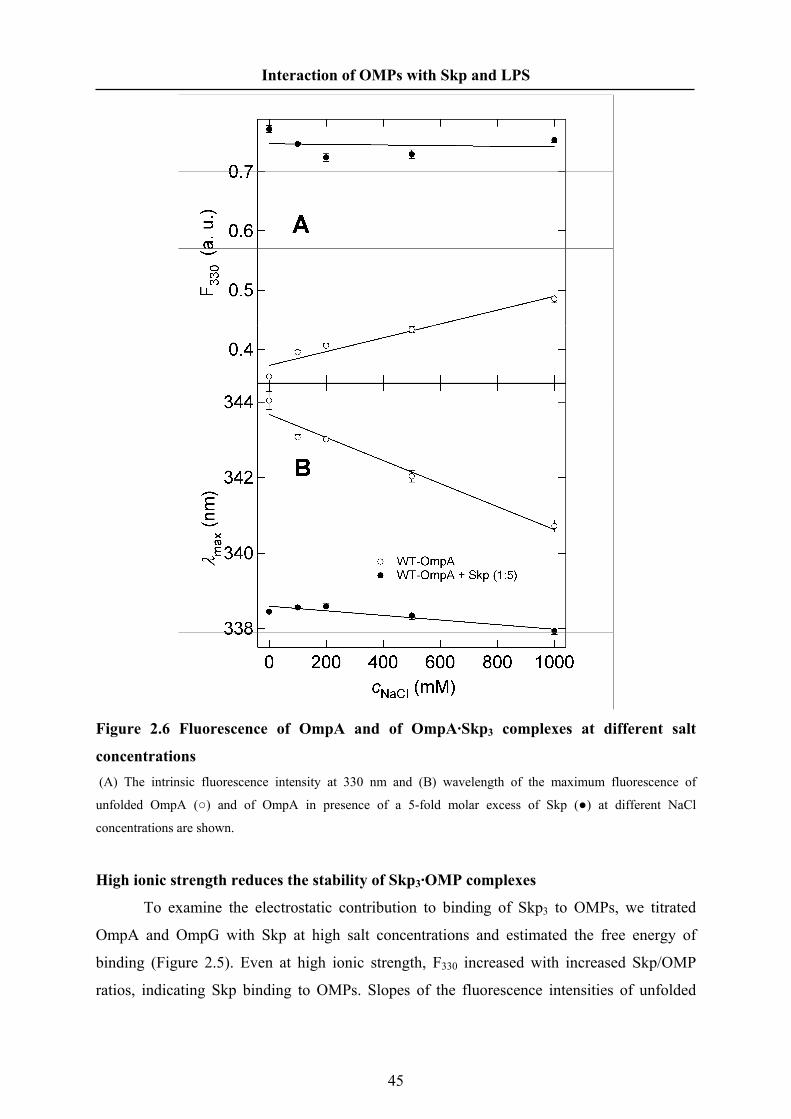
Interaction of OMPs with Skp and LPS
Figure 2.6 Fluorescence of OmpA and of OmpA·Skp3 complexes at different salt
concentrations (A) The intrinsic fluorescence intensity at 330 nm and (B) wavelength of the maximum fluorescence of
unfolded OmpA (○) and of OmpA in presence of a 5-fold molar excess of Skp (●) at different NaCl
concentrations are shown.
High ionic strength reduces the stability of Skp3·OMP complexes
To examine the electrostatic contribution to binding of Skp3 to OMPs, we titrated
OmpA and OmpG with Skp at high salt concentrations and estimated the free energy of
binding (Figure 2.5). Even at high ionic strength, F330 increased with increased Skp/OMP
ratios, indicating Skp binding to OMPs. Slopes of the fluorescence intensities of unfolded
45

Interaction of OMPs with Skp and LPS
OmpA or unfolded OmpG upon Skp binding were less steep than at low ionic strength, but
the stoichiometry of the complexes remained unaltered. The more gradual leveling of the
fluorescence intensities suggested weaker interactions and fits of binding functions to the data
resulted in lower association constants. Free energies of binding were reduced by ~3 kJ/mol
for OmpA and by ~5 kJ/mol for OmpG (Table 2.2). The fluorescence intensity of OmpA
increased at higher concentrations of NaCl, while λAQ decreased suggesting that screening of
the negative charges on the surface of OmpA reduces the exposure of the tryptophan residues
to the aqueous space. This effect was less pronounced for the fluorescence of OmpA·Skp3
complexes, since the tryptophans in these complexes are already well shielded from the
aqueous space at low salt concentrations (Figure 2.6). The results indicate that while
electrostatic interactions may be necessary for complex formation, the high stability of
OMP·Skp3 complexes is caused by hydrophobic interactions.
LPS binds to OmpA·Skp3 complexes
We previously demonstrated that Skp binding to OmpA results in smaller rate
constants of membrane insertion and folding of OmpA and in lower yields of folded OmpA.
The simultaneous presence of LPS at 2 to 7 mol per mol OmpA·Skp3 complex was required
for fast folding kinetics of OmpA and high yields of membrane inserted and folded OmpA
[Bulieris et al. 2003]. The crystal structure of Skp [Korndörfer et al. 2004; Walton et al. 2004]
indicated an LPS binding site in the tentacle domain of Skp [Walton et al. 2004]. To probe
the interaction, we gradually titrated OmpA·Skp3 complexes with LPS and recorded the
fluorescence spectra. Increased LPS binding to OmpA·Skp3 resulted in an initial steep
reduction of the fluorescence intensity (Figure 2.7A, filled circles) and in an initial steep
increase of the wavelength of the intensity maximum λmax (Figure 2.7B, filled circles) up to a
molar ratio of about 3 LPS/(OmpA·Skp3). At higher ratios, between 5 and 15
LPS/(OmpA·Skp3) both, the decrease in intensity and the increase in leveled off at ~65% of
the original intensity and at λmax ~342 nm. The LPS binding stoichiometry to OmpA·Skp3 is a
far lower than the molar ratio of 25 LPS/OmpA observed previously for LPS binding to
OmpA in absence of Skp [Bulieris et al. 2003]. In absence of Skp, LPS binding to unfolded
OmpA led to a fluorescence increase and a λmax decrease to ~335 nm (Figure 2.7, open
circles).
46

Interaction of OMPs with Skp and LPS
Figure 2.7 LPS binds to the OmpA·Skp3 complex (A) The intrinsic fluorescence intensity at 330 nm and (B) the wavelength of maximum fluorescence of the
complex of OmpA and Skp were recorded after addition of LPS at molar LPS/(OmpA·Skp3) ratios ranging from
0 to 30. Three separate samples were measured at each LPS concentration and an average value was calculated.
F330 and λmax of OmpA after addition of LPS at molar LPS/OmpA ratios ranging from 0 to 30 are shown for
comparison (○). The data of LPS binding to WT-OmpA were taken from reference [Bulieris et al. 2003].
Skp and LPS shield the Trps of OmpA in the acrylamide quenching experiments
To test the average exposure of the 5 tryptophans of OmpA to the aqueous quencher
acrylamide for both aqueous and Skp-bound forms of OmpA, we performed fluorescence
quenching studies with acrylamide. We also examined the effect of LPS on tryptophan
47

Interaction of OMPs with Skp and LPS
accessibility for aqueous and Skp-bound OmpA (Figure 2.8). In absence of both Skp and LPS
OmpA fluorescence was heavily quenched and a Stern-Volmer constant of KSV = 6.0 M–1 was
calculated, consistent with previous results [Kleinschmidt et al. 1999c]. In presence of LPS
and in absence of Skp, the Stern-Volmer constant is 4.5 M–1 indicating LPS binding to
aqueous OmpA, which partially shields it against quenching by acrylamide. In presence of
Skp and in absence of LPS, the quenching was weakest with a Stern-Volmer constant of 2.7
M–1. When LPS was added to OmpA·Skp3 complex, the Stern-Volmer constant increased to
3.6 M–1. Upon addition of LPS, tryptophans of OmpA, which have been shielded in complex
with Skp, became more accessible to the fluorescence quencher, but were still better shielded
than in presence of LPS, but in absence of Skp. These fluorescence-quenching results further
supported the formation of a ternary complex between LPS, Skp and OmpA at low ratios of
LPS per OmpA·Skp3 complex complementing the observed stoichiometry of LPS binding to
OmpA·Skp3 complexes.
Figure 2.8 In presence of LPS or Skp, the tryptophans of OmpA are less accessible to
aqueous fluorescence quenchers Fluorescence spectra of OmpA were recorded at different concentrations of the fluorescence quencher
acrylamide to obtain the Stern-Volmer plots for OmpA either in aqueous solution (●), in presence of Skp (■), in
presence of LPS (○) or in presence of Skp and LPS (□). F and F0 are fluorescence intensities in the presence and
in the absence of acrylamide, respectively. Stern-Volmer constants of 6.0 M-1 (OmpA), 4.5 M–1 (OmpA+LPS),
2.7 M–1 (OmpA+Skp) and 3.6 M–1 (OmpA+Skp+LPS) were obtained from the slopes of linear fits.
48

Interaction of OMPs with Skp and LPS
2.5 Discussion
Our present observations on the interactions of Skp with unfolded β-barrel membrane
proteins provide detailed new insights into the properties and function of this periplasmic
chaperone. Unfolded transmembrane proteins are bound with nanomolar affinity in stable
complexes with trimeric Skp. Independent of the size of the transmembrane domain of the
OMP, a 1:1 binding-stoichiometry was observed. Bacterial OMPs were bound independent of
their origin, but binding did not occur with mitochondrial hVDAC1. Both, hydrophobic and
electrostatic interactions contributed to the formation of OMP·Skp3 complex. We finally
directly observed LPS binding to OmpA·Skp3 complexes, which induces a conformation
change in the OmpA·Skp3 complex that could explain facilitated folding of OmpA into lipid
membranes in presence of both, Skp and LPS [Bulieris et al. 2003].
Trimeric Skp binds OMPs with nanomolar affinity and 1:1 stoichiometry
We have shown that Skp of E. coli binds five unfolded OMPs of varying sizes (32-89
kDa), OmpA, OmpG, YaeT, NalP, and FomA all at 1:1 stoichiometry. Binding affinities
ranged from 0.3 to 50 nM (Table 2.2) at low ionic strength and were therefore about three
orders of magnitude higher than the binding affinities of another periplasmic chaperone,
SurA of E. coli, to OmpG and OmpF [Bitto et al. 2004]. Thermodynamic binding equilibria
of OMPs in presence of both, Skp and SurA therefore favor OMP·Skp3 complexes over
OMP·SurA complexes, suggesting that Skp is the primary chaperone for OMPs, if both Skp
and SurA are present in sufficient and similar quantity. A recent study indicated that a major
role of SurA is the correct assembly of type 1 and P-pili of uropathogenic strains of E. coli
[Justice et al. 2005]. However, SurA depletion in cells still leads to reduced concentrations of
OMPs [Lazar et al. 1996; Rouvière et al. 1996], suggesting that typical concentrations of Skp
may not be high enough to bind all OMPs during their passage through the periplasm.
In contrast to bacterial OMPs, Skp did neither bind to hVDAC1 nor to BSA. There are
no homologs of Skp in eukaryotic cells, but Skp is also found in other Gram-negative bacteria.
Skp of N. meningitides and Skp of F. nucleatum share about 22% and 23% sequence identity
with Skp of E. coli. It is therefore not unexpected that Skp of E. coli also binds NalP of N.
meningitides and FomA of F. nucleatum. However, Skp generally recognizes unfolded
structures, since previous studies demonstrated that it prevents aggregation of lysozyme from
49

Interaction of OMPs with Skp and LPS
hen eggs [Walton et al. 2004] and promoted expression of soluble antibody fragments
[Bothmann et al. 1998]. Presumably, soluble BSA folds too fast upon urea-dilution [Johanson
et al. 1981] to be recognized by Skp. Similarly, Skp also did not bind to hVDAC1 because
upon urea-dilution, this mitochondrial OMP rapidly develops large amounts of β-sheet
secondary structure already in aqueous solution [Shanmugavadivu et al. 2007].
Figure 2.9 Crystal Structure of Skp (PDB 1SG2 [Korndörfer et al. 2004]) (A) The Skp trimer is composed of a tentacle domain, formed by an association domain that contains a 9-
stranded β-barrel, which is surrounded by three pairs of long α-helices, which are about 65 Å long and form the
tentacle domain. Each 162 residue Skp monomer contains a putative LPS binding site composed of residues
K77, R87, and R88, which at a root-mean-square (rms) deviation of 1.75 Å [Walton et al. 2004], matches a
conserved LPS binding motif previously described for the OMP FhuA [Ferguson et al. 2000]. The surface of the
trimer is strongly positively charged, because each monomer contains 28 lysines and arginines, but only 19
aspartates and glutamates. (B) The charged residues are asymmetrically distributed and most of the negatively
charged aspartates and glutamates are found in the association domain, while the positively charged residues are
found at the other end at the tips of the tentacle domain, leading to a large dipole moment of ~3 700 Debye (770
eÅ) [Korndörfer et al. 2004].
50

Interaction of OMPs with Skp and LPS
Skp binds OMPs by hydrophobic and by electrostatic interactions
The flexible three-pronged forcep-like structure of Skp suggests that OMPs are bound
inside the basket defined by the six α-helices of Skp (Figure 2.9). As pointed out previously
[Korndörfer et al. 2004; Walton et al. 2004], the abundance of positively charged residues at
the end of the tentacles likely ensures that the inside of the tentacle domain is accessible in
solution. The surface of the Skp tentacle domain has a positive charge overall. However
charged and hydrophobic areas form patches over the entire surface of the tentacle domain
(Figure 2.9). Inside the tentacle basket, there exist hydrophobic patches formed in between
helices 1 and 2. Two of them are composed of residues F13, A17, V22, and L26 or of
residues F30, A34, L37, F74, and F81. These hydrophobic residues also form the interface
between the two helices of the Skp monomer (Figure 2.9C). The patches are not large enough
for the hydrophobic surface of a completely folded OMP and they alternate with polar and
charged areas. For example Arginin 85, faces L26 and F30 on the interior surface and
separates two hydrophobic patches. The alternating surface polarity of Skp is consistent with
the amphipathic nature of OMP transmembrane β-strands, in which hydrophobic and polar
residues also occur in an alternating pattern.
The formation of the OmpA·Skp3 complexes is pH-dependent, and does not occur
above pH 10 and below pH 5 (Figure 2.3A). These upper and lower limits already indicate
that the interaction between Skp and OmpA is in part electrostatic, since these pH values are
close to the two isoelectric points, which have been calculated to pI ~ 5.6 for OmpA and to pI
~ 9.5 for Skp. Stable complexes are formed when opposite charges in both proteins are
present (Figure 2.3 B and C). While the role of charge-charge interactions is obvious for the
pH dependence of OmpA·Skp3 complex formation, the titrations also suggest charge-charge
interactions for NalP and FomA although these OMPs have higher isoelectric points (Table
2.1), leading to the positive net-charges in both Skp3 and FomA or NalP below pH 8 and pH
8.6, respectively. Our binding experiments still demonstrate binding of Skp to NalP down to
pH 6, suggesting charge-charge interactions with specific regions of the OMPs, in which the
acidic residues aspartate and glutamate dominate. OMP regions rich in aspartate or glutamate
are the periplasmic turns (Table 2.3) and here we propose that the turns of OMPs that often
contain aspartate or glutamate, interact with the most basic regions in Skp. The Skp trimer
has a high positive charge density at the tips of the three tentacles (Figure 2.9) containing 7
basic, but only 3 acidic residues (Figure 2.9C). The three tip regions are quite likely the sites
in Skp3 that contribute to the electrostatic interactions observed in this work.
51

Interaction of OMPs with Skp and LPS
Table 2.3 Acidic and Basic Residues in the Periplasmic Turns of some OMPs OMP PDB ZTMβs
a ZTb Dc Ec Kc Rc T-Charged
OmpA 1BXW 8 3 2 1 0 0 –3 NalPd 1UYN 12 5 2 1 0 0 –3 OmpG 2IWW 14 6 5 1 0 0 –6 OmpW 2F1V 8 3 3 0 0 1 –2 OmpT 1I78 10 4 1 2 0 0 –3 FadL 1T16 14 6 6 0 0 2 –4 OmpF 2OmF 16 7 4 1 1 0 –5 LamB 1MAL 18 8 3 2 1 0 –4 FhuA 2FCP 22 10 11 1 2 2 –8 aNumber of transmembrane β-strands of the OMP. This number is predicted for YaeT. bNumber of periplasmic β-turns of the OMP. This number is predicted for YaeT. cFrequencies of aspartes (D), glutamates (E), lysines (K), and arginines (R) in the periplasmic turns of these OMPs. dTotal charge of all turns. etranslocator domain of NalP, residues 776-1083
The following NaCl effect experiments showed that the binding was sensitive to the
salt concentration and electrostatic forces contributed to the binding only about 10%. Thereby
the hydrophobic force should give main contribution to the interaction between Skp and
OMPs. From the crystal structure, Skp contains several hydrophobic patches at the surface of
its cavities [Korndörfer et al. 2004]. As for OMPs, the hydrophobic residues are very
abundant at the transmembrane domain of OMPs [Diederichs et al. 1998]. Large quantity of
hydrophobic residues exposed to the external environment after translocated from the inner
membrane is one of the reasons that make OMPs aggregate. After OMPs bind to Skp, the
hydrophobic cavities of Skp are the best place to protect the hydrophobic residues of the
OMPs. Similarly, SecB and Prefoldin also have hydrophobic region in their ligand binding
site [Martin-Benito et al. 2002; Dekker et al. 2003]. The predominated interactions between
SecB or PFD and their substrate involve hydrophobic interactions [Lundin et al. 2004; Zhou
et al. 2005].
Structural Similarity of Skp to Cytosolic Prefoldins of Archaea and Eukarya
Jellyfish-like structures similar to Skp have been reported for prefoldins (PFDs) of
archaea like Methanobacterium thermoautotrophicum [Siegert et al. 2000] and of eukarya
[Martin-Benito et al. 2002]. PFDs bind and prevent the aggregation of denatured substrate
proteins of varying size (14-75 kDa) [Lundin et al. 2004]. For example, PFD of Pyrococcus
52

Interaction of OMPs with Skp and LPS
horikoshii strongly binds to denatured soluble proteins such as citrate synthase and green
fluorescent protein with dissociation constants of 20 nM and 5 nM, respectively [Zako et al.
2005]. In contrast to the highly basic Skp, PFDs have a low pI at ~4.5 and their surface is
considerably more hydrophobic than the surface of Skp, in particular at the tips of the
tentacles, which in contrast to Skp do not carry a strong net-charge.
Similarity to Cytosolic SecB
Similar to Skp and PFDs, SecB binds non-native proteins of varying size (16-100
kDa), although it has totally different structure than these two chaperones [Baars et al. 2006].
SecB is of interest, because it binds outer membrane protein precursors like proOmpA in the
cytoplasm independent of a signal-sequence [Lecker et al. 1990; Knoblauch et al. 1999].
Binding to tetrameric SecB (68 kDa) is also characterized by a 1:1 stoichiometry and
nanomolar affinity. For example, the affinities of SecB binding to denatured maltose-binding
protein, RNase A, the α subunit of tryptophan synthase and ribose-binding protein ranged
from 5 nM to 50 nM [Hardy et al. 1991]. Substrate proteins must have a critical length to
allow isolation of stable SecB·protein complexes [Randall et al. 1997]. SecB (calculated pI
4.1) has a net-negative charge and a selectivity for positively charged or aromatic peptides
[Knoblauch et al. 1999] consistent with an electrostatic component in SecB-substrate binding.
SecB-binding peptides contained at least three aromatic or three positive residues
independent of their position along the 13 residue sequence. SecB-binding peptides derived
from OmpA [Knoblauch et al. 1999] were rich in aromatic residues which are present in the
OmpA transmembrane strands.
LPS modulates the conformation of the OmpA·Skp3 complex
To obtain a crude estimate for the binding stoichiometry and binding constant, we
again assumed a simple mass action law with identical LPS binding sites within the
Skp3·OmpA. The fits gave a low binding stoichiometry of 1.5 ± 2 LPS/OmpA·Skp3 and a
dissociation constant of KD ≈ 500 nM. However, these are very crude estimates, since LPS is
known to bind to both OmpA and Skp and the assumption of equivalent binding sites may be
incorrect.
It was previously shown that Skp alone inhibits folding of OmpA and that LPS reverts
this retarding effect of Skp [Bulieris et al. 2003] by interaction with the Skp-OMP complex.
Here we showed experimentally that LPS changes the conformation of the OmpA·Skp3
complex and partially exposes the β-strands containing the fluorescent tryptophans of OmpA
53

Interaction of OMPs with Skp and LPS
to the aqueous environment (Figure 2.7 and 2.8). The stoichiometry obtained here for LPS
binding to OmpA·Skp3 complexes (Figure 2.7) corresponds well to our previous observations
of optimal OmpA folding kinetics and folding yields at an LPS content of 2 to 7 moles per
mol OmpA·Skp3 complex [Bulieris et al. 2003], suggesting that LPS is not able to completely
displace Skp from OmpA. It is quite possible that LPS binds to Skp, when reacted with the
OmpA·Skp3 complex as suggested [Walton et al. 2004]. LPS binding to OmpA·Skp3
complexes could result in a conformational change in Skp that partially exposes the
tryptophans of OmpA to a more polar environment, leading to a decrease in the fluorescence
intensity and to an increase in λmax for those tryptophans that become more solvent exposed.
The acrylamide quenching experiments also showed that the OmpA·Skp3 complex is more
compact than the OmpA·Skp3·LPSn complex (Figure 2.8). The strong interaction between
Skp and OMPs may be the obstacle of OMPs insertion into the OM. This may be compared,
to the ATP-dependent folding pathway, where ATP modulates the conformation of the
chaperone-MP complex and initiates folding [Hartl et al. 2002]. LPS may play a similar role
in the Skp-assisting folding pathway. Although chemical energy is not provided, binding
energy the interaction of LPS with Skp is exergonic and may provide a significant amount of
energy for a conformational change in the OMP. After OMPs enter the periplasm in an
unfolded form through the SecYEG translocon, Skp binds OMPs to prevent their aggregation.
However, this binding is tight and insertion and folding of OMPs into the outer membrane are
inhibited. LPS modulates the binding between Skp and OMP thereby the state of OMP is
adjusted and OMP goes into a ready-state of insertion and folding.
54

Interaction of OMPs with Skp and LPS
3 The interaction of OmpA with LPS and the periplasmic
chaperone Skp of Escherichia coli studied by site-directed
mutagenesis
3.1 Abstract
The outer membrane protein of Escherichia coli, OmpA, is an important model
protein for the membrane protein folding studies. The periplasmic chaperone Skp and
lipopolysaccharides (LPS) facilitate OmpA fold and insert into lipid bilayers. Here we
intensively studied the interaction of OmpA with Skp and/or LPS by site-directed
mutagenesis and fluorescence spectroscopy. Thirteen single tryptophan mutants of OmpA
were developed and the single tryptophan is located in the different regions of OmpA: strands,
loops, turns of transmembrane domain and periplasmic domain. All of them can fold into the
lipid bilayers as wild type OmpA spontaneously. The intrinsic tryptophan fluorescence of
these mutants was recorded to show the details of the interaction between OmpA mutants and
Skp or LPS or both. In 8 M urea, the spectra of all mutants have similar line shapes,
intensities and wavelengths of fluorescence emission maxima (λmax ≈ 347 nm). In aqueous
solution, the periplasmic domain of OmpA folds to its native conformation while the
transmembrane domain folds to a water-soluble intermediate. Skp significantly increased the
fluorescence intensities of all mutants with single Trp in the TM domain and shifted λmax
towards lower wavelength, suggesting that Skp binds the entire transmembrane domain of
OmpA by multivalent interaction and change its conformation. LPS gave less effect on the
OmpA mutants than Skp did. The loop 1 and loop 3 of OmpA was found to interact with LPS
and the strands and turns of OmpA are not the binding region of LPS. Skp and LPS together
help OmpA turn into a folding-competent state and LPS plays a key role on the conformation
change of OmpA. LPS loosed the binding of Skp to OmpA and especially exposed the loops
L1, L2 and L4 of OmpA to a more polar environment. The turn regions of OmpA remain
tightly bound to Skp independent of LPS. MTSSL quenching Trp fluorescence of double
mutants only in DOPC indicated that there is no tertiary structure in urea-denatured form, in
aqueous form, in Skp and/or LPS bound form of OmpA.
55

Interaction of OMPs with Skp and LPS
3.2 Introduction
In recent years, several gene-deletion studies indicated that the assembly of outer
membrane proteins (OMPs) of Gram-negative bacteria is facilitated by molecular chaperones
that are present in the periplasm. Removal of the periplasmic proteins Skp and SurA resulted
in reduced concentrations of some OMPs in the outer membrane of E. coli [Chen et al. 1996;
Lazar et al. 1996; Missiakas et al. 1996; Rouvière et al. 1996; Rizzitello et al. 2001].
Periplasmic proteins and OMPs Gram-negative bacteria are synthesized in the cytoplasm and
then translocated across the cytoplasmic membrane [Danese et al. 1998]. How molecular
chaperones interact with unfolded OMPs in the periplasm is not well understood. The
periplasmic Seventeen kDa Protein, Skp bound to unfolded outer membrane protein F (OmpF)
linked to sepharose on an affinity column [Chen et al. 1996] while SurA was shown to bind
peptides containing the Ar-X-Ar motif frequently found in outer membrane proteins [Bitto et
al. 2003; Hennecke et al. 2005]. There is no ATP in the periplasm [Wülfing et al. 1994] and
therefore periplasmic chaperones are expected to function differently from most cytoplasmic
chaperones, which utilize ATP in their catalytic cycles [Craig 1993].
Periplasmic Skp of E. coli (141 residues, 15.7 kDa) forms a stable homo trimer in
solution as determined by gel-filtration and crosslinking experiments [Schlapschy et al. 2004].
The structure of the Skp trimer [Korndörfer et al. 2004; Walton et al. 2004] resembles a
jellyfish with α-helical tentacles protruding about 60 Å from a β-barrel body and defining a
central cavity (Figure 1.12). The crystal structures (1U2M, 1SG2) show that Skp has clamp-
like structure similar to prefoldin [Siegert et al. 2000], trigger factor [Ferbitz et al. 2004],
DnaK [Zhu et al. 1996] and Hsp90 [Prodromou et al. 1997]. Clamp-like structures are
evolutionarily favored by both ATP-dependent and ATP-independent molecular chaperones.
The clamps present a multivalent binding surface ideal for protecting unfolded proteins until
they reach the native state or are transferred to another component of the folding machinery
[Stirling et al. 2006]. Many OMPs interact directly with Skp, see e.g. reports on OmpF [Chen
et al. 1996], maltoporin (LamB) [Harms et al. 2001], phosphoporin (PhoE) [Schäfer et al.
1999], and the outer membrane proteins A (OmpA) [Bulieris et al. 2003], G (OmpG) and
YaeT of E. coli, on the autotransporter NalP of N. meninigitidis, and on the major porin
FomA of F. nucleatum, [Qu et al. 2007]. Skp also prevented the aggregation of the water-
56

Interaction of OMPs with Skp and LPS
soluble protein lysozyme [Walton et al. 2004], similar to another periplasmic, SurA, which
functioned as a chaperone towards soluble citrate synthase [Behrens et al. 2001].
Our previous work showed that Skp strongly binds to OMPs at a 3:1 stoichiometry
consistent with the trimeric structure of Skp, i.e. independent of the size of the OMP
transmembrane domain, each Skp trimer binds one unfolded OMP monomer [Qu et al. 2007].
The dissociation constants of these stable complexes are in the nanomolar range. They form
via both, hydrophobic and electrostatic interactions [Qu et al. 2007].
Lipopolysaccharide (LPS) is a major component of the outer membrane of Gram-
negative bacteria, consisting of three domains, namely lipid A, core region and O-specific
chain [Rietschel et al. 1992]. OmpA can partially refold into LPS in vitro [Vogel et al. 1986b;
Bulieris et al. 2003]. In cells, LPS with a defect polysaccharide core prevented assembly of
OmpF and OmpC [Ried et al. 1990].
However, after translocation across the cytoplasmic membrane, unfolded OMPs like
OmpA [Schäfer et al. 1999] or PhoE [Harms et al. 2001] are first bound by periplasmic Skp.
OmpA bound to Skp did not fold in presence of LPS micelles [Bulieris et al. 2003]. However,
the simultaneous presence of LPS and Skp facilitated folding of OmpA into phospholipid
bilayers [Bulieris et al. 2003]. While in absence of LPS, Skp partially inhibited insertion and
folding of OmpA into lipid bilayers. Obviously, LPS weakened the interaction between Skp
and OmpA, partially exposing OmpA to the external environment [Qu et al. 2007].
In this detailed study, we used site-directed fluorescence spectroscopy to examine the
interactions of unfolded OmpA with Skp and LPS on the level of individual amino acid
residues within OmpA. We prepared a range of single tryptophan mutants of OmpA, in which
tryptophan can be selectively excited to study the changes in its microenvironment upon
interactions with its binding partners or upon folding. The residues that we replaced by
tryptophan were chosen to determine whether Skp or LPS preferentially bind to either β-
strand, β-turn, or outer space loop regions of OmpA. We determined the effect of urea-
dilution on the aqueous form of these OmpA mutants and then examined their interactions in
separate experiments first with Skp then with LPS. We next investigated how addition of LPS
to preformed Skp-OmpA complexes alters the environment of the mutated residues in OmpA.
We finally isolated five different single-tryptophan/single cysteine mutants of OmpA. These
mutants were labeled with a fluorescence quencher at the cysteine. Intramolecular site-
directed fluorescence quenching was used to examine whether OmpA adopts partially folded
structure in complexes with Skp and LPS.
57

Interaction of OMPs with Skp and LPS
3.3 Materials and Methods
Materials
E. coli strain XL1-blue recA1 endA1 gyrA96 thi-1 hsdR17 supE44 relA1 lac [F´
proAB lacIqZΔM15 Tn10 (Tetr)] (Stratagene) was used for plasmid manipulations; E. coli
strain BL21 (DE3) omp8 fhuA [F–, ompT hsdSB (rB– mB
–) gal dcm (DE3) ΔlamB ompF::Tn5
ΔοmpA ΔοmpC ΔfhuA] [Prilipov et al. 1998], which is the OmpA deficiency strain, was used
for the expression of the OmpA mutants. Plasmids and primers used in this study are listed in
Table 1 and 2. All oligonucleotide primers were purchased from MWG Biotech AG
(Germany). The spin labeling reagent (1-oxyl-2,2,5,5-tetramethylpyrroline-3-methyl)
methanethiosulfonate (MTSSL) was obtained from Reanal (Budapest, Hungary). Methyl- 4-
nitrobenzenesulfonate, 5,5´-dithiobis(2-nitrobenzoic acid) (DTNB), Tris(2-
carboxyethyl)phosphine hydrochloride (TCEP) were purchased from Sigma-Aldrich
(Germany). 1,2-dioleoyl-sn-glycero-3-phosphocholine (DOPC) was purchased from Avanti
Polar Lipids (Alabaster, AL).
Construction of a plasmid to express OmpA mutants into inclusion bodies
To obtain better expression yields and to simplify the purification of single tryptophan
mutants, we first subcloned a gene enconding a proOmpA mutant, in which all tryptophan
residues were replaced by phenylalanines, into a new vector, deleting the signal sequence for
expression of OmpA mutants in form of inclusion bodies. The sequence of the gene was
amplified from plasmid pET185 [Kleinschmidt et al. 1999a] by PfuUltra polymerase
(stratagene) chain reaction (PCR) using primers that included the cut-sites for the restriction
enzymes NdeI and XhoI (Table 3.2). In the forward primer, the signal sequence of proompA
was removed. The PCR product was then cloned into the pET22b vector (Novagen) to obtain
plasmid pET22b185.
Construction of the plasmids of single tryptophan mutants
The preparation of 5 plasmids which were marked in Table 3.1 was described
previously [Kleinschmidt et al. 1999a; Kleinschmidt et al. 2007]. The eight new plasmids
prepared in this work, were based on the plasmid pET22b185. Site-directed mutagenesis was
58

Interaction of OMPs with Skp and LPS
performed using the QuickChange II kit as described (Strategene). The resulting plasmids
were isolated and sequenced to verify the desired mutation.
Table 3.1 list of the plasmids and proteins of single tryptophan mutants of OmpA Plasmids Vector Trp position Products Source pET1102 pTRC99A 7 s1W7 [Kleinschmidt et al. 1999a]
pET1115 pTRC99A 15 s1W15 [Kleinschmidt et al. 1999a]
pET187 pUC18 57 s3W57 [Kleinschmidt et al. 1999a]
pET186 pTRC99A 102 s5W102 [Kleinschmidt et al. 1999a]
pET1103 pTRC99A 143 s7W143 [Kleinschmidt et al. 1999a]
pET24 pET22b 24 l1W24 this work
pET67 pET22b 67 l2W67 this work
pET110 pET22b 110 l3W110 this work
pET153 pET22b 153 l4W153 this work
pET48 pET22b 48 t1W48 this work
pET91 pET22b 91 t2W91 this work
pET131 pET22b 131 t3W131 this work
pET263 pET22b 263 PW263 this work
pET185 pTRC99A - - [Kleinschmidt et al. 1999a]
pET22b185 pET22b - - this work
Table 3.2 Oligonucleotide primers for site-directed mutagenesis and plasmid
construction Plasmids Sequence of primers* pET24 5'-CCATGACACTGGTTTCTGGAACAACAATGGCCCG-3'
pET67 5'-GCCGTACAAAGGCAGCTGGGAAAACGGTGCATAC-3'
pET110 5'-CGACACTAAATCCAACTGGTACGGTAAAAACCACG-3'
pET153 5'-GGTGACGCACACACCTGGGGCACTCGTCCGGACAAC-3'
pET48 5'-GTTACCAGGTTAACCCGTGGGTTGGCTTTGAAATG-3'
pET91 5'-CAATCACTGACGACTGGGACATCTACACTC-3'
pET131 5'-CGGTGTTGAGTACGCGTGGACTCCTGAAATCGC-3'
pET263 5'-CAGTCTGTTGTTGATTGGCTGATCTCCAAAGGTATC-3'
pET22b185 5'-CGGCATATGGCTCCGAAAGATAACACCTG-3'
5'-CTGCTCGAGTTAAGCCTGCGGCTGAGTTACA-3'
* A pair of complementary primers was used to create each mutant; however, only the sequence of the sense
strand is shown. Changes in the sequences are shown in boldface type. NdeI and XhoI cut site sequences are
shown underlined.
59

Interaction of OMPs with Skp and LPS
Expression and purification of OmpA mutants
Each OmpA mutant was purified after transformation of the corresponding plasmid
into an E. coli BL21 (DE3) strain for expression. For purification, 2 L LB medium were
inoculated with 20 ml overnight culture and grown for 3 h. IPTG was then added to a final
concentration of 0.1 mM. After 4-6 h of induction, cells were harvested by 30 min of
centrifugation (1500 g, 4°C). The wet cell paste was resuspended in 40 ml Tris buffer (20
mM Tris, 0.1% 2-ME, pH 8.0) using an ice/water cooling bath. Lysozyme was added to a
concentration of 50 μg/ml and the mixture was stirred for 30 min at room temperature. The
solution was then sonified for 30 min using a Branson ultrasonifier W-450D (20%power,
50 % pulse cycle) with a macrotip in an ice/water bath. The buffer and soluble proteins were
removed by centrifugation at 3000g (4°C, 30 min). The pellet was resuspended in 20 ml 8 M
urea buffer (20 mM Tris, pH8.0, 0.1% 2-ME). An equal volume isopropanol was added and
the mixture should be incubated in 55°C waterbath immediately for exactly 30 min. The
pellet was removed by centrifugation (5000 g x 30min, 25°C). The supernatant was loaded
onto a Q-sepharose FF column (Amersham), which was pre-equilibrated by 4 M urea/50%
isopropanol buffer, and the proteins were eluted from the column by a NaCl gradient (0-
100mM).
The OmpA mutant plasmids constructed in previous work expressed the OmpA
mutant proteins into the outer membrane of E. coli. The purification protocol for these
mutants is the same as reported previously for wild type OmpA [Surrey et al. 1992].
Folding of single Trp mutants detected by SDS-PAGE
3 mg of DOPC were dissolved in chloroform, dried on the bottom of a glass test tube
under a stream of nitrogen, and desiccated under high vacuum for at least 3 h to remove
residual solvent. The lipid was dispersed in 10 mM glycine buffer, pH 8.3, containing 1 mM
EDTA and 150 mM NaCl, to a concentration of 1.5 mg/ml. Small unilamellar vesicles (SUVs)
were prepared by sonicating the lipid dispersions for 30 min with a Branson W450D
ultrasonicator, operating at 40 W with a 50% pulse cycle. To confirm folding of each OmpA
mutant, 3 μM were incubated with 400-fold molar excess of DOPC in 200 μl of glycine
buffer at 40°C for 16 h. About 2 μg proteins were loaded for SDS-PAGE [Laemmli 1970].
MTSSL spin-labeling and S-Methylation of single tryptophan/single cysteine mutants
60

Interaction of OMPs with Skp and LPS
The single tryptophan/single cysteine mutants of OmpA (WnCm mutants) were
expressed and purified as previously described [Kleinschmidt et al. 2007]. S-Methylation was
carried out using a modified procedure of Hunziker [Hunziker 1991]. A 5-fold molar excess
of TCEP was added to denatured WnCm mutants in 8 M urea buffer (50 mM borate, 1 mM
EDTA, pH9.0). After 30 min of incubation at room temperature, a 40-fold molar excess of
methyl-4-nitrobenzenesulfonate in acetonitrile was added. The sample tube was incubated at
37 °C for 60 min. Unreacted methyl-4-nitrobenzenesulfonate was removed by exhaustive
dialysis.
MTSSL spin-labeling was performed as published [Berliner et al. 1982]. About 10 mg
of denatured protein was diluted in 1 ml of 10 mM Tris buffer, pH 7.2, containing 8 M urea
and 1 mM EDTA. Disulfide bonds that may form between WnCm OmpA mutants, were
reduced by reacting denatured OmpA with a 5-fold molar excess of TCEP for 30 min at room
temperature. Subsequently, 90 μl MTSSL in absolute ethanol was added to the reaction tube
at a molar ratio of MTSSL/OmpA of 10. The mixture was reacted at least 12 h in the dark at
room temperature. Excess MTSSL was removed by exhaustive dialysis.
To determine the degree of S-methylation and MTSSL spin-labeling, an aliquot of the
solution was taken and DTNB was added in a 10-fold molar excess followed by
spectrophotometric determination of free thiol groups at 412 nm, using ε412 = 13700 M-1 cm-1
[Riddles et al. 1983].
Table 3 list of the plasmids and proteins of single Trp/single Cys mutants Plasmids Vector Trp position Cys position* Products Source pTB001dc pTRC99A 7 43 W7C43 [Kleinschmidt et al. 2007]
pTB003rdc pTRC99A 57 35 W57C35 [Kleinschmidt et al. 2007]
pTB004dc pTRC99A 7 170 W7C170 [Kleinschmidt et al. 2007]
pTB005dc pTRC99A 15 162 W15C162 [Kleinschmidt et al. 2007]
pTB008dc pTRC99A 15 35 W15C35 [Kleinschmidt et al. 2007]
* Two native cysteins Cys290 and Cys302 are replaced by alanine by site-direct mutagenesis.
Purification of Skp
The Skp protein was purified as described [Bulieris, 2003 #82] from the periplasmic
fraction of E. coli CAG16037 [Mecsas et al. 1993] harboring the plasmids pSkp (pQE60
from Qiagen carrying the skp gene). Cells were grown in LB medium at 37 °C and were
61
Interaction of OMPs with Skp and LPS
induced at an absorbance of A(600 nm) = 0.3 with 1 mM IPTG. After 4 h, the cells were
harvested and resuspended in sucrose buffer (100 mM Tris-HCl, pH 8.0, 20% sucrose) and
equilibrated on ice for 10 min. Cells were centrifuged and then resuspended in sucrose buffer
with 10 mM EDTA and incubated with lysozyme (10μg/ml) for 30 min on ice. After addition
of MgSO4 (20 mM), Spheroplasts were sedimented at low speed after. The resulting
periplasmic fraction was dialyzed overnight at 4 °C against buffer A (20 mM Tris-HCl, pH
8.0, 100 mM NaCl), run through a DEAE-sepharose FF column and then loaded onto a CM-
sepharose FF column. The DEAE column was removed, and the CM column was washed
with 3 volumes of buffer A. Skp was eluted from the column by a NaCl gradient (100–500
mM NaCl in buffer A).
Purification of LPS
E. coli rough mutant F576 was cultivated as described previously [Vinogradov et al.
1999], and its LPS (R2 core type, M≈ 3900 g/mol) was isolated as reported [Müller-Loennies
et al. 1994].
Fluorescence spectroscopy
Fluorescence spectra were recorded as described previously [Bulieris et al. 2003] on a
Spex Fluorolog-3 spectrofluorometer with double monochromators in the excitation and
emission pathways. The excitation wavelength was 295 nm, and the bandwidths of the
excitation monochromators were 2.5 nm. The bandwidths of the emission monochromators
were 5 nm. The integration time was 0.05 s, and an increment of 0.5 nm was used to scan
spectra in the range of 310-380 nm. All of the experiments were performed at pH 8.0 and at
25 °C. The final concentrations were 1.15 μM of each OmpA mutant, 4.6 μM Skp and
5.75 μM LPS. Fluorescence data analyses were performed using Igor Pro 6.0 (WaveMetrics)
and the spectra were fitted to a log-normal function to obtain the fluorescence intensities at
330 nm (F330) and wavelengths of fluorescence emission maxima (λmax).
Binding of single Trp mutants of OmpA to Skp
4.5 μl Skp (8 mg/ml) was added into 490 μl of Tris buffer (10 mM, pH 8.0) and the
background spectrum was recorded. 10 μl OmpA mutant proteins (2 mg/ml) were then added
and the spectrum of Skp-mOmpA complex was recorded after 30 seconds incubation.
Binding of single Trp mutants of OmpA to LPS
62

Interaction of OMPs with Skp and LPS
10 μl unfolded OmpA mutant (2 mg/ml) were diluted into 490 μl Tris buffer (10 mM,
pH 8.0) and the spectrum of this aqueous form was recorded. After addition of 4.5 μl LPS
(2.5 mg/ml), the spectrum was recorded again for the LPS-mOmpA complex after 30 seconds
incubation.
Binding of LPS to complexes of OmpA muntants and Skp
4.5 μl Skp (8 mg/ml) was added into 490 μl of Tris buffer (10 mM, pH 8.0) and the
background spectrum was recorded. 10 μl OmpA mutant proteins (2 mg/ml) were added. 4.5
μl LPS (2.5 mg/ml) were then added into the complex of OmpA and Skp. The spectrum of
the mOmpA-Skp-LPS complex was recorded after 30 seconds incubation. All the
experiments were performed at 25 °C and repeated three times.
Fluorescence experiments of the WnCm mutants All fluorescence experiments of WnCm mutants were performed as same as the
described process of single Trp mutants of OmpA.
63

Interaction of OMPs with Skp and LPS
3.4 Results
Single Trp mutants of OmpA
To explore the sites within OmpA that are in contact with Skp or LPS upon complex
formation, we expressed and purified 13 different single tryptophan mutants of OmpA. The
expression systems were derived from a previously prepared plasmid encoding a tryptophan-
free mutant of OmpA, in which all native Trps were replaced by phenylalanine [Kleinschmidt
et al. 1999a]. Figure 1 shows the topology of OmpA. The residues that were replaced by a
tryptophan to obtain single Trp mutants of OmpA for the present study are typeset in bold.
The corresponding plasmids and OmpA mutants are listed in Table 3.1. These mutants were
from four categories: in the first, sxWn category amino acid n in β-strand x of the eight-
stranded transmembrane (TM) domain of OmpA was replaced by a tryptophan. The mutants
were s1W7, s1W15, s3W57, s5W102 and s7W143 [Kleinschmidt et al. 1999b]. In four
mutants of the second, lyWn category, the unique tryptophan was located in one of the four
loops y of OmpA. Here we prepared l1W24, l2W67, l3W110 and l4W153. In the third, tzWn
category of mutants, the single Trp was introduced to replace a residue in one of the three
periplasmic turns z of the TM β-barrel, to obtain t1W48, t2W91 and t3W131. The product of
plasmid pET263 was named PW263 because the unique Trp was introduced in the
periplasmic domain of OmpA. PW263 represents the fourth category. All mutants were
isolated in their unfolded forms in 8 M urea.
The folding of the sxWn mutants is already described in previous studies
[Kleinschmidt et al. 1999b; Arora et al. 2000]. We confirmed folding of the eight single Trp
mutants prepared in this study by incubating them in presence of preformed lipid bilayers of
DOPC under concurrent urea dilution (Figure 3.2). The unfolded mutants migrated at 35 kDa
on SDS-polyacrylamide gels whereas after incubation, all mutants migrated at 30 kDa. The
30 kDa form of OmpA was shown previously by Raman, FTIR, and CD spectroscopy [Vogel
et al. 1986a; Dornmair et al. 1990; Surrey et al. 1992; Rodionova et al. 1995; Kleinschmidt et
al. 1999a], by phage inactivation assays [Schweizer et al. 1978], by proteolysis [Surrey et al.
1992; Rodionova et al. 1995; Pocanschi et al. 2006b], and by single channel conductivity
measurements [Arora et al. 2000] to correspond to completely folded and functionally active
OmpA. All single-Trp mutants prepared here developed this 30 kDa form indicating their
folding into lipid bilayers of DOPC.
64

Interaction of OMPs with Skp and LPS
Figure 3.1 Transmembrane topology of OmpA and sites selected to probe binding of
Skp and LPS Residues replaced by tryptophan to for site-directed fluorescence spectroscopy are shown in boldface. The β-
strands of OmpA are numbered starting from the N-terminus as β1 - β8, the loops as L1 - L4, and the turns as T1 -
T3.
Figure 3.2 Folding of single Trp mutants of OmpA into DOPC bilayers at 40 °C SDS-PAGE demonstrates that the single Trp mutants prepared in this study, namely all lyWn and tzWn,
carrying the single tryptophan either in the outer loops or in the periplasmic turns, and PW263, folded into lipid
bilayers of DOPC. Each mutant was incubated with sonicated vesicles (SUVs) of DOPC for 16 h at 40°C.
Folded and unfolded WT-OmpA are shown for comparison. The samples were either boiled (+) or not (-) before
loading them onto the gel.
65
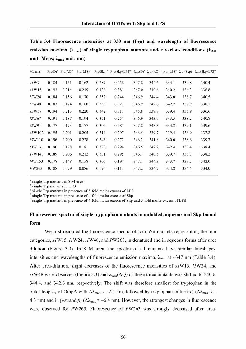
Interaction of OMPs with Skp and LPS
Table 3.4 Fluorescence intensities at 330 nm (F330) and wavelength of fluorescence
emission maxima (λmax) of single tryptophan mutants under various conditions (F330
unit: Mcps; λmax unit: nm)
Mutants F330(D)a F330(AQ)b F330(LPS)c F330(Skp)d F330(Skp+LPS)e λmax(D)a λmax(AQ)b λmax(LPS)c λmax(Skp)d λmax(Skp+LPS)e
s1W7 0.184 0.151 0.162 0.287 0.258 347.8 344.6 344.1 339.8 340.4
s1W15 0.193 0.214 0.219 0.438 0.381 347.0 340.6 340.2 336.3 336.8
l1W24 0.184 0.156 0.170 0.352 0.244 346.9 344.4 343.0 338.7 340.5
t1W48 0.183 0.174 0.180 0.353 0.322 346.9 342.6 342.7 337.9 338.1
s3W57 0.194 0.213 0.220 0.342 0.311 345.8 339.8 339.4 335.9 336.6
l2W67 0.191 0.187 0.194 0.371 0.257 346.9 343.9 343.5 338.2 340.8
t2W91 0.177 0.173 0.177 0.302 0.287 347.8 343.3 343.2 339.1 339.6
s5W102 0.195 0.201 0.205 0.314 0.297 346.5 339.7 339.4 336.9 337.2
l3W110 0.196 0.200 0.228 0.346 0.272 346.2 341.8 340.0 338.6 339.7
t3W131 0.190 0.178 0.181 0.370 0.294 346.5 342.2 342.4 337.4 338.4
s7W143 0.189 0.206 0.212 0.331 0.295 346.7 340.5 339.7 338.3 338.2
l4W153 0.178 0.148 0.158 0.306 0.197 347.1 344.3 343.7 339.2 342.0
PW263 0.188 0.079 0.086 0.096 0.113 347.2 334.7 334.8 334.4 334.0
a single Trp mutants in 8 M urea b single Trp mutants in H2O c single Trp mutants in presence of 5-fold molar excess of LPS d single Trp mutants in presence of 4-fold molar excess of Skp e single Trp mutants in presence of 4-fold molar excess of Skp and 5-fold molar excess of LPS
Fluorescence spectra of single tryptophan mutants in unfolded, aqueous and Skp-bound
form
We first recorded the fluorescence spectra of four Wn mutants representing the four
categories, s1W15, l1W24, t1W48, and PW263, in denatured and in aqueous forms after urea
dilution (Figure 3.3). In 8 M urea, the spectra of all mutants have similar lineshapes,
intensities and wavelengths of fluorescence emission maxima, λmax at ~347 nm (Table 3.4).
After urea-dilution, slight decreases of the fluorescence intensities of s1W15, l1W24, and
t1W48 were observed (Figure 3.3) and λmax(AQ) of these three mutants was shifted to 340.6,
344.4, and 342.6 nm, respectively. The shift was therefore smallest for tryptophan in the
outer loop L1 of OmpA with Δλmax ≈ –2.5 nm, followed by tryptophan in turn T1 (Δλmax ≈ –
4.3 nm) and in β-strand β1 (Δλmax ≈ –6.4 nm). However, the strongest changes in fluorescence
were observed for PW263. Fluorescence of PW263 was strongly decreased after urea-
66

Interaction of OMPs with Skp and LPS
dilution and the intensity maximum shifted by Δλmax ≈ –12 nm to ~335 nm. These large
differences between the fluorescence changes of mutants of the transmembrane and
periplasmic domains suggested that they independently develop structure in aqueous solution.
It is consistent with the hypothesis that the soluble aqueous domain already folds to its native
conformation while the TM domain folds to a water-soluble intermediate.
Skp affected the fluorescence of PW263 only slightly, but strongly increased the
fluorescence intensities of all TMWn mutants and shifted λmax towards shorter wavelength
(Table 4). Upon Skp binding, fluorescence intensities at 330 nm (F330) of the mutants s1W15,
l1W24, and t1W48 were increased by +105 %, +125 %, and +103 % respectively (Figure 3).
The intensity maxima, λmax(Skp), of these mutants were shifted by Δλmax ≈ –4.3, ≈ –5.7, and
≈ –4.7 nm, respectively, to 336.3, 338.7, and 337.9 nm (Figure 3.3). In contrast, F330 of the
mutant PW263 was increased by just 20% and Δλmax was only 0.4 nm (Table 4). The
relatively minor change indicated little or no interaction of Skp with the periplasmic domain,
in agreement with previous observations [Qu et al. 2007].
Figure 3.3 Fluorescence spectra of single Trp mutants of OmpA in denatured form and
after denaturant dilution in aqueous solution in absence and presence of Skp Fluorescence spectra of 4 representative single Trp mutants of OmpA are shown: (A) s1W15, (B) l1W24, (C)
t1W48 and (D) PW263. For each OmpA mutant, spectra were recorded in 8 M urea (- - - -), in Tris buffer (10
mM, pH 8.0) (. . . .) after 50-fold dilution of urea, and in presence of 4-fold molar excess of Skp (—).
67

Interaction of OMPs with Skp and LPS
Skp binds strands, loops and turns of the unfolded transmembrane domain of OmpA
To investigate whether the changes in the fluorescence spectra observed for unfolded,
aqueous and Skp-bound forms of s1W15, l1W24, and t1W48, are representative for the other
loops, turns, and strands, we recorded the fluorescence spectra of 9 additional TMWn mutants
in unfolded, aqueous and Skp-bound forms. The mutants contained a single tryptophan in all
other loops and turns (l2W67, l3W110, l4W153, t2W67, t3W131) and in 4 of the β-strands
(s1W7, s3W57, s5W102, s7W143). Figure 3.4 shows the emission maxima λmax of the
spectra of the aqueous and Skp-bound forms of OmpA as a function of the position of the
introduced single-Trp. For the aqueous forms (Figure 3.4A, open symbols), shifts of the
emission maxima were strongest (from λmax(D) = 346-348 nm to λmax(AQ) ~340 nm, i.e.
Δλmax = -6 to -8 nm) for the spectra of s1W15, s3W57, s5W102, and s7W143, which
contained the mutation in the β-strands. The s1W7 mutant containing tryptophan 7 close to
the N-terminus was an exception and within the TM domain, its emission maxima (λmax(AQ)
~345 nm) displayed the smallest change, Δλmax = –3 nm compared to the denatured form
(Table 4). Similar to s1W7, only small shifts in λmax were observed for l1W24, l2W67,
l4W153, t1W48, and t2W91 (λmax(D) = 346-348 nm to λmax(AQ) = ~343-344 nm). Shifts in
λmax of l3W110 and t3W131 were slightly larger (λmax(AQ) = ~342 nm). These data indicated
that in aqueous OmpA, the β-strands are less exposed to water than the loops and the turns.
These observations on the shifts in λmax were also reflected in changes in the fluorescence
intensities (Figure 3.5A).
In presence of a 4-fold molar excess of Skp (Figure 3.4A, filled symbols), emission
maxima of all TMWn mutants were shifted towards even shorter wavelength and the shortest
were observed for s1W15, s3W57, s5W102 and t3W131 with λmax at ~336-337 nm.
Compared to the aqueous forms none of the mutants displayed a λmax larger than 340 nm. The
changes in λmax were largest for lyWn and tzWn mutants with tryptophan in the loops and in
the turns. This is most obvious in a plot of the difference in λmax in presence and in absence of
Skp as a function of the position of the mutated residue (Figure 3.4B). The changes in the
fluorescence intensities at 330 nm upon the addition of Skp showed similar results as the
shifts in λmax (Figure 3.5B). In summary, Skp increased F330 of the single Trp mutants from
60% to 125% and Δλmax ranged from 2.1 nm to 5.7 nm. These results indicated a strong
change in the polarity of the microenvironment of the fluorescent tryptophans of OmpA,
demonstrating that Skp replaces water from the surface the entire TM domain of OmpA,
including its more polar or even charged loop and turn regions.
68

Interaction of OMPs with Skp and LPS
Figure 3.4 The wavelength changes of the single tryptophan mutants of OmpA upon the
addition of Skp (A) The wavelengths of fluorescence emission maxima λmax of single Trp mutants as a function of the position
of the inserted tryptophan exhibit a similar periodicity across the transmembrane domain in aqueous solution
(open symbols) and in presence of Skp (filled symbols). (B) The difference of λmax of each mutant in absence
and presence of Skp. For the β-strands (□, ■), fluorescence maxima are mostly located at shorter wavelength,
λmax, than for β-turns (◊, ♦) and outer loops (○, ●). In the annotation of the lower graph, symbols ‘s’, ‘l’, and ‘t’
represent the β-strands (■), the outer loops (●) and the turns (♦), respectively while Rn is the residue at position
n in OmpA (e.g. ‘l1R24’ denotes residue 24 of loop 1).
69

Interaction of OMPs with Skp and LPS
Figure 3.5 Fluorescence ratios of single Trp mutants of OmpA in absence and presence
of Skp (A) The fluorescence intensities at 330 nm of single Trp mutants in aqueous solution (open symbols) and in
presence of Skp (filled symbols). (B) The fluorescence ratio is the quotient of fluorescence intensity of OmpA
mutants at 330 nm in the presence and in the absence of Skp.
LPS effect on the single Trp OmpA mutants
We previously observed that Skp inhibits folding of OmpA into lipid bilayers, while
facilitating it when LPS was simultaneously present [Bulieris et al. 2003]. We also observed
LPS binding to a preformed OmpA·Skp3 complex [Qu et al. 2007]. This raised the question,
70

Interaction of OMPs with Skp and LPS
Figure 3.6 Fluorescence spectra of single Trp mutants of OmpA in absence and
presence of LPS Fluorescence spectra are shown for 4 representative single Trp mutants of OmpA: s1W15,l1W24, t1W48, and
PW263. Two curves in each graph are the OmpA mutant, in H2O (......) and in presence of 5-fold molar excess
of LPS (—), respectively.
whether the interaction with LPS changes the topology of the OmpA·Skp3 complex, which
could be the reason for improved folding kinetics and yields of OmpA observed in our
previous study [Bulieris et al. 2003]. We here used the prepared single tryptophan mutants of
OmpA to first examine the effect of LPS on OmpA in absence of Skp (Representative spectra
are shown in Figure 3.6). Figure 3.7 and 3.8 shows λmax and F330 of the fluorescence spectra
of the TMWn mutants as a function of the position of the tryptophan in absence and in
presence of a 5-fold molar excess of LPS. The addition of LPS had only minor effects on the
fluorescence spectra of the aqueous forms of the OmpA mutants. Slight increases of the
fluorescence intensities, ranging from 2% to 15%, and slight decreases of the λmax ranging
from 0 nm to -1.8 nm, were observed for all single tryptophan mutants (see Table 3.4). This
is consistent with the previous report that a 5-fold molar excess of LPS increases F330 of WT-
OmpA by ~12% [Bulieris et al. 2003]. The strongest changes upon LPS binding were
71

Interaction of OMPs with Skp and LPS
observed for mutants l1W24 and l3W110, for which F330 increased by about +10 % and
+15 %, respectively. The λmax of fluorescence spectra of l1W24 and l3W110 was decreased
by about 1.5 nm. This suggests that the microenvironments of residue 24 and 110, which are
located in loop L1 and L3 of OmpA may contain sites of interaction with LPS. This is not
surprising, since the outer loops of OmpA are facing LPS in the outer leaflet of the outer
membrane, which is composed of LPS. In contrast, tryptophans in turn and strand regions of
OmpA indicated little or no interaction with LPS although the strands of OmpA are more
hydrophobic than the turns.
Figure 3.7 The wavelength changes of the single tryptophan mutants of OmpA upon the
addition of LPS (A) The wavelengths of fluorescence emission maxima λmax of single Trp mutants in aqueous solution and in
presence of LPS. (B) The differences of λmax of each mutant in the absence and in presence of LPS (panel B)
were not very significant, except for loops l1 and l3, with ~1.5 and ~2 nm, respectively.
72

Interaction of OMPs with Skp and LPS
Figure 3.8 Fluorescence ratios of single Trp mutants of OmpA in absence and presence
of LPS (A) The fluorescence intensities at 330 nm of single Trp mutants in aqueous solution and in presence of LPS. (B)
The fluorescence ratio is the quotient of fluorescence intensity of OmpA mutants at 330 nm in the presence and
in the absence of LPS. Only minor changes of the fluorescence spectra of Wn mutants were observed when a 5-
fold molar excess of LPS was added
Fluorescence spectra of single Trp mutants of OmpA in presence of LPS and Skp
To examine the effect of LPS on OmpA·Skp3 complexes, we first recorded the
fluorescence spectra of single Trp mutants s1W15, l1W24, t1W48, and PW263, each in
complex with Skp in absence and presence of a 5-fold molar excess of LPS (Figure 3.9).
Upon LPS binding to these complexes, the fluorescence intensities of s1W15, l1W24, and
73

Interaction of OMPs with Skp and LPS
t1W48 were decreased, by –13 %, –21 %, and –9 % respectively, and the λmax were slightly
shifted towards longer wavelength by Δλmax ≈ 0.5, ≈1.8, and ≈0.3 nm, respectively. The
fluorescence of mutant PW263 displayed a relative fluorescence increase by +17 % and a
Δλmax ≈ –0.4 nm.
Figure 3.9 Fluorescence spectra of single Trp mutants of OmpA in presence of Skp and
LPS Fluorescence spectra are shown for 4 representative single Trp mutants of OmpA: s1W15, l1W24, t1W48, and
PW263. Three curves in each graph are the OmpA mutant in H2O (......), in presence of 4-fold molar excess of
Skp (– – –) and in presence of both Skp and LPS (—), respectively.
LPS interacts with OmpA·Skp3 complexes in the loop regions of OmpA.
To compare the effect of LPS on the complexes of Skp and OmpA for all TMWn
mutants, we plotted the fluorescence intensity (F330) of these complexes in absence and
presence of LPS (Figure 3.10) and also the corresponding emission maxima, λmax, (Figure
3.11) as a function of the position of the introduced tryptophan. Addition of LPS resulted in
only small changes of the fluorescence spectra of Trps either in the transmembrane strands or
in the first two periplasmic turns of OmpA in complex with Skp. In stark contrast,
fluorescence spectra of Trps placed into the loops 1, 2 and 4 indicated a much stronger effect
of LPS binding on these regions of OmpA when bound to Skp. For residues 24, 67, and 153,
74
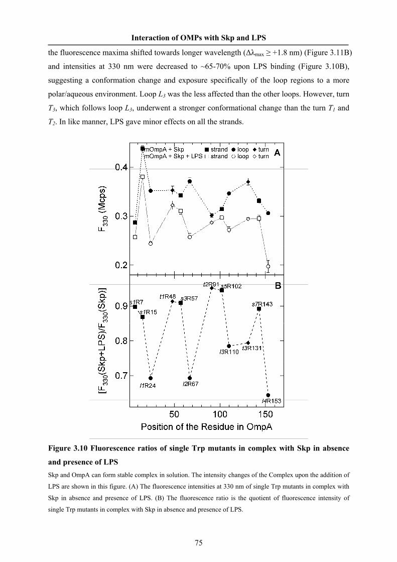
Interaction of OMPs with Skp and LPS
the fluorescence maxima shifted towards longer wavelength (Δλmax ≥ +1.8 nm) (Figure 3.11B)
and intensities at 330 nm were decreased to ~65-70% upon LPS binding (Figure 3.10B),
suggesting a conformation change and exposure specifically of the loop regions to a more
polar/aqueous environment. Loop L3 was the less affected than the other loops. However, turn
T3, which follows loop L3, underwent a stronger conformational change than the turn T1 and
T2. In like manner, LPS gave minor effects on all the strands.
Figure 3.10 Fluorescence ratios of single Trp mutants in complex with Skp in absence
and presence of LPS Skp and OmpA can form stable complex in solution. The intensity changes of the Complex upon the addition of
LPS are shown in this figure. (A) The fluorescence intensities at 330 nm of single Trp mutants in complex with
Skp in absence and presence of LPS. (B) The fluorescence ratio is the quotient of fluorescence intensity of
single Trp mutants in complex with Skp in absence and presence of LPS.
75

Interaction of OMPs with Skp and LPS
Figure 3.11 The wavelength changes of the single Trp mutants in complex with Skp
upon the addition of LPS (A) The wavelengths of fluorescence emission maxima λmax of single Trp mutants in presence of Skp and in
presence of both Skp and LPS. (B) The Δλmax is the difference of λmax of the Complex of mOmpA·Skp in
presence and absence of LPS.
76

Interaction of OMPs with Skp and LPS
The turn regions of OmpA remain tightly bound when LPS interacts with OmpA·Skp3
complexes
To characterize the impact of the presence of both, Skp and LPS, in complex with
OmpA, we compared the fluorescence spectra of single tryptophan mutants of OmpA in the
complex to the fluorescence spectra of OmpA in aqueous solution after urea dilution.
Fluorescence intensities at 330 nm and emission maxima λmax obtained from the spectra are
shown in Figure 3.12 and 3.13 as a function of the location of the mutated residue.
Tryptophans that were introduced into the periplasmic turns were most strongly influenced by
the presence of both, Skp and LPS, with wavelength shifts of the emission maxima in
between Δλmax ≈ 3.7 and 4.5 nm and relative fluorescence increases between 66 and 86 %. In
comparison, fluorescence emissions of mutants containing the single tryptophan either in the
β-strands or in the outer loops, were less strongly affected. Only residues of the N-terminal
first β-strand and first outer loop displayed stronger changes in presence of both, Skp and
LPS, with Δλmax ranging from ~3.8 nm to ~4.2 nm. The presence of LPS and Skp showed a
gradually decreasing effect on fluorescence spectra of tryptophans in strands 3 to 7 and in
loops 2 to 4. The shifts and the fluorescence ratios (FR330) decreased towards the C-terminus
and were only Δλmax ≈ 2.3 nm and ΔFR330 ≈ +33 to +44 % for s7W143 and l4W153, i.e.
about half of ΔFR330 observed for the periplasmic turns.
In comparison to binding of the single tryptophan mutants to Skp only (Figure 3.4B),
the fluorescence spectra in presence of both, Skp and LPS (Figure 3.13B) revealed that
addition of LPS to preformed Skp-OmpA complexes causes the most dramatic changes in the
loops of OmpA. In particular, binding of loops 1, 2, and 4 to Skp is strongly weakened upon
addition of LPS. The fluorescence of the tryptophans in these loops indicated a much more
polar environment after LPS binding. The effect of LPS addition on the β-strands and β-turns
of OmpA is comparably small. From the comparison of the data sets in Figures 3.4B and
3.13B, it is clear that in contrast to the long and flexible loops, the periplasmic turns remain
tightly associated with Skp when LPS is added. These data indicate a large change in
orientation and conformation of Skp-bound OmpA that is induced by the interaction with
LPS.
77

Interaction of OMPs with Skp and LPS
Figure 3.12 Fluorescence intensity changes of single Trp mutants of OmpA due to both
Skp and LPS (A) The fluorescence intensities at 330 nm of single Trp mutants in aqueous solution and in presence of both
Skp and LPS. (B) The fluorescence ratio is the quotient of fluorescence intensity of OmpA mutants at 330 nm in
absence and presence of both Skp and LPS.
78

Interaction of OMPs with Skp and LPS
Figure 3.13 The wavelength changes of the single Trp mutants of OmpA upon the
addition of both Skp and LPS This figure shows the changes of the wavelength of fluorescence emission maxima λmax of OmpA mutants after
both Skp and LPS were added. (A) The wavelengths of fluorescence emission maxima λmax of single Trp
mutants in aqueous solution and in presence of both Skp and LPS. (B) Δλmax is shown, which is the difference of
the wavelength of fluorescence emission maxima λmax in absence and presence of both Skp and LPS.
79

Interaction of OMPs with Skp and LPS
MTSSL quenching Trp fluorescence of double mutants only in DOPC
To explore formation of structure in OmpA upon binding to Skp, we used
intramolecular fluorescence quenching. Tryptophan fluorescence quenching by nitroxyl spin
labels is distance dependent [London et al. 1981; Chattopadhyay et al. 1987] and can be used
to monitor protein folding [Kleinschmidt et al. 2007]. We therefore prepared a second set of
OmpA mutants, based on several single-Trp mutants into which a single sulfhydryl-reactive
cysteine was introduced for site-directed spin-labeling. To prepare these mutants, site-
directed mutagenesis was first used to substitute the two native cysteines of the periplasmic
domain of OmpA by alanines. Based on the high-resolution structure of OmpA (1bxw), a
new single cysteine residue was then introduced, replacing a residue in a direct neighbor
strand in close proximity to the single tryptophan residue, also facing the outer surface of the
OmpA barrel. The fluorescence of the spin-labeled mutants is only quenched in later stages
of OmpA folding [Kleinschmidt et al. 2007]. We prepared 5 such single Trp/single Cys
mutants (WnCm mutants), which are shown in Table 3.3.
Figure 3.14 OmpA remains unfolded in complex with Skp and LPS Proximity of the single Trp and the single Cys residue in the mutant W15C162 was tested by intramolecular
fluorescence quenching via a cysteine-linked spin-label. Spectra of the spin-labeled mutant W15C162 in 8 M
urea (– – –), after 100-fold urea dilution in Tris buffer (· · · ·), in presence of a 4-fold molar excess of Skp (—
—)in presence of a 5-fold molar excess of LPS (– – –), at the same molar ratios of both, LPS and Skp (· · · ·)
and of W15C162 folded into DOPC bilayers are shown in panel B and D. For comparison, the spectra of the S-
methylated mutant W15C162 are shown under otherwise identical conditions in panels A and C.
80

Interaction of OMPs with Skp and LPS
To monitor the degree of folding of OmpA in 8 M urea, in water, in complex with
either Skp or LPS or both, and in lipid bilayers, we recorded the fluorescence spectra of spin-
labeled WnCm mutants. For reference, spectra were also recorded for all mutants in absence
of the fluorescence quenching spin-label under otherwise identical conditions. To avoid
unwanted disulfide dimerization of the mutants, the cysteines were methylated instead of
spin-labeled (Figure 3.14). The fluorescence spectra of W15C162 in 8 M urea, in aqueous
buffer, and in presence of either LPS or Skp or both, Skp and LPS were very similar to the
spectra of s1W15. There was no obvious difference (FR330 ≈ 1) between the fluorescence
intensities at 330 nm (F330) of S-methylated and spin-labeled WnCm mutants under these
conditions, indicating that OmpA did not fold to its native structure in the complexes with
either Skp or Skp and LPS. However, when these WnCm mutants were incubated with
preformed DOPC bilayers, the fluorescence intensities of the S-methylated WnCm mutants
were ~25% - 100% higher than those of the spin-labeled WnCm mutants, indicating folding
of OmpA into lipid membranes (Figure 3.15).
Figure 3.15 Skp and LPS do not induce native-like formation of antiparallel β-strands Structure formation in the transmembrane domain of OmpA was probed as described in the legend to Figure
3.14 for 4 additional WnCm mutants, namely W7C43, W7C170, W15C35, and W57C35. The fluorescence
intensity ratios of each mutant in methylated and in quencher-labeled form in 8 M urea, in Tris buffer, in
presence of 5-fold molar excess of LPS, 4-fold molar excess of Skp, and in presence of both, LPS and Skp, and
finally after folding into DOPC bilayers indicate formation of native antiparallel β-sheet structure only in lipid
bilayers.
81

Interaction of OMPs with Skp and LPS
3.5 Discussion
The periplasmic chaperones prevent the outer membrane proteins aggregation and
help them insert and fold into the outer membrane in the periplasm in absence of ATP
[Behrens 2003]. The periplasmic chaperone Skp assisting OmpA folding in presence of LPS
was presented by our group in 2003 [Bulieris et al. 2003]. Here our sequential works about
the interaction OmpA with Skp and LPS demonstrated some details of this pathway: i) Skp
binds to transmembrane domain of OmpA and changes its conformation; ii) LPS interacts
with the loops of OmpA; iii) LPS changes the conformation of OmpA·Skp3 complex.
Skp binds to coiled transmembrane domain of OmpA by multivalent interaction
In aqueous solution, OmpA formed coiled conformation to prevent from aggregation.
It kept the hydrophobic strands inside and the loops and the turns outside in more polar
environment (Figure 3.4A). λmax of PW263 was 334.7 nm in aqueous solution, suggesting the
periplasmic domain of OmpA was almost folded. The intrinsic fluorescence of Trp263 was
strongly quenched by the neighbor amino acids possibly by a disulfide group formed by two
native cysteines of the periplasmic domain.
Trimeric Skp binds unfolded OmpA and forms stable complex as 1:1 stoichiometry
[Bulieris et al. 2003]. Skp only binds to the transmembrane domain of OmpA, not
periplasmic domain [Chen et al. 1996; Qu et al. 2007]. We found that Skp almost hold the
entire transmembrane domain of OmpA, not only part of it. All the tryptophans, which are
located in the transmembrane domain, were in more hydrophobic environment after Skp
bound OmpA, suggesting these Trps were shielded by Skp. The volume of the cavity of Skp
which contains three flexible tentacles is ~33,000 Å3 which could accommodate a folded
protein of ~25 kDa [Walton et al. 2004]. The transmembrane domain of OmpA is only 19
kDa. Considering the significant conformational flexible, Skp could occupy the substrate
which is much bigger than 25 kDa.
The formation of the β-barrel of OmpA resulted in the MTSSL quenching. The
quenching results (Figure 3.15) indicated that OmpA didn’t form any tertiary structure when
OmpA formed the complex with Skp or both Skp and LPS. Circular dichroism had shown
that OmpA developed minor amounts of secondary structure in the complex of OmpA and
Skp [Bulieris et al. 2003]. When Skp bound to the transmembrane domain of OmpA, it was
82

Interaction of OMPs with Skp and LPS
still in a coiled state. It may form a globular coil to fit the cavity of Skp. However, the
conformation of the transmembrane domain was different from it in aqueous solution
according to the hydrophobicity of the residues. The relatively hydrophobicity of residues 91
to 153 was altered totally (Figure 3.4A). Prefoldin (PFD), which has similar crystal structure
and function with Skp, was confirmed recently by electron microscopy that it binds to the
globular unfolded substrates [Martin-Benito et al. 2007]. In sharp contrast with Skp and PFD,
chaperone Dnak and SurA prefer to bind linear polypeptide [Zhu et al. 1996; Hennecke et al.
2005]. The crystal structures of these four chaperones revealed the reason that both Skp and
PFD have much larger cavities than DnaK and SurA (PDB ID for Skp:1SG2; PFD:1FXK;
DnaK:1DXk; SurA:1M5Y).
Figure 3.4, 3.5, 3.12 and 3.13 indicated that the interaction between Skp and OmpA
was multivalent. The strong binding sites were located in the loops and turns of OmpA, not
on the β-strands, suggesting that β-strand sequence motif was not the specific recognition
motif for Skp. Our previously result had demonstrated that the hydrophobic force played a
key role on the binding of Skp to the substrate and electrostatic interaction were also involved
in the binding [Qu et al. 2007]. The transmembrane domain of OmpA containing large
quantity of hydrophobic residues and three hydrophobic patches in the cavity of Skp resulted
in a multivalent hydrophobic interaction. This kind of multivalent interaction was found very
recently in Prefoldin [Martin-Benito et al. 2007]. Prefoldin has six hydrophobic patches on
the tips of its tentacles [Lundin et al. 2004] and the substrates interact with several tentacles
of PFD upon their masses [Martin-Benito et al. 2007]. The hydrophobic patches of Skp are
close to the middle of the tentacles in the interior surface [Korndörfer et al. 2004]. The
substrates of Skp have to enter into the cavity and interact with the hydrophobic patches.
Besides, positive charged Skp under neutral pH could interact with negative charged regions
of OmpA. We assumed in previous work [Qu et al. 2007] that the turns of OMPs that often
contained aspartate or glutamate, interacted with the most basic regions in Skp. Now we have
further experimental results from OmpA to support this assumption. In the strong binding
regions of OmpA, turn T2 and T3 are more negative charged than other regions. These two
regions should interact with the positive charged tips of Skp and contain specific recognition
motif for Skp. Further, the negative charged turn regions of OMPs contain specific
recognition motif for Skp binding.
LPS interacts with the loop regions of OmpA
83

Interaction of OMPs with Skp and LPS
LPS was found to interact with OmpA in 1978 [Schweizer et al. 1978]. Lipid A
moiety alone can renature the unfolded OmpA [Dornmair et al. 1990]. Our works showed for
the first time that LPS interacts with the loop regions of OmpA, not with the turn regions of
OmpA (Figure 3.7 and 3.8). The loop L1 and L3 regions of OmpA contain 4 and 3 consecutive
hydrophobic residues respectively. The hydrophobic force may drive the hydrophobic lipid A
to interact with these two hydrophobic regions. The loop L1 region was confirmed acting as
bacteriophage receptor site [Morona et al. 1985] and phage requires LPS fro activity [Mutoh
et al. 1978]. Thereby, this interaction may still function as the phage receptor site even after
OmpA assembles into the outer membrane [Puspurs et al. 1983]. Besides, the hydrophobic
interaction should give some help for OmpA assembly.
LPS may is the trigger for the Skp-assisting folding pathway
LPS decreased the fluorescence ratio and shifted λmax of the OmpA·Skp3 complex
towards higher wavelength, suggesting that LPS weakened the interaction of OmpA with Skp
and exposed the tryptophans to the polar/external environment. The results of single Trp
mutants is consistent with same experiments that we performed by using WT-OmpA before
[Qu et al. 2007]. In Figure 3.10 and 3.11, the concrete changes were that LPS loosened the
interaction between the loops of OmpA and Skp and exposed them to the external
environments (water or the outer membrane). This adjustment should be indispensable to
membrane insertion and folding of OmpA because the insertion of OmpA began from the
loop direction. LPS did not compete for binding to OmpA with Skp and released OmpA from
Skp. LPS only binds to Skp and weakens the interaction between Skp and OmpA because the
LPS binding sites on Skp is very close to the hydrophobic patch in the cavity of Skp
[Korndörfer et al. 2004; Walton et al. 2004]. This kind of loose binding enable the
chaperone to help the proteins fold [Jewett et al. 2006].
Now I can give the reasons that Skp alone inhibits the insertion and folding of OmpA.
One is the binding is so tightly that OmpA is not easy to release from Skp [Qu et al. 2007].
Another is OmpA is not in the right orientation for the insertion and folding. LPS binding to
OmpA·Skp3 complex brings on the conformational changes of both Skp and OmpA. The
conformational change was also found in the other chaperone-assisting folding pathway, such
as GroEL-GroES [Weissman et al. 1996] and DnaK-DnaJ [Moro et al. 2006]. For the ATP-
dependent chaperones, the binding of ATP triggers a critical conformational change leading
to release of the bound substrate protein [Fink 1999]. LPS may play a similar role with ATP
84

Interaction of OMPs with Skp and LPS
as a trigger during the Skp-assisting folding pathway. In summary, one work model for Skp
and LPS assisting OmpA in traversing the periplasm was presented in Figure 3.16.
Figure 3.16 Scheme for OmpA traversing the periplasm assisted by Skp and LPS (A) Unfolded OmpA in 8 M urea forms a random coil structure. The black line represents the transmembrane
domain of OmpA and the gray line represents the periplasmic domain of OmpA. (B) The transmembrane
domain of OmpA forms a water-soluble intermediate state in water. (C) Skp binds the entire transmembrane
domain of OmpA. (D) LPS interacts with OmpA·Skp3 complexes in the loop regions of OmpA and weakens the
interaction between OmpA and Skp. The turn regions of OmpA remains tightly bound to Skp. (E) LPS orients
the conformation of OmpA and orients the loops of OmpA towards the aqueous space. Skp and LPS together
therefore induce a preferred direction of OmpA for insertion into lipid bilayers.
85
Interaction of OMPs with Skp and LPS
4 Orientation of β-barrel proteins OmpA and FhuA in lipid
membranes. Chainlength dependence from infrared
dichroism
4.1 Abstract
The outer membrane proteins OmpA and FhuA of E. coli are monomeric β-barrels of
widely differing size. Polarised attenuated total reflection infrared spectroscopy has been
used to determine the orientation of the β-barrels in phosphatidylcholine host matrices of
different lipid chainlengths. The linear dichroism of the amide I band from OmpA and FhuA
in hydrated membranes generally increases with increasing chainlength from diC12:0 to
diC17:0 phosphatidylcholine, in both the fluid and gel phases. Measurements of the amide I
and amide II dichroism from dry samples are used to deduce the strand tilt (β = 46° for
OmpA, and β = 44.5° for FhuA). These values are then used to deduce the order parameters,
⟨P2(cosα)⟩, of the β-barrels from the amide I dichroic ratios of the hydrated membranes. The
orientational ordering of the β-barrels, and their assembly in the membrane, are discussed in
terms of hydrophobic matching with the lipid chains.
86

Interaction of OMPs with Skp and LPS
4.2 Introduction
The integral proteins of the outer membrane of Gram-negative bacteria perform a
variety of functions, including those of receptors, lipases, passive diffusion pores, sugar
transport pores, and ligand-gated transport systems. In spite of the diversity in function, the
outer-membrane proteins are mainly, if not exclusively, of the β-barrel type: the second
structural paradigm, in addition to the α-helix, for transmembrane proteins. The size of the β-
barrel ranges from 8 to 22 β-strands, the shear number of the barrel ranges from 8 to 24, and
the degree of oligomerisation varies from monomer to trimer (for a review, see ref. [Schulz
2002]. The β-strands are tilted, relative to the barrel axis by 36° to 42° [Páli et al. 2001]. This
structural information is obtained mostly from x-ray diffraction of crystals produced from
purified proteins in detergent [Cowan et al. 1992; Weiss et al. 1992a; Kreusch et al. 1994a;
Schirmer et al. 1995; Meyer et al. 1997; Ferguson et al. 1998b; Forst et al. 1998; Buchanan
et al. 1999b; Dutzler et al. 1999; Snijder et al. 1999; Vogt et al. 1999b; Pautsch et al. 2000],
and to a much lesser extent from solution-state NMR of proteins in detergent micelles [Arora
et al. 2001b; Fernandez et al. 2001; Fernandez et al. 2002; Hwang et al. 2002]. These
environments approximate those of the lipid bilayer in biological membranes, but do not
provide information on the orientation of the β-barrel proteins in the membrane. The latter
feature not only determines the way in which the protein is integrated into the membrane, but
also might be responsible at least partly for the functional differentiation between the various
β-barrel proteins.
In principle, the dichroism of the amide bands in the infrared spectra from aligned
membranes can be used to determine both the β-strand tilt and the orientation of the β-sheet
plane or β-barrel axis [Marsh 1997, 1998]. Combination of the dichroism from amide I and
amide II bands allows determination of both orientational parameters. If the strand tilt is
known from x-ray diffraction, only one dichroic ratio is required to determine the orientation
of the β-sheet or β-barrel. Several previous analyses of infrared dichroism from β-barrel
membrane proteins have not exploited this possibility [Nabedryk et al. 1988; Goormaghtigh
et al. 1990; Rodionova et al. 1995]. Recently, this method of analysis has been applied
successfully to ATR-FTIR studies on two isoforms of the VDAC voltage-dependent anion-
selective channel from the outer membrane of mitochondria [Abrecht et al. 2000]. In the
present work, we investigate the dependence on lipid chainlength of the orientational tilt of
87

Interaction of OMPs with Skp and LPS
two β-barrel transmembrane proteins with very different sizes. One of these outer membrane
proteins from E. coli is OmpA, which has 8 β-strands; the other is the β-barrel domain of the
ferrichrome-iron receptor FhuA mutant without the cork domain, which has 22 β-strands.
Both proteins are monomers and therefore are those most likely to respond to the lipid
environment. The β-barrel orientation displays a characteristic dependence on the lipid
chainlength that is determined by the hydrophobic span of the outer membrane proteins and
correlates well with the lipid requirement for spontaneous membrane insertion of OmpA
[Kleinschmidt et al. 2002].
88

Interaction of OMPs with Skp and LPS
4.3 Theoretical background
The order parameter, ⟨P2(cosα)⟩, for the axis of a β-barrel protein with circular cross-
section is related to the amide band dichroic ratio, R(Θ), by [Tamm et al. 1997; Marsh 1998]:
( )[ ]22222
2222
2 /2)(/cosP/)(/
)(cosPyzyx
yzyx
EEREEEEREE
−Θ−Θ+Θ−
=α (Eq. 4.1)
where Ex, Ey, Ez are the components of the electric field vector in the sample, normalised to
those of the incident infrared beam. The z-axis is normal to the ATR plate; the x-axis lies in
the plane of incidence, and the y-axis is orthogonal to the latter. The order parameter is
defined in terms of the Legendre polynomial: ( )1cos3)(cosP 221
2 −= αα , where angular
brackets indicate an ensemble average over all amides. The orientation of the amide transition
moment, relative to the β-strand axis, is given by Θ = 90°-β for the amide I band, and by Θ =
β for the amide II band, where β is the tilt angle of the strands to the barrel axis (see left-hand
panel of Figure 4.1). The angle α is the inclination of the barrel axis to the normal to the
orienting substrate (i.e., the ATR crystal), and the order parameter is determined by the
distribution in α. In the case of partial disorder in alignment of the sample on the substrate,
the order parameter, ⟨P2(cosα)⟩, is the product of the order parameter, ⟨P2(cosα)⟩o, of the
barrel axis relative to the membrane normal, and an order parameter, Sdisorder, that
characterises the misalignment of the membrane. For a model in which a fraction (1-f) of the
sample is totally disordered, and the remainder is perfectly ordered: ⟨P2(cosα)⟩ = f⟨P2(cosα)⟩o
[Fraser 1953].
The dichroism of a flattened β-barrel, or of β-strands extending beyond the barrel
domain, may be depicted better in terms of a planar β-sheet. For the latter, the dichroic ratio
of the amide I band is given by [Marsh 1997]:
2
2
22
22
2
2
sincos1
sincos2
y
z
y
xI E
EEER
βα
βα
−+= (Eq. 4.2)
and that of the amide II band by:
89

Interaction of OMPs with Skp and LPS
2
2
22
22
2
2
coscos1
coscos2
y
z
y
xII E
EEER
βα
βα
−+= (Eq. 4.3)
where α is the inclination of the β-sheet to the normal to the orienting substrate, and β is the
tilt of the β-strands within the β-sheet (see right-hand panel of Figure 4.1). For a partially
oriented sample:
3/)1(coscos 22 ffo
−+= αα (Eq. 4.4)
where (1-f) is the fraction unoriented, and ⟨cos2α⟩o is referred to the membrane normal, just
as for the β-barrel case.
Figure 4.1 Orientation of transition moments, ΘM, in β-sheets (right) and β-barrels (left) The β-strand tilt is β; the tilt of the barrel or sheet axis, relative to the membrane normal (z), is α. For the amide I band: ΘI = 90°-β, and for the amide II band: ΘII = β.
90
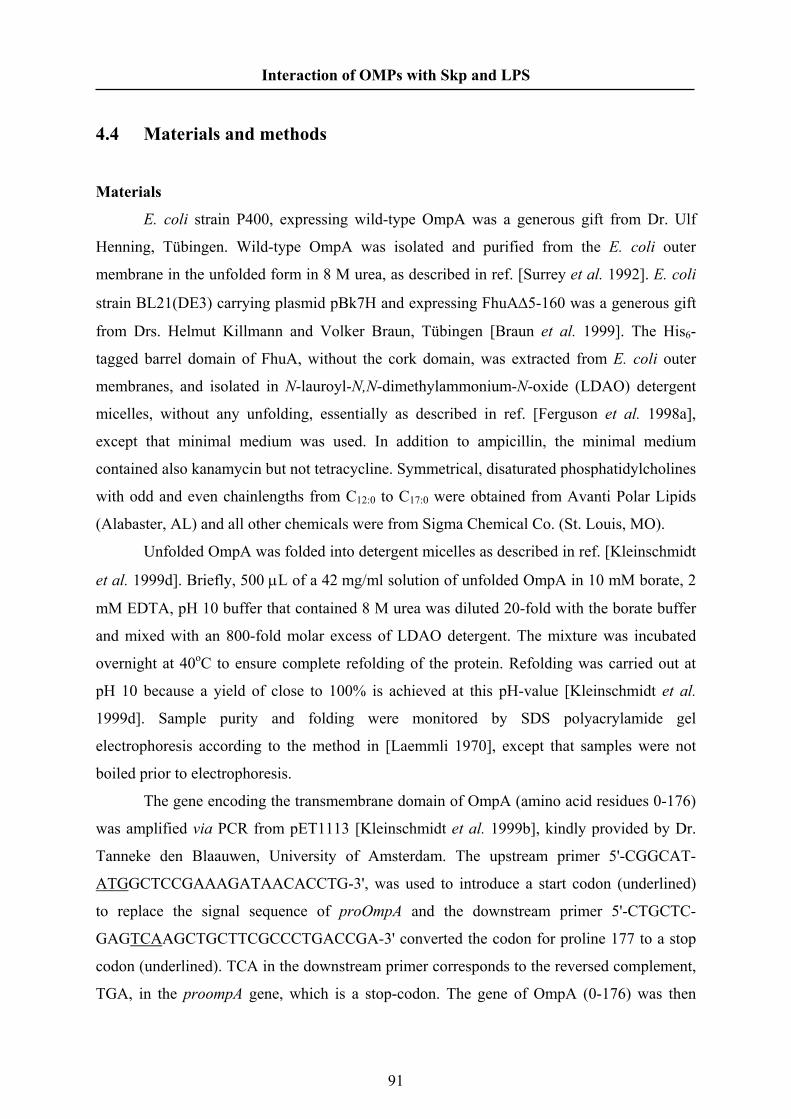
Interaction of OMPs with Skp and LPS
4.4 Materials and methods
Materials
E. coli strain P400, expressing wild-type OmpA was a generous gift from Dr. Ulf
Henning, Tübingen. Wild-type OmpA was isolated and purified from the E. coli outer
membrane in the unfolded form in 8 M urea, as described in ref. [Surrey et al. 1992]. E. coli
strain BL21(DE3) carrying plasmid pBk7H and expressing FhuAΔ5-160 was a generous gift
from Drs. Helmut Killmann and Volker Braun, Tübingen [Braun et al. 1999]. The His6-
tagged barrel domain of FhuA, without the cork domain, was extracted from E. coli outer
membranes, and isolated in N-lauroyl-N,N-dimethylammonium-N-oxide (LDAO) detergent
micelles, without any unfolding, essentially as described in ref. [Ferguson et al. 1998a],
except that minimal medium was used. In addition to ampicillin, the minimal medium
contained also kanamycin but not tetracycline. Symmetrical, disaturated phosphatidylcholines
with odd and even chainlengths from C12:0 to C17:0 were obtained from Avanti Polar Lipids
(Alabaster, AL) and all other chemicals were from Sigma Chemical Co. (St. Louis, MO).
Unfolded OmpA was folded into detergent micelles as described in ref. [Kleinschmidt
et al. 1999d]. Briefly, 500 μL of a 42 mg/ml solution of unfolded OmpA in 10 mM borate, 2
mM EDTA, pH 10 buffer that contained 8 M urea was diluted 20-fold with the borate buffer
and mixed with an 800-fold molar excess of LDAO detergent. The mixture was incubated
overnight at 40oC to ensure complete refolding of the protein. Refolding was carried out at
pH 10 because a yield of close to 100% is achieved at this pH-value [Kleinschmidt et al.
1999d]. Sample purity and folding were monitored by SDS polyacrylamide gel
electrophoresis according to the method in [Laemmli 1970], except that samples were not
boiled prior to electrophoresis.
The gene encoding the transmembrane domain of OmpA (amino acid residues 0-176)
was amplified via PCR from pET1113 [Kleinschmidt et al. 1999b], kindly provided by Dr.
Tanneke den Blaauwen, University of Amsterdam. The upstream primer 5'-CGGCAT-
ATGGCTCCGAAAGATAACACCTG-3', was used to introduce a start codon (underlined)
to replace the signal sequence of proOmpA and the downstream primer 5'-CTGCTC-
GAGTCAAGCTGCTTCGCCCTGACCGA-3' converted the codon for proline 177 to a stop
codon (underlined). TCA in the downstream primer corresponds to the reversed complement,
TGA, in the proompA gene, which is a stop-codon. The gene of OmpA (0-176) was then
91

Interaction of OMPs with Skp and LPS
ligated into pET22b (Novagen) using the NdeI and XhoI restriction enzyme cut sites to yield
the plasmid pET22bB1. The plasmid was transformed into E. coli strain BL21(DE3) ΔlamB
ΔompF::Tn5 ΔompA ΔompC ΔfhuA [Prilipov et al. 1998] using the protocol “One-step
preparation and transformation of competent cells” [Ausubel et al. 1999]. OmpA (0-176) was
subsequently purified as described in refs. [Surrey et al. 1992] and [Pautsch et al. 1999].
For refolding of the transmembrane domain of OmpA, 12 mg of denatured OmpA(0-
176) were diluted into 20 mL 25 mM LDAO in 10 mM borate buffer containing 2 mM
EDTA (pH 10). The proteins were incubated at 40 °C for 12 hours. The yield of refolding
was 100% as confirmed by SDS-PAGE and CD spectroscopy. In SDS-PAGE, OmpA0-176
displayed the typical band-shift from 19 kDa (unfolded) to 23 kDa (folded form) as observed
previously [Pautsch et al. 1999], if samples were not boiled prior to electrophoresis. The full-
length wt-OmpA similarly displayed a band-shift from 35 kDa to 30 kDa in SDS-PAGE as
described previously [Surrey et al. 1992], if samples were not heat-denatured prior to
electrophoresis. Folding was quantitative, because the bands for the unfolded proteins
disappeared after refolding, but appeared again, when samples were boiled (heat-denatured)
in SDS prior to electrophoresis. After refolding, OmpA was concentrated 20-fold in an
Amicon ultrafiltration chamber using Amicon YM-10 membranes.
Reconstitution into membranes
Lipid stock solutions were prepared in CHCl3 and dried under a stream of dry
nitrogen gas. The resulting lipid film was desiccated overnight under vacuum and then
covered with argon. The dry lipid film was hydrated with 10 mM Hepes, 2 mM EDTA buffer,
pH 7.0, and frozen (in liquid nitrogen) and thawed (in a water bath at ~5oC above the
transition temperature of the phospholipid) seven times, to obtain uniform lipid vesicles.
The reconstitution was carried out by mixing 1 mg of the above lipid vesicles with
protein to the desired lipid-protein ratio, and then 10% sodium cholate solution was added to
give a final concentration of 0.2% sodium cholate, in a total volume of 500 µL. The sample
was mixed well and incubated at room temperature for 1 hour, vortexing from time to time.
After 1 hour incubation, the protein was precipitated with 52.5% ammonium sulfate solution
that was added to 35% final concentration. The pellet containing the reconstituted protein was
then centrifuged for 30 minutes at 35000 rpm in a 50TI rotor. The pellet contains the
reconstituted sample and, because the outer membrane proteins have a low density, the
reconstituted membrane pellets tend to float when centrifuged. This decreases the protein and
lipid content and invariably changes the lipid-protein (L/P) ratios from the expected range.
92

Interaction of OMPs with Skp and LPS
L/P ratios were therefore determined after ammonium-sulfate precipitation [Lowry et al.
1951; Rouser et al. 1970], and then the samples were adjusted to achieve a L/P ratio close to
50 mol/mol by adding solubilised lipid vesicles. The pellet was dissolved and vortexed in 500
µL 10 mM Hepes buffer pH 7.0, containing 2 mM EDTA and 250 mM NaCl. Detergent
removal was achieved by extensive dialysis at 8°C against 10 mM Hepes buffer pH 7.0
containing 2 mM EDTA and 250 mM NaCl, using 10-kDa cut-off dialysis membranes. Four
or five changes of 2 L buffer were made every 7-8 h, with the last dialysis step overnight.
ATR spectroscopy
OmpA was purified and reconstituted as described above. The reconstituted sample
was layered on a clean ZnSe ATR crystal. Initially, the sample was dried with dry nitrogen
purge and then desiccated overnight with a vacuum pump. The dry lipid film was incubated
in a Bruker IFS 25 FTIR spectrometer by purging with dry nitrogen at 1.5 kp/cm2 flow rate.
ATR spectra were recorded at a nominal resolution of 2 cm-1 with parallel and perpendicular
polarisation of the incident beam. Then the sample was hydrated with 10 mM Hepes pH 7.0
buffer containing 2 mM EDTA and 250 mM NaCl prepared in D2O, and washed (using a
pipette) again with the same buffer in order to remove noninserted lipid and protein
[Rodionova et al. 1995]. Meanwhile, ATR spectra were recorded at both polarisations for
every wash. The sample holder was temperature controlled using a recirculating water bath
and the spectra were recorded in the gel phase (~10o below the lipid chain-melting
temperature) and in the fluid phase (~10o above the lipid chain-melting temperature) of the
membranes. Further details of the ATR spectroscopy are given in ref. [Kóta et al. 2004].
To determine the dichroic ratio each spectrum was analysed using OPUS-Version
3.02 software from Bruker (Karlsruhe, Germany). If necessary, spectra were smoothed with
11-point Savitsky-Golay smoothing to remove the noise from the residual water vapour. A
local baseline was established and curve fitting using the Levenberg- Marquardt algorithm
was carried out from 1500 to 1708 cm-1 and from 1600 to 1700 cm-1, for dry and hydrated
samples, respectively. The intensity ratios of parallel and perpendicular polarisation were
taken as ATR dichroic ratio (R) from the intensities obtained at 1630 cm-1 and 1530 cm-1
from the curve fitting analysis, for the amide I and amide II bands, respectively. The dichroic
ratio changes with the first two to three washes and then becomes stable, which is taken to
correspond to the respective hydrated samples. To calculate molecular orientations from
dichroic ratios, intensities of the infrared electric field components were obtained from the
93

Interaction of OMPs with Skp and LPS
thick film approximation: = 0.450 and = 1.550 for a ZnSe ATR crystal (see
e.g. ref. [Marsh 1999b].
22 / yx EE 22 / yz EE
94
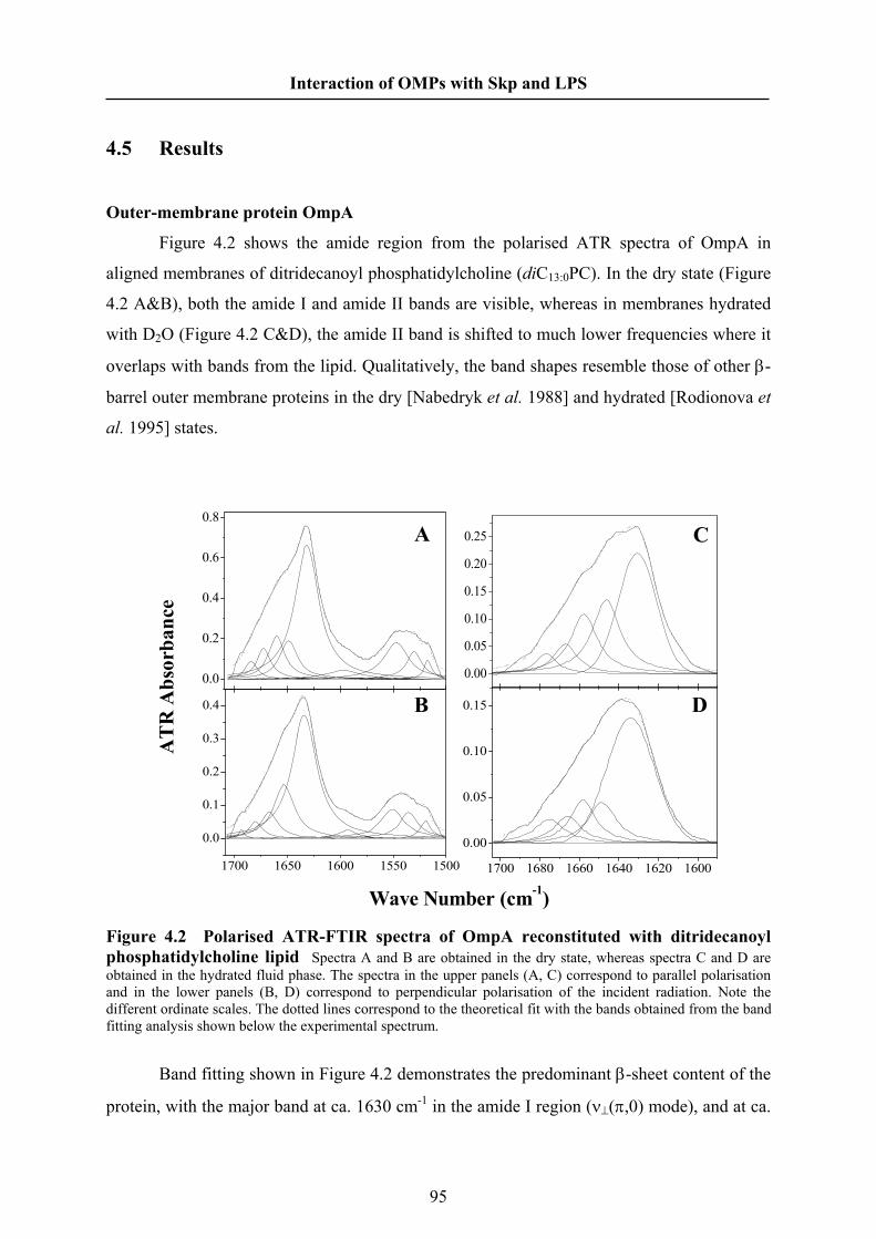
Interaction of OMPs with Skp and LPS
4.5 Results
Outer-membrane protein OmpA
Figure 4.2 shows the amide region from the polarised ATR spectra of OmpA in
aligned membranes of ditridecanoyl phosphatidylcholine (diC13:0PC). In the dry state (Figure
4.2 A&B), both the amide I and amide II bands are visible, whereas in membranes hydrated
with D2O (Figure 4.2 C&D), the amide II band is shifted to much lower frequencies where it
overlaps with bands from the lipid. Qualitatively, the band shapes resemble those of other β-
barrel outer membrane proteins in the dry [Nabedryk et al. 1988] and hydrated [Rodionova et
al. 1995] states.
0.00
0.05
0.10
0.15
0.20
0.25
1700 1680 1660 1640 1620 1600
0.00
0.05
0.10
0.15
1700 1650 1600 1550 1500
0.0
0.1
0.2
0.3
0.4
0.0
0.2
0.4
0.6
0.8
AT
R A
bsor
banc
e
Wave Number (cm-1)
D
C
B
A
Figure 4.2 Polarised ATR-FTIR spectra of OmpA reconstituted with ditridecanoyl phosphatidylcholine lipid Spectra A and B are obtained in the dry state, whereas spectra C and D are obtained in the hydrated fluid phase. The spectra in the upper panels (A, C) correspond to parallel polarisation and in the lower panels (B, D) correspond to perpendicular polarisation of the incident radiation. Note the different ordinate scales. The dotted lines correspond to the theoretical fit with the bands obtained from the band fitting analysis shown below the experimental spectrum.
Band fitting shown in Figure 4.2 demonstrates the predominant β-sheet content of the
protein, with the major band at ca. 1630 cm-1 in the amide I region (ν⊥(π,0) mode), and at ca.
95

Interaction of OMPs with Skp and LPS
1530-1550 cm-1 in the amide II region (ν//(0,π) mode). The minor band at ca. 1675 cm-1 in the
amide I region (v//(0,π) mode) from hydrated membranes is characteristic of antiparallel β-
sheets [Miyazawa 1960].
Table 4.1 gives the results of band fitting for the amide I region from OmpA in
disaturated phosphatidylcholines of different chain lengths. Fitting data are given for
hydrated membranes in the gel and fluid phases, recorded at temperatures 10° C below and
10° C above the respective chain-melting transitions. Transition temperatures for diC12:0,
diC13:0, diC14:0, diC15:0 and diC17:0PCs are: -2, 14, 23, 34 and 48 °C, respectively [Marsh
1990].
To obtain the true absorbed intensity that reflects relative populations in aligned
samples, it is necessary to combine absorbances, A// and A⊥, for parallel and perpendicular
polarised radiation [Marsh 1999a]. The appropriate admixture with A⊥ in the present
geometry is ( ) ⊥−+ AEEEEA yxyz2222
// //2 . This is used to calculate the percentage populations
that are presented in Table 4.1. Generally speaking, the populations are rather similar in the
different lipid hosts and in the fluid and gel phases. There is, however, a tendency to a
decrease in β-sheet population at shorter chain lengths, with a corresponding increase in the
1645 cm-1 band corresponding to disordered structures. With the exception of diC12:0PC, the
mean β-sheet population of OmpA in the different lipid hosts is 59 ± 6%, from Table 4.1 for
hydrated membranes. For comparison, the β-sheet content of OmpA truncated to residues 22-
192 is 63% from the crystal structure [Pautsch et al. 2000]; PDB:1QJB). This is similar to the
values obtained in the longer lipid hosts. The full-length protein has 325 residues. Therefore
an appreciable part (ca. 55%) of the sizeable periplasmic domain must also be β-sheet. For
comparison, the resolved 127 residues from the homologous C-terminal domain of RmpM
(which shares 35% sequence identity with the periplasmic C-terminal domain of OmpA)
contains 25% β-strands and 25% β-turns [Grizot et al. 2004].
From the dichroic ratios of the amide I and amide II bands of the dry sample, it is
possible to determine the tilt, β, of the β-strands within the β-sheet. From equation. 4.2 and
4.3 for a β-sheet one obtains a consistent mean value of β = 46.2 ± 0.8° (N = 5) from
measurements in disaturated phosphatidylcholines of different chainlengths from C12 to C17.
Analysis according to equation 4.1 for axially symmetric barrels yields a similar value for the
strand tilt: β = 48° ± 5°, averaged over membranes with host lipids of different chainlengths.
96

Interaction of OMPs with Skp and LPS
For the barrel domain alone of OmpA, a somewhat smaller value of β = 43.9 ± 0.9° (N = 5) is
obtained for the mean strand tilt.
Table 4.1 Band fitting of the polarised ATR spectra from the amide I band of OmpA in hydrated disaturated phosphatidylcholines of different chainlengths, C(n:0), in the gel and fluid phases
C(n:0) Gel Fluid position (cm-1) normalised area (%)a position (cm-1) normalised area (%)a
C(12:0) 1631 1645 1653 1663 1674
41 36 16
5 2
C(13:0) 1631 1646 1657 1667 1676
42 30 18
7 3
1633 1645 1658 1666 1676
49 18 17
9 7
C(14:0) 1631 1647 1657 1665 1673
58 19 14
7 2
1632 1645 1653 1663 1672
59 10 18 10
3
C(15:0) 1632 1645 1653 1663 1673
56 10 19 11
4
1633 1646 1655 1663 1672
58 14 15 10
3
C(17:0) 1633 1645 1655 1664 1673
63 7
15 11
4
1633 1652 1663 1672 1682
56 28 10
5 1
aRelative band intensities are obtained by combining integrated absorbances, A// and A⊥, with radiation polarised parallel and perpendicular, respectively, to the plane of the incident beam. The appropriate combination that reflects the full intensity is ( ) ⊥−+ AEEEEA yxyz
2222// //2 [Marsh 1999a].
The upper panel of Figure 4.3 shows the dichroic ratios of the amide I band from
OmpA in bilayer membranes hydrated in D2O, as a function of chain length of the disaturated
97
Interaction of OMPs with Skp and LPS
phosphatidylcholine. Values are given for membranes in the gel phase and for those in the
fluid phase, above the lipid chain-melting temperature.
1.6
1.7
1.8
1.9
2.0
12 13 14 15 16 17
1.6
1.8
2.0
2.2
2.4
2.6
2.8
wt-OmpA
Gel Fluid
Fhu-A Δ5-160 Gel Fluid
chainlength, n-PtdCho
dich
roic
ratio
Figure 4.3 Dependence of the dichroic ratios from the amide I band of OmpA (upper panel) and of FhuA barrel domain (lower panel) on chainlength, n, of the disaturated phosphatidylcholine membrane in which the protein is incorporated. Dichroic ratios are given for samples in the gel phase (squares) and in the fluid phase (circles). Membranes are hydrated in D2O buffer.
The general trend is an increase in amide I dichroic ratio with increasing lipid chain
length. Use of equation 4.2, together with the mean value of the strand tilt, β, deduced from
the dichroic ratio of the dry sample, yields the values for the order parameter, ⟨P2(cosα)⟩, and
mean tilt, α, of the β-sheets that are given in Table 4.2. The trend is similar to that of the
dichroic ratios: the order parameters increase, and the tilt angles decrease, with increasing
lipid chainlength. The differences in order parameters between gel- and fluid-state
membranes are not large.
98

Interaction of OMPs with Skp and LPS
Previous ATR measurements on full-length OmpA in single supported bilayers of
diC14:0PC at room temperature [Rodionova et al. 1995] yield comparable degrees of order
when analysed by the present methods (⟨P2(cosα)⟩ = 0.35-0.38). Also measurements on
diC16:0PC from the same work, are consistent with the present observed trends with
chainlength for phosphatidylcholines in the gel phase.
Table 4.2 Order parameters, ⟨P2(cosα)⟩, and mean effective inclinations, α, of the β-sheets of OmpA reconstituted in disaturated phosphatidylcholines, diC(n:0)PC, with different chainlengths, n, in the gel and fluid phases
C(n:0) Gel Fluid ⟨P2(cosα)⟩ α (°) ⟨P2(cosα)⟩ α (°)
C(12:0) - - 0.26 (0.25)b 45 (45)b
C(13:0) 0.32 (0.35)b 42 (41)b 0.34 (0.30)b 42 (43)b
C(14:0)a 0.40 (0.52)b 39 (35)b 0.39 (0.53)b 40 (34)b
C(15:0) 0.39 (0.51)b 40 (35)b 0.42 (0.57)b 38 (32)b
C(17:0) 0.43 (0.62)b 38 (30)b 0.42 (0.61)b 38 (30)b
aFor full-length OmpA, data obtained with 10% dimyristoylphosphatidylglycerol in dimyristoylphosphatidylcholine host matrix. bValues in parentheses are obtained with the barrel domain of OmpA (residues 0-176).
Similar conclusions to those drawn from Table 4.2 can also be deduced by analysing
the dichroic ratios according to equation 4.1 for cylindrical β-barrels. The net values for the
tilt, α, of the barrel are considerably larger, however, than those given in Table 4.2. Note that
these values include the tilt of the β-strands in the periplasmic domains, in addition to that of
the transmembrane β-barrel.
Experiments were also performed with a truncated form of OmpA, consisting of
residues 0-176, that corresponds essentially to the transmembrane barrel domain. Order
parameters and effective values for the tilt of the barrel axis are given by the values in
parentheses in Table 4.2. The order parameters are higher (and the tilt angles corresponding
are lower) for the barrel domain of OmpA than for the full-length protein, at least in the lipids
of longer chainlengths. The effective tilt angles, θ, of the lipid chains, relative to the substrate
normal, are given in Table 4.3.
99
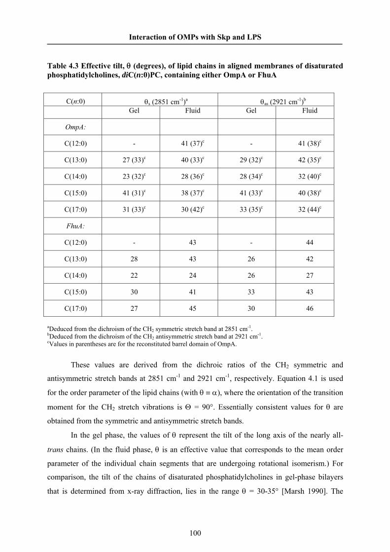
Interaction of OMPs with Skp and LPS
Table 4.3 Effective tilt, θ (degrees), of lipid chains in aligned membranes of disaturated phosphatidylcholines, diC(n:0)PC, containing either OmpA or FhuA
C(n:0) θs (2851 cm-1)a θas (2921 cm-1)b
Gel Fluid Gel Fluid
OmpA:
C(12:0) - 41 (37)c - 41 (38)c
C(13:0) 27 (33)c 40 (33)c 29 (32)c 42 (35)c
C(14:0) 23 (32)c 28 (36)c 28 (34)c 32 (40)c
C(15:0) 41 (31)c 38 (37)c 41 (33)c 40 (38)c
C(17:0) 31 (33)c 30 (42)c 33 (35)c 32 (44)c
FhuA:
C(12:0) - 43 - 44
C(13:0) 28 43 26 42
C(14:0) 22 24 26 27
C(15:0) 30 41 33 43
C(17:0) 27 45 30 46
aDeduced from the dichroism of the CH2 symmetric stretch band at 2851 cm-1. bDeduced from the dichroism of the CH2 antisymmetric stretch band at 2921 cm-1. cValues in parentheses are for the reconstituted barrel domain of OmpA.
These values are derived from the dichroic ratios of the CH2 symmetric and
antisymmetric stretch bands at 2851 cm-1 and 2921 cm-1, respectively. Equation 4.1 is used
for the order parameter of the lipid chains (with θ ≡ α), where the orientation of the transition
moment for the CH2 stretch vibrations is Θ = 90°. Essentially consistent values for θ are
obtained from the symmetric and antisymmetric stretch bands.
In the gel phase, the values of θ represent the tilt of the long axis of the nearly all-
trans chains. (In the fluid phase, θ is an effective value that corresponds to the mean order
parameter of the individual chain segments that are undergoing rotational isomerism.) For
comparison, the tilt of the chains of disaturated phosphatidylcholines in gel-phase bilayers
that is determined from x-ray diffraction, lies in the range θ = 30-35° [Marsh 1990]. The
100

Interaction of OMPs with Skp and LPS
values of θ that are found for the gel-phase membranes in Table 4.3 are of a similar size and
therefore suggest that the OmpA-containing membranes are reasonably well aligned.
Outer-membrane iron siderophore receptor FhuA
Figure 4.4 shows the polarised ATR spectra of the FhuA barrel domain in aligned
membranes of dipentadecanoyl phosphatidylcholine. The amide I bands are particularly sharp
in the dry state. The low-intensity ν//(0,π) mode of the antiparallel β-sheet at ca. 1695 cm-1 is
readily visible, in addition to the high intensity ν⊥(π,0) mode at ca. 1630 cm-1. Intermediate
bands correspond to β-turns and unordered loops. Considerably broader amide I bands are
obtained for the hydrated membranes, even in the gel phase. This corresponds to an increase
in the protein dynamics, compared with the dry state. The overall band frequencies and
secondary structure are, however, conserved.
D
C
B
A
Wave Number (cm-1)
AT
R A
bsor
banc
e
1680 1660 1640 1620 1600
0.00
0.04
0.08
0.12
0.00
0.05
0.10
0.15
0.20
0.25
1710 1665 1620 1575 1530
0.00
0.05
0.10
0.15
0.20
0.25
0.0
0.1
0.2
0.3
0.4
0.5
0.6
Figure 4.4 Polarised ATR-FTIR spectra of the β-barrel domain of FhuA reconstituted with dipentadecanoyl phosphatidylcholine lipid Spectra A and B are obtained in the dry state, whereas spectra C and D are obtained in the hydrated gel phase. The spectra in the upper panels (A, C) correspond to parallel polarisation, and in the lower panels (B, D) correspond to perpendicular polarisation of the incident radiation. Note the different ordinate scales. The dotted lines correspond to the theoretical fit with the bands obtained from the band fitting analysis shown below the experimental spectrum.
101

Interaction of OMPs with Skp and LPS
Table 4.4 gives the results of band fitting for the amide I region from FhuA in
hydrated membranes of disaturated phosphatidylcholines with different chain lengths. The β-
sheet population displays a pattern similar to that found with OmpA in the same lipids, with a
small decrease at shorter chain lengths. The effective β-sheet population is slightly less than
that of OmpA. With the exception of diC12:0PC, the mean β-sheet population deduced for the
β-barrel domain of FhuA in the different lipids of Table 4.3 is 55 ± 3%. For comparison, the
β-sheet content for residues 161-723 that is deduced from the crystal structure of FhuA is
63% [Ferguson et al. 2000]; PDB:1QFG). The lower panel of Figure 4.3 shows the
dependence on lipid chainlength of the amide I dichroic ratios for the β-barrel domain of
FhuA.
The dichroic ratios increase progressively with increasing chainlength of the
reconstituting disaturated phosphatidylcholine phospholipid. From the amide I and amide II
dichroic ratios of the dry samples, a consistent mean tilt of β = 44.5° ± 0.7° (N = 5) can be
deduced from measurements in phosphatidylcholines of chainlengths from C(12:0) to C(17:0),
by using equation 4.2 and 4.3. Together with this value, the order parameters and mean tilts
of the β-sheets of FhuA that are obtained from the amide I dichroic ratios, by using equation
4.2, are given in Table 4.5.
Similar to the situation with OmpA, the orientation of FhuA improves progressively
with increasing chainlength of the reconstituting lipid. However, the degree of orientation of
FhuA is considerably greater in the fluid phase than in the gel phase, unlike for OmpA. Also,
the order parameters for FhuA are greater than those for OmpA in the same lipid system, in
both fluid and gel phases. Again, analysis of the amide dichroism according to equation 4.1
for axially symmetric barrels leads to similar conclusions.
The mean strand tilt in the different host lipids is β = 42° ± 4°, similar to that obtained
with the β-sheet formalism, but the order parameters for the barrel axis are considerably
lower. Table 4.5 gives the effective tilt angles of the lipid chains for the various
phosphatidylcholine membranes containing FhuA. As for membranes containing OmpA, the
effective chain tilts in the gel phase are similar to those in gel-phase phosphatidylcholine
bilayers. This suggests that the membranes containing FhuA are also reasonably well oriented.
102

Interaction of OMPs with Skp and LPS
Table 4.4 Band fitting of the polarised ATR spectra from the amide I band of FhuA in hydrated disaturated phosphatidylcholines of different chainlengths, C(n:0), in the gel and fluid phases
C(n:0) Gel Fluid position (cm-1) normalised area (%)a position (cm-1) normalised area (%)a
C(12:0) - - 1625 1641 1652 1662 1671
40 29 21
8 2
C(13:0) 1624 1640 1649 1657 1666
48 22 16
9 5
1626 1641 1652 1662 1671
48 12 19 14
7
C(14:0) 1624 1639 1647 1655 1665
52 12 16 13
7
1625 1642 1653 1663 1669
52 23 19
4 2
C(15:0) 1625 1641 1648 1658 1667
52 12 18 13
5
1626 1641 1648 1658 1667
51 11 20 12
6
C(17:0) 1625 1640 1647 1656 1665
48 17 16 12
7
1626 1643 1651 1660 1668
46 27 14
9 4
aRelative band intensities are obtained by combining integrated absorbances, A// and A⊥, with radiation polarised parallel and perpendicular, respectively, to the plane of the incident beam. The appropriate combination that reflects the full intensity is ( ) ⊥−+ AEEEEA yxyz
2222// //2 [Marsh 1999a].
103

Interaction of OMPs with Skp and LPS
Table 4.5 Order parameters, ⟨P2(cosα)⟩, and mean effective inclinations, α, of the β-sheets of FhuA reconstituted in disaturated phosphatidylcholines, diC(n:0)PC, with different chainlengths, n, in the gel and fluid phases
C(n:0) Gel Fluid ⟨P2(cosα)⟩ α (°) ⟨P2(cosα)⟩ α (°)
C(12:0) - - 0.48 36
C(13:0) 0.31 43 0.51 35
C(14:0) 0.48 36 0.65 29
C(15:0) 0.57 33 0.79 22
C(17:0) 0.63 30 0.80 21
104

Interaction of OMPs with Skp and LPS
4.6 Discussion
Measurements from the amide infrared dichroism yield consistent values for the β-
strand tilt in the different phosphatidylcholine lipid hosts. A somewhat larger value of the
strand tilt (β ≈ 46 ± 1°) was obtained for OmpA than for FhuA (β ≈ 44.5 ± 1°). For
comparison, the mean value of the strand tilt derived from the value of ⟨cos2β⟩ calculated
using the crystal structure of OmpA (PDB: 1QJP) is β = 43.1° [Páli et al. 2001]. This latter
value includes only residues 1-171 of native mature OmpA [Pautsch et al. 2000], without the
comparably large periplasmic domain that constitutes residues 172-325, and is closer to the
value of β = 44 ± 1° obtained here for OmpA also truncated to residues 0-176. In the case of
FhuA, a mean value of β = 38.3° [Páli et al. 2001] was obtained from the crystal structure
(PDB:2FCP) for the β-barrel domain comprising residues 161-723 [Ferguson et al. 1998b].
This is significantly smaller than the value of the strand tilt obtained for FhuA truncated to
the barrel domain from infrared dichroism. To the extent that the truncated barrel domain
represents the whole protein, the somewhat larger tilt obtained in membranes may represent a
slight relaxation of the FhuA barrel structure, relative to packing of the whole protein in the
crystal. Correspondingly, from the change in bandwidths in Figures 4.2 and 4.4, a greater
flexibility of the protein structure is evidenced in the hydrated membrane than in the dry
membrane. Unfortunately, it is not possible to measure the dichroic ratio of the amide II band
reliably in the hydrated state. Therefore, we used the strand tilt determined from the dry
samples to deduce the order parameter of the protein in hydrated membranes. The above
considerations suggest that this might introduce some uncertainty into the values obtained for
the order parameter. However, this is less likely to affect the dependence of the protein order
parameter on lipid chainlength. (Note that the strand tilt determined in the dry state is
independent of chainlength.)
The values obtained for the strand tilt, β, from the polarised ATR measurements can
be used to obtain estimates of the sheet twist, θ, and the strand coiling, ε, by using the
expressions derived for idealised β-barrels [Marsh 2000]:
105

Interaction of OMPs with Skp and LPS
ββ
βββπθ
θ
22
2
2
cos1
tan1
sintancos2
+⎟⎠⎞
⎜⎝⎛
⎥⎥⎦
⎤
⎢⎢⎣
⎡+⎟
⎠⎞
⎜⎝⎛+
=
dh
dh
no
(Eq. 4.5)
and
ββ
θπε cossin
2⎟⎟⎠
⎞⎜⎜⎝
⎛−⎟
⎠⎞
⎜⎝⎛=
ndh (Eq. 4.6)
where the number of strands is n = 8 and 22 for OmpA and FhuA, respectively. Optimised
values for the twist in an unstrained sheet of long strands, and for the characteristic
dimensions of a β-sheet in this model are: θo = -3.4° and h/d = 0.719, respectively [Páli et al.
2001]. This yields values for the twist of θ = 18° and for the coiling of ε = 10°, from the IR
dichroism of OmpA in lipid membranes. For comparison, the twist and coiling angles
obtained from the x-ray coordinates of crystalline OmpA are: θ = 18.1° and ε = 12.0° (see ref.
[Páli et al. 2001]). For the FhuA barrel domain, θ = 6° and ε = 4° from IR dichroism, as
compared with θ = 6.8° and ε = 3.5° deduced from the x-ray coordinates [Páli et al. 2001].
The barrel geometry is therefore similar in the two environments.
The crystal structure [Pautsch et al. 1998] or hydropathy analysis [Vogel et al. 1986b]
of the OmpA transmembrane barrel shows that the hydrophobic belt and aromatic girdles
comprise an average of five outward-facing residues in each of the eight strands of the β-
barrel. With a mean strand tilt of 44±1°, the transmembrane thickness of this hydrophobic
stretch is therefore predicted to be 10×hcosβ = 2.5±0.05 nm, where h = 0.345 nm [Arnott et
al. 1967] is the rise per residue in an antiparallel β-sheet. This agrees well with recent
estimates for a range of β-barrel proteins from the E. coli outer membrane [Lee 2003].
Whereas there is little systematic change in lipid order with lipid chainlength (see Table 4.3),
progressive changes are found in the protein orientation (see Tables 4.2 and 4.4). The
increasing orientational order, or decreasing tilt, of the β-barrels with increasing chainlength
of the host phosphatidylcholine lipid therefore arises undoubtedly from hydrophobic
matching (or mismatching) between the protein and lipid. The tilt of the barrel will be
reduced when the hydrophobic span of the lipid bilayer increases relative to that of the
protein, and will increase when the lipid bilayer thickness decreases. The greater orientational
106

Interaction of OMPs with Skp and LPS
freedom of OmpA in diC12:0PC than in phosphatidylcholines with longer chains correlates
well with the optimal folding and insertion of OmpA into large unilamellar vesicles
composed of this lipid [Kleinschmidt et al. 2002]. The membrane ordering of FhuA is greater
than that of OmpA, in the fluid phase of any particular lipid host. This presumably is the
result of the difference in barrel size. FhuA has cross-sectional dimensions 3.9 nm × 4.6 nm
[Ferguson et al. 1998b] that considerably exceed the membrane thickness, whereas OmpA
has an outer diameter of ca. 2.4 nm [Pautsch et al. 1998]. In the gel phase, however, the order
of FhuA is more comparable to that of OmpA, most probably because the protein ordering is
dominated in both cases by that of the close-packed all-trans lipid chains.
For the trimeric β-barrel porin OmpF from E. coli, the lipid binding constants of di
cis-monounsaturated phosphatidylcholines have been measured as a function of lipid
chainlength by competition experiments with brominated dioleoyl phosphatidylcholine
[O'Keeffe et al. 2000]. A maximum in affinity was found for diC14:1PC, with a progressive
decrease for longer chainlengths. In the latter regime, the lipid hydrophobic thickness is
expected to exceed that of the protein, which is consistent with the increasing order of OmpA
and FhuA with increasing chainlength. Note that a maximum in ordering of the protein is not
expected under conditions of hydrophobic matching. In a single-component lipid system, the
protein order will either increase or remain constant as the hydrophobic length of the lipid
exceeds that of the protein.
The bilayer thickness for a diunsaturated lipid is less than that for a disaturated lipid
of equal chainlength [Lewis et al. 1983]. Consequently, that of diC14:1PC is comparable to
that of diC12:0PC for the disaturated lipids used in this study. Extrapolation of the optimised
data given in ref. [Nagle et al. 2000] for diC14:0PC, diC16:0PC and diC18:1PC predicts a
hydrophobic thickness of 2Dc = 2.4 nm for diC12:0PC and 2Dc = 2.3 nm for diC14:1PC. Thus
the monotonic increase in ordering of OmpA and FhuA with lipid chainlength (Tables 4.2
and 4.5) is consistent with the chainlength dependence of the lipid affinities for OmpF.
Highest affinity, i.e., best matching, is found for a bilayer hydrophobic thickness of 2.3 nm,
which corresponds to diC12:0PC in the saturated phosphatidylcholine series. From the crystal
structures, it has been deduced that the hydrophobic thicknesses of OmpA, OmpF and FhuA
are each ca. 2.4 nm [Lee 2003]. This presumably corresponds to the hydrophobic thickness of
the E. coli outer membrane that is reasonably well mimicked by diC12:0PC or diC14:1PC,
according to the data in ref. [Nagle et al. 2000]. It is worth noting, however, that
extrapolation of the x-ray scattering data of ref. [Lewis et al. 1983] yields estimates of 2.15
107

Interaction of OMPs with Skp and LPS
nm and 1.95 nm for the hydrophobic thicknesses of diC14:1PC and diC12:0PC bilayer
membranes, respectively. Until now, this latter more extensive data set has been used
routinely in the discussion of hydrophobic matching. These lower values would imply that
diC14:1PC and diC12:0PC bilayers are appreciably shorter than the hydrophobic thickness of
the E. coli outer membrane. According to ref. [Lewis et al. 1983], the hydrophobic thickness
of diC14:0PC is 2.3 nm. This would correlate better with the chainlength dependence of the
infrared order parameters that is given in Tables 4.2 and 4.5. The onset of maximum ordering,
which presumably corresponds to achieving hydrophobic matching, occurs for
phosphatidylcholine chainlengths C(14:0)-C(15:0). Significantly, the lipopolysaccharide
component of the outer membrane is characterised by relatively short chains, both C12 and
C14 [Harwood et al. 1984].
It is significant to note that recent simulations of OmpA and OmpT in dimyristoyl
phosphatidylcholine bilayers by means of molecular dynamics have revealed a dynamic tilt of
the 済²-barrel axis, relative to the membrane normal [Bond et al. 2002; Baaden et al. 2004].
A tilt of ca. 5-10° was detected for OmpA and of ca. 20° for OmpT, although the duration of
the simulation was probably insufficient to ensure a true thermodynamic average for this
cooperative motion of the lipids plus protein. Because this represents a rigid body motion,
off-axis rotations of finite amplitude are likely to be a general feature of small transmembrane
proteins, including α-helical bundles. In the latter case, infrared dichroism cannot distinguish
off-axis motion from the static tilt of individual helices, relative to the protein symmetry axis
[Marsh 1998]. This dynamic feature of protein incorporation into fluid lipid membranes may
have functional significance. Our results with FhuA indicate that such effects will be less
important for larger transmembrane proteins.
108

Interaction of OMPs with Skp and LPS
Summary
Biological membranes are needed for maintaining the integrity of cells and cell
organelles as well as their shape. All membranes are composed of a lipid bilayer and proteins.
Up to 50% of the total mass of the outer membrane of Gram-negative bacteria consists of
proteins, outer membrane proteins (OMPs). OMPs play key roles in cells e.g. as ion channels,
drug receptors, and solute transporters. After their biosynthesis in the cytoplasm, OMPs bind
to the chaperone SecB and are then targeted in concert with the ATPase SecA to the
cytoplasmic membrane. The OMPs are translocated in an unfolded form across the
cytoplasmic (inner) membrane via the SecYEG translocon requiring ATP and
electrochemical energy. In the periplasm, a signal peptidase cleaves off the signal sequence
of the OMPs. After signal sequence cleavage, the mature OMP traverses the periplasm
towards the outer membrane (OM) for integration. How OMPs traverse the periplasm and
then assemble into the OM is largely unknown.
Molecular chaperones are a set of conserved protein families that protect nonnative
proteins from misfolding and aggregation. In recent years, periplasmic chaperones were
discovered by gene deletion studies that assist the folding and assembly of outer membrane
proteins in the periplasm. There is no ATP in the periplasm and the periplasmic chaperones
do not require it to keep OMPs unfolded. Skp, the seventeen-kDa protein, is one of the best-
studied periplasmic chaperones. Skp was identified and separated from other periplasmic
proteins by affinity chromatography with sepharose-linked unfolded OmpF and played a role
in OMP biogenesis in vivo and in vitro. A first study of an Skp-assisted OmpA pathway of
folding and membrane insertion was described in 2003. Unfolded OmpA binds to Skp and
forms a stable complex, OmpA·Skp3. This complex interacts with lipopolysaccharides (LPS)
to lead to a folding-competent intermediate form of OmpA, which subsequently inserts and
folds into lipid bilayers.
This present thesis describes several important advancements in the study of OMPs
interacting with Skp and LPS. In the first study, we characterized the complexes that Skp
forms with OMPs and how LPS modulates the surface exposure of Skp-bound OmpA. We
used tryptophan fluorescence spectroscopy to study the interactions of wild-type Skp, which
is devoid of tryptophan, with several OMPs, namely outer membrane proteins A (OmpA), G
109

Interaction of OMPs with Skp and LPS
(OmpG), and YaeT (also called Omp85) from E. coli, the translocator domain of the
autotransporter NalP from Neisseria meningitides, the major outer membrane protein A
(FomA) from Fusobacterium nucleatum, and the voltage-dependent anion-selective channel,
human isoform 1 (hVDAC1) from mitochondrial OMs. The fluorescence spectroscopic
analysis revealed:
(i) The Skp trimer binds the bacterial OMPs, but not the mitochondrial hVDAC1.
(ii) Skp of E. coli binds bacterial OMPs independent of the bacterium, from which
they are isolated
(iii) The Skp trimer forms stable complexes with YaeT, OmpG, OmpA and NalP at a
1:1 stoichiometry. The stoichoimetry by which Skp binds its substrates does not
depend on the size of the bound protein.
(iv) Skp binds OMPs with nanomolar affinity. The dissociation constants of
OMP·Skp3 complexes were 0.3, 12, 22, 50 nM for YaeT, OmpG, OmpA and NalP,
respectively. The affinity for E. coli OMPs is therefore at least twice as high as for
the neisserial NalP.
(v) Skp binding to OMPs is pH-dependent and OMPs did not display binding with
Skp at extreme pH values of ~4 and of ~11. Light scattering experiments indicate
that Skp forms a stable trimer from pH 3 to pH 11, indicating an essential
electrostatic component in binding of OMPs to Skp.
(vi) The free energy of binding of Skp to OMP was reduced by ~4 kJ/mol at high salt
concentration and at 25°C. This reduction indicates that electrostatic interactions
of oppositely charged amino acid residues in OMPs and in Skp contribute to
binding. These interactions are screened at high ionic strength.
The pH and the salt dependencies of OmpA binding to Skp indicated that complexes
are kept together by both hydrophobic and electrostatic forces. In particular the pH
dependence demonstrates that the electrostatic attraction is very necessary for complex
formation. Although the reduction of the free binding energy at high ionic strength was
estimated to only about 4 kJ/mol, this number can be considered a minimal value (i.e. lower
limit) and the real contribution of electrostatic and polar interactions may in fact be much
larger. From the pH-dependence of OmpA binding to Skp3, it is obvious that hydrophobic
forces alone do not cause the formation of a stable complex.
The surface exposure of the 5 fluorescent tryptophans of OmpA was determined using
acrylamide as a water soluble fluorescence quencher. Skp efficiently shielded the tryptophans
of bound OmpA against interaction with acrylamide as determined in fluorescence quenching
110

Interaction of OMPs with Skp and LPS
experiments. However, the addition of a 5-fold molar excess of LPS weakened the shielding
effect. This LPS binding experiment indicated that LPS modulates the conformation of the
OmpA·Skp3 complex, partially exposing the fluorescent tryptophans to a more polar
environment.
In a second study, I prepared 8 single tryptophan mutants of OmpA by site-directed
mutagenesis. These mutants, together with 5 additional single tryptophan mutants for which
plasmids were available, were expressed in E. coli and isolated in unfolded form in 8 M urea.
In these 13 single-Trp OmpA mutants the single tryptophan residue was either located in each
of the four outer loops, in each of the three periplasmic turns, in four of the eight
transmembrane β-strands, or in the periplasmic domain. All of these mutants folded into
preformed lipid bilayers of dioleoylphosphatidylcholine. The interactions of the unfolded
mutants with the chaperone Skp and with LPS were then studied in detail by fluorescence
spectroscopy, which is sensitive to the microenvironment to the tryptophan residue. This
study led to the following interesting results:
(i) For the first time, the topology of the aqueous folding intermediate of OmpA was
described on the level of single amino acid residues. For unfolded single
tryptophan mutants in 8 M urea, the fluorescence spectra of all mutants have
similar line shapes, intensities and wavelengths of fluorescence emission maxima
(λmax) at ~347 nm. When the single tryptophan was introduced into the outer loops
or into the periplasmic turns, the Stokes-shift of λmax towards shorter wave length
was smaller (by ~ –3 to ~ –5 nm) compared to the Stokes-shift observed for
tryptophan residues in the transmembrane β-strands (~ –6 to –7 nm), suggesting
that loops and turns are more surface-oriented than β-strands.
(ii) The periplasmic domain is an autonomous folding unit. In aqueous solution, the
fluorescence of tryptophan 263, introduced into the periplasmic domain, exhibits a
strongly quenched fluorescence that has a maximum at 334 nm. The location of
this fluorescence maximum is not altered upon Skp binding.
(iii) Binding of Skp significantly increased the fluorescence intensities of all mutants
containing the single Trp in the transmembrane (TM) domain and shifted λmax
towards lower wavelength, indicating that Skp binds the entire transmembrane
domain of OmpA by multivalent interaction.
(iv) At low LPS/OmpA ratios of 5 mol/mol, the fluorescence spectra of the single
tryptophan mutants of OmpA showed only small changes, with Stokes-shifts of
111

Interaction of OMPs with Skp and LPS
about –1.5 and –2 nm for tryptophan in loops 1 and 3. All other mutants did not
exhibit any significant changes in the fluorescence spectra.
(v) LPS binding to complexes of Skp and OmpA led to a change in the interactions of
the outer loops of OmpA with Skp, since fluorescence spectra indicated a more
polar environment of the corresponding tryptophans introduced into the outer
loops of OmpA. Likely, these tryptophans are directly interacting with LPS.
(vi) A comparison of the fluorescence properties of the single tryptophan mutants of
the TM domain of OmpA in the ternary OmpA/Skp/LPS complex and in aqueous
solution indicates that the periplasmic β-turns of OmpA are in the most
hydrophobic environment, while the loops are in polar environment, with only
small Stokes-shifts compared to the aqueous form of OmpA. The interaction of
both, Skp and LPS, gives OmpA a preferred direction, explaining that both Skp
and LPS are necessary to facilitate membrane insertion and folding of OmpA into
lipid bilayers.
(vii) To test folding of OmpA in presence of either Skp or LPS or both, a set of single
Trp/single cysteine mutants was isolated and labeled with MTSSL, which results
in a nitroxyl spin-label group that is connected via a disulfide bridge to the single
cysteine. Spin-labels are known to efficiently quench fluorescence when in
proximity to the fluorophore. The cysteines were introduced into OmpA at
location that are in close proximity to the tryptophans in the folded form of OmpA.
They were labeled with a fluorescence quencher. Fluorescence experiments
indicated that OmpA did not develop native structure, neither in binary complexes
with Skp nor in ternary complexes with both, Skp and LPS. Fluorescence was
only quenched for spin-labeled mutants of OmpA after these were reacted with
preformed lipid bilayers, indicating that membrane insertion is a requirement for
folding of OmpA.
In the third chapter, I describe work performed in collaboration with another research
group for which I provided the membrane proteins. In this project, polarised attenuated total
internal reflection Fourier-transform infrared spectroscopy (ATR-FTIR) was used to
determine the orientation of the β-barrels in phosphatidylcholine host matrices of different
lipid chainlengths. The linear dichroism of the amide I band from OmpA and FhuA in
hydrated membranes generally increased with increasing chain length from diC12:0PC to
diC17:0PC phosphatidylcholine, in both the fluid and gel phases. Measurements of the
112

Interaction of OMPs with Skp and LPS
dichroisms of the amide I and amide II bands from dry samples were used to deduce the
strand tilt (β = 46° for OmpA, and β = 44.5° for FhuA). These values were then used to
deduce the order parameters, ⟨P2(cos α)⟩, of the β-barrels from the amide I dichroic ratios of
the hydrated membranes. The orientational ordering of the β-barrels, and their assembly in
the membrane, are discussed in terms of hydrophobic matching with the lipid chains.
113

Interaction of OMPs with Skp and LPS
Zusammenfassung
Biologische Membranen sind zur Erhaltung der Integrität und der Form von Zellen
und Zellorganellen essentiell. Alle Membranen sind aus einer Phospholipiddoppelschicht und
aus Membranproteinen zusammengesetzt. Die Außenmembranen Gram-negativer Bakterien
enthalten so genannte Außenmembranproteine (OMPs), die bis zu 50% der Gesamtmasse der
Membran ausmachen. OMPs spielen Schlüsselrollen in diesen Bakterien, zum Beispiel als
Ionenkanäle, Rezeptoren, Enzyme und Transporter. Nach ihrer Biosynthese im Cytoplasma
binden OMPs an die Chaperone SecB und werden dann durch die ATPase SecA zur
Cytoplasmamembran gebracht. Die OMPs werden in entfalteter Form durch den
Proteintranslokationskanal SecYEG über die innere (Cytoplasma-) Membran transportiert.
Dieser Prozess benötigt Energie in Form von ATP und elektrochemischen
Potentialdifferenzen. Im Periplasma spaltet eine Signalpeptidase die Signalsequenz der OMPs
ab. Die OMPs gelangen dann zur Außenmembran, um dort einzubauen. Dieser Schritt ist
weitgehend ungeklärt.
Molekulare Chaperone bilden eine Reihe von konservierten Proteinfamilien und
schützen nicht-native Proteine vor Fehlfaltung und Aggregation. In den letzten Jahren wurden
in genetischen Studien eine Reihe von periplasmatischen Chaperonen gefunden, die
Fehlfaltung und Aggregation von Außenmembranproteinen verhindern. Im Periplasma
existiert kein ATP und die periplasmatischen Chaperone benötigen es nicht, um OMPs im
entfalteten Zustand zu halten. Skp, das ’Seventeen kiloDalton Protein’, ist eine der am besten
charakterisierten periplasmatischen Chaperone. Skp wurde durch Affinitätschromatographie
von Periplasma-Extrakt identifiziert, da es durch entfaltetes, Sepharose-gebundenes
Außenmembranprotein F (OmpF) auf einer entsprechenden Säule von anderen Proteinen
getrennt werden konnte. Eine erste Studie über die Rolle von Skp im Faltungsweg von
Außenmembranprotein A (OmpA) erschien in 2003. Entfaltetes OmpA bindet Skp und bildet
einen stabilen 3:1 Komplex, OmpA·Skp3. Dieser Komplex bindet Lipopolysaccharid (LPS),
um eine Faltungs-kompetente Form des OmpA auszubilden, die in Phospholipid-
Doppelschichten inserieren und falten kann.
Die vorliegende Dissertationsschrift beschreibt mehrere wichtige Fortschritte für das
Verständnis der Wechselwirkung von Skp und LPS mit entfalteten Außenmembranproteinen.
114

Interaction of OMPs with Skp and LPS
In einer ersten Studie habe ich die Komplexe charakterisiert, die Skp mit verschiedenen
Außenembranproteinen ausbildet und wie die Wechselwirkung mit LPS zur einer Änderung
der Mikroumgebung von Skp-gebundenem OmpA führt. Tryptophan-Fluoreszenzspektros-
kopie wurde benutzt, um die Wechselwirkungen von Wild-Typ Skp, das keine Tryptophan-
reste enthält, mit mehreren Außenmembranproteinen zu untersuchen. Zu diesen gehörten die
Außenmembranproteine A (OmpA) und G (OmpG) sowie YaeT (Omp85) aus E. coli, die
Translokatordomäne des Autotransporters NalP von Neisseria meningitidis, das Haupt-
Außenmembranprotein FomA von Fusobacterium nucleatum, und der Spannungs-abhängige
Anionen-selektive Kanal VDAC, human isoform 1. Die fluoreszenzspektroskopischen
Untersuchungen zeigten:
(i) Das trimere Skp bindet die bakteriellen Außenmembranproteine, aber nicht den
mitochondrialen VDAC.
(ii) Skp aus E. coli bindet OMPs unabhängig von dem Bakterium, aus dem diese
OMPs stammen.
(iii) Das Skp Trimer bildet stabile 1:1 Komplexe mit YaeT, OmpG, OmpA, und NalP.
Diese Stöchiometrie hängt nicht von der Größe des gebundenen Proteins ab.
(iv) Skp bindet OMPs mit nanomolarer Affinität. Die Dissoziationskonstanten der
OMP·Skp3 Komplexe waren 0.3, 12, 22 und 50 nM für YaeT, OmpG, OmpA bzw.
für NalP. Die Affinität für die E. coli OMPs war daher mindestens doppelt so groß
wie für NalP aus N. meningitidis.
(v) Die Bindung von Skp an OMPs ist pH-abhängig. Bei extremen pH-Werten von 4
oder kleiner, bzw. von 11 oder größer, bildet Skp keine Komplexe mit OMPs aus.
Licht-Streuungs-Experimente zeigten, dass Skp auch ein stabiles Trimer bei pH 3
bzw. bei pH 11 ist. Untersuchungen mit der CD-Spektroskopie demonstrierten,
dass extreme pH-Werte die Sekundärstruktur von Skp nicht beeinflussen. Da die
Bindung von Skp an OMPs dennoch vom pH-Wert abhängt, muss es eine
essentielle elektrostatische Komponente bei der Bindung von Skp an OMPs geben.
(vi) Die freie Enthalpie der Bindung von Skp an OMPs war bei hohen Ionenstärken
und bei 25 °C um etwa 4 kJ/mol reduziert. Dies bestätigte die Rolle
entgegengesetzt geladener Aminosäurereste bei der Komplexbildung.
Die pH- und Salzabhängigkeit der Bindung von OmpA an Skp zeigte, dass die
Komplexe sowohl durch elektrostatische als auch durch hydrophobe Wechselwirkungen
zusammengehalten werden. Insbesondere die pH-Abhängigkeit demonstriert, dass die
elektrostatische Anziehung für die Ausbildung der Komplexe sehr wichtig ist. Obgleich die
115

Interaction of OMPs with Skp and LPS
Reduktion der freien Bindungsenthalpie bei hohen Ionenstärken zu 4 kJ/mol bestimmt wurde,
muss dieser Wert als ein Minimalwert angesehen werden. Der Beitrag von elektrostatischen
oder polaren Wechselwirkungen zur Komplexbildung dürfte tatsächlich um einiges größer
sein. Die pH-Abhängigkeit der Bindung zeigt, dass die hydrophobe Wechselwirkung allein
nicht zur Ausbildung von Komplexen führt.
Die Oberflächenexposition der 5 fluoreszierenden Tryptophanreste von OmpA wurde
durch Fluoreszenz-Quenching-Experimente untersucht. Skp schirmte die Tryptophanreste des
OmpA effizient gegen Fluoreszenzlöschung durch den wässerigen Quencher Acrylamid ab.
Die Zugabe eines 5-fachen molaren Überschusses von LPS führte jedoch zur Schwächung
dieses Abschirmeffektes. Dies zeigt an, dass die Segmente des OmpA, in den sich die 5
Tryptophanreste befinden, durch die Bindung von LPS an den Komplex der wässerigen
Umgebung ausgesetzt werden.
In einer 2. Studie habe ich mit Hilfe von ortsgerichteter Mutagenese Plamide für die
Gewinnung von 8 Einzeltryptophanmutanten des OmpA präpariert und diese, zusammen mit
5 bereits vorhandenen Einzeltryptophanmutanten des OmpA aus E. coli isoliert. In den
insgesamt 13 isolierten Einzeltryptophanmutanten des OmpA, war der einzelne
Tryptophanrest entweder in jeder der vier äußeren Schleifen, in jeder der drei
periplasmatischen Turns, in vier der acht transmembranen β-Stränge, oder aber in der
periplasmatischen Domäne lokalisiert. Alle Mutanten falteten in vorpräparierte Phospholipid-
Doppelschichten aus Dioleoylphosphatidylcholin. Die Wechselwirkungen der entfalteten
Mutanten mit der Chaperone Skp und mit LPS wurden im Detail mit Hilfe der
Fluoreszenzspektroskopie studiert, die sehr sensitiv von der Mikroumgebung der
Tryptophanreste abhängt. Diese Studie erbrachte die folgenden wichtigen Ergebnisse:
(i) Die Topologie des wässerigen Faltungsintermediates von OmpA konnte erstmals
auf der Ebene einzelner Aminosäurereste beschrieben werden. Für die entfalteten
Einzeltryptophanmutanten in 8 M Harnstoff waren alle Fluoreszenzspektren durch
ähnliche Linienformen, Intensitäten und Wellenlängen der Emissionsmaxima
(λmax) bei 347 nm gekennzeichnet. Für Mutanten, in denen der einzelne
Tryptophanrest entweder in den äußeren Schleifen oder in den periplasmatischen
β-Turns lokalisiert war, wurden Fluoreszenzspektren erhalten, die durch eine
Stokes-Verschiebung zu kürzeren Wellenlängen von Δλmax = ~ –3 bis ~ –5 nm
charakterisiert waren. Hingegen zeigten Tryptophanreste in den β-Strängen eine
Verschiebung von ~ –6 bis ~ –7 nm. Demnach sind die Schleifen und β-Turns in
116

Interaction of OMPs with Skp and LPS
der wässerigen Form des OmpA im Vergleich zu den β-Strängen eher
oberflächen-orientiert.
(ii) Die periplasmatische Domäne faltete unabhängig von der Transmembrandomäne.
In wässeriger Lösung zeigten die Fluoreszenzspektren von Tryptophan 263, eine
stark gequenchte Fluoreszenz mit einem Maximum bei 334 nm. Diese Wellen-
länge des Maximums war im Skp-gebundenen Zustand des OmpA unverändert.
(iii) Die Bindung von Skp an die Transmembrandomäne des OmpA erhöhte die
Fluoreszenzintensitäten aller Einzel-Tryptophanmutanten der Transmembran-
domäne und verschob λmax gegenüber der wässerigen Form um –2.5 bis –6 nm zu
kürzeren Wellenlängen.
(iv) Bei niedrigen LPS/OmpA Verhältnissen von 5 mol/mol zeigten die Fluoreszenz-
spektren der Einzel-Tryptophan-Mutanten des OmpA nur geringe durch LPS
induzierte Änderungen. Die Stokes-Verschiebungen der Emissionsmaxima lagen
zwischen –1 und –2 nm, für Tryptophan-Reste in den Schleifen 1 und 3. Alle
anderen Mutanten zeigten keine signifikanten Änderungen.
(v) Die Bindung von LPS an Komplexe aus Skp und OmpA führten zu Änderungen
in den Wechselwirkungen der äußeren Schleifen des OmpA mit Skp, da die
Fluoreszenzspektren eine polarere Umgebung für Tryptophanreste in den
Schleifen anzeigten. Sehr wahrscheinlich stehen diese in direkter Wechselwirkung
mit LPS.
(vi) Ein Vergleich der Fluoreszenzeigenschaften der Einzel-Tryptophanmutanten der
Transmembrandomäne des OmpA in wässeriger Lösung und im ternären
Komplex mit Skp und LPS zeigte, dass die Mikroumgebung der periplasma-
tischen Turns des OmpA die geringsten Polarität aufwies, während die äußeren
Schleifen und β-Strang-Regionen vergleichsweise geringe Stokes-Verschiebungen
gegenüber der wässerigen Form des OmpA aufwiesen. Die Wechselwirkung des
Komplexes aus Skp und OmpA mit LPS resultiert daher in einer Ausrichtung des
OmpA. Diese Ausrichtung könnte erklären, warum Skp und LPS zusammen
benötigt werden, um die Faltungs- und Einbaukinetik des OmpA in Lipid-
Doppelschichten zu erleichtern.
(vii) Um zu testen, ob OmpA in Gegenwart von Skp, LPS oder bei gleichzeitiger
Anwesenheit von Skp und LPS faltet, wurden eine Reihe von Einzel-
Tryptophan/Einzel-Cystein Mutanten des OmpA isoliert. Diese wurden jeweils
am Cystein mit MTSSL reagiert, um diese Mutanten mit einer Disulfid-
117

Interaction of OMPs with Skp and LPS
gebundenen Nitroxyl-Spin-Label Gruppe zu markieren. Es ist bekannt, dass Spin-
Label die Emission eines Fluorophors quenchen, wenn sich die Label in naher
Nachbarschaft zum Fluorophor befinden. Fluoreszenzmessungen zeigten, dass
OmpA weder in Gegenwart von Skp oder LPS, noch bei gleichzeitiger Gegenwart
beider vollständig faltet. Eine Fluoreszenzlöschung wurde nur beobachtet, wenn
OmpA in Lipid-Doppelschichten inseriert und gefaltet wurde.
Im letzten Kapitel dieser Dissertationsschrift beschreibe ich Resultate, die in
Zusammenarbeit mit einer anderen Forschungsgruppe erzielt wurden, für die ich die
notwendigen Proteine isoliert habe. In dem Projekt wurde polarisierte abgeschwächte interne
Totalreflexions Fourier-Transformations Infrarot- (ATR-FTIR) Spektroskopie benutzt, um
die Orientierung von β-Fass Membranproteinen in Lipid-Doppelschichten aus
Phosphatidylcholinen mit verschiedenen hydrophoben Acylkettenlängen zu bestimmen. Der
Lineardichroismus der Amid I Banden von OmpA und FhuA in hydratisierten Membranen
nahm in diesen Doppelschichten mit zunehmender Kettenlänge von diC12:0PC zu diC17:0PC
sowohl in der Gel- als auch in der fluiden Phase zu. Messungen der dichroismen der Amid I
und Amid II Banden von getrockneten Proben wurden benutzt, um die Neigung der β-Stränge,
β = 46° für OmpA und β = 44.5° für FhuA, zu bestimmen. Diese Werte wurden dann benutzt,
um die Ordnungsparameter, ⟨P2(cos α)⟩, der β-Fässer aus den dichroitischen Verhältnissen
der Amid I Bande in hydratisierten Membranen zu berechnen. Die Ordnung der Orientierung
der β-Fässer und ihr Einbau in Membranen werden in diesem Kapitel hinsichtlich der
Übereinstimmung der hydrophoben Länge der β-Fässer und der hydrophoben Schichtdicke
der Membran diskutiert.
118

Interaction of OMPs with Skp and LPS
References Abrecht, H., E. Goormaghtigh, J. M. Ruysschaert and F. Homble (2000). "Structure and
orientation of two voltage-dependent anion-selective channel isoforms. An attenuated total reflection fourier-transform infrared spectroscopy study." J. Biol. Chem. 275(52): 40992-9.
Adams, V., L. Griffin, J. Towbin, et al. (1991). "Porin interaction with hexokinase and glycerol kinase: metabolic microcompartmentation at the outer mitochondrial membrane." Biochem. Med. Metab. Biol. 45(3): 271-91.
Ahn, V. E., E. I. Lo, C. K. Engel, et al. (2004). "A hydrocarbon ruler measures palmitate in the enzymatic acylation of endotoxin." EMBO J. 23(15): 2931-41.
Alba, B. M. and C. A. Gross (2004). "Regulation of the Escherichia coli σE-dependent envelope stress response." Mol. Microbiol. 52(3): 613-9.
Anfinsen, C. B. (1973). "Principles that govern the folding of protein chains." Science 181(96): 223-30.
Arnott, S., S. D. Dover and A. Elliott (1967). "Structure of beta-poly-L-alanine: refined atomic co-ordinates for an anti-parallel beta-pleated sheet." J Mol Biol 30(1): 201-8.
Arora, A., F. Abildgaard, J. H. Bushweller and L. K. Tamm (2001a). "Structure of outer membrane protein A transmembrane domain by NMR spectroscopy." Nat. Struct. Biol. 8(4): 334-8.
Arora, A., F. Abildgaard, J. H. Bushweller and L. K. Tamm (2001b). "Structure of outer membrane protein A transmembrane domain by NMR spectroscopy." Nat Struct Biol 8(4): 334-8.
Arora, A., D. Rinehart, G. Szabo and L. K. Tamm (2000). "Refolded outer membrane protein A of Escherichia coli forms ion channels with two conductance states in planar lipid bilayers." J. Biol. Chem. 275(3): 1594-600.
Ausubel, F. M., R. Brent, R. E. Kinston, et al. (1999). Short Protocols in Molecular Biology. New York, John Wiley & Sons Inc. .
Baaden, M. and M. S. Sansom (2004). "OmpT: molecular dynamics simulations of an outer membrane enzyme." Biophys J 87(5): 2942-53.
Baars, L., A. J. Ytterberg, D. Drew, et al. (2006). "Defining the role of the Escherichia coli chaperone SecB using comparative proteomics." J Biol Chem 281(15): 10024-34.
Bates, J. B. (1976). "Fourier transform infrared spectroscopy." Science 191(4222): 31-7. Bedzyk, W. D., K. M. Weidner, L. K. Denzin, et al. (1990). "Immunological and structural
characterization of a high affinity anti-fluorescein single-chain antibody." J. Biol. Chem. 265(30): 18615-20.
Behrens, S. (2003). "Periplasmic chaperones--preservers of subunit folding energy for organelle assembly." Cell 113(5): 556-7.
Behrens, S., R. Maier, H. de Cock, F. X. Schmid and C. A. Gross (2001). "The SurA periplasmic PPIase lacking its parvulin domains functions in vivo and has chaperone activity." EMBO J. 20(1-2): 285-94.
Benz, R. (1994). "Permeation of hydrophilic solutes through mitochondrial outer membranes: review on mitochondrial porins." Biochim. Biophys. Acta 1197(2): 167-96.
Berliner, L. J. (1976). Spin labeling : theory and applications. New York, Academic Press. Berliner, L. J., J. Grunwald, H. O. Hankovszky and K. Hideg (1982). "A novel reversible
thiol-specific spin label: papain active site labeling and inhibition." Anal Biochem 119(2): 450-5.
119

Interaction of OMPs with Skp and LPS
Beveridge, T. J. (1999). "Structures of gram-negative cell walls and their derived membrane vesicles." J Bacteriol 181(16): 4725-33.
Bitto, E. and D. B. McKay (2003). "The periplasmic molecular chaperone protein SurA binds a peptide motif that is characteristic of integral outer membrane proteins." J. Biol. Chem. 278(49): 49316-22.
Bitto, E. and D. B. McKay (2004). "Binding of phage-display-selected peptides to the periplasmic chaperone protein SurA mimics binding of unfolded outer membrane proteins." FEBS Lett. 568(1-3): 94-8.
Bond, P. J., J. D. Faraldo-Gomez and M. S. Sansom (2002). "OmpA: a pore or not a pore? Simulation and modeling studies." Biophys. J. 83(2): 763-75.
Booth, P. J. and A. R. Curran (1999). "Membrane protein folding." Curr. Opin. Struct. Biol. 9(1): 115-21.
Bos, M. P. and J. Tommassen (2004). "Biogenesis of the Gram-negative bacterial outer membrane." Curr Opin Microbiol 7(6): 610-6.
Bothmann, H. and A. Plückthun (1998). "Selection for a periplasmic factor improving phage display and functional periplasmic expression." Nat. Biotechnol. 16(4): 376-80.
Bothmann, H. and A. Plückthun (2000). "The periplasmic Escherichia coli peptidylprolyl cis,trans-isomerase FkpA. I. Increased functional expression of antibody fragments with and without cis-prolines." J. Biol. Chem. 275(22): 17100-5.
Braun, M., H. Killmann and V. Braun (1999). "The beta-barrel domain of FhuADelta5-160 is sufficient for TonB-dependent FhuA activities of Escherichia coli." Mol Microbiol 33(5): 1037-49.
Braun, V. (1998). "Pumping iron through cell membranes." Science 282(5397): 2202-3. Breyton, C., W. Haase, T. A. Rapoport, W. Kühlbrandt and I. Collinson (2002). "Three-
dimensional structure of the bacterial protein-translocation complex SecYEG." Nature 418(6898): 662-5.
Buchanan, S. K., B. S. Smith, L. Venkatramani, et al. (1999a). "Crystal structure of the outer membrane active transporter FepA from Escherichia coli." Nat. Struct. Biol. 6(1): 56-63.
Buchanan, S. K., B. S. Smith, L. Venkatramani, et al. (1999b). "Crystal structure of the outer membrane active transporter FepA from Escherichia coli." Nat Struct Biol 6(1): 56-63.
Bulieris, P. V., S. Behrens, O. Holst and J. H. Kleinschmidt (2003). "Folding and insertion of the outer membrane protein OmpA is assisted by the chaperone Skp and by lipopolysaccharide." J. Biol. Chem. 278(11): 9092-9.
Calhoun, D. B., J. M. Vanderkooi, G. R. Holtom and S. W. Englander (1986). "Protein fluorescence quenching by small molecules: protein penetration versus solvent exposure." Proteins 1(2): 109-15.
Campbell, E. A., J. L. Tupy, T. M. Gruber, et al. (2003). "Crystal structure of Escherichia coli σE with the cytoplasmic domain of its anti-σ RseA." Mol. Cell 11(4): 1067-78.
Cantor, C. R. and P. Schimmel (1980). Biophysical Chemistry, Part II: Techniques for the study of biological structure and function. New York, W. H. Freeman and Co.
Caroff, M. and D. Karibian (2003). "Structure of bacterial lipopolysaccharides." Carbohydr Res 338(23): 2431-47.
Casadio, R., I. Jacoboni, A. Messina and V. De Pinto (2002). "A 3D model of the voltage-dependent anion channel (VDAC)." FEBS Lett. 520(1-3): 1-7.
Chattopadhyay, A. and E. London (1987). "Parallax method for direct measurement of membrane penetration depth utilizing fluorescence quenching by spin-labeled phospholipids." Biochemistry 26(1): 39-45.
Chen, R. and U. Henning (1996). "A periplasmic protein (Skp) of Escherichia coli selectively binds a class of outer membrane proteins." Mol. Microbiol. 19(6): 1287-94.
120

Interaction of OMPs with Skp and LPS
Cobessi, D., H. Celia, N. Folschweiller, et al. (2005). "The crystal structure of the pyoverdine outer membrane receptor FpvA from Pseudomonas aeruginosa at 3.6 Å resolution." J. Mol. Biol. 347(1): 121-34.
Colombini, M. (1989). "Voltage gating in the mitochondrial channel, VDAC." J. Membr. Biol. 111(2): 103-11.
Colombini, M. (2004). "VDAC: the channel at the interface between mitochondria and the cytosol." Mol. Cell. Biochem. 256-257(1-2): 107-15.
Colombini, M., E. Blachly-Dyson and M. Forte (1996). "VDAC, a channel in the outer mitochondrial membrane." Ion Channels 4: 169-202.
Conlan, S. and H. Bayley (2003). "Folding of a monomeric porin, OmpG, in detergent solution." Biochemistry 42(31): 9453-65.
Costerton, J. W., J. M. Ingram and K. J. Cheng (1974). "Structure and function of the cell envelope of gram-negative bacteria." Bacteriol Rev 38(1): 87-110.
Coulton, J. W., P. Mason, D. R. Cameron, et al. (1986). "Protein fusions of beta-galactosidase to the ferrichrome-iron receptor of Escherichia coli K-12." J Bacteriol 165(1): 181-92.
Cowan, S. W., T. Schirmer, G. Rummel, et al. (1992). "Crystal structures explain functional properties of two E. coli porins." Nature 358(6389): 727-33.
Craig, E. A. (1993). "Chaperones: helpers along the pathways to protein folding." Science 260(5116): 1902-3.
Danese, P. N. and T. J. Silhavy (1998). "Targeting and assembly of periplasmic and outer-membrane proteins in Escherichia coli." Annu. Rev. Genet. 32: 59-94.
Dartigalongue, C., D. Missiakas and S. Raina (2001). "Characterization of the Escherichia coli σE-regulon." J. Biol. Chem. 276(24): 20866-75.
Dartigalongue, C. and S. Raina (1998). "A new heat-shock gene, ppiD, encodes a peptidyl-prolyl isomerase required for folding of outer membrane proteins in Escherichia coli." EMBO J. 17(14): 3968-80.
de Cock, H., K. Brandenburg, A. Wiese, O. Holst and U. Seydel (1999a). "Non-lamellar structure and negative charges of lipopolysaccharides required for efficient folding of outer membrane protein PhoE of Escherichia coli." J. Biol. Chem. 274(8): 5114-9.
de Cock, H., U. Schäfer, M. Potgeter, et al. (1999b). "Affinity of the periplasmic chaperone Skp of Escherichia coli for phospholipids, lipopolysaccharides and non-native outer membrane proteins. Role of Skp in the biogenesis of outer membrane protein." Eur. J. Biochem. 259(1-2): 96-103.
de Cock, H. and J. Tommassen (1996). "Lipopolysaccharides and divalent cations are involved in the formation of an assembly-competent intermediate of outer-membrane protein PhoE of E.coli." EMBO J. 15(20): 5567-73.
Dekker, C., B. de Kruijff and P. Gros (2003). "Crystal structure of SecB from Escherichia coli." J Struct Biol 144(3): 313-9.
Dicker, I. B. and S. Seetharam (1991). "Cloning and nucleotide sequence of the firA gene and the firA200(Ts) allele from Escherichia coli." J. Bacteriol. 173(1): 334-44.
Diederichs, K., J. Freigang, S. Umhau, K. Zeth and J. Breed (1998). "Prediction by a neural network of outer membrane beta-strand protein topology." Protein Sci 7(11): 2413-20.
Dixon, D. R. and R. P. Darveau (2005). "Lipopolysaccharide heterogeneity: innate host responses to bacterial modification of lipid a structure." J Dent Res 84(7): 584-95.
Doerrler, W. T. and C. R. Raetz (2005). "Loss of outer membrane proteins without inhibition of lipid export in an Escherichia coli YaeT mutant." J. Biol. Chem. 280(30): 27679-87.
Dornmair, K., H. Kiefer and F. Jähnig (1990). "Refolding of an integral membrane protein. OmpA of Escherichia coli." J. Biol. Chem. 265(31): 18907-11.
121
Interaction of OMPs with Skp and LPS
Driessen, A. J., E. H. Manting and C. van der Does (2001). "The structural basis of protein targeting and translocation in bacteria." Nat. Struct. Biol. 8(6): 492-8.
Duguay, A. R. and T. J. Silhavy (2004). "Quality control in the bacterial periplasm." Biochim. Biophys. Acta. 1694(1-3): 121-34.
Dutzler, R., G. Rummel, S. Alberti, et al. (1999). "Crystal structure and functional characterization of OmpK36, the osmoporin of Klebsiella pneumoniae." Structure Fold. Des. 7(4): 425-34.
Eftink, M. R. and C. A. Ghiron (1981). "Fluorescence quenching studies with proteins." Anal Biochem 114(2): 199-227.
Ehrmann, M. and T. Clausen (2004). "Proteolysis as a regulatory mechanism." Annu. Rev. Genet. 38: 709-24.
Ellis, R. J. and S. M. van der Vies (1991). "Molecular chaperones." Annu Rev Biochem 60: 321-47.
Engelman, D. M. and T. A. Steitz (1981). "The spontaneous insertion of proteins into and across membranes: the helical hairpin hypothesis." Cell 23(2): 411-22.
Ernst-Fonberg, M. L., L. M. Worsham and S. G. Williams (1993). "Comparison of acyl-carrier protein and other protein structures in aqueous solutions by Fourier-transform infrared spectroscopy." Biochim Biophys Acta 1164(3): 273-82.
Fajardo, D. A., J. Cheung, C. Ito, et al. (1998). "Biochemistry and regulation of a novel Escherichia coli K-12 porin protein, OmpG, which produces unusually large channels." J. Bacteriol. 180(17): 4452-9.
Faller, M., M. Niederweis and G. E. Schulz (2004). "The structure of a mycobacterial outer-membrane channel." Science 303(5661): 1189-92.
Feldman, D. E. and J. Frydman (2000). "Protein folding in vivo: the importance of molecular chaperones." Curr. Opin. Struct. Biol. 10(1): 26-33.
Ferbitz, L., T. Maier, H. Patzelt, et al. (2004). "Trigger factor in complex with the ribosome forms a molecular cradle for nascent proteins." Nature 431(7008): 590-6.
Ferguson, A. D., J. Breed, K. Diederichs, W. Welte and J. W. Coulton (1998a). "An internal affinity-tag for purification and crystallization of the siderophore receptor FhuA, integral outer membrane protein from Escherichia coli K-12." Protein Sci 7(7): 1636-8.
Ferguson, A. D., E. Hofmann, J. W. Coulton, K. Diederichs and W. Welte (1998b). "Siderophore-mediated iron transport: crystal structure of FhuA with bound lipopolysaccharide." Science 282(5397): 2215-20.
Ferguson, A. D., W. Welte, E. Hofmann, et al. (2000). "A conserved structural motif for lipopolysaccharide recognition by procaryotic and eucaryotic proteins." Structure 8(6): 585-92.
Fernandez, C., C. Hilty, S. Bonjour, et al. (2001). "Solution NMR studies of the integral membrane proteins OmpX and OmpA from Escherichia coli." FEBS Lett 504(3): 173-8.
Fernandez, C., C. Hilty, G. Wider, P. Guntert and K. Wüthrich (2004). "NMR structure of the integral membrane protein OmpX." J. Mol. Biol. 336(5): 1211-21.
Fernandez, C., C. Hilty, G. Wider and K. Wuthrich (2002). "Lipid-protein interactions in DHPC micelles containing the integral membrane protein OmpX investigated by NMR spectroscopy." Proc Natl Acad Sci U S A 99(21): 13533-7.
Fink, A. L. (1999). "Chaperone-mediated protein folding." Physiol Rev 79(2): 425-49. Forst, D., W. Welte, T. Wacker and K. Diederichs (1998). "Structure of the sucrose-specific
porin ScrY from Salmonella typhimurium and its complex with sucrose." Nat. Struct. Biol. 5(1): 37-46.
122

Interaction of OMPs with Skp and LPS
Foulds, J. and T. J. Chai (1978). "Defeat of colicin tolerance in Escherichia coli ompA mutants: evidence for interaction between colicin L-JF246 and the cytoplasmic membrane." J Bacteriol 133(1): 158-64.
Fraser, R. D. B. (1953). "The interpretation of infrared dichroism in fibrous protein structures." J Chem Phys 21: 1511-1515.
Freudl, R., H. Schwarz, Y. D. Stierhof, et al. (1986). "An outer membrane protein (OmpA) of Escherichia coli K-12 undergoes a conformational change during export." J. Biol. Chem. 261(24): 11355-61.
Gasteiger E., Hoogland C., Gattiker A., et al. (2005). Protein Identification and Analysis Tools on the ExPASy Server. The proteomics protocols handbook. J. M. Walker. Totowa, N.J., Humana Press: 571-607.
Genevrois, S., L. Steeghs, P. Roholl, J. J. Letesson and P. van der Ley (2003). "The Omp85 protein of Neisseria meningitidis is required for lipid export to the outer membrane." EMBO J. 22(8): 1780-9.
Goormaghtigh, E., V. Cabiaux and J. M. Ruysschaert (1990). "Secondary structure and dosage of soluble and membrane proteins by attenuated total reflection Fourier-transform infrared spectroscopy on hydrated films." Eur J Biochem 193(2): 409-20.
Goormaghtigh, E., V. Cabiaux and J. M. Ruysschaert (1994). "Determination of soluble and membrane protein structure by Fourier transform infrared spectroscopy. III. Secondary structures." Subcell Biochem 23: 405-50.
Greenfield, N. J. (1996). "Methods to estimate the conformation of proteins and polypeptides from circular dichroism data." Anal Biochem 235(1): 1-10.
Greenfield, N. J. (2004). "Analysis of circular dichroism data." Methods Enzymol 383: 282-317.
Grizot, S. and S. K. Buchanan (2004). "Structure of the OmpA-like domain of RmpM from Neisseria meningitidis." Mol. Microbiol. 51(4): 1027-37.
Hannun, Y. A. and R. M. Bell (1989). "Functions of sphingolipids and sphingolipid breakdown products in cellular regulation." Science 243(4890): 500-7.
Hardy, S. J. and L. L. Randall (1991). "A kinetic partitioning model of selective binding of nonnative proteins by the bacterial chaperone SecB." Science 251(4992): 439-43.
Harms, N., G. Koningstein, W. Dontje, et al. (2001). "The early interaction of the outer membrane protein phoe with the periplasmic chaperone Skp occurs at the cytoplasmic membrane." J. Biol. Chem. 276(22): 18804-11.
Hartl, F. U. and M. Hayer-Hartl (2002). "Molecular chaperones in the cytosol: from nascent chain to folded protein." Science 295(5561): 1852-8.
Harwood, J. L. and N. J. Russell (1984). Lipids in Plants and Microbes. London, George Allen and Unwin.
Heinrikson, R. L. (1971). "The selective S-methylation of sulfhydryl groups in proteins and peptides with methyl-p-nitrobenzenesulfonate." J Biol Chem 246(12): 4090-6.
Hendrick, J. P. and F. U. Hartl (1993). "Molecular chaperone functions of heat-shock proteins." Annu Rev Biochem 62: 349-84.
Hennecke, G., J. Nolte, R. Volkmer-Engert, J. Schneider-Mergener and S. Behrens (2005). "The periplasmic chaperone SurA exploits two features characteristic of integral outer membrane proteins for selective substrate recognition." J. Biol. Chem. 280(25): 23540-8.
Houry, W. A. (2001). "Chaperone-assisted protein folding in the cell cytoplasm." Curr Protein Pept Sci 2(3): 227-44.
Huang, K. S., H. Bayley, M. J. Liao, E. London and H. G. Khorana (1981). "Refolding of an integral membrane protein. Denaturation, renaturation, and reconstitution of intact bacteriorhodopsin and two proteolytic fragments." J. Biol. Chem. 256(8): 3802-9.
123

Interaction of OMPs with Skp and LPS
Hubbell, W. and C. Altenbach (1994). "Investigation of structure and dynamics in membrane proteins using site-directed spin labeling." Curr. Opin. Struct. Biol 4: 566-573.
Hubbell, W. L., A. Gross, R. Langen and M. A. Lietzow (1998). "Recent advances in site-directed spin labeling of proteins." Curr Opin Struct Biol 8(5): 649-56.
Hunziker, P. E. (1991). "Cysteine modification of metallothionein." Methods Enzymol 205: 399-400.
Hwang, P. M., W. Y. Choy, E. I. Lo, et al. (2002). "Solution structure and dynamics of the outer membrane enzyme PagP by NMR." Proc. Natl. Acad. Sci. U. S. A. 99(21): 13560-5.
Jewett, A. I. and J. E. Shea (2006). "Folding on the chaperone: yield enhancement through loose binding." J Mol Biol 363(5): 945-57.
Johanson, K. O., D. B. Wetlaufer, R. G. Reed and T. Peters, Jr. (1981). "Refolding of bovine serum albumin and its proteolytic fragments. Regain of disulfide bonds, secondary structure, and ligand-binding ability." J Biol Chem 256(1): 445-50.
Justice, S. S., D. A. Hunstad, J. R. Harper, et al. (2005). "Periplasmic peptidyl prolyl cis-trans isomerases are not essential for viability, but SurA is required for pilus biogenesis in Escherichia coli." J. Bacteriol. 187(22): 7680-6.
Kahn, T. W. and D. M. Engelman (1992). "Bacteriorhodopsin can be refolded from two independently stable transmembrane helices and the complementary five-helix fragment." Biochemistry 31(26): 6144-51.
Kelly, S. M. and N. C. Price (1997). "The application of circular dichroism to studies of protein folding and unfolding." Biochim Biophys Acta 1338(2): 161-85.
Kinder, S. A. and S. C. Holt (1993). "Localization of the Fusobacterium nucleatum T18 adhesin activity mediating coaggregation with Porphyromonas gingivalis T22." J Bacteriol 175(3): 840-50.
Kleinschmidt, J. H. (2003). "Membrane protein folding on the example of outer membrane protein A of Escherichia coli." Cell Mol. Life Sci. 60(8): 1547-58.
Kleinschmidt, J. H. (2005). Folding and stability of monomeric β-barrel membrane proteins. Protein-lipid interactions: From membrane domains to cellular networks. L. K. Tamm, Wiley-VCH: 27-56.
Kleinschmidt, J. H. (2006a). Assembly of Integral Membrane Proteins from the Periplasm into the Outer Membrane. The Periplasm. M. Ehrmann. Washington, D.C., ASM Press: p33-60.
Kleinschmidt, J. H. (2006b). Assembly of Integral Membrane Proteins from the Periplasm into the Outer Membrane. The periplasm. M. Ehrmann. Washington, D.C., ASM Press.
Kleinschmidt, J. H., T. den Blaauwen, A. Driessen and L. K. Tamm (1999a). "Outer membrane protein A of E. coli inserts and folds into lipid bilayers by a concerted mechanism." Biochemistry 38(16): 5006-16.
Kleinschmidt, J. H., T. den Blaauwen, A. J. Driessen and L. K. Tamm (1999b). "Outer membrane protein A of Escherichia coli inserts and folds into lipid bilayers by a concerted mechanism." Biochemistry 38(16): 5006-16.
Kleinschmidt, J. H., J. Qu, P. V. Bulieris, M. Dogterom and T. den Blauuwen (2007). "Formation of the β-Barrel Domain of Outer Membrane Protein A (OmpA) analyzed by Intramolecular Site-Directed Fluorescence Quenching." in preparation.
Kleinschmidt, J. H. and L. K. Tamm (1999c). "Time-resolved distance determination by tryptophan fluorescence quenching: probing intermediates in membrane protein folding." Biochemistry 38(16): 4996-5005.
124

Interaction of OMPs with Skp and LPS
Kleinschmidt, J. H. and L. K. Tamm (2002). "Secondary and tertiary structure formation of the beta-barrel membrane protein OmpA is synchronized and depends on membrane thickness." J Mol Biol 324(2): 319-30.
Kleinschmidt, J. H., M. C. Wiener and L. K. Tamm (1999d). "Outer membrane protein A of E. coli folds into detergent micelles, but not in the presence of monomeric detergent." Protein Sci 8(10): 2065-71.
Kleivdal, H., R. Benz and H. B. Jensen (1995). "The Fusobacterium nucleatum major outer-membrane protein (FomA) forms trimeric, water-filled channels in lipid bilayer membranes." Eur. J. Biochem. 233(1): 310-6.
Kleivdal, H., R. Benz, J. Tommassen and H. B. Jensen (1999). "Identification of positively charged residues of FomA porin of Fusobacterium nucleatum which are important for pore function." Eur. J. Biochem. 260(3): 818-24.
Knoblauch, N. T., S. Rudiger, H. J. Schonfeld, et al. (1999). "Substrate specificity of the SecB chaperone." J Biol Chem 274(48): 34219-25.
Koch, A. L. (1998). "The biophysics of the gram-negative periplasmic space." Crit Rev Microbiol 24(1): 23-59.
Koebnik, R. (1995). "Proposal for a peptidoglycan-associating alpha-helical motif in the C-terminal regions of some bacterial cell-surface proteins." Mol Microbiol 16(6): 1269-70.
Korndörfer, I. P., M. K. Dommel and A. Skerra (2004). "Structure of the periplasmic chaperone Skp suggests functional similarity with cytosolic chaperones despite differing architecture." Nat. Struct. Mol. Biol. 11(10): 1015-20.
Koronakis, V., A. Sharff, E. Koronakis, B. Luisi and C. Hughes (2000). "Crystal structure of the bacterial membrane protein TolC central to multidrug efflux and protein export." Nature 405(6789): 914-9.
Kóta, Z., T. Páli and D. Marsh (2004). "Orientation and lipid-peptide interactions of gramicidin A in lipid membranes: polarized attenuated total reflection infrared spectroscopy and spin-label electron spin resonance." Biophys J 86(3): 1521-31.
Kreusch, A., A. Neubuser, E. Schiltz, J. Weckesser and G. E. Schulz (1994a). "Structure of the membrane channel porin from Rhodopseudomonas blastica at 2.0 A resolution." Protein Sci 3(1): 58-63.
Kreusch, A. and G. E. Schulz (1994b). "Refined structure of the porin from Rhodopseudomonas blastica. Comparison with the porin from Rhodobacter capsulatus." J. Mol. Biol. 243(5): 891-905.
Kurisu, G., S. D. Zakharov, M. V. Zhalnina, et al. (2003). "The structure of BtuB with bound colicin E3 R-domain implies a translocon." Nat. Struct. Biol. 10(11): 948-54.
Laemmli, U. K. (1970). "Cleavage of structural proteins during the assembly of the head of bacteriophage T4." Nature 227: 680–685.
Laskey, R. A., B. M. Honda, A. D. Mills and J. T. Finch (1978). "Nucleosomes are assembled by an acidic protein which binds histones and transfers them to DNA." Nature 275(5679): 416-20.
Lazar, S. W. and R. Kolter (1996). "SurA assists the folding of Escherichia coli outer membrane proteins." J. Bacteriol. 178(6): 1770-3.
Lecker, S. H., A. J. Driessen and W. Wickner (1990). "ProOmpA contains secondary and tertiary structure prior to translocation and is shielded from aggregation by association with SecB protein." Embo J 9(7): 2309-14.
Lee, A. G. (2003). "Lipid-protein interactions in biological membranes: a structural perspective." Biochim Biophys Acta 1612(1): 1-40.
Lehninger, A. L., D. L. Nelson and M. M. Cox (2005). Lehninger principles of biochemistry. New York, W.H. Freeman.
125

Interaction of OMPs with Skp and LPS
Lewis, B. A. and D. M. Engelman (1983). "Lipid bilayer thickness varies linearly with acyl chain length in fluid phosphatidylcholine vesicles." J Mol Biol 166(2): 211-7.
Liu, J. and C. T. Walsh (1990). "Peptidyl-prolyl cis-trans-isomerase from Escherichia coli: a periplasmic homolog of cyclophilin that is not inhibited by cyclosporin A." Proc. Natl. Acad. Sci. U.S.A. 87(11): 4028-32.
Locher, K. P., B. Rees, R. Koebnik, et al. (1998). "Transmembrane signaling across the ligand-gated FhuA receptor: crystal structures of free and ferrichrome-bound states reveal allosteric changes." Cell 95(6): 771-8.
Lodish, H. (2003). Molecular cell biology. New York, W. H. Freeman. London, E. and G. W. Feigenson (1981). "Fluorescence quenching in model membranes. 1.
Characterization of quenching caused by a spin-labeled phospholipid." Biochemistry 20(7): 1932-8.
Lowry, O. H., N. J. Rosebrough, A. L. Farr and R. J. Randall (1951). "Protein Measurement with the Folin Phenol Reagent." J. Biol. Chem. 193: 265-275.
Lundin, V. F., P. C. Stirling, J. Gomez-Reino, et al. (2004). "Molecular clamp mechanism of substrate binding by hydrophobic coiled-coil residues of the archaeal chaperone prefoldin." Proc. Natl. Acad. Sci. U. S. A. 101(13): 4367-72.
Malinverni, J. C., J. Werner, S. Kim, et al. (2006). "YfiO stabilizes the YaeT complex and is essential for outer membrane protein assembly in Escherichia coli." Mol Microbiol 61(1): 151-64.
Marsh, D. (1990). Handbook of Lipid Bilayers. Boca Raton, FL, CRC Press. Marsh, D. (1997). "Dichroic ratios in polarized Fourier transform infrared for nonaxial
symmetry of beta-sheet structures." Biophys J 72(6): 2710-8. Marsh, D. (1998). "Nonaxiality in infrared dichroic ratios of polytopic transmembrane
proteins." Biophys J 75(1): 354-8. Marsh, D. (1999a). "Quantitation of secondary structure in ATR infrared spectroscopy."
Biophys J 77(5): 2630-7. Marsh, D. (1999b). "Spin-label electron spin resonance and Fourier transform infrared
spectroscopy for structural/dynamic measurements on ion channels." Methods Enzymol 294: 59-92.
Marsh, D. (2000). "Infrared dichroism of twisted beta-sheet barrels. The structure of E. coli outer membrane proteins." J Mol Biol 297(3): 803-8.
Marsh, D. and T. Pali (2001). "Infrared dichroism from the X-ray structure of bacteriorhodopsin." Biophys J 80(1): 305-12.
Martin-Benito, J., J. Boskovic, P. Gomez-Puertas, et al. (2002). "Structure of eukaryotic prefoldin and of its complexes with unfolded actin and the cytosolic chaperonin CCT." EMBO J. 21(23): 6377-86.
Martin-Benito, J., J. Gomez-Reino, P. C. Stirling, et al. (2007). "Divergent substrate-binding mechanisms reveal an evolutionary specialization of eukaryotic prefoldin compared to its archaeal counterpart." Structure 15(1): 101-10.
McCabe, E. R. (1994). "Microcompartmentation of energy metabolism at the outer mitochondrial membrane: role in diabetes mellitus and other diseases." J Bioenerg Biomembr 26(3): 317-25.
Mecsas, J., P. E. Rouviere, J. W. Erickson, T. J. Donohue and C. A. Gross (1993). "The activity of σE, an Escherichia coli heat-inducible σ-factor, is modulated by expression of outer membrane proteins." Genes. Dev. 7(12B): 2618-28.
Meyer, J. E., M. Hofnung and G. E. Schulz (1997). "Structure of maltoporin from Salmonella typhimurium ligated with a nitrophenyl-maltotrioside." J. Mol. Biol. 266(4): 761-75.
126

Interaction of OMPs with Skp and LPS
Missiakas, D., J. M. Betton and S. Raina (1996). "New components of protein folding in extracytoplasmic compartments of Escherichia coli SurA, FkpA and Skp/OmpH." Mol. Microbiol. 21(4): 871-84.
Miyazawa, T. (1960). "Perturbation treatment of the characteristic vibrations of polypeptide chains in various configurations." J Chem Phys 32: 1647-1652.
Mogensen, J. E., J. H. Kleinschmidt, M. A. Schmidt and D. E. Otzen (2005a). "Misfolding of a bacterial autotransporter." Protein Sci. 14(11): 2814-27.
Mogensen, J. E. and D. E. Otzen (2005b). "Interactions between folding factors and bacterial outer membrane proteins." Mol. Microbiol. 57(2): 326-46.
Moro, F., V. Fernandez-Saiz and A. Muga (2006). "The allosteric transition in DnaK probed by infrared difference spectroscopy. Concerted ATP-induced rearrangement of the substrate binding domain." Protein Sci 15(2): 223-33.
Morona, R., C. Kramer and U. Henning (1985). "Bacteriophage receptor area of outer membrane protein OmpA of Escherichia coli K-12." J Bacteriol 164(2): 539-43.
Müller-Loennies, S., O. Holst and H. Brade (1994). "Chemical structure of the core region of Escherichia coli J-5 lipopolysaccharide." Eur. J. Biochem. 224(2): 751-60.
Müller, M., H. G. Koch, K. Beck and U. Schäfer (2001). "Protein traffic in bacteria: multiple routes from the ribosome to and across the membrane." Prog. Nucleic Acid Res. Mol. Biol. 66: 107-57.
Mutoh, N., H. Furukawa and S. Mizushima (1978). "Role of lipopolysaccharide and outer membrane protein of Escherichia coli K-12 in the receptor activity for bacteriophage T4." J Bacteriol 136(2): 693-9.
Nabedryk, E., R. M. Garavito and J. Breton (1988). "The orientation of beta-sheets in porin. A polarized Fourier transform infrared spectroscopic investigation." Biophys J 53(5): 671-6.
Nagle, J. F. and S. Tristram-Nagle (2000). "Structure of lipid bilayers." Biochim Biophys Acta 1469(3): 159-95.
Nikaido, H. and M. Vaara (1985). "Molecular basis of bacterial outer membrane permeability." Microbiol. Rev. 49(1): 1-32.
O'Keeffe, A. H., J. M. East and A. G. Lee (2000). "Selectivity in lipid binding to the bacterial outer membrane protein OmpF." Biophys. J. 79(4): 2066-74.
Onufryk, C., M. L. Crouch, F. C. Fang and C. A. Gross (2005). "Characterization of six lipoproteins in the σE regulon." J. Bacteriol. 187(13): 4552-61.
Oomen, C. J., P. Van Ulsen, P. Van Gelder, et al. (2004). "Structure of the translocator domain of a bacterial autotransporter." EMBO J. 23(6): 1257-66.
Páli, T. and D. Marsh (2001). "Tilt, twist, and coiling in beta-barrel membrane proteins: relation to infrared dichroism." Biophys J 80(6): 2789-97.
Pautsch, A. and G. E. Schulz (1998). "Structure of the outer membrane protein A transmembrane domain." Nat. Struct. Biol. 5(11): 1013-7.
Pautsch, A. and G. E. Schulz (2000). "High-resolution structure of the OmpA membrane domain." J. Mol. Biol. 298(2): 273-82.
Pautsch, A., J. Vogt, K. Model, C. Siebold and G. E. Schulz (1999). "Strategy for membrane protein crystallization exemplified with OmpA and OmpX." Proteins 34(2): 167-72.
Pebay-Peyroula, E., G. Rummel, J. P. Rosenbusch and E. M. Landau (1997). "X-ray structure of bacteriorhodopsin at 2.5 angstroms from microcrystals grown in lipidic cubic phases." Science 277(5332): 1676-81.
Pegues, J. C., L. S. Chen, A. W. Gordon, L. Ding and W. G. Coleman, Jr. (1990). "Cloning, expression, and characterization of the Escherichia coli K-12 rfaD gene." J. Bacteriol. 172(8): 4652-60.
127

Interaction of OMPs with Skp and LPS
Pocanschi, C. L., H.-J. Apell, P. Puntervoll, et al. (2006a). "The major outer membrane protein of Fusobacterium nucleatum (FomA) folds and inserts into lipid bilayers via parallel folding pathways." J. Mol. Biol. 355: 548-561.
Pocanschi, C. L., T. Dahmane, Y. Gohon, et al. (2006b). "Amphipathic polymers: tools to fold integral membrane proteins to their active form." Biochemistry 45(47): 13954-13961.
Popot, J. L., S. E. Gerchman and D. M. Engelman (1987). "Refolding of bacteriorhodopsin in lipid bilayers. A thermodynamically controlled two-stage process." J. Mol. Biol. 198(4): 655-76.
Prasadarao, N. V., C. A. Wass, J. N. Weiser, et al. (1996). "Outer membrane protein A of Escherichia coli contributes to invasion of brain microvascular endothelial cells." Infect Immun 64(1): 146-53.
Prilipov, A., P. S. Phale, P. Van Gelder, J. P. Rosenbusch and R. Koebnik (1998). "Coupling site-directed mutagenesis with high-level expression: large scale production of mutant porins from E. coli." FEMS Microbiol. Lett. 163(1): 65-72.
Prince, S. M., M. Achtman and J. P. Derrick (2002). "Crystal structure of the OpcA integral membrane adhesin from Neisseria meningitidis." Proc. Natl. Acad. Sci. U.S.A. 99(6): 3417-21.
Prodromou, C., S. M. Roe, P. W. Piper and L. H. Pearl (1997). "A molecular clamp in the crystal structure of the N-terminal domain of the yeast Hsp90 chaperone." Nat Struct Biol 4(6): 477-82.
Puntervoll, P., M. Ruud, L. J. Bruseth, et al. (2002). "Structural characterization of the fusobacterial non-specific porin FomA suggests a 14-stranded topology, unlike the classical porins." Microbiology 148(Pt 11): 3395-403.
Puspurs, A., P. Medon, C. Corless, J. Hackett and P. Reeves (1983). "A class of ompA mutants of Escherichia coli K12 affected in the interaction of ompA protein and the core region of lipopolysaccharide." Mol Gen Genet 189(1): 162-5.
Qu, J., S. Behrens, O. Holst and J. H. Kleinschmidt (2007). "The periplasmic chaperone Skp of E. coli forms 1:1 complexes with outer membrane proteins via hydrophobic interactions." submitted.
Raina, S. and C. Georgopoulos (1991). "The htrM gene, whose product is essential for Escherichia coli viability only at elevated temperatures, is identical to the rfaD gene." Nucleic Acids Res. 19(14): 3811-9.
Ramakrishnan, M., J. Qu, C. L. Pocanschi, J. H. Kleinschmidt and D. Marsh (2005). "Orientation of β-Barrel Proteins OmpA and FhuA in Lipid Membranes. Chain Length Dependence from Infrared Dichroism." Biochemistry 44(9): 3515-23.
Ramm, K. and A. Plückthun (2000). "The periplasmic Escherichia coli peptidylprolyl cis,trans-isomerase FkpA. II. Isomerase-independent chaperone activity in vitro." J. Biol. Chem. 275(22): 17106-13.
Ramm, K. and A. Plückthun (2001). "High enzymatic activity and chaperone function are mechanistically related features of the dimeric E. coli peptidyl-prolyl-isomerase FkpA." J. Mol. Biol. 310(2): 485-98.
Randall, L. L., T. B. Topping, S. J. Hardy, et al. (1997). "Binding of SecB to ribosome-bound polypeptides has the same characteristics as binding to full-length, denatured proteins." Proc Natl Acad Sci U S A 94(3): 802-7.
Riddles, P. W., R. L. Blakeley and B. Zerner (1983). "Reassessment of Ellman's reagent." Methods Enzymol. 91: 49-60.
Ried, G., I. Hindennach and U. Henning (1990). "Role of lipopolysaccharide in assembly of Escherichia coli outer membrane proteins OmpA, OmpC, and OmpF." J. Bacteriol. 172(10): 6048-53.
128

Interaction of OMPs with Skp and LPS
Rietschel, E. T. and H. Brade (1992). "Bacterial endotoxins." Sci Am 267(2): 54-61. Rizzitello, A. E., J. R. Harper and T. J. Silhavy (2001). "Genetic Evidence for Parallel
Pathways of Chaperone Activity in the Periplasm of Escherichia coli." J. Bacteriol. 183(23): 6794-800.
Rodionova, N. A., S. A. Tatulian, T. Surrey, F. Jähnig and L. K. Tamm (1995). "Characterization of two membrane-bound forms of OmpA." Biochemistry 34(6): 1921-9.
Rouser, G., S. Fkeischer and A. Yamamoto (1970). "Two dimensional then layer chromatographic separation of polar lipids and determination of phospholipids by phosphorus analysis of spots." Lipids 5(5): 494-6.
Rouvière, P. E., A. De Las Penas, J. Mecsas, et al. (1995). "rpoE, the gene encoding the second heat-shock σ-factor, σE, in Escherichia coli." EMBO J. 14(5): 1032-42.
Rouvière, P. E. and C. A. Gross (1996). "SurA, a periplasmic protein with peptidyl-prolyl isomerase activity, participates in the assembly of outer membrane porins." Genes Dev. 10(24): 3170-82.
Roy, A. M. and J. Coleman (1994). "Mutations in firA, encoding the second acyltransferase in lipopolysaccharide biosynthesis, affect multiple steps in lipopolysaccharide biosynthesis." J. Bacteriol. 176(6): 1639-46.
Schäfer, U., K. Beck and M. Müller (1999). "Skp, a molecular chaperone of gram-negative bacteria, is required for the formation of soluble periplasmic intermediates of outer membrane proteins." J. Biol. Chem. 274(35): 24567-74.
Schirmer, T., T. A. Keller, Y. F. Wang and J. P. Rosenbusch (1995). "Structural basis for sugar translocation through maltoporin channels at 3.1 A resolution." Science 267(5197): 512-4.
Schlapschy, M., M. K. Dommel, K. Hadian, et al. (2004). "The periplasmic E. coli chaperone Skp is a trimer in solution: biophysical and preliminary crystallographic characterization." Biol. Chem. 385(2): 137-43.
Schnaitman, C. A. and J. D. Klena (1993). "Genetics of lipopolysaccharide biosynthesis in enteric bacteria." Microbiol. Rev. 57(3): 655-82.
Schulz, G. E. (1996). "Porins: general to specific, native to engineered passive pores." Curr Opin Struct Biol 6(4): 485-90.
Schulz, G. E. (2002). "The structure of bacterial outer membrane proteins." Biochim. Biophys. Acta 1565(2): 308-17.
Schweizer, M., I. Hindennach, W. Garten and U. Henning (1978). "Major proteins of the Escherichia coli outer cell envelope membrane. Interaction of protein II with lipopolysaccharide." Eur. J. Biochem. 82(1): 211-7.
Seltmann, G. and O. Holst (2002). The bacterial cell wall. Berlin, Springer. Shanmugavadivu, B., H. J. Apell, T. Meins, K. Zeth and J. H. Kleinschmidt (2007). "Correct
Folding of the beta-Barrel of the Human Membrane Protein VDAC Requires a Lipid Bilayer." J Mol Biol 368(1): 66-78.
Siegert, R., M. R. Leroux, C. Scheufler, F. U. Hartl and I. Moarefi (2000). "Structure of the molecular chaperone prefoldin: unique interaction of multiple coiled coil tentacles with unfolded proteins." Cell 103(4): 621-32.
Singer, S. J. and G. L. Nicolson (1972). "The fluid mosaic model of the structure of cell membranes." Science 175(23): 720-31.
Snijder, H. J., I. Ubarretxena-Belandia, M. Blaauw, et al. (1999). "Structural evidence for dimerization-regulated activation of an integral membrane phospholipase." Nature 401(6754): 717-21.
Song, J., C. Midson, E. Blachly-Dyson, M. Forte and M. Colombini (1998). "The topology of VDAC as probed by biotin modification." J. Biol. Chem. 273(38): 24406-13.
129

Interaction of OMPs with Skp and LPS
Song, L., M. R. Hobaugh, C. Shustak, et al. (1996). "Structure of staphylococcal α-hemolysin, a heptameric transmembrane pore." Science 274(5294): 1859-66.
Sonntag, I., H. Schwarz, Y. Hirota and U. Henning (1978). "Cell envelope and shape of Escherichia coli: multiple mutants missing the outer membrane lipoprotein and other major outer membrane proteins." J Bacteriol 136(1): 280-5.
Sorgato, M. C. and O. Moran (1993). "Channels in mitochondrial membranes: knowns, unknowns, and prospects for the future." Crit Rev Biochem Mol Biol 28(2): 127-71.
Stirling, P. C., S. F. Bakhoum, A. B. Feigl and M. R. Leroux (2006). "Convergent evolution of clamp-like binding sites in diverse chaperones." Nat Struct Mol Biol 13(10): 865-70.
Subbarao, G. V. and B. van den Berg (2006). "Crystal structure of the monomeric porin OmpG." J Mol Biol 360(4): 750-9.
Surewicz, W. K., H. H. Mantsch and D. Chapman (1993). "Determination of protein secondary structure by Fourier transform infrared spectroscopy: a critical assessment." Biochemistry 32(2): 389-94.
Surrey, T. and F. Jähnig (1992). "Refolding and oriented insertion of a membrane protein into a lipid bilayer." Proc. Natl. Acad. Sci. USA 89(16): 7457-61.
Surrey, T., A. Schmid and F. Jähnig (1996). "Folding and membrane insertion of the trimeric β-barrel protein OmpF." Biochemistry 35(7): 2283-8.
Tamm, L. K. and S. A. Tatulian (1997). "Infrared spectroscopy of proteins and peptides in lipid bilayers." Q Rev Biophys 30(4): 365-429.
Thome, B. M., H. K. Hoffschulte, E. Schiltz and M. Müller (1990). "A protein with sequence identity to Skp (FirA) supports protein translocation into plasma membrane vesicles of Escherichia coli." FEBS Lett. 269(1): 113-6.
Turner, D. P., K. G. Wooldridge and D. A. Ala'Aldeen (2002). "Autotransported serine protease A of Neisseria meningitidis: an immunogenic, surface-exposed outer membrane, and secreted protein." Infect Immun 70(8): 4447-61.
Tuteja, R. (2005). "Type I signal peptidase: an overview." Arch. Biochem. Biophys. 441(2): 107-11.
van den Berg, B., P. N. Black, W. M. Clemons, Jr. and T. A. Rapoport (2004a). "Crystal structure of the long-chain fatty acid transporter FadL." Science 304(5676): 1506-9.
Van den Berg, B., W. M. Clemons, Jr., I. Collinson, et al. (2004b). "X-ray structure of a protein-conducting channel." Nature 427(6969): 36-44.
Van Holde, K. E., W. C. Johnson and P. S. Ho (2006). Principles of physical biochemistry. Upper Saddle River, NJ, Pearson/Prentice Hall.
van Ulsen, P., L. van Alphen, J. ten Hove, et al. (2003). "A Neisserial autotransporter NalP modulating the processing of other autotransporters." Mol Microbiol 50(3): 1017-30.
Vandeputte-Rutten, L., M. P. Bos, J. Tommassen and P. Gros (2003). "Crystal structure of Neisserial surface protein A (NspA), a conserved outer membrane protein with vaccine potential." J. Biol. Chem. 278(27): 24825-30.
Vandeputte-Rutten, L., R. A. Kramer, J. Kroon, et al. (2001). "Crystal structure of the outer membrane protease OmpT from Escherichia coli suggests a novel catalytic site." EMBO J. 20(18): 5033-9.
Vinogradov, E. V., K. Van Der Drift, J. E. Thomas-Oates, et al. (1999). "The structures of the carbohydrate backbones of the lipopolysaccharides from Escherichia coli rough mutants F470 (R1 core type) and F576 (R2 core type)." Eur. J. Biochem. 261(3): 629-39.
Vogel, H. and F. Jahnig (1986a). "Models for the structure of outer-membrane proteins of Escherichia coli derived from raman spectroscopy and prediction methods." J Mol Biol 190(2): 191-9.
130

Interaction of OMPs with Skp and LPS
Vogel, H. and F. Jähnig (1986b). "Models for the structure of outer-membrane proteins of Escherichia coli derived from Raman spectroscopy and prediction methods." J. Mol. Biol. 190(2): 191-9.
Vogt, J. and G. E. Schulz (1999a). "The structure of the outer membrane protein OmpX from Escherichia coli reveals possible mechanisms of virulence." Structure Fold. Des. 7(10): 1301-9.
Vogt, J. and G. E. Schulz (1999b). "The structure of the outer membrane protein OmpX from Escherichia coli reveals possible mechanisms of virulence." Structure Fold Des 7(10): 1301-9.
Voulhoux, R., M. P. Bos, J. Geurtsen, M. Mols and J. Tommassen (2003). "Role of a highly conserved bacterial protein in outer membrane protein assembly." Science 299(5604): 262-5.
Walton, T. A. and M. C. Sousa (2004). "Crystal structure of Skp, a prefoldin-like chaperone that protects soluble and membrane proteins from aggregation." Mol. Cell 15(3): 367-74.
Wang, Y. F., R. Dutzler, P. J. Rizkallah, J. P. Rosenbusch and T. Schirmer (1997). "Channel specificity: structural basis for sugar discrimination and differential flux rates in maltoporin." J. Mol. Biol. 272(1): 56-63.
Weiss, M. S. and G. E. Schulz (1992a). "Structure of porin refined at 1.8 A resolution." J Mol Biol 227(2): 493-509.
Weiss, M. S. and G. E. Schulz (1992b). "Structure of porin refined at 1.8 Å resolution." J. Mol. Biol. 227(2): 493-509.
Weissman, J. S., H. S. Rye, W. A. Fenton, J. M. Beechem and A. L. Horwich (1996). "Characterization of the active intermediate of a GroEL-GroES-mediated protein folding reaction." Cell 84(3): 481-90.
Werner, J. and R. Misra (2005). "YaeT (Omp85) affects the assembly of lipid-dependent and lipid-independent outer membrane proteins of Escherichia coli." Mol. Microbiol. 57(5): 1450-9.
Whitlow, M., A. J. Howard, J. F. Wood, E. W. Voss, Jr. and K. D. Hardman (1995). "1.85 A structure of anti-fluorescein 4-4-20 Fab." Protein Eng. 8(8): 749-61.
Whittaker, A. G., A. R. Mount and M. R. Heal (2000). Physical chemistry. Oxford New York, BIOS Scientific ;Springer.
Woody, R. W. (1995). "Circular dichroism." Methods Enzymol 246: 34-71. Wu, T., J. Malinverni, N. Ruiz, et al. (2005). "Identification of a multicomponent complex
required for outer membrane biogenesis in Escherichia coli." Cell 121(2): 235-45. Wülfing, C. and A. Plückthun (1994). "Protein folding in the periplasm of Escherichia coli."
Mol. Microbiol. 12(5): 685-92. Ye, J. and B. van den Berg (2004). "Crystal structure of the bacterial nucleoside transporter
Tsx." EMBO J. 23(16): 3187-95. Yildiz, O., K. R. Vinothkumar, P. Goswami and W. Kuhlbrandt (2006). "Structure of the
monomeric outer-membrane porin OmpG in the open and closed conformation." Embo J 25(15): 3702-13.
Yue, W. W., S. Grizot and S. K. Buchanan (2003). "Structural evidence for iron-free citrate and ferric citrate binding to the TonB-dependent outer membrane transporter FecA." J. Mol. Biol. 332(2): 353-68.
Zako, T., R. Iizuka, M. Okochi, et al. (2005). "Facilitated release of substrate protein from prefoldin by chaperonin." FEBS Lett 579(17): 3718-24.
Zeth, K., K. Diederichs, W. Welte and H. Engelhardt (2000). "Crystal structure of Omp32, the anion-selective porin from Comamonas acidovorans, in complex with a periplasmic peptide at 2.1 Å resolution." Structure Fold. Des. 8(9): 981-92.
131

Interaction of OMPs with Skp and LPS
Zhou, J. and Z. Xu (2005). "The structural view of bacterial translocation-specific chaperone SecB: implications for function." Mol Microbiol 58(2): 349-57.
Zhu, X., X. Zhao, W. F. Burkholder, et al. (1996). "Structural analysis of substrate binding by the molecular chaperone DnaK." Science 272(5268): 1606-14.
132

List of publications
List of publications
Qu, J., S. Behrens, O. Holst, C. Mayer and J. H. Kleinschmidt (2007).
"The periplasmic chaperone Skp of E. coli forms 1:1 complexes with outer membrane
proteins via hydrophobic and electrostatic interactions." submitted.
Qu, J., S. Behrens, O. Holst, and J. H. Kleinschmidt (2007).
"The interactions of OmpA with LPS and the periplasmic chaperone Skp studied by
site-directed mutagenesis." in preparation.
Kleinschmidt, J. H., J. Qu, P. V. Bulieris, M. Dogterom and T. den Blauuwen (2007).
"Formation of the β-Barrel Domain of Outer Membrane Protein A (OmpA) analyzed
by Intramolecular Site-Directed Fluorescence Quenching." in preparation.
Ramakrishnan, M., J. Qu, C. L. Pocanschi, J. H. Kleinschmidt and D. Marsh (2005).
"Orientation of β-Barrel Proteins OmpA and FhuA in Lipid Membranes. Chain
Length Dependence from Infrared Dichroism." Biochemistry 44(9): 3515-23.
133

Acknowledgements
Acknowledgements
I’m grateful to PD Dr. Jörg H. Kleinschmidt for offering me the study
chance. His working spirit and deep love for scientific research always inspire
me. I learned a lot of knowledge from his patient guidance, discussions,
valuable scientific and non-scientific tips, and carefully revising my
manuscripts.
I thank my colleagues Dr. Cosmin Pocanschi, Paula Bulieris, Dr.
Shanmugavadivu Baladhandapani, Dr. Regina Pape, Vijaya Shankara
Narayanan Narasimhan and Geetika Patel. They kindly offer help to me all the
time.
I would also like to extend my sincere thanks to all people who help me
during my stay in Germany.
And last but not least, my very special thanks go to my wife, Jia Wang,
who always backed me up.
Jian Qu
June 2007
134