biogenesis and function of mitochondrial outer membrane ... · biogenesis and function of...
TRANSCRIPT
Biogenesis and Function of Mitochondrial Outer
Membrane Proteins
Dissertation zur Erlangung des Doktorgrades des Fachbereichs für
Biologie der Ludwig-Maximilians Universität München
von
Shukry James Habib
aus
Jish/Israel
München
2006

Mündliche Prüfung am: 13.12.2006
Sondergutachter: Herr Prof. Dr. Dr. Walter Neupert
1. Gutachter: Herr Prof. Dr. Reinhold Herrmann
2. Gutachter: Herr Prof. Dr. Hugo Scheer
I
CONTENTS
1. Introduction 1 1.1 Mitochondrial structure and function 1 1.2 Mitochondrial biogenesis 2 1.2.1 Overview on protein translocation into mitochondria 2 1.2.2 Mitochondrial targeting signals 3 1.2.3 Interaction of cytosolic chaperones with precursor proteins 5 1.2.4 Cotranslational versus posttranslatioinal import 6 1.2.5 Translocation across the outer membrane 7 1.2.6 The TIM23 translocase 10 1.2.7 The TIM22 translocase 12 1.2.8 The OXA1 translocase 13 1.3 Biogenesis of mitochondrial outer membrane proteins 14 1.3.1 Topologies of mitochondrial outer membrane proteins 14 1.3.2 Targeting sequences of mitochondrial outer membrane proteins 15 1.3.2.1 The targeting sequence of signal anchored proteins 15 1.3.2.2 The sorting sequence of tail anchored proteins 15 1.3.3 Biogenesis of β-barrel membrane proteins 17 1.3.3.1 The import pathway of mitochondrial β-barrel membrane proteins 17 1.3.3.2 The TOB complex 18 1.3.4 Biogenesis of bacterial β-barrel membrane proteins 19 1.4 Aims of the present study 20 2. Material and Methods 21 2.1 Methods in molecular biology 21 2.1.1 Small and large scale isolation of plasmid DNA from E. coli 21 2.1.2 Preparation of yeast DNA 22 2.1.3 Polymerase chain reaction (PCR) 22 2.1.4 Enzymatic manipulation of DNA 23 2.1.4.1 Digestion of DNA with restriction endonuleases 23 2.1.4.2 Ligation 23 2.1.5 DNA purification and analysis 23 2.1.6 Preparation and transformation of E. coli competent cells 24 2.1.6.1 Preparation of competent cells 24 2.1.6.2 Transformation of E. coli 24 2.1.7 Over-view of used plasmids 24 2.2 Methods in yeast genetics 28 2.2.1 Over-view of used S. cerevisiae strains 28 2.2.2 Cultivation of S. cerevisiae 29 2.2.2.1 Media for S. cerevisiae 29 2.2.2.2 S. cerevisiae growth conditions 30 2.2.2.3 Transformation of S. cerevisiae by the lithium acetate method 30
II
2.3 Methods in cell biology 31 2.3.1 Isolation of mitochondria from S. cerevisiae 31 2.3.2 Preparation of mitoplasts 32 2.3.3 Isolation of crude mitochondria from S. cerevisiae 32 2.3.4 In vitro synthesis of radioactive labeled proteins 32 2.3.5 Import of preprotein into isolated mitochondria 33 2.3.6 Carbonate extraction 33 2.3.7 Antibody shift 34 2.3.8 Fluorescence microscopy 34 2.3.9 Pull down of radiolabeled preprotein via His-tagged Tob38 34 2.3.10 Interaction of radiolabeled preprotein with the N-terminal domain
of Tob55 35 2.3.11 Binding assay with water soluble porin 35 2.4 Methods in protein biochemistry 36 2.4.1 Purification of recombinant MBP-fusion proteins expressed in E.coli 36 2.4.2 Purification of porin from N. crassa 36 2.4.3 Preparation of water-soluble porin 36 2.4.4 Reductive methylation of water-soluble porin 37 2.4.5 Protein precipitation with trichloroacetic acid 37 2.4.6 Protein precipitation with ammonium sulphate 37 2.4.7 Determination of protein concentration 38 2.4.8 SDS-Polyacrylamide gel electrophoresis (SDS-PAGE) 38 2.4.9 Semi native SDS-PAGE 39 2.4.10 Blue-Native gel electrophoresis (BNGE) 39 2.4.11 Staining SDS-PA gels with Coomassie brilliant blue 40 2.4.12 Transfer of protein to nitrocellulose or PVDF membrane (Western blot) 40 2.4.13 Autoradiography and quantification 41 2.5 Methods in immunology 41 2.5.1 Generation of Fis1-poly clonal antisera in a rabbit 41 2.5.2 Immunoblotting 41 2.5.3 Binding of water soluble porin to MBP-Tob55(1-120) on a blot 42 2.5.4 Purification of immunoglobulin G (IgG) 42 2. Results 43 3.1 Structural and functional characterization of tail-anchor domains of mitochondrial outer membrane proteins 43 3.1.1 A net positive charge at the C-terminus of Fis1 is crucial for Mitochondrial targeting 43 3.1.2 The tail-anchor domain of Fis1 does not has a sequence- specific role 45 3.1.3 The tail-anchor domain of Tom6 plays a role in the stability of the TOM complex 47 3.1.4 The tail-anchor domain of Tom5 plays an essential role in the function of the protein 47 3.2 Tob38, a novel essential component of the TOB complex 49

III
3.2.1 Identification of Tob38 49 3.2.2 Tob38 is part of the TOB complex and essential for the biogenesis of β-barrel proteins 49 3.3 Assembly of the TOB complex 51 3.3.1 The establishment of an in vitro import assay 51 3.3.2 The TOM machinery in involved in the import of Tob55 but is dispensable for the import of Mas37 52 3.3.3 The small Tim proteins in the IMS are involved in the import of Tob55 55 3.3.4 Analyzing the import intermediates of Tob55 by blue native gel electrophoresis 57 3.3.5 Assembly of Tob55 and Mas37 precursors into pre-existing TOB complexes 59 3.4 The N-terminal domain of Tob55 has a receptor-like function In the biogenesis of mitochondrial β-barrel proteins 62 3.4.1 Tob55 precursor devoid of its N-terminal domain is targeted to and assembled into the outer membrane of mitochondria 63 3.4.2 The truncated variants of Tob55 become assembled into pre- existing TOB complexes 66 3.4.3 Deletion of the N-terminal domain of Tob55 results in a growth Phenotype of yeast cells 69 3.4.4 Deletion of the N-terminal domain of Tob55 results in impaired Biogenesis of β-barrel proteins 69 3.4.5 Purified N-terminal domain of Tob55 binds β-barrel precursors 74 3.5.6 Translocation of porin precursor across the TOM complex is required for its efficient insertion into the outer membrane 82 4. Discussion 83 4.1 Multiple functions of tail-anchor domains of mitochondrial outer membrane 83 4.2 Tob38, a novel component of the TOB complex 86 4.3 Assembly of the TOB complex 87 4.4 The N-terminal domain of Tob55 has a receptor-like function in the biogenesis of mitochondrial β-barrel proteins 89 5. Summary 93 6. Abbreviations 95 7. References 97


1
1 Introduction 1.1 Mitochondrial structure and function
Most eukaryotic cells contain many mitochondria, which occupy up to 25 % of the
cytoplasm. Each mitochondrion contains two highly specialized membranes, an outer and
an inner membrane, that play a crucial part in its activities. These membranes define two
separate mitochondrial compartments: the innermost matrix space and the intermembrane
space. The outer membrane contains proteins that render the membrane permeable to
molecules having molecular masses as high as 10,000 Daltons. The inner membrane,
which is less permeable, is composed of approximately 20 % lipids and 80 % proteins-
the highest ratio of proteins to lipids in cellular membranes (Lodish et al. 1999). The
inner membrane is composed of two topologically continuous but distinct domains. The
inner boundary membrane is closely juxtaposed to the outer membrane around the
circumference and it appears to be the preferred region where nuclear encoded
preproteins are imported into and across the inner membrane. The cristae, tubular or
lamellar structures which protrude into the matrix, are connected to the inner boundary
membrane by narrow tubular cristae junctions (Reichert and Neupert, 2002).
Mitochondria play essential rules in cell life and cell death. Besides being the main
site of ATP production under aerobic conditions, these complex organelles carry out
many other functions such as the synthesis of lipids, heme and amino acids. They have
essential roles in the iron-sulfur cluster biogenesis (Muhlenhoff and Lill, 2000) and
perform functions related to cell stress response, programmed cell death and aging (Jiang
and Wang, 2004; Trifunovic et al., 2004). Mitochondria are also important for the
maintenance of cellular Ca2+ homeostasis (Gunter et al., 2004). Mitochondrial
dysfunction has been implicated in many different aspects of diseases. For example, a
certain defect in the biogenesis of iron-sulfur cluster leads to the neurodegenerative
disease Friedreich ataxia (Puccio and Koenig, 2000) and mutations in the OPA1 gene,
which encode a dynamin-related mitochondrial protein, cause autosomal dominant optic
atrophy (ADOA) (Alexander et al., 2000).
Mitochondria are highly dynamic organelles. They actively move along cytoskeletal
tracks and frequently change their shape and size due to fission and fusion events.
Mitochondrial motility, fission and fusion play important roles in the adaptation of the
cell’s energy requirements and in the inheritance of mitochondria by daughter cells
during cell division (Reichert and Neupert, 2002; Yoon and McNiven, 2001). In line with

2
the previous notion, it is important to note that no de novo synthesis of the organelle
occurs.
It is widely accepted that present day mitochondria represent the remnant of an α-
proteobacterium that had become a partner in a symbiotic relationship with another cell
early in the evolution of life on earth (Gray et al., 1999). As the symbiotic relationship
evolved over time, it was accompanied by the loss of redundant genes and the transfer of
prokaryotic genes to the eukaryotic nucleus. As a result, the present mitochondria are no
longer autonomous and are totally dependent on their host. About 98-99% of
mitochondrial proteins are nuclear encoded. For example it is estimated that the yeast
Saccharomyces cerevisiae, contains 600-800 different mitochondrial polypeptides
(Lithgow, 2000). Amongst these mitochondrial proteins, only eight are encoded and
synthesized within the mitochondria itself, while the rest are encoded by nuclear genes
and synthesized on ribosomes in the cytosol (Lithgow, 2000). Evidence from studies in
vitro with isolated mitochondria shows that completely synthesized preproteins can be
released from ribosomes and imported in a post translational manner (Neupert et al.,
1990). The majority of preprotein import in vivo probably also occurs by a post-
translational mechanism (Schatz and Dobberstein, 1996; Wienhues et al., 1991), although
it cannot be ruled out that part of the import occurs contranslationally.
1.2 Mitochondrial biogenesis
1.2.1 Overview on protein translocation into mitochondria
Transport of nuclear-encoded proteins into the mitochondria is mediated by distinct
multi-subunit translocation machineries located in the outer and inner membranes of
mitochondria (Fig 1). The mitochondrial entry gate for these preproteins is formed by
high molecular weight machinery, termed the translocase of the outer mitochondrial
membrane (TOM). From the TOM complex, β-barrel precursors are relayed to another
complex in the outer membrane which was termed TOB complex (topogenesis of
mitochondrial outer membrane β-barrel proteins) or SAM complex (sorting and assembly
machinery). The latter complex mediates the insertion of the β-barrel precursors into the
outer membrane (Kozjak et al., 2003; Paschen et al., 2003; Wiedemann et al., 2003). For
import of preproteins across or into the inner membrane, the TOM complex cooperates
with TIM23 and TIM22 complexes in the inner membrane, which differ in their substrate
specificity for preproteins (Fig. 1). Whereas the TIM22 complex mediates the membrane

3
potential-dependent insertion of the multitopic proteins (AAC proteins) into the inner
membrane, the TIM23 complex mediates translocation of preproteins with a matrix-
targeting signal into or across the inner membrane (Paschen and Neupert, 2001). Another
pathway involves the export of proteins from the matrix into the inner membrane and is
used by both proteins synthesized within the mitochondria, as well as by a subset of
nuclear encoded proteins. The protein translocase involved in this pathway is the OXA1
complex (Hartl and Neupert, 1990).
TOBcomplex
IMS
OM
IMTIM22
complexTIM23
complexOXA
complex
+++
TOMcomplex
Cytosol
Matrix
Figure 1. The general import pathways of mitochondrial preproteins The TOM complex mediates the translocation of virtually all mitochondrial preproteins. From the TOM complex β-barrel proteins are relayed to the TOB complex which mediates their insertion into the outer membrane. Preproteins with a matrix targeting signal are translocated further via the TIM23 machinery. The inner membrane multispanning proteins use TIM22 complex for membrane insertion. OXA complex mediates the insertion into the inner membrane of proteins coming from the matrix side. 1.2.2 Mitochondrial targeting signals
Mitochondrial biogenesis is dependent upon the import of nucleus-encoded,
cytoplasmically synthesized proteins. Thus, mitochondrial proteins must be targeted
specifically to the organelle and imported into the correct sub-mitochondrial
compartment. About half of mitochondrial precursor proteins possess a presequence at
their N-terminus that contains sufficient information to be recognized by the
mitochondrial import apparatus, leading to import into the mitochondria. Presequences of
mitochondrial preproteins are commonly 10-80 amino acid residues in length, enriched in
positively charged, hydrophobic and hydroxylated amino-acid residues (von Heijne,

4
1989). They are able to form amphipathic α-helices that present one positively charged
surface and one hydrophobic surface (Abe et al., 2000; Epand et al., 1986). Specific
primary sequence motifs have not been found. Previously, it was believed that the
positive charges were required for recognition by the receptors and the amphipathic
nature of the presequence favored insertion into the outer membrane. However, new
studies show that different surfaces of the presequence are recognized by different
receptors of the TOM complex: the hydrophobic side by Tom20 and the positively
charged side by Tom22 (Brix et al., 1999). Furthermore, the NMR structure of
presequence-receptor complex shows that hydrophobic residues of the presequence are
required for the interaction with Tom20 (Abe et al., 2000). Presequences are cleaved, in
most cases, upon import into the matrix by the mitochondrial processing peptidase, MPP
(Braun et al., 1992). However, several matrix proteins such as rhodanese, 3-oxo-CoA-
thiolase and chaperonin 10 (Hsp10) are synthesized with a non-cleavable N-terminal
targeting signal which has characteristics very similar to those of the cleaved signals
(Hammen et al., 1996; Jarvis et al., 1995; Waltner and Weiner, 1995). One matrix
protein, the DNA helicase Hmil, has a presequence-like targeting signal at its carboxyl
terminus. In contrast to the usual amino-to-carboxy terminal translocation, this preprotein
seems to be translocated in the reverse orientation, showing that the mitochondrial import
system is flexible (Lee et al., 1999).
Several other preproteins contain signals resembling presequences that are present
mainly in proteins of the mitochondrial membranes and the intermebrane space. In these
cases the positively charged sequences are followed by hydrophobic sorting signals that
lead to the specific arrest of the preprotein in the outer or inner membranes (Gärtner et
al., 1995; Glick et al., 1992a; McBride et al., 1992). Some inner membrane proteins (e.g.
cytochrome c1) and intermembrane space proteins (e.g. cytochrom b2) are sorted via a
bipartite presequence. This presequence consists of the N-terminal matrix targeting
sequences followed by the hydrophobic sorting sequences which are preceded by a few
positively charged residues. The sorting sequences are cleaved off at the outer surface of
the inner membrane by the heterodimeric inner membrane peptidase (Imp1-Imp2) (Glick
et al., 1992b). Two alternative mechanisms for sorting proteins into the inner membrane
were proposed: the stop transfer route, where proteins are arrested in the inner membrane
during import, and the conservative sorting pathway, where proteins are first translocated
into the matrix and then directed to the inner membrane (Fölsch et al., 1996; Hartl et al.,
1987). Two critical characteristics of the sorting signal were recently reported to play a

5
role in determining the insertion pathway of a protein. Accordingly, a strong hydrophobic
character and the absence of proline residues in the transmembrane segment would favor
sorting by the stop transfer mechanism (Meier et al., 2005).
Proteins of the metabolite carrier family of the inner membrane do not contain a
cleavable presequence, but have multiple signals distributed over the entire length of the
preprotein (Endres et al., 1999; Pfanner and Neupert, 1987; Smagula and Douglas, 1988).
Other membrane proteins, such as components of the inner membrane translocase
(Tim23, Tim17 and Tim22), also contain several internal targeting and sorting signals,
including hydrophobic segments and positively charged loops (Davis et al., 2000; Káldi
et al., 1998; Paschen and Neupert, 2001). The targeting signal of outer membrane
proteins will be discussed in detail in the chapter dealing with the biogenesis of
mitochondrial outer membrane proteins (see 1.3.2).
1.2.3 Interaction of cytosolic chaperones with precursor proteins
The eukaryotic cytoplasm contains several different molecular chaperones that bind
newly synthesized precursors and prevent their aggregation or misfolding. These
chaperones include members of the 70 kDa heat shock protein family (hsp70), which
maintain the newly synthesized preproteins in import competent conformations using an
ATP-dependent mechanism (Mihara and Omura, 1996). The function of Hsp70 is not
specific to mitochondrial preproteins; it is also involved in targeting preproteins to other
subcellular compartments. The Ydj1 protein, a yeast homolog of Hsp40, also plays a role
in mitochondrial protein import (Caplan et al., 1992a; Caplan et al., 1992b). It may assist
cytosolic Hsp70 to cycle on and off unfolded (or partially folded) preproteins, and help to
target preprotein-Hsp70 complexes to the surface of the outer membrane. Another
cytosolic chaperone was reported to be dedicated to mitochondrial protein import; the
heterodimeric protein termed mitochondrial import stimulating factor (MSF), which has
been purified from rat liver cytosol. The ATPase activity of MSF is greatly stimulated by
binding of non-native proteins (Hachiya et al., 1993). Recently, it was shown that the
chaperone Hsp90, in cooperation with Hsp70, mediates the targeting of a subset of
mitochondrial preproteins in mammals (Young et al., 2003).
One component that assists the co-translational import is the nascent polypeptide –
associated complex (NAC), which was originally identified in mammalian cell extracts as
a ribosome associated factor that interacts with nascent polypeptides (Wiedemann et al.,

6
2004a). In yeast, disruption of the genes encoding the subunits of the NAC heterodimer
leads to defects in protein targeting to the mitochondria (George et al., 1998).
1.2.4 Cotranslational versus posttranslational import
The question of whether mitochondrial protein import in vivo occurs co- or post-
tranlationally is still open. Cytosolic ribosomes were found to be associated with yeast
mitochondria in vivo and in vitro under certain conditions and some biochemical data
were taken as evidence of a cotranslational insertion of nascent polypeptide chains into
mitochondria (Fujiki and Verner, 1991; Fujiki and Verner, 1993; Verner, 1993). When
translation is slowed-down by the addition of cycloheximide, yeast mitochondria are
covered with ribosomes, suggesting that ribosome–bound precursors are accumulated on
the surface of mitochondria (Kellems et al., 1975). Thus it seems that the relative kinetics
of translation and translocation probably determines the enrichment of polysomes
encoding mitochondrial precursors on the surface of the organelle (Beddoe and Lithgow,
2002). Several recent observations support the idea that a co-translational process is
involved in the mitochondrial import of at least some proteins. It was proposed that
mRNA localization to the vicinity of mitochondria plays a critical role in organelle
biogenesis (Knox et al., 1998; Marc et al., 2002; Stein et al., 1994). On the other hand, a
large body of evidence provides convincing support for post-translational import. First,
many mitochondrial precursor proteins synthesized in a cell-free system can be imported
post-translationally into isolated mitochondria (Harmey et al., 1977; Neupert, 1997).
Second, mitochondrial precursor proteins that accumulate in the cytosol can be chased
subsequently into mitochondria (Hallermayer et al., 1977; Reid and Schatz, 1982), Third,
translatable mRNAs for imported mitochondrial proteins are present in free as well as
mitochondria-bound cytoplasmic polysomes (Suissa and Schatz, 1982). Fourth, some
mitochondrial proteins contain targeting information at their C-terminus suggesting that
import must occur after a complete synthesis of the protein (Borgese et al., 2003; Fölsch
et al., 1998; Suissa and Schatz, 1982). Taken together, the translation of mitochondrial
precursor proteins in the cytosol is generally not coupled to their import into the
organelle, and the vast majority of precursor proteins can be imported post-
translationally.
7
1.2.5 Translocation across the outer membrane
The TOM complex mediates the translocation across and insertion into the outer
membrane of virtually all nuclear-encoded mitochondrial precursors. The TOM complex
is a multi-subunit complex of ca. 450 kDa composed of seven subunits: Tom70, Tom40
Tom22, Tom20, Tom7, Tom6 and Tom5 (Neupert, 1997; Pfanner and Geissler, 2001).
When purified without the receptor subunits Tom20 and Tom70, it is referred to the
TOM core complex or the GIP (general import pore) (Ahting et al., 1999; Pfanner and
Geissler, 2001). The two receptor proteins, Tom20 and Tom70, show different but
partially overlapping specificities for preproteins (Lithgow et al., 1995). Single deletion
of either receptor can be tolerated but double deletion is lethal (Ramage et al., 1993).
Genetic and biochemical studies led to the idea that Tom20, together with Tom22 is a
general receptor for most preproteins with a cleavable presequence (Harkness et al.,
1994; Lithgow et al., 1995). In recent studies some proteins were found to interact with
Tom20 although they lacked a mitochondrial presequence, like the outer membrane
proteins porin (Schleiff et al., 1999), Tom40 (Rapaport and Neupert, 1999) and the
intermembrane space protein cytochrome c heme lyase (Diekert et al., 1999). Tom70
forms a dimeric receptor for hydrophobic preproteins that contain internal targeting
information, especially the carrier protein family (Brix et al., 1999; Schlossmann et al.,
1994). An intriguing property of Tom20 and Tom70 is a repetitive, degenerate motif of
34 amino acid residues called the tetratricopeptide repeat (TPR). While Tom70 contains
seven predicted TPR motifs, Tom20 contains one such domain (Haucke et al., 1996).
This motif is present in the cytosol-exposed domains of these proteins and might have a
role in protein–protein interaction (Haucke et al., 1996; Young et al., 2003).
Tom20 and Tom70 interact with cytosolic chaperones (Yano et al., 2003; Young et
al., 2003). This interaction is essential to deliver a set of chaperone-associated preproteins
to the receptor for subsequent membrane translocation. In yeast, Hsp70 interacts with
Tom70. In mammals, preprotein in the cytosol is associated with both Hsp90 and Hsp70
in a multichaperone complex, and docking of both chaperones onto Tom70 is essential
for preprotein targeting (Young et al., 2003). In the case of human Tom20, the extreme
C-terminus of the receptor interacts with tetratricopeptide repeats of arylhydrocarbon
receptor-interacting protein (AIP). It was further demonstrated that AIP specifically binds
to mitochondrial preproteins, suggesting that AIP functions as a cytosolic factor that
mediates preprotein import into mitochondria (Yano et al., 2003). In S. cerevisiae another
receptor component was identified, Tom71. Despite the fact that Tom71 is closely related
8
to Tom70 (53% sequence identity and 70% similarity) the two receptors do not perform
identical functions. Tom71 is expressed in minor amounts and it loosely associates with
the TOM machinery. The import of Tom70 dependent preproteins is minimally affected
by the deletion of Tom71, regardless of the presence or the absence of the Tom70
receptor (Schlossmann et al., 1996). Up to now the function of Tom71 has not been
elucidated.
After recognition of the preproteins on the mitochondrial surface by the receptor
subunits, the preproteins are transferred into the protein-conducting channel of the TOM
core complex. In this context Tom22 plays an important role. The cytosolic domain of
Tom22 not only functions as the receptor for preproteins but also acts as the docking
point of the GIP complex to which the peripheral receptors Tom20 and Tom70 can
associate (Brix et al., 1997; Pfanner and Geissler, 2001).
The preprotein conducting channel of the GIP is probably formed by several
molecules of Tom40, where dimers were suggested to be the basic structure unit
(Rapaport et al., 1998a). This protein spans the membrane presumably with several β-
strands that form a β-barrel (Hill et al., 1998; Künkele et al., 1998). Reconstitution of
purified Tom40 molecules into liposomes showed that Tom40 alone is able to form a
cation selective high-conductance channel, which is voltage gated and binds
mitochondrial presequences in a specific manner (Ahting et al., 2001; Hill et al., 1998;
Stan et al., 2000). Tom40, the only Tom subunit that is essential for cell viability under
all growth conditions, is present in about six copies per GIP complex. Electron
microscopy analysis revealed that a complete GIP complex contains two to three
channels (Ahting et al., 1999; Künkele et al., 1998; Model et al., 2002). The channel has
a pore diameter of ~ 22Å, a size that is sufficient for the passage of up to two α-helical
segments, but not folded domains. (Hill et al., 1998; Schwartz and Matouschek, 1999).
Site-specific photocrosslinking studies indicate that Tom40 seems to interact
preferentially with unfolded segments of the preprotein in the translocation channel.
Furthermore, purified Tom40 binds to non-native proteins and suppresses their
aggregation (Esaki et al., 2003). Taken together, the Tom40 channel offers an optimized
environment for translocating non-native precursor proteins by preventing their
aggregation.
The functions of small Tom proteins are only partially understood. Tom5 is closely
associated with Tom40 and was proposed to represent the connecting link between
import receptors and the general import pore. It is also needed subsequently for

9
polypeptide chain insertion into the translocation pore. Up to now, Tom5 is seen as a
receptor, which functions by taking over preproteins from the Tom22 receptor (Dietmeier
et al., 1997). However, new experimental evidence (including this thesis) contradicting
the receptor function of Tom5 have accumulated in more recent years (Habib et al.,
2003a; Horie et al., 2003; Schmitt et al., 2005a). Possible role(s) that Tom5 may play will
be discussed in more detail in the discussion chapter.
Tom6 and Tom7 seem to be involved in the dynamic modulation of assembly and
disassembly of receptor proteins with the GIP (Meisinger et al., 2001; Sherman et al.,
2005). Both proteins were found in N. crassa to be in the vicinity of Tom40, while Tom6
probably forms the link between Tom40 and Tom22 (Dembowski et al., 2001). Tom7
plays a role in sorting and accumulation of the preproteins at the outer membrane and
supports dissociation of the translocase components which may maintain the continuous
exchange of the Tom proteins (Dekker et al., 1998; Rapaport, 2002).
The driving force for translocation across the outer membrane is not completely
understood. It seems possible, if not likely, that the translocation across the outer
membrane is driven by sequential, non-covalent interactions of presequences with
different modules of the TOM complex. Presequences interact initially with the primary
receptor Tom20. The cytosolic domain of Tom22 takes part in the formation of this
surface binding site, termed cis (Honlinger et al., 1996; Mayer et al., 1995a; Rapaport et
al., 1997). Cross-linking experiments suggest that the presequence is already in the
vicinity of Tom40 at this early stage of import (Kanamori et al., 1999; Rapaport et al.,
1997). Movement of the presequence to the inner side of the outer membrane results in
the formation of a second intermediate bound at the trans site of the outer membrane. The
trans site consists of Tom40, the C-terminal domain of Tom22 and Tom7 (Bolliger et al.,
1995; Endo and Kohdab, 2002; Esaki et al., 2004; Meisinger et al., 2001; Moczko et al.,
1997; Rapaport et al., 1997; Schatz, 1997). Trans site binding occurs with much higher
affinity than cis site binding (Mayer et al., 1995b; Rapaport et al., 1998b; Stan et al.,
2000). Binding of the preprotein to the cis or trans sites induces distinct structural
alterations in Tom40 and influences the interactions of Tom6 with both Tom40 and
Tom22 (Dembowski et al., 2001; Rapaport et al., 1998a). The current model suggests that
a chain of presequence binding sites with increased affinity towards the presequence
provide the driving force for translocation across the outer membrane and facilitates
vectorial movement of the protein precursor.

10
After passage through the TOM complex, proteins are sorted either to the outer
membrane, the intermembrane space, or to one of the translocases of the inner membrane
(TIMs).
1.2.6 The TIM23 translocase
The TIM23 translocase and TOM complex form independent structural and functional
units. If the outer membrane is ruptured, preproteins can be directly translocated across
the inner membrane without a need for the TOM machinery (Hwang et al., 1989).
The main building blocks of the TIM23 machinery are the inner membrane channel
for preproteins and an import motor located in the matrix (also known as PAM-
presequence translocase associated motor). The translocase consists of the two
phylogenetically related membrane-embedded subunits Tim17 and Tim23 to which six
largely hydrophilic subunits, Tim50, Tim44, Tim16 (Pam16), Tim14 (Pam18), Tim21
and Pam17 are attached (Rehling et al., 2004).
Tim50 is the first Tim component that interacts with the preprotein merging from the
TOM complex. This component has a large domain in the intermembrane space, which
binds to Tim23. Tim50 seems to associate with presequence-containing proteins when
they enter the intermembrane space and directs them to the TIM23 translocase (Geissler
et al., 2002; Mokranjac et al., 2003a; Yamamoto et al., 2002). Tim21 is specific for a
TIM23 form that cooperates with TOM and promotes inner membrane insertion (see
below) (Chacinska et al., 2005). The 90 kDa core of the TIM23 translocase is formed by
equimolar amounts of Tim23 and Tim17. Both proteins contain a membrane-integrated
domain with four predicted transmembrane segments. Although the membrane spanning
regions of Tim23 and Tim17 are homologous, they cannot substitute for each other; each
protein is essential for cell viability (Emtage and Jensen, 1993; Kübrich et al., 1994;
Maarse et al., 1994). Tim23 additionally exposes an amino terminal domain of
approximately 100 amino acid residues, which can be subdivided into two parts. Residues
~50 to 100 dimerize and thereby form a presequence receptor domain that is exposed to
the intermembrane space (Donzeau et al., 2000). The N-terminal domain (amino acids
~1-49) was shown to be integrated into the outer membrane. Tethering the inner
membrane translocase to the outer membrane may facilitate the transfer of precursor
proteins from the TOM complex and increases the efficiency of protein import.
Biochemical studies indicate that Tim17 and Tim23 are in close contact with a
preprotein in transit (Kübrich et al., 1994; Ryan and Jensen, 1993). Accordingly, it is

11
thought that the intermembrane space domain of Tim23 forms the presequence receptor,
and the integral membrane domains of Tim17 and Tim23 form the translocation channel
for preproteins. Purified recombinant Tim23 forms a voltage-sensitive high-conductance
channel with specificity for mitochondrial presequences. The reported channel, which is
formed by the C-terminal domain alone, is cation selective with a width of ~13-24 Å
(Truscott et al., 2001). Functional reconstitution of Tim17 alone has not been
demonstrated yet. Thus, the role of Tim17 remains elusive. However, it is possible that
Tim17 also forms a channel due to the homology between its membrane domain and that
of Tim23. Recently it was demonstrated that two conserved negative charges in the
amino terminus of Tim17 are critical for its function (Meier et al., 2004). It was
suggested that these charged amino acids are required for the preprotein mediated
opening of TIM23 translocase.
Preprotein translocation across the inner membrane requires two driving forces. One
is a membrane potential (negative on the matrix side) for presequence translocation and
activation of Tim23. The other is an ATP-dependent motor system to drive the
translocation of the mature portion of preproteins into the matrix. The ATP-dependent
import motor consists of a subcomplex that is represented by the membrane proteins
Tim14 and Pam17 (van der Laan et al., 2005), the two peripheral associated components
Tim16 and Tim44, the mitochondrial heat shock protein (mtHsp70) and the cochaperone
mGrpE (Mge1). Tim44 functions as a membrane anchor for the ATPase domain of
mtHsp70 directly at the exit site of the import channel, whereas, mtHsp70 is required for
translocation of the remainder of the polypeptide precursor across the inner membrane in
an ATP-dependent manner. The J protein Tim14 stimulates the ATPase activity of
mtHsp70 and thereby enables efficient binding of mtHsp70 to preproteins (Mokranjac et
al., 2003b; Truscott et al., 2003).
Yeast cells contain a close homologue of Tim14, known as Mdj2, which was also
found to be a component of the mitochondrial import motor. This protein stimulates, in
vitro, the ATPase activity of mtHsp70 to the same extent as Tim14 does (Mokranjac et
al., 2005b). mGrpE is a nucleotide exchange factor that promotes the reaction cycle of
mtHsp70. Two models were proposed to explain the role of mtHsp70 in the protein
import, the Brownian Ratchet Model and the Translocation Motor Model. According to
the Brownian Ratchet Model, a precursor polypeptide oscillates randomly within the
translocation channel due to Brownian motion. After an inward oscillation, mtHsp70
passively traps a segment of the precursor chain. A series of such events would lead to

12
complete import of the precursor (Neupert and Brunner, 2002). Meanwhile, the
Translocation Motor Model proposes that mtHsp70, bound to Tim44, interacts with the
precursor polypeptide. mtHsp70 is an ATPase that undergoes conformational changes
upon binding ATP. The conformational changes of membrane-bound Hsp70 generate a
mechanical force that pulls the preprotein into the matrix (Matouschek et al., 2000).
Recently a new Tim component was identified, Tim21 (Chacinska et al., 2005;
Mokranjac et al., 2005a). It was shown that Tim21 interacts with the TOM complex and
plays an important role in coordinating the TOM complex, the TIM23 translocase and the
import motor. Two forms of TIM23 seem to occur. In one form Tim21 associates with
TIM23 and the import motor dissociates. This form promotes the TIM23 dependent
insertion into the inner membrane. The other form is required for translocation of matrix
proteins. In the latter, Tim21 dissociates from the TIM23 tranlocase and the import motor
is recruited. Tim17 plays a crucial role in this switch, as it is required for both sorting into
the inner membrane and direct interaction with the import motor (Chacinska et al., 2005).
The journey of preproteins from the cytosol to the matrix ends in most cases by
proteolytic removal of the preprotein presequence by the mitochondrial processing
peptidase (MPP) (Gakh et al., 2002). Then, the critical step of protein refolding begins. A
number of folding helpers (mtHsp70, the chaperonin Hsp60, Peptidyl-Prolyl cis/trans
isomerase) in the matrix can assist in refolding the imported proteins. Depending on the
type and the conformation of imported proteins, the dependence on the various folding
helpers can be quite different (Cheng et al., 1989; Ostermann et al., 1989).
1.2.7 The TIM22 translocase
While the TIM23 complex inserts inner membrane proteins, which have presequences
and contain only one transmembrane segment, the TIM22 complex is required for the
insertion of multiple membrane-spanning domain proteins like Tim23, Tim17, Tim22 and
the metabolite carried proteins family. This preprotein translocase inserts the proteins into
the inner membrane in a membrane potential-dependent manner (Kerscher et al., 1997;
Kerscher et al., 2000; Koehler et al., 2000; Sirrenberg et al., 1996). To pass through
Tom40, the precursor adopts a hairpin-like conformation and the small soluble Tim
proteins in the intermembrane space bind to it. Whereas the essential 70 kDa Tim9-
Tim10 complex is required for the transport of carrier proteins, the non-essential 70 kDa
Tim8-Tim13 complex was found to interact with Tim23 precursors (Bauer et al., 2000).
Recently, it was demonstrated that in N. crassa the Tim23 precursor is also one of the

13
substrates of the Tim9-Tim10 complex (Vasiljev et al., 2004). The small Tim proteins
function in a chaperone-like manner to prevent aggregation of the imported precursors
and are required for further translocation from the outer membrane to the TIM22
complex. This complex is organized into peripheral and membrane integrated units. The
peripheral unit, which consists of the 70 kDa Tim9-Tim10-Tim12, associates with the
integral portion of the complex on the intermembrane-space side. The membrane-
integrated unit consists of three proteins: Tim18, Tim54 and the essential Tim22. Little is
known about the molecular roles of Tim18 and Tim54 in protein insertion, but Tim22
was shown to form a pore in the inner membrane. A single-particle, electron microscopy
analysis of the 300 kDa TIM22 highlighted two stain filled pits that are reminiscent of
two pores, each with a diameter of about 16 Å (Rehling et al., 2003).
Electrophysiological analysis on the purified and reconstituted Tim22 showed
characteristics of a single pore with a diameter of 12-17 Å, depending on the open state
(Kovermann et al., 2002; Rehling et al., 2004). Furthermore, Tim22 was found to
recognize internal targeting signals and responds to the membrane potential with rapid
gating in a signal dependent manner.
1.2.8 The OXA1 translocase
The OXA1 translocase of the mitochondrial inner membrane facilitates the insertion
of both mitochondrially and nuclear-encoded proteins from the matrix into the inner
membrane. Examples for such precursors are the mitochondrially encoded subunit 2 of
the cytochrome oxidase complex (Cox2p) which spans the inner membrane twice, and
Oxa1p itself, a nuclear encoded polytopic protein (He and Fox, 1997; Hell et al., 1998;
Hell et al., 2001; Nargang et al., 2002). Oxa1p is a member of the highly conserved
Oxa1p/YidC/Alb3 protein family found throughout prokaryotes and eukaryotes (Kuhn et
al., 2003). Oxa1 spans the inner membrane five times, exposing a long α-helical C-
terminal domain into the matrix. The ability of this domain to bind mitochondrial
ribosomes was suggested to tether the precursors to the site of its integration into the lipid
bilayer (Jia et al., 2003; Szyrach et al., 2003).

14
1.3 Biogenesis of mitochondrial outer membrane proteins
1.3.1 Topologies of mitochondrial outer membrane proteins
The mitochondrial outer membrane harbors a variety of proteins that include porins,
components of the protein translocation machinery, enzymes for the lipid biosynthesis
and other processes as well as components that control the morphology of the organelle
(Schmitt et al., 2006; Zahedi et al., 2006). The topology of these proteins can be divided
in different classes (Fig 2):
Figure 2. Topologies of outer membrane proteins of mitochondria.
(1) Signal anchor proteins like Tom70, Tom20 and OM45. These proteins expose a
large domain to the cytosol and only a small N-terminal segment protrudes the outer
membrane. (2) Tail-anchored proteins such as Tom5, Bcl-2 and Fis1, have a single
transmembrane domain (TMD) at the carboxy terminus and their large N-terminal region
is exposed to the cytosol. (3) Fzo1 (a component of the fusion machinery) which spans
the outer membrane twice, exposing a small loop to the IMS. (4) β-barrel proteins that are
predicted to traverse the outer membrane as a series of anti parallel β-strands that form a
β-barrel structure. To date, only structures of membrane β-barrel proteins from bacteria
are resolved (Tamm et al., 2004). Accordingly, the number of β strands varies from eight
to 22, but is always an even number. On average, a β strand consists of 8-11 amino acid
Outermembrane
Cytosol
• Tom20• Tom70• OM45
• Tom5• Tom6• Tom22• Bcl-2, Bcl-xL
• Fis1• VAMP-1B
• Fzo1 • Porin• Tom40• mdm10• Tob55• mmm2
N
NC
C
C
N-terminalanchored
Tailanchored
ß-barrelTwo TMD Membrane associated
N
• Mas37
Outermembrane
Cytosol
• Tom20• Tom70• OM45
• Tom5• Tom6• Tom22• Bcl-2, Bcl-xL
• Fis1• VAMP-1B
• Fzo1 • Porin• Tom40• mdm10• Tob55• mmm2
N
NC
C
C
N-terminalanchored
Tailanchored
ß-barrelTwo TMD Membrane associated
N
• Mas37

15
residues, a number sufficient to span a biological membrane (Paschen, 2004; Rapaport,
2003). (5) Peripheral associated membrane proteins, like Mas37, which are attached to
the cytosolic side of the outer membrane (Gratzer et al., 1995; Wiedemann et al., 2003).
1.3.2 Targeting sequences of mitochondrial outer membrane proteins
1.3.2.1 The targeting sequence of signal anchored proteins
Amino-terminally anchored proteins are also known as ‘signal anchored’ proteins
because their transmembrane domain (TMD) and its flanking regions function both as an
intracellular sorting signal and as an anchor to the membrane. The signal sequence of S.
cerevisiae Tom70 was analyzed in detail. The targeting information resides in a linear
sequence that include the predicted transmembrane domain (residues 10-29) as well as
residues 1-9, which comprise a hydrophilic, positively charged segment (Shore et al.,
1995). The TMD is required for both mitochondrial targeting and membrane anchoring of
the protein, whereas the positively charged residues are required to enhance the import
rate (McBride et al., 1992). Analysis of the signal anchor sequence of rat Tom20 revealed
that both moderate TMD hydrophobicity and a net positive charge within five residues of
the COOH-terminal flanking region were critical for mitochondria targeting. (Kanaji et
al., 2000). Recently, it was also shown in yeast that moderate hydrophobicity of the TMD
is the most important requirement for mitochondrial targeting and anchoring.
Furthermore, signal anchor domains of outer membrane proteins were shown to be
functionally interchangeable. Hence, they seem to play only a minor role in the specific
function of these proteins but have a decisive role in topogenic signaling, although they
do not posses sequence homology (Waizenegger et al., 2003).
1.3.2.2 The sorting sequence of tail anchored proteins
Like the N-terminally anchored proteins, tail anchored proteins do not share any
sequence conservation in their tail region (Fig 3), and the mitochondrial targeting
information is encoded instead in the structural features of this part. The importance of
the positive charges in the TMD-flanking regions was emphasized in several proteins.
The Bcl-2 family of proteins is central regulators of apoptosis. In cells, it was shown that
only Bcl-xL is specifically targeted to the mitochondrial outer membrane, whereas Bcl-2
is distributed on several intracellular membranes. The TMD of both proteins have the
same length and hydrophobicity. However, Bcl-xL contains a particular mitochondrial
signal sequence. This signal requires two basic amino acids at both ends of the TMDs
Bcl-2 lacks this signal, as it contains only one basic residue on either side, and thereby is

16
inserted into different membranes in non-specific manner (Kaufmann et al., 2003). Cytb5
and VAMP1 are other examples of dual-membrane localization. Both proteins exist in
two isoforms: one in the ER membrane and the other in the mitochondrial outer
membrane. Proper targeting and insertion in the mitochondrial outer membrane require a
short TMD within the tail domain and the lack of negatively charged amino acids in its
C-terminal flanking region (Borgese et al., 2003; Isenmann et al., 1998).
Figure 3. Sequences of tail–anchor domains of mitochondrial and endoplasmic reticulum proteins. Positively charged amino-acid residues are shown in grey and negatively charged residues are shown in bold, underlined letters.Cytb5, cytochrome b5; ER, endoplasmic reticulum; IMS, intermembrane space; mit, mitochondrial; Net+, net positive charge; OM, outer membrane; Tom, translocase of the outer membrane of mitochondria; VAMP, vesicle-associated membrane protein/synaptobrevin (Rapaport, 2003)
Single residues within the TMD were also shown to play a role in the targeting of tail
anchored proteins such as the components of the TOM core complex: Tom5, Tom6,
Tom7 and Tom22. The TMD of these proteins harbour a conserved proline residue. In the
case of Tom7, this residue was shown to be important for efficient targeting of the protein
(Allen et al., 2002). It is possible that proline, which is known as an α-helical destabilizer,
introduces flexibility within the TMD. This flexibility might further help the tail domain
to be anchored in the membrane. Further information was obtained from studying the
targeting of the tail anchored yeast Tom5 in mammalian system. This study revealed that
the moderate length of the TMD, the positive charges in the C-segment, and the distance
between or context of the TMD and C-segment are critical for the targeting signal (Horie
cytosol outer membrane IMS
Tom5 (mit) …….TEKTLKQ AAYVAAFLWVSPMIWHLV KKQWK
Tom7 (mit) .........LTLTHN VAHYGWIPFVLYLGWAHTSN RPNFLNLL……
Fis1 (mit) ………IQKETLK GVVVAGGVLAGAVAVASFFL RNKRR
VAMP-1A (ER) …...WKNCK MIMLGAICAIIVVVIVIYFFT
VAMP-1B (mit) ........WKNCK MIMLGAICAIIVVVIV RRD
Cyt b5 (ER) .......IDSSSSW WTNWVIPAISAVAVALMY RLYMAED
Cyt b5 (mit) ........KNDTCK SCWAYWILPIIGAV LLGFLY RYYTSESKSS
BCL-2 (mit+ER) ...FSWLSLK TLLSLALVGACITLGAYLG HK
Bcl-x (mit) ...GQERFNR WFLTGMTVAGVVLLGSLFS RK

17
et al., 2002). Importantly, yeast Tom5 was targeted to the mammalian mitochondria but
did not assemble into the TOM complex. In one part of this thesis, the possible roles of
the TMD in the biogenesis of tail anchored proteins were investigated and the importance
of positively charged residues in the flanking region of the TMD was accentuated.
1.3.2.3 The targeting information of β-barrel proteins
The targeting information of membrane proteins, such as porin and Tom40 is spread
throughout different regions of the protein. In the case of N.crassa Tom40, deletion of the
N- or the C-terminus did not affect protein targeting to mitochondria, thus indicating that
the targeting information is not in the termini of the protein (Rapaport and Neupert,
1999). Studies on yeast porin show that deletions of residues 17-98 (Hamajima et al.,
1988) or 9-156 (Smith et al., 1995) abrogate the import process. The import efficiency is
also decreased when one of the two residues, Lys234 or Lys236, is mutated to a neutral
or negatively charged amino acid (Smith et al., 1995). Additionally, deletion of the last
62 residues prevents its import (Hamajima et al., 1988). On the other hand, it appears that
N. crassa porin contains import targeting and/or assembly information at its C-terminus,
rather than at the N-terminus (Court et al., 1996). Taken together, these studies suggest
that the targeting information in β-barrel proteins may be encoded in a structural element
that involves different regions rather than a linear sequence (Rapaport, 2003).
1.3.3 Biogenesis of β-barrel membrane proteins
1.3.3.1 The import pathway of mitochondrial β-barrel membrane proteins
Mitochondria and chloroplasts contain β-barrel proteins in their outer membranes
(Gabriel et al., 2001; Rapaport, 2003; Schleiff et al., 2003) The only other biological
membrane known to harbor β-barrel proteins is the outer membrane of gram-negative
bacteria (Tamm et al., 2001; Wimley, 2003). This situation is believed to reflect the
evolutionary origin of mitochondria and chloroplasts from endosymbionts that belong to
the class of gram-negative bacteria. Despite their central role in bacterial and organelle
biogenesis very little is known about how newly synthesized β-barrel proteins are sorted
within the cell, integrated into lipid bilayers and assembled into oligomeric structures.
In the case of mitochondria, β-barrel precursors are initially recognized by the receptors
Tom20 and Tom70. They are then translocated via the general pore of TOM complex
(Krimmer et al., 2001; Model et al., 2001; Rapaport, 2002; Rapaport and Neupert, 1999;
Schleiff et al., 1999). From the TOM complex β-barrel precursors are transferred to the

18
TOB complex which mediates the topogenesis of these proteins in the outer membrane.
On their way from the TOM to the TOB complex β-barrel precursors are exposed to the
intermembrane space (IMS) where they were reported to interact with small Tim
components residing in this compartment (Hoppins and Nargang, 2004; Wiedemann et
al., 2004b).
1.3.3.2 The TOB complex
The major component of the TOB complex is Tob55 (also known as Sam50) (Gentle et
al., 2004) (Kozjak et al., 2003; Paschen et al., 2003). Its sequence is similar to that of the
highly conserved bacterial protein Omp85/YaeT, which was proposed to mediate the
insertion of β-barrel proteins into the bacterial outer membrane (see below) (Voulhoux et
al., 2003; Wu et al., 2005b). Furthermore, Tob55 apparently has homologous proteins
throughout the entire eukaryotic kingdom. Tob55 was found to be essential for viability
in yeast cells. Depletion of Tob55 or the growth of a conditional mutant at restrictive
temperature leads to reduced levels of β-barrel membrane proteins in mitochondria.
Furthermore, isolated mitochondria lacking Tob55 are unable to import β-barrel
membrane proteins. Taken together, Tob55 plays a specific role in the biogenesis of
mitochondrial β-barrel membrane proteins. The 55 kDa protein is composed of two parts:
the membrane-integrated β-barrel domain in its C-terminal region and the predicted
hydrophilic N-terminal domain which is exposed to the IMS. The N-terminal domain is
rich in POTRA (polypeptide-transport-associated domain) repeats which are assumed to
have a chaperone-like function (Gentle et al., 2005; Sanchez-Pulido et al., 2003a) Thus,
this domain was proposed to present the interaction site for β-barrel precursors before
there TOB-mediated insertion into the membrane (Pfanner et al., 2004). This proposal
was investigated in this thesis in detail.
Electron microscopy analyses of the recombinant and the purified Tob55 show ring-
shaped assemblies with an outer diameter of approximately 15 nm and an inner diameter
of about 7–8 nm. The central density measured approximately 4–5 nm (Paschen et al.,
2003). Tob55 is the main component of a complex of ~ 250 kDa, termed TOB/SAM
complex. Mas37 is another component of this complex, and its role in the biogenesis of
mitochondrial β-barrel membrane proteins has not yet been identified. Mas37 is not an
essential protein in yeast. In its absence, a functional TOB core complex is present, yet
imported β-barrel precursors accumulate as TOB-bound species (Paschen et al., 2003;
Wiedemann et al., 2003). An important point which was addressed in this thesis together

19
with Waizenegger T. et al is the identification and characterization of a new component
of the TOB complex. Furthermore, the assembly of the TOB complex into the outer
membrane was analyzed in detail.
1.3.4 Biogenesis of bacterial β-barrel membrane proteins
Proteins present in the outer membrane (OM) of gram-negative bacteria belong to one
of two classes: lipoproteins, which are anchored to the OM with an N-terminal lipid tail,
and integral proteins that contain membrane-spanning regions that form β-barrel
structures (OMPs, outer membrane proteins). All proteins destined to the OM are
synthesized in the cytosol with an N-terminal signal sequence (Bos and Tommassen,
2004). The Sec machinery of the inner membrane recognizes the signal sequence and
mediates the translocation of the proteins across the inner membrane. Several chaperons
in the periplasm, such as Skp (Harms et al., 2001) and SurA (Rouviere and Gross, 1996)
interact with the β-barrel precursor. The insertion of OMPs into the OM is mediated by
Tob55 homolog, Omp85. This component was discovered in N. meningitides and was
shown to be essential for viability (Voulhoux and Tommassen, 2004). Depletion of
Omp85 from cells leads to accumulation of outer-membrane β-barrel proteins in the
periplasm. Moreover, overlay experiments showed that Omp85 bound non-native porin,
indicating a direct role of Omp85 in OMP assembly. It was also suggested that Omp85
has a role in lipid transport to the outer membrane (Genevrois et al., 2003). However, in a
temperature-sensitive mutant of YaeT, the homolog of Omp85 in E. coli, the lipid
synthesis and export was (essentially) not affected, whereas levels of β-barrel proteins
were reduced (Doerrler and Raetz, 2005). Recently, YaeT was shown to be in a
multimeric protein complex that contains additionally three conserved lipoproteins:
YfgL, YfiO and NlpB (Wu et al., 2005a). Taken together, it seems that Omp85/YaeT
exerts a similar function(s) to Tob55 in the biogenesis of β-barrel membrane proteins.

20
1.4 Aims of the present study
The main purpose of this study was to investigate the biogenesis of the TOB
complex. In particular two points were addressed in detail: (1) The mechanisms by
which Tob55 and Mas37 are targeted to mitochondria and become assembled into the
TOB complex. (2) The role of the N-terminal domain of Tob55 in the topogenesis of
mitochondrial β-barrel membrane proteins.
Another goal of this study was to investigate the targeting information of
mitochondrial tail-anchored proteins and the role(s) that these domains play in the
function of the individual protein.

21
2. Material and Methods 2.1 Methods in molecular biology
Standard molecular biology methods were performed according to Sambrook et al.
(Sambrook et al., 1989).
2.1.1 Small and large scale isolation of plasmid DNA from E. coli
Small scale preparation of plasmid DNA was performed according to the alkaline
lysis method (Birnboim and Doly, 1979). Two millilitres LB-medium (Luria- Bertani
medium: 10 g/l bactotrypton, 5 g/l bacto-yeast-extract, 10 g/l NaCl supplemented with
100 mg/l Ampicillin) inoculated with a single bacterial colony and incubated (ON at
37°C) under vigorous agitation conditions. Cells from 1.5 ml culture were harvested by
centrifugation (7,500xg, 30 sec, room temperature (RT)). The resulting cell pellet was
resuspended in 300 µl buffer E1 (50 mM Tris-HCl, 10 mM EDTA, 100 mg/l RNase, pH
8.0) and cell lysis was performed by adding 300 µl buffer E2 (0.2 M NaOH, 1% (w/v)
SDS). The samples were mixed by inverting the tubes 5 times and left for 5 min at RT.
For neutralization, 300 µl buffer E3 (3.1 M KOAc, pH 5.5) was added, and the samples
were mixed immediately by inverting the tubes 5 times. After centrifugation (35,000 x g,
10 min, RT), the supernatant (containing the plasmid DNA) was transferred to a new tube
and the DNA was precipitated by adding 600 µl isopropanol (96%). The samples were
pelleted via centrifugation, washed with 70% cold ethanol and, after drying at RT,
resuspended in 30 µl H2O and used for further analysis.
For large scale preparation of plasmid DNA (up to 0.5 mg) a “Jetstar” Kit (Genomed)
was used. LB-medium (50 ml) supplemented with Ampicillin was inoculated with
bacteria carrying the required plasmid and incubated (ON, 37°C) under vigorous agitation
conditions. The bacteria were harvested by centrifugation (3,000 x g, 10 min, RT) and
resuspended in 4 ml of buffer E1 and cells lysis was performed by adding 4 ml of buffer
E2. The samples were mixed by inverting the tubes 5 times and left for 5 min at RT.
After neutralization by adding 4 ml of buffer E3, samples were centrifuged (31,000xg, 10
min, RT). The supernatant was applied to an anion-exchange column, previously
equilibrated with 10 ml buffer E4 (0.15% v/v Triton X-100, 0.6 M NaCl, 100 mM
NaOAc, pH 5.0). The column was washed twice with 10 ml buffer E5 (0.8 M NaCl, 100
mM NaOAc, pH 5.0) and the plasmid was eluted by adding 5 ml buffer E6 (1.25 M
NaCl, 100 mM Tris-HCl, pH 8.5). The DNA was precipitated with isopropanol,
22
sedimented (23,700xg, 30 min, 4°C), washed with 70% ethanol, dried at RT and
resuspended in 100µl of water.
2.1.2 Preparation of yeast DNA
Isolation of yeast DNA was performed as described previously by Rose et al. (Rose et
al., 1990). YPD-medium (see 2.2.2.1) (5 ml) was inoculated with S. cerevisiae cells and
incubated overnight at 30ºC, while shaking (140 rpm). The cells were harvested by
centrifugation (7,500xg, 30 sec, RT), washed with water, and resuspended in 200 µl of
breaking buffer (2% Triton-X100, 1% SDS, 100 mM NaCl, 1 mM EDTA, 10 mM Tris-
HCl, pH 8.0). Then, phenol/chloroform/isoamyl alcohol (25:24:1) mix (200 µl) and 0.3 g
glass beads were added, and the samples were vortexed for 2 min. The probes were then
centrifuged (36,670xg, 5 min, RT) and the supernatant (the aqueous phase) transferred to
new tubes. DNA was precipitated by adding 2.5 vol. of 100% ethanol. Samples were
incubated for 10 min at –20°C, centrifuged (36,670xg, 10 min, 2°C), and washed with
70% ethanol. Pellets were dried at RT, resuspended in 20 µl H2O and stored at –20°C.
2.1.3 Polymerase chain reaction (PCR)
DNA sequences were amplified by Polymerase Chain Reaction (PCR), using
thermostable DNA polymerase. PCR mix contains (total 100 µl): 1-2 U DNA polymerase
(Taq-polymerase and/or Pfu-polymerase), 10 µl PCR-buffer (1% Triton X-100, 100 mM
Tris-HCl, 500 mM KCl, 15 mM MgCl2, pH 8.8), 2 µl [10 mM] dNTPs, 100 pmol primers
and 100 ng plasmid DNA template or 1 µg genomic DNA as template.
The following program was used:
1) 5 min, 94°C nuclease inactivation and complete DNA denaturation;
2) 30 cycles: 1 min, 94°C DNA denaturation;
1 min, 45-65°C annealing of oligonucleotide primers;
1-3.5 min, 72°C new DNA synthesis (extension)
(The duration of this step is determined by the length of
the DNA fragment to be amplified)
Taq-polymerase: 1 min/1 kb
Pfu-polymerase: 2.5 min/1 kb;
3) 10 min, 72°C completion of the last reaction.
The amplified DNA fragments were analyzed by agarose gel electrophoresis.

23
Annealing temperature for primers was calculated by arithmetically adding the
number of A and T nucleotides (in primer’s sequence), multiplied by two, to the number
of G and C nucleotides multiplied by 4. The calculation includes only the part of primer
which fully anneals with the template. Temperature that is ~5 degrees lower than the
lowest calculated annealing temperature of the two primers was chosen.
2.1.4 Enzymatic manipulation of DNA
2.1.4.1 Digestion of DNA with restriction endonucleases
For analytical and preparative purposes plasmid DNA and PCR products were
digested with specific restriction endonuclease (up to 5 U of enzyme for 1 µg DNA). The
incubation time, temperature and the buffer used in different reactions were according to
the manufacturer’s recommendations. The fragments obtained were analyzed by agarose
gel electrophoresis or directly isolated using anion-exchange chromatography (Qiagen).
2.1.4.2 Ligation
Linearized DNA vector (50-200 ng) and a 5 fold molar excess of DNA fragment to be
inserted, were incubated in a 10 µl reaction with 1 µl of 10x ligation buffer (10 mM
MgCl2, 5% (w/v) PEG-8000, 1 mM DTT, 1 mM ATP, 50 mM Tris-HCl, pH 7.6), and 0.5
µl (1 U) T4-DNA ligase (Gibco-BRL). Reactions were performed at 14ºC for 16 h and
0.5-1 µl of this mixture was used for transformation into E. coli cells.
2.1.5 DNA purification and analysis
DNA fragments were separated by electrophoresis in a horizontal agarose gel (0.8-
2%) according to their molecular weight. The samples were mixed with loading buffer
(6% (v/v) glycerol, 0.05% bromphenolblue, 0.05% xylencyanol) and electrophoresis was
performed in TAE-buffer (4.84 g/l Tris-Base, 1.14 ml/l acetic acid, 1 mM EDTA, pH
8.0). The agarose solution contained 0.5 µg/ml ethidium bromide, to allow visualization
of DNA in gel under UV light. The agarose was stored at 65C until use. The 1 kb and the
100 b DNA –ladders from NEB (New England Biolabs, Beverly, USA) were used as
markers. DNA fragments required for further work were cut out from the gel with a clean
scalpel and DNA was extracted from the gel and isolated using anion-exchange
chromatography kit (Qiagen).
For DNA concentration measurements, the absorption of DNA solutions was
measured at 260 nm. An OD of 1.0 corresponds to a concentration of 50 µg/ml of double

24
stranded DNA, 33 µg/ml single stranded DNA, 40 µg/ml RNA or 20 µg/ml
oligonucleotides.
2.1.6 Preparation and transformation of E. coli competent cells
2.1.6.1 Preparation of competent cells
A small culture, usually 10 ml of LB-medium + Ampicillin (LBamp), inoculated with
a single colony of the corresponding E. coli strain (MH-1 or XL-1 Blue), was grown
overnight at 37ºC under moderate shaking conditions. The following day, 500 ml of
liquid LBamp medium was inoculated with the overnight culture. The bacterial cells were
grown further until they reached the logarithmic growth phase (OD600 ~ 0.5). Then, they
were incubated on ice for 30 min, harvested by centrifugation (4,400 x g, 5 min, 4ºC) and
washed sequentially with 500 ml, 250 ml, and 50 ml of 10% (v/v) glycerol. The
competent cells were finally resuspended in 500 µl 10% (v/v) glycerol, aliquoted, and
stored at –80ºC.
2.1.6.2 Transformation of E. coli
E. coli competent cells were incubated with 1-5 µl ligation mixture for 30 sec on ice.
The suspension was transferred to a pre-chilled cuvette and the cuvette was introduced in
an electroporation apparatus, Gene Pulser (BioRad). The instrument was set at 2.5 kV,
400 Ω, 25 µF, time constant 8-9 ms. After a brief application of a high electric voltage to
the cells, the suspension was diluted with 800 µl LB-medium, and incubated for 30-60
min at 37°C under moderate shaking conditions. The transformed cells were harvested by
centrifugation and plated on LBamp plates. The plates were incubated ON at 37°C.
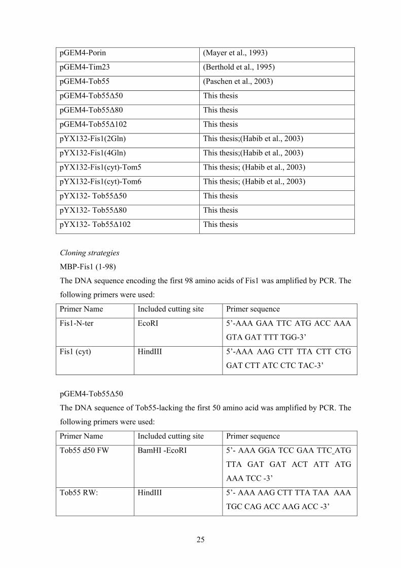
2.1.7 Over-view of used Plasmids
Plasmid Reference
MBP (pMal cRI) New England BioLabs
MBP-Fis1 (1-98) This thesis
MBP-Tob55 (1-120) (Paschen et al., 2003)
pGEM4 (empty) Promega
pGEM4-Mas37 (Habib et al., 2005)
pGEM4-Mdm10 (Paschen et al., 2003)
pGEM4-N.c. F1β (Rassow et al., 1990)
25
pGEM4-Porin (Mayer et al., 1993)
pGEM4-Tim23 (Berthold et al., 1995)
pGEM4-Tob55 (Paschen et al., 2003)
pGEM4-Tob55∆50 This thesis
pGEM4-Tob55∆80 This thesis
pGEM4-Tob55∆102 This thesis
pYX132-Fis1(2Gln) This thesis;(Habib et al., 2003)
pYX132-Fis1(4Gln) This thesis;(Habib et al., 2003)
pYX132-Fis1(cyt)-Tom5 This thesis; (Habib et al., 2003)
pYX132-Fis1(cyt)-Tom6 This thesis; (Habib et al., 2003)
pYX132- Tob55∆50 This thesis
pYX132- Tob55∆80 This thesis
pYX132- Tob55∆102 This thesis
Cloning strategies
MBP-Fis1 (1-98)
The DNA sequence encoding the first 98 amino acids of Fis1 was amplified by PCR. The
following primers were used:
Primer Name Included cutting site Primer sequence
Fis1-N-ter EcoRI 5’-AAA GAA TTC ATG ACC AAA
GTA GAT TTT TGG-3’
Fis1 (cyt) HindIII 5’-AAA AAG CTT TTA CTT CTG
GAT CTT ATC CTC TAC-3’
pGEM4-Tob55∆50
The DNA sequence of Tob55-lacking the first 50 amino acid was amplified by PCR. The
following primers were used:
Primer Name Included cutting site Primer sequence
Tob55 d50 FW BamHI -EcoRI 5’- AAA GGA TCC GAA TTC ATG
TTA GAT GAT ACT ATT ATG
AAA TCC -3’
Tob55 RW:
HindIII 5’- AAA AAG CTT TTA TAA AAA
TGC CAG ACC AAG ACC -3’

26
pGEM4-Tob55∆102
The DNA sequence of Tob55-lacking the first 50 amino acid was amplified by PCR. The
following primers were used:
Primer Name Included cutting site Primer sequence
Tob55 d102 FW BamHI -EcoRI 5’- AAA GGA TCC GAA TTC ATG
CAT GAT GTG GTG CCT TTG
ATG G -3’
Tob55 RW
HindIII 5’- AAA AAG CTT TTA TAA AAA
TGC CAG ACC AAG ACC -3’
pYX132-Fis1(2Gln)
The DNA sequence of Fis1, where the Arg154 and Arg155 were replaced by glutamine
residues, was amplified by PCR. The following primers were used:
Primer Name Included cutting site Primer sequence
Fis1-N-ter EcoRI 5’-AAA GAA TTC ATG ACC AAA
GTA GAT TTT TGG-3’
C-ter 2Gln
HindIII 5’- AAA AAA AAG CTT TCA TTG
TTG CTT GTT TCT TAA GAA
GAA ACT AGC-3’
pYX132-Fis1(4Gln)
The DNA sequence of Fis1, where Arg151, Lys153, Arg154,and Arg155 were replaced
by glutamine residues, was amplified by PCR. The following primers were used:
Primer Name Included cutting site Primer sequence
Fis1-N-ter EcoRI 5’-AAA GAA TTC ATG ACC AAA
GTA GAT TTT TGG-3’
C-ter 4Gln
HindIII 5’- AAA AAA AAG CTT TTA TTG
TTG TTG GTT TTG TAA GAA
GAA ACT AGC C-3’
27
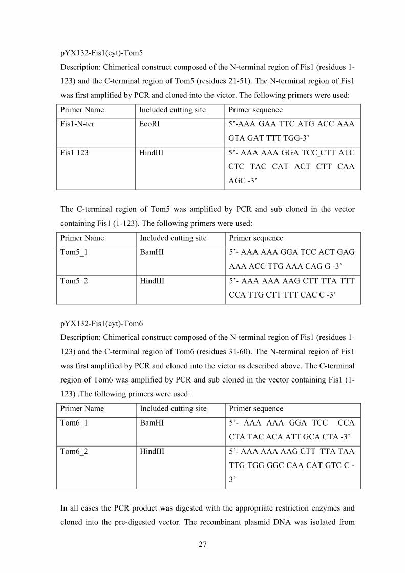
pYX132-Fis1(cyt)-Tom5
Description: Chimerical construct composed of the N-terminal region of Fis1 (residues 1-
123) and the C-terminal region of Tom5 (residues 21-51). The N-terminal region of Fis1
was first amplified by PCR and cloned into the victor. The following primers were used:
Primer Name Included cutting site Primer sequence
Fis1-N-ter EcoRI 5’-AAA GAA TTC ATG ACC AAA
GTA GAT TTT TGG-3’
Fis1 123
HindIII 5’- AAA AAA GGA TCC CTT ATC
CTC TAC CAT ACT CTT CAA
AGC -3’
The C-terminal region of Tom5 was amplified by PCR and sub cloned in the vector
containing Fis1 (1-123). The following primers were used:
Primer Name Included cutting site Primer sequence
Tom5_1 BamHI 5’- AAA AAA GGA TCC ACT GAG
AAA ACC TTG AAA CAG G -3’
Tom5_2
HindIII 5’- AAA AAA AAG CTT TTA TTT
CCA TTG CTT TTT CAC C -3’
pYX132-Fis1(cyt)-Tom6
Description: Chimerical construct composed of the N-terminal region of Fis1 (residues 1-
123) and the C-terminal region of Tom6 (residues 31-60). The N-terminal region of Fis1
was first amplified by PCR and cloned into the victor as described above. The C-terminal
region of Tom6 was amplified by PCR and sub cloned in the vector containing Fis1 (1-
123) .The following primers were used:
Primer Name Included cutting site Primer sequence
Tom6_1 BamHI 5’- AAA AAA GGA TCC CCA
CTA TAC ACA ATT GCA CTA -3’
Tom6_2
HindIII 5’- AAA AAA AAG CTT TTA TAA
TTG TGG GGC CAA CAT GTC C -
3’
In all cases the PCR product was digested with the appropriate restriction enzymes and
cloned into the pre-digested vector. The recombinant plasmid DNA was isolated from
28
positive cultures and the construct was sequenced to confirm proper in-frame ligation and
fidelity of the polymerase.
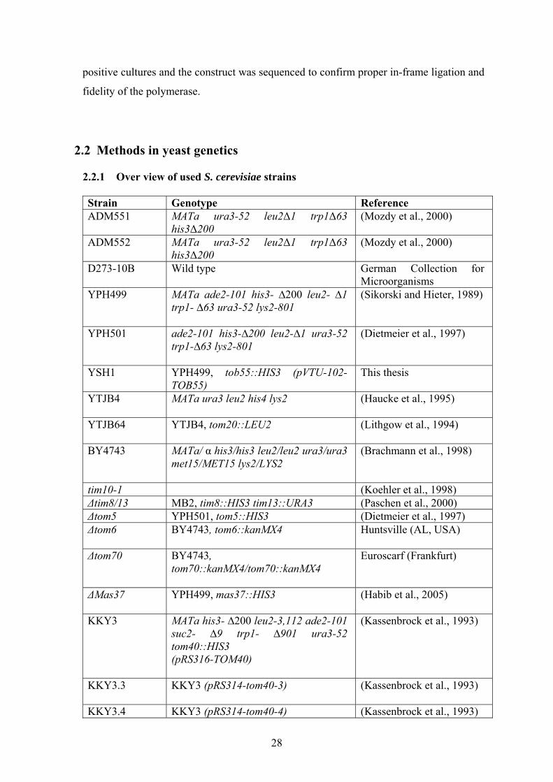
2.2 Methods in yeast genetics 2.2.1 Over view of used S. cerevisiae strains Strain Genotype Reference ADM551 MATa ura3-52 leu2∆1 trp1∆63
his3∆200 (Mozdy et al., 2000)
ADM552 MATa ura3-52 leu2∆1 trp1∆63 his3∆200
(Mozdy et al., 2000)
D273-10B Wild type German Collection for Microorganisms
YPH499 MATa ade2-101 his3- ∆200 leu2- ∆1 trp1- ∆63 ura3-52 lys2-801
(Sikorski and Hieter, 1989)
YPH501 ade2-101 his3-∆200 leu2-∆1 ura3-52 trp1-∆63 lys2-801
(Dietmeier et al., 1997)
YSH1 YPH499, tob55::HIS3 (pVTU-102-TOB55)
This thesis
YTJB4 MATa ura3 leu2 his4 lys2
(Haucke et al., 1995)
YTJB64 YTJB4, tom20::LEU2
(Lithgow et al., 1994)
BY4743 MATa/ α his3/his3 leu2/leu2 ura3/ura3 met15/MET15 lys2/LYS2
(Brachmann et al., 1998)
tim10-1 (Koehler et al., 1998) ∆tim8/13 MB2, tim8::HIS3 tim13::URA3 (Paschen et al., 2000) ∆tom5 YPH501, tom5::HIS3 (Dietmeier et al., 1997) ∆tom6 BY4743, tom6::kanMX4
Huntsville (AL, USA)
∆tom70 BY4743, tom70::kanMX4/tom70::kanMX4
Euroscarf (Frankfurt)
∆Mas37 YPH499, mas37::HIS3
(Habib et al., 2005)
KKY3 MATa his3- ∆200 leu2-3,112 ade2-101 suc2- ∆9 trp1- ∆901 ura3-52 tom40::HIS3 (pRS316-TOM40)
(Kassenbrock et al., 1993)
KKY3.3 KKY3 (pRS314-tom40-3)
(Kassenbrock et al., 1993)
KKY3.4 KKY3 (pRS314-tom40-4) (Kassenbrock et al., 1993)

29
GAL-His8-Tob55
YPH499, tob55::HIS3-pGAL-His8-TOB55
(Paschen et al., 2003)
GAL-Tob38 YPH499, tob38::HIS3-pGAL-TOB38
(Waizenegger et al., 2004)
Tob38-6HA YPH499, tob38::TOB38-6HA-HIS3
(Habib et al., 2005)
Construction of TOB55 genomic disruption strain
The TOB55 gene was cloned into the yeast expression vector pVTU-102 which
contains the selectable marker URA3. The resulting plasmid was transformed into the
wild type strain YPH499. Next, the genomic TOB55 open reading frame was replaced
with the HIS3 marker gene by homologous recombination. The HIS3 marker gene-
containing the flanking regions of TOB55 gene was amplified from the plasmid pFA6s-
His3MX6 (Wach et al., 1997) by using the following primers:
F1-OM55 5’ TAC GTG GCA AAA GTT TTG ATG CCA AAT AGA CAA
AAG TAG CTC AAT CGT ACG CTG CAG GTC GAC-3’
R1-OM55 5’ AAT GGG AAG CTA GGC GAT AGC TTC ACC TTG ACA
TTA AAA GGA ATG TAT TCT ATC GAT GAA TTC GAG CTC
G-3’
The resulting His+Ura+ strain containing a complete coding sequence deletion of the
genomic TOB55 gene and a wild type TOB55 gene on a 2µ plasmid was termed YSH1.
2.2.2 Cultivation of S. cerevisiae strains
2.2.2.1 Media for S. cerivisiae
Lactate medium: 3 g yeast extract, 1g KH2PO4, 1 g NH4Cl, 0.5 g CaCl2 x 2 H2O, 0.5g
NaCl, 1.1 g MgSO4 x 6 H2O, 0.3 ml 1% FeCl3, 22 ml 90% lactic acid, H2O to 1 l, pH
5.5 (adjusted with 10 M KOH). Usually supplemented with 0.1% glucose or 0,1 %
galactose.
YP –medium: 10 g yeast extract, 20 g bacto-pepton, 5.5 ml 90% lactic acid, H2O to 930
ml pH 5.5 (adjusted with 10 M KOH) after autoclaving add 67 ml 2% glucose (YPD) or
100 ml 20% galactose (YPGal) or 100 ml 30% glycerol (YPG).
S-medium: 1.7 g yeast nitrogen base, 5 g ammonium sulfate, 20 g glucose, 1.5 g “Drop-
out mix” powder” (mix containing equal weight of all amino acids; for selecting one
auxothrophic marker, the corresponding amino acid was left out), H2O to 900 ml. After

30
autoclaving add 100 ml 20% galactose (SGal) or 100 ml 20% glucose (SD) or 30%
glycerol (SG). To eliminate wild type (WT)-URA3-allele, yeast were grown on SD
medium containing 0.1% (w/v) 5-fluoro-orotic acid (5-FOA)
To prepare plates with solid media, 2% (w/v) agar was added before autoclaving. For
selective media, amino acids solutions (His, Leu, Lys, all 10 mg/ml) and uracil and
adenine solutions (both 2 mg/ml) were separately autoclaved for 20 min at 120°C, with
the exception of tryptophan (10 mg/ml) which was filter sterilized. The amino acids were
added to the mixture before pouring the plates.
2.2.2.2 S. cerivisiae growth conditions
S. cerevisiae growth was performed as described in Sambrook et al. (Sambrook et al.,
1989) in YPD complete medium or, when a selection on the auxotrophic marker was
necessary, on SD medium. The cells were incubated at 30ºC, under moderate shaking
conditions. Temperature-sensitive mutants were grown at 24°C. For isolation of
mitochondria, cells were propagated for ca. 3 days while the OD600 never exceeded 1. For
depletion of an essential protein, a yeast strain harbouring the corresponding gene under
GAL promoter was grown for ca. 3 days on galactose-containing media. Cells were then
collected, washed with water and resuspended in glucose-containing media. Cells were
grown in glucose medium till the gene of interest was not detected but the mitochondria
still have basal import activity.
2.2.2.3 Transformation of S .cerivisiae by the lithium acetate method
The corresponding yeast strain was grown overnight in YPD-medium and diluted the
next morning to 50 ml medium with an OD600 of 0.2. Cells were grown further, till they
reached an OD600 of 0.8. Then, cells were transferred to a sterile centrifuge tube, and
harvested by centrifugation (1,000 x g, 3 min, RT). After washing with 25 ml of sterile
water, cells were recollected, resuspended in 1 ml 100 mM lithium acetate and
transferred to an Eppendorf tube. Cells were centrifuged again (7,500 x g, 15 sec, RT)
and were resuspended in 400 µl 100 mM lithium acetate. For each transformation 50 µl
of the cell suspension was centrifuged (7,500 x g, 5 min, RT) and the supernatant
removed. Next, a mixture was added to the cells in the following order: 240 µl PEG 3350
(50% v/v), 36 µl 1 M lithium acetate, 5 µl single stranded salmon sperm DNA (10
mg/ml; previously incubated for 5 min at 95ºC), 70 µl H2O containing 0.1-10 µg of DNA

31
to be transformed. The mixture was vortexed for 1 min and incubated for 30 min at 30ºC,
with moderate shaking, followed by another 20-25 min at 42ºC. The cells were harvested
by centrifugation (7,000xg, 15 sec, RT), washed with sterile water, resuspended in a
small volume of sterile water (150 µl), and spread on plates with the appropriate selective
media. The plates were incubated for 2-4 days at 30ºC to recover transformants.
2.3 Methods in cell biology
2.3.1 Isolation of mitochondria from S. cerevisiae
Mitochondria were isolated from S. cerevisiae following the previously described
method (Daum et al., 1982). Yeast cells were cultivated to OD600 of 1-1.5 and collected
by centrifugation (4,400 x g, 5 min, RT). The pellets were washed with H2O and
resuspended to a final concentration of 0.5 g/ml in DTT buffer (100 mM Tris-SO4, 10
mM dithiotreitol (DTT), pH 9.4). The cell suspension was incubated for 15 min at 30°C
with gentle shaking, followed by a new centrifugation step and resuspended in 100 ml of
1.2 M sorbitol buffer (1.2 M sorbitol, 20mM potassium phosphate-KOH, pH 7.4). The
cell wall was digested by 2.5 mg Zymolyase per gram yeast dissolved in Sorbitol buffer.
Cells were incubated for 30-45 min at 30°C, under moderate shaking conditions. To test
the cell wall digestion (obtaining of spheroplasts), 50 µl cell suspension was diluted with
2 ml H2O or into a solution of 1.2 M sorbitol. Formation of spheroplasts was complete
when the OD of the H2O dilution was 10-20% of the OD of the sorbitol dilution. The
solution of spheroplasts in pure H2O becomes clear because spheroplasts burst under
these conditions. All the subsequent steps were performed at 4°C. The spheroplasts were
isolated by centrifugation (3,000 x g, 5 min, 4°C), resuspended (0.15 g/ml) in
homogenizing buffer (0.6 M sorbitol, 10 mM Tris-HCl, 1 mM EDTA, 0.2% (w/v) BSA, 1
mM PMSF, pH 7.4) and homogenized 10 times in a Dounce-Homogeniser. The cell
remnants and unopened cells were sedimented by double centrifugation (2,000 x g, 5
min, 4°C). The supernatant was spun (17,400xg, 12 min, 4°C) and the sedimented
mitochondria were resuspended in SEM buffer (10 mM MOPS/KOH, 250 mM sucrose, 1
mM EDTA, pH 7.4) and separated again from cell’s remnants (2,000xg, 5 min, 4ºC). The
mitochondria were sedimented again as above (17,400xg, 12 min, 4°C). Finally,
mitochondria were resuspended in a small volume of SEM buffer to a concentration of 10
mg/ml protein, aliquoted, frozen in liquid nitrogen, and stored at – 80ºC till use.

32
2.3.2 Preparation of mitoplasts
Mitochondria whose outer membrane has been disrupted are known as mitoplasts. To
obtain mitoplasts, the outer mitochondrial membrane is ruptured by swelling of
mitochondria in a hypotonic solution which keeps the inner membrane intact.
Mitochondria in SEM buffer were diluted 1:10 in 20 mM HEPES-KOH, pH 7.3
containing 2 mM EDTA and incubated on ice for 30 min. If necessary, mitoplasts (or
mitochondria) were reisolated by centrifugation (10 min, 17,000xg, 4ºC) and resuspended
in a desired buffer.
2.3.3 Isolation of crude mitochondria from S. cerevisiae
Cells corresponding to 10-20 OD units were harvested by centrifugation (3,000 x g, 5
min, RT) and washed with water. The cells were resuspended in 400 µl SEM buffer and
0.3 g glass beads (diameter 0.3 mm) were added. The samples were vortexed four times
30 sec each, with 30 sec breaks in between (during this break the samples were incubated
on ice). After centrifugation (1,000 x g, 3 min, 4ºC), the supernatant was transferred to a
new tube and the protein concentration was determined. The desired amount of protein
was centrifuged (10,000xg, 10 min, 4ºC), resulting in crude mitochondrial pellets.
2.3.4 In vitro synthesis of radioactive labelled proteins
Transcription/Translation in a cell-free system
For in vitro synthesis of 35S labelled proteins, the constructs contained in pGEM4
plasmid were first transcribed in mRNA using SP6-RNA-polymerase. The transcription
mixture (100µl) contained: 10-20 µg DNA, 20 µl 5x transcription buffer (200 mM Tris-
HCl, 50 mM MgCl2, 10 mM spermidine, pH 7.5), 10 µl 0.1 M DTT, 4 µl RNasin
nuclease inhibitor (40 U/µl), 20 µl 2.5 mM rNTP, and 5.2 µl 2.5 mM M7G(5`)ppp(5`)G.
After adding 3 µl of SP6-Polymerase (25 U/ml) the mixture was incubated for 1 h at
37°C. The RNA was precipitated by adding 10 µl of LiCl (10 M) and 300 µl ethanol
(96%), centrifuged, and washed with 70% ethanol. After ethanol evaporation at RT, the
RNA was resuspended in water supplemented with 1 µl RNasin (40 U/µl), aliquoted and
kept at – 80°C till use.
For translation using rabbit reticulocyte lysate, the following mix was incubated for
60 min at 30°C: 25 µl RNA, 3.5 µl amino acid mix (without methionine), 7 µl 15 mM
MgOAc, 12 µl 35S (10 mCi/ml) and 100 µl rabbit reticulocyte lysate (Promega). At the

33
end of the translation reaction 5 mM of cold methionine and 250 mM sucrose were
added.
TNT Coupled reticulocyte lysate system
This system combines transcription and translation in the same reaction mixture. TNT
mix (50 µl) contained: 25µl TNT rabbit reticulocyte lysate (Promega), 2 µl TNT reaction
buffer, 1 µl TNT SP6 RNA polymerase, 1 µl amino acid mix without methionine, 2 µl 35S 45 methionine (10 mCi/ml), 1 µl RNasin ribonuclease inhibitor (40 U/µl) and 2 µl
DNA template (0.5 µg/µl). The reaction was incubated for 90 min at 30ºC, and further
treatment was as described for the translation reaction alone.
2.3.5 Import of preprotein into isolated mitochondria
Precursor proteins were synthesized in the presence of 35S-methionine as mentioned
above. Mitochondria were resuspended at 0.5-1 mg/ml in F5 import buffer (0.03-3%
(w/v) fatty acid- free BSA, 250 mM sucrose, 80 mM KCl, 5 mM MgCl2, 2 mM ATP, 2
mM NADH, 100 µg/ml ceratin kinase, 5 mM ceratine phosphate, 10 mM MOPS-KOH,
pH 7.2). Import reactions were performed by addition of lysate (1-3% (v/v)) for the
indicated time periods at 25°C (or in some cases at 15°C). Samples were divided and
diluted in SEM buffer. In some experiments protease treatment was performed by
incubation of the import reaction with 15-100 µg/ml proteinase K or 100 µg/ml trypsin
for 15 min on ice. Protease treatment was stopped by addition of 1 mM PMSF.
Mitochondria were reisolated and the import reaction was analyzed by SDS-PAGE,
transferred to nitrocellulose membrane, autoradiography and quantification.
In some experiments import was competed by addition of recombinant proteins like:
pSu9 (1-69)-DHFR (fusion protein of mitochondrial targeting sequence of subunit 9 of
Fo-ATPase from N. crasa and dihydrofolate reductase (DHFR) from mouse) were added
to 50 µg mitochondria or MBP-Tob55(1-120) (maltose binding protein fused to the first
120 amino acids of Tobb55).
2.3.6 Carbonate extraction
Carbonate extraction was used to check whether a protein is inserted into a membrane
or it is in a soluble form. For this purpose, mitochondria were pelleted via centrifugation,
resuspended in buffer containing 10 mM HEPES-KOH, 100 mM Na2CO3, pH 11.5 and
incubated for 30 min at 0ºC. The probes were then centrifuged for 30 min at 125,000 x g,
2ºC. The soluble proteins were found in the supernatant and were precipitated with

34
Trichloroacetic acid (TCA) and membrane-embedded proteins were found in the pellet.
The samples were analyzed by SDS-PAGE.
2.3.7 Antibody shift
The radiolabelled preproteins were imported into mitochondria and the organelles
were sedimented. The resulting pellet was resuspended in 30-45 µl buffer N (20 mM
Tris-HCl, 50 mM NaCl, 10% (v/v) glycerol, 0.1 mM EDTA, 1 mM PMSF, pH 7.4)
supplemented with 1% (w/v) digitonin and incubated for 15 min on ice. Next, 5 µl of the
desired immunoglobulin G (IgGs) or the purified anti-bodies were added and the mixture
was further incubated for 1 h at 4ºC with gentle shaking (over-head shaker). Insoluble
materials and aggregates were removed by centrifugation step (36700 x g, 20 min, 4ºC).
The samples were further analysed by BNGE.
2.3.8 Fluorescence microscopy
Yeast cells were transformed with plasmid pVT100U-mtGFP expressing
mitochondria-targeted green fluorescent protein (Westermann and Neupert, 2000). Cells
were grown to exponential phase in liquid selective glucose medium at 30°C. Fixation of
the samples was achieved by mixing 7 µl transformed cells together with 7 µl of 0.5 %
(w/v) low melting point agarose. The samples were analyzed by Axioplan microscope
with Plan-Neoflur 100 x/1,30 oil-objective using 100 W Quecksilber-lamp (Carl zeiss
Gmbh, Jena). Classification and quantification of the morphology phenotypes were
performed without knowledge of strain identity at the time of analysis. For quantification
of the phenotype, at least 100 cells were analyzed in three independent experiments.
2.3.9 Pull down of radiolabeled preprotein via His-tagged Tob38
Mitochondria harbouring a hepta-histidin tag at the C-terminus of Tob38 were used to
study the interaction of Tob38 with radiolabeled precursors of the TOB complex
components. Mitochondria (150 µg) were incubated with radiolabeled precursors for 15
min at 0°C. Mitochondria were then reisolated and lysed in 300 µl buffer N containing
0.25% digitonin and 10 mM Imidazole for 1 h at 0°C. Aggregates and insoluble
membranes were removed by clarifying spin (36,700xg, 20 min, 4°C). A portion of 20 %
was taken from the resulted supernatant as a control. The rest of the supernatant was
incubated with 50 µl pre-equilibrated Ni-NTA beads (equilibration with buffer N) for 1 h
at 4°C. Finally the beads were washed 3 times with buffer N (0.05% (w/v) digitonin) and

35
one time with SEMK20 (SEM + 20 mM KCl). Bound proteins were eluted with 2x
Laemmli buffer and analysed by SDS-PAGE followed by autoradiography.
2.3.10 Interaction of radiolebeled preprtoeins with the N-terminal domain of Tob55
The DNA sequences encoding either the N-terminal domain of Tob55 (amino acid
residues 1-120) or the cytosolic domain of Fis1 (amino acid residues 1-98) were cloned
into the pMalCRI plasmid (New England Biolabs), and expressed in E. coli BL21 cells as
soluble fusion proteins with maltose binding protein (MBP). Purification of the protein
was according to the manufacturer instructions (see 2.5.1). For in vitro binding assays, 4
ml induced E. coli cells were lysed and proteins were applied to 40 µl amylose resin
(New England Biolabs). Next, unbound proteins were washed twice with 1 ml MBP-
column buffer (20 mM HEPES, 100 mM NaCl, 2 mM EDTA, 1 mM PMSF, pH 7.4). To
minimize unspecific binding the resin was washed with 500 µl import buffer containing
3% BSA, followed by 20 min incubation of the resin with 50 µl reticulocyte lysate
diluted in 500 µl F5 import buffer containing 0.03 % BSA. The resin was washed twice
with 1 ml F5 import buffer and then incubated with 50 µl reticulocyte lysate containing
the radiolabeled proteins 500 µl in import buffer for 20 min at 4°C. At the end of the
incubation period the resin was washed twice with import buffer and eluted with 1 M
NaCl. The eluted material was precipitated by TCA and analyzed on SDS-PAGE.
2.3.11 Binding assay with water soluble porin
Initially, all the eppindorf tubes were washed with 340 µl 0.25% BSA to prevent
unbound and aggregated proteins to bind to the surface of the tube. The procedure of
binding MBP or MBP Tob55 to amylose resin is according to (2.3.10).Briefly, lysed cells
were applied to 15-25 µl (1:12) amylase resin which were prewashed with MBP column
buffer containing 0.25% BSA. Unbound proteins were washed twice with 1 ml MBP-
column buffer. For binding experiments, various amounts of radiolabeled ws-porin (see
2.4.4) were added in binding buffer (100 mM KCl, 0.025% BSA, 10% glycerol, 100 mM
sodium phosphate, pH 6.8) to MBP or MBP-Tob55 pre-bound to amylose beads. After
incubation at 4°C for 35 min the beads were washed once with binding buffer, then with
binding buffer without BSA, and finally with buffer containing 100 mM NaCl and 90
mM Tris-base. Bound proteins were eluted with sample buffer and analyzed by SDS-
PAGE and autoradiography. For quantification of the binding reactions increasing

36
amounts of radiolabeled ws-porin were analyzed directly by SDS-PAGE and
autoradiography.
2.4 Methods in protein biochemistry 2.4.1 Purification of recombinant MBP-fusion proteins expressed in E. coli
The first 120 amino acids residues of Tob55 or the first 98 amino acids residues of
Fis1 fused to maltose binding protein (MBP) were expressed in E.coli from pMAL-cRI
vector (New England Biolabs). Purification of the recombinant proteins out of E. coli was
done as described before (Luciano et al., 1997) and according to the manufacturer
instructions. An overnight E. coli culture (10 ml) were diluted into 250 ml LB-Amp and
grown until OD600 of ca. 0.5- 0.8 at 37°C. Expression of the recombinant protein was
induced by 1 mM isopropyl-β, D-tiogalactopyranoside (IPTG) and the culture grown for
additional 3 h at 37°C. Cells were pelleted by centrifugation (4,400xg, 10 min, RT) and
resuspended in 20 ml MBP-column buffer. In order to degrade the cell wall and to obtain
spheroplasts, 1 mg/ml Lysozyme was added and the mixture was incubated for 45 min at
4°C. Cells were completely broken by sonication (10x12 s, Branson sonifier, setting 4,
80% duty cycle). After centrifugation (27,200xg, 15 min, 4°C), clear supernatant was
loaded on a 5 ml Amylose-column (New England Biolabs) preequilibrated in MBP-
column buffer. Column was washed with 30 ml of MBP-column buffer and bound
proteins eluted with 10 mM maltose in MBP-column buffer. Expression and purification
were monitored by SDS-PAGE.
2.4.2 Purification of porin from N. crassa
Native porin from N. crassa was isolated by modification of a published procedure
(Pfaller et al., 1985). Shortly, 5 mg outer membrane vesicles (OMV) which were isolated
as described elsewhere (Schmitt et al., 2006) were solubilized in 1 ml buffer containing
50 mM HEPES-KOH, 1 mM PMSF, 10% glycerol and 2% Triton X-100 for 1h at 4ºC.
After a clarifying spin (36670xg, 10 min, 2 °C), the supernatant was applied to anion-
exchange column (ResQ, Amersham). The flow-through (4ml) which contains porin was
collected.
2.4.3 Preparation of water –soluble porin.
Water soluble porin was obtained according to a published protocol (Pfaller et al.,
1985). After addition of 1/10 volume of 3 M TCA and 1/3 volume of methanol, purified

37
porin was precipitated for 1 h at 0 ºC. The precipitated porin was pelted (48,800xg, 1h,
4ºC) and washed twice with ethanol (48,800xg, 30 min, 4ºC). The resulting protein pellet
was solubilzed by vortixing with 1 volume of 0.1 M NaOH for 60 s and immediately
neutralized by an equal volume of 0.2 M NaH2PO4 . This procedure yielded water-
soluble porin in 100 mM NaPi, pH 6.8. the final concentration was determined to be
0.007 µg/ml, 21 µg/3 ml)
2.4.4 Reductive Methylation of water-soluble porin.
Reductive methylation was carried out according to published protocols (Jentoft and
Dearborn, 1979; Pfaller et al., 1985) employing [14C] formaldehyde as a radioactive label
and sodium cyanoborohydride as reducing agent. [14C] Formaldehyde (specific activity
52.5 µCi/µmol, PerkinElmer LAS GmbH) at a concentration of 270 nmol/nmol of porin
in 100 mM NaPi, pH 6.8, and sodium cyanoborohydride ( Sigma, 334 nmol/nmol of
formaldehyde, 48 mM stock solution in 100 mM NaPi, pH 6.8) were consecutively added
to a sample of water-soluble porin 0.007 µg/ml. After 2 hours incubation at 25 ºC, the
reaction was stopped by adding 100 mM Tris.HCl, pH 7.0 followed by 10 min incubation
at 25 ºC. Then water soluble porin was precipitated with 3 M TCA and methanol for 1 h
at 0 ºC and centrifuged, and the pellet was washed once with ethanol and once with
acetone. The protein pellet was resolubilized as described to a final protein concentration
of 0.007 µg/ml
2.4.5 Protein precipitation with trichloroacetic acid (TCA)
Proteins from aqueous solutions were precipitated by adding TCA to a final
concentration of 12% (w/v). The samples were incubated for 10 min on ice or at –20ºC,
and then centrifuged (36,700xg, 20 min, 2ºC). The precipitated proteins were washed
with acetone (kept at –20ºC), and re-centrifuged (36,700xg, 10 min, 2ºC). Protein pellet
was shortly dried at RT and dissolved in 2 x Laemmli buffer.
2.4.6 Protein precipitation with ammonium sulphate
Aqueous solutions containing proteins were mixed with 2 volume of saturated
solution of ammonium sulphate (4°C). The samples were incubated for 30 min at 4°C and
then centrifuged (36,700xg, 10 min, 4°C). The pellets containing the precipitated proteins
were dissolved in the appropriate buffer. To prepare the saturated ammonium sulphate
solution, 76.7 g of ammonium sulphate was dissolved in 100 ml 100 mM Tris-HCl pH

38
7.0, was heated until it dissolved and then chilled to 4°C, so that the ammonium sulphate
crystals sediment.
2.4.7 Determination of protein concentration
Protein concentration was determined according to (Bradford, 1976) using the “Bio-
Rad- Protein assay” reagent. Protein solution was mixed with 1 ml reagent (1:5 dilution)
and incubated for 10 min at RT. The absorbance was measured at 595 nm using a 1-cm-
path length microcuvette. Protein concentration was calculated according to a standard
curve (Bovine from IgGs (Biorad) were used as standard proteins).
2.4.8 SDS-Polyacrylamide gel electrophoresis (SDS-PAGE)
Proteins were separated via one-dimensional vertical slab SDS Polyacrylamide gel
electrophoresis (SDS-PAGE), under denaturing conditions, as described (Laemmli,
1970). Two different electrophoresis systems were used. Normally, big gels were used
(running gel: 9 x 15 x 0.1 cm; stacking gel: 1 x 15 x 0.1 cm) and in some cases Mini gels
(Mini-PROTEAN II, Bio-Rad) (running gel: 7 x 7.2 x 0.075 cm; stacking gel: 1 x 7.2 x
0.075 cm). The concentration of acrylamide and bis-acrylamide in the separating gel was
chosen according to the molecular size of the proteins to be separated. Glass plates of 160
x140 mm and spacers of 1 mm thickness were used. The samples were dissolved in 35 µl
1 x Laemmli buffer (60 mM Tris-HCl, pH 6.8, 2% (w/v) SDS, 10% glycerol, 5% (v/v) β-
mercaptoethanol, 0.05% (w/v) bromphenol-blue) and incubated at 95°C for 5 min, before
loading. The electrophoresis was performed at 25 mA for 3 h (1 h for mini gels). Protein
molecular weight standards were used in each gel.
Bottom gel 2% (w/v) agar in running buffer
Separating gel 8-16% (w/v) acrylamide, 0.16-0.33% (w/v) bis-acrylamide, 375
mM Tris-HCl (pH 8.8), 0.1% (w/v) SDS, 0.05% (w/v) APS,
0.05% (v/v) TEMED
Stacking gel 5% (w/v) acrylamide , 0.1% (w/v) bis-acrylamide, 60 mM Tris-
HCl (pH 6.8), 0.1% (w/v) SDS, 0.05% (w/v) APS, 0.05% (v/v)
TEMED
Running buffer 50 mM Tris-HCl (pH 8.3), 384 mM Glycin, 0.1% (w/v) SDS

39
2.4.9 Semi native SDS-PAGE
Recombinant proteins were dissolved in 1x Laemmli buffer (containing 0.05% SDS)
at 4°C and separated via Semi native SDS-PAGE (Stacking gel and running buffer do not
contain SDS). The electrophoresis was performed at 15 mA for 40 min at 4°C.
2.4.10 Blue-Native gel electrophoresis (BNGE)
For separation of proteins under non-denaturing (native) conditions, Blue-Native gel
electrophoresis (BNGE) was use (Schägger et al., 1994). For this purpose, mitochondria
(50-150 µg) were pelleted, resuspended in 30-45 µl of buffer N containing 0.4-1%
digitonin. The solubilisation was performed at 4C for 15 min. After a clarifying spin
(36,700 x g, 10 min, 4C), 5 µl of sample buffer (5% (w/v) Coomassie-Brilliant-blue G-
250, 100 mM bis-Tris pH 7.0, 500 mM 6-amino-n-Capronic acid) were added to the
soluble material, mixed and finally loaded onto a 6%-13% gradient gel. The
electrophoresis was performed at 4ºC. For the first step of electrophoresis, cathode buffer
containing Coomassie-Brilliant-blue and anode buffer were used and the voltage was set
for 1 h at 100V and then shifted to 500V. When the blue front had migrated to about two
thirds of the separation distance, the cathode buffer with Coomassie-Brilliant-blue was
replaced by a cathode buffer without Coomassie-Brilliant-blue. Marker proteins like
Apoferritin (440 kDa), Alcohol dehydrogenase (ADH) (monomer: 50 kDa, dimer: 100
kDa, trimer: 150 kDa), and bovine serum albumin (BSA) (monomer: 66 kDa, dimer: 136
kDa) were used.
Bottom gel 20% (w/v) acrylamide, 0,13% (w/v) bis-acrylamide, 0.5 M 6-
amino-n-Capronic acid, 50 mM bis Tris-HCl, pH 7,0, 0.05%
(w/v) APS, 0,025% (v/v) TEMED
Separating gel 0.15-0.3% (w/v) bis-acrylamide, 5% (v/v) glycerol, 0.5 M 6-
amino-n-Capronic acid, 50 mM bis Tris-HCl, pH 7,0, 0.04%
(w/v) APS, 0.04% (v/v) TEMED
Stacking gel 3.8% (w/v) acrylamide, 0,12% (w/v) bis-acrylamide, 0.5 M 6-
amino-n-Capronic acid, 50 mM bis Tris-HCl, pH 7.0, 0.08%
(w/v) APS, 0.08% (v/v) TEMED
Cathode buffer 15 mM bis-Tris, pH 7.0, 50 mM Tricine, with or without 0.02%
Coomassie-Brilliant-blue G250
Anode buffer 50 mM bis Tris, pH 7.0

40
2.4.11 Staining SDS-PA gels with Coomassie brilliant blue
After SDS-PAGE, and removal of the bottom and stacking gels, separating gel was
stained at RT for 30-40 min, with an aqueous solution containing 30% (v/v) methanol,
10% (v/v) acetic acid, and 0.1 (w/v) Coomassie-Brilliant-blue R250. The gel was
destained with destaining solution (30% (v/v) methanol and10% (v/v) acetic acid in H2O
) until the protein bands appeared against a clear background. The contrast between
proteins background was enhanced through further 15 minutes incubation with 7% (v/v)
acetic acid solution. This step was followed by further 10-15 minutes incubation in 50%
(v/v) methanol solution in water. The gel was dried overnight between two gel-drying-
films (Promega).
2.4.12 Transfer of proteins to nitrocellulose or PVDF membrane (Western-blot)
Proteins separated via SDS-PAGE were transferred onto nitrocellulose membrane
using the semi-dry blotting method (Kyshe-Anderson, 1984; Towbin et al., 1979). The
gel, the membrane, and six sheets of Whatman filter paper (3MM) were incubated in
transfer buffer (20 mM Tris, 150 mM glycine, 20% (v/v) methanol, 0.08% (w/v) SDS).
Three sheets of filter paper were placed on the anode electrode followed by the
membrane and on top of it the gel. This was covered with other three filter papers and
with the cathode electrode. The transfer was performed at 200 mA for 1.5 h or at 110 mA
for 40 min (for mini gels). To verify transfer efficiency, the nitrocellulose membranes
were reversibly stained with Ponceau S solution (0.2% (w/v) Ponceau S in 3% (w/v)
TCA).
Proteins separated via BNGE were transferred onto a PVDF membrane using the
semi-dry blotting method described above. Before blotting, the PVDF membrane was
activated for 5 min in methanol and 10 min in transfer buffer. After electrophoresis the
PVDF membrane was reversibly stained with Coomassie-Brilliant-blue R solution (30%
(v/v) methanol, 10% (v/v) acetic acid, 0.1 (w/v) Coomassie-Brilliant-blue R250) for 2
min in order to visualize the marker proteins. For destaining, a solution of 30% (v/v)
methanol and 10% (v/v) acetic acid was used. After completely removing the methanol
by washing with TBS buffer (9 g/l NaCl, 10 mM Tris/HCl, pH 7.4) the membrane was
immunodecorated or the radioactive material was visualized by autoradiography.

41
2.4.13 Autoradiography and quantification
Radiolabeled proteins were detected by autoradiography. A dried PVDF or a
nitrocellulose membrane was exposed to Röntgen film (Kodak Bio Max MM) for
different time points (ranging between few hours to 15 days). Period of exposure
depended on signal intensities. Next, the exposed Film was developed in a developing
machine (Gevamatic 60, AGFAGevaert). The films were scanned and the intensity of the
bands was quantified using theAida Image Analyzer 3.4.3 software.
2.5 Methods in immunology 2.5.1 Generation of Fis1-polyclonal antisera in a rabbit
The first 98 amino acids of Fis1 fused to maltose binding protein (MBP) were
expressed in and purified from E. coli as mentioned before (see 2.5.1) Polyclonal antisera
were made in rabbits. Purified recombinant proteins were analyzed by SDS-PAGE,
blotted on nitrocellulose membranes and stained with Ponceau S. Pro injection, 3-7 bands
(ca. 200 µg protein) were cut and the nitrocellulose dissolved in 300 µl DMSO (Knudsen,
1985). Dissolved proteins were mixed with 300 µl Freunds incomplete adjuvant (or
Injection TiterMaxTM for the first injection) until a stable emulsion was obtained. This
emulsion was subcutaneously injected into a rabbit (nr. 309). The antigen was injected
twice within ten days before the first bleeding was taken. Ten days after each injection,
25 -35 ml blood was taken from the ear vane and left at RT for ca. 3 h. After coagulation
was complete, serum was obtained by two centrifugation steps (3,000 x g, 5 min, RT and
27,200 x g, 15 min, RT). Inactivation of the complement was achieved by 20-30 min
incubation at 56°C and the serum was stored at -20°C.
2.5.2 Immunoblotting
To visualize the immobilized proteins on PVDF or nitrocellulose membrane,
immonodecoration with specific antibodies was carried out. Membranes were first
incubated for 1 h in 5% (w/v) milk powder in TBS (135 mM NaCl, 10 mM Tris-HCl, pH
7.5) to block all non-specific binding sites. The immunodecoration was done for 1 h at
RT or over night at 4°C, with specific antiserum (1:200 to 1:1000 dilutions in milk/TBS).
The membrane was then washed 3 times (each wash lasts 10 min), with TBS, TBS/0.05%
(w/v) Triton X-100 and again with TBS, and incubated for 1 h with horseradish
peroxidase coupled to secondary goat anti-rabbit-IgG or anti-mouse-IgG (Bio-Rad)

42
(diluted 1:10.000 dilutions in milk/TBS). The membrane was again washed (as above)
and treated with ECL reagents: luminol (2.5 mM 3-aminophtalhydrazide and 0.4 mM p-
cumaric acid in 0.1 M Tris-HCl, pH 8.5), mixed with equal volume of H2O2 (0.018%
(v/v) in 0.1 M Tris-HCl, pH 8.5). The luminescence reactions were detected with
Röntgen films (FujiNewRX).
For detection of HA-tagged Tob38, anti-HA antibody (Santa Cruz, Roche) was used.
Blots were blocked in the supplied blocking solution and secondary antibody (goat anti-
mouse) was diluted 1:5000 in the blocking reagent in TBS. Further treatment was as
described above.
2.5.3 Binding of water soluble porin to MBP–Tob55(1-120) on a blot
An overlay method described in (Voulhoux and Tommassen, 2004) was used to
evaluate the binding of water soluble porin to the first 120 amino acids residues of Tob55
on blot. Different amounts of purified fusion protein MBP-Tob55 (1-120), and the control
proteins MBP-Fis1(1-98) and MBP were separated by SDS-PAGE under seminative
conditions and the proteins were blotted onto nitrocellulose membranes. The membrane
was blocked in TBS pH 7.6 containing 2.5% non-fat dried milk for 30 min and incubated
2h at 4°C with water soluble porin (11 µg/ml). The membrane was washed twice with 100
mM NaCl and 90 mM Tris base followed by 3 time standard washes as mentioned above.
The immunodecoration was done for 1 h at RT with antibodies against N. crassa porin.
Further treatments were as described in (2.5.2)
2.5.4 Purification of immunoglobulin G (IgG)
Antiserum (4 ml) was centrifuged (20,000xg, 20 min, 4°C) and the white surface film
(corresponding to aggregated lipids) was removed. The antiserum was diluted with 10 ml
of buffer C (100 mM KPi, pH 8.5), filtrated, and loaded onto a 5 ml Protein A-Superose
column (Pharmacia), previously equilibrated with buffer C. The column was washed with
25 ml buffer C and the bound IgGs were eluted with buffer E (100 mM Citrate/NaOH,
pH 3.0). The elute was immediately neutralized with 2 ml 2 M Tris-HCl, pH 8.0, dialyzed
ON against 5 l of water and concentrated via lyophilization. The IgGs were resuspended
in 10 mM MOPS-KOH, pH 7.2 to a final protein concentration of 10-50 mg/ml,
aliquoted, and stored at – 20ºC till use.

43
3. Results
3.1 Structural and functional characterization of tail anchor domains of
mitochondrial outer membrane proteins
Tail anchored proteins form a distinct class of integral membrane proteins that reside
in the outer membrane of mitochondria. These proteins are anchored to the outer
membrane via a single transmembrane domain (TMD) at the C-terminus and expose a
hydrophilic N-terminal domain of variable size to the cytosol. The TMDs of
mitochondrial tail-anchored proteins are relatively short, moderately hydrophobic and do
not exhibit sequence similarity (Fig. 3). Both flanking regions of the TMDs have
positively charged residues (Borgese et al., 2003).
3.1.1 A net positive charge at the C terminus of Fis1 is crucial for mitochondrial
targeting
The importance of the positive charges in targeting tail-anchored proteins to
mitochondria was investigated. To that end, the tail-anchored protein Fis1 was used as a
model protein. Fis1 is anchored to the outer membrane through a 20 amino acid residues
C-terminal TMD. A short segment of five residues is facing the IMS whereas four of
those are basic ones (Fig. 4A). To study the role of these basic residues, two charge
variants were constructed. In the first case, two arginine residues Arg154 and Arg155,
were replaced by glutamine residues reducing the net positive charge to +2 (Fis1(2Gln),
Fig. 4A). In the second case, the net charge in the C-terminal segment was made neutral
by mutating all four basic amino acids to glutamine residues (Fis1(4Gln), Fig. 4A). Figure 4. A net positive charge at the C terminus of Fis1 is crucial for mitochondrial targeting. A. The sequences of the tail domains of native Fis1 and two charge-mutation variants are presented. B. Fis1 null cells transformed with a vector encoding either authentic Fis1 or the indicated variant were ruptured by vortexing in the presence of glass beads. A mitochondrial and a post-mitochondrial fraction were obtained by differential centrifugation (M and S, respectively) and were subjected to SDS–PAGE and immunoblotting. The antibodies used were directed against the cytosolic domain of Fis1, a control marker protein for the cytosol (hexokinase), and the mitochondrial outer membrane protein Tom40. C. As B, but the variant was Fis1(4Gln).

44
The Fis1(2Gln) variant was targeted to mitochondria in vivo as was demonstrated by
subcellular fractionation (Fig. 4B). In contrast, the vast majority of the Fis1(4Gln) protein
was in non-mitochondrial compartments (Fig. 4C). Hence, a net positive charge in the C-
terminal segment appears to be crucial for mitochondrial targeting of Fis1.
Fis1 is involved in mitochondrial fission and thus, deletion of this protein causes
changes in mitochondrial morphology. In the absence of Fis1, mitochondria form a net-
like structure instead of the normal reticulum structure (Mozdy et al., 2000). When
Fis1(2Gln) variant was transformed into fis1 null strain, the wild type morphology of
mitochondria was completely restored (Table 1). The small amount of Fis1(4Gln) protein
that was targeted to mitochondria was sufficient to complement the morphology
phenotype of fis1 null strain (Table 1). These results suggest that the positive charge at
the C-terminal segment is required for targeting to mitochondria but not for the function
of Fis1, once it is present in the membrane.
Recently, the involvement of human Fis1 (hFis1) in the fission machinery of
mammalian mitochondria was investigated. It was demonstrated that high expression
levels of hFis1 strongly promote fission and the resulting mitochondria are fragmented
(Yoon et al., 2003). Notably, the expression levels of Fis1(4Gln) in cells were higher than
those of Fis1(2Gln) and Fis1wt (Fig. 4B,C). The remarkable differences in the expression
levels among the various variants of Fis1 in yeast may hint to a tight regulation of the
functional levels of Fis1 in the outer membrane. Over-expression of the functional
variants Fis1wt and Fis1(2Gln) might harm the cells; therefore their expression is
regulated. Since only a small fraction of Fis1(4Gln) is functional in the outer membrane
of mitochondria, this variant can be expressed in high amounts in the cell.
Table 1. Complementation analysis of Fis1 variants Yeast strain: wild type or ∆fis1 transformed with a plasmid encoding the indicated protein
% of cells with normal reticular mitochondrial morphologya
Wild type 96
∆fis1 + Fis1 96
∆fis1 + Fis1(4Gln) 95
∆fis1 + Fis1(2Gln) 89
∆fis1 + Fis1(cyt)-Tom5C 79
∆fis1 + Fis1(cyt)-Tom6C 69
∆fis1 + empty plasmid 0
a The percentage of cells with wild type-like mitochondrial morphology was determined using fluorescence microscopy

45
3.1.2 The tail anchor-domain of Fis1 does not have a sequence-specific role
To explore some features of tail anchor domains we asked whether these domains
could functionally replace each other. To address this point, the first 123 amino acid
residues of Fis1 (located in the cytosol) were fused to the tail anchor domain of either
Tom5 (residues 21-51) or Tom6 (residues 31-60). Subcellular fractionation shows that
both variants, Fis1(cyt)–Tom5C and Fis1(cyt)-Tom6C were targeted to mitochondria
(Fig. 5A). Thus, tail anchor domains of different proteins can be exchanged without
losing their targeting and anchoring functions. As the cytosolic domain of Fis1 does not
contain a mitochondrial targeting signal (Mozdy et al., 2000), these results imply that the
signal-anchor domains of both Tom5 and Tom6 are sufficient for mitochondrial targeting.
Since Tom5 and Tom6 are components of the TOM core complex (Ahting et al.,
1999; Dekker et al., 1998), the ability of their tail anchor domains to assemble into the
TOM complex was tested. The TOM core complex from strains expressing either
Fis1(cyt)–Tom5C or Fis1(cyt)-Tom6C, was analyzed by BNGE. In both cases, the vast
majority of the fusion protein assembled into the 410 kDa TOM core complex and only a
minor fraction (~6%) migrated as unassembled low-molecular-weight species (Fig. 5B).
Taken together, the tail anchor domains of Tom5 and Tom6 are sufficient for the
assembly of the fusion proteins into the TOM complex. Based on quantitative Western
blotting, mitochondria expressing Fis1(cyt)-Tom5C or Fis1(cyt)-Tom6C were estimated
to harbour 7-8 fold more Fis1 molecules as compared to Fis1wt. Although only minor
amounts of these molecules were not assembled into the TOM complex (see above), both
proteins were able to partially complement the morphology phenotype of ∆fis1 strain
(Fig. 5C and Table 1). It seems that those molecules of Fis1 fusion proteins, which were
not assembled into the TOM complex, can be as active as native Fis1 in mediating fission
of mitochondria. The tail anchor domain of Fis1 is probably not involved in sequence-
specific interactions that are essential for the function of the protein. These conclusions
are in line with data suggesting that the region mediating mitochondrial fission resides
within the first 31 amino acids of the N-terminal cytosolic domain of human Fis1 (Yoon
et al., 2003).
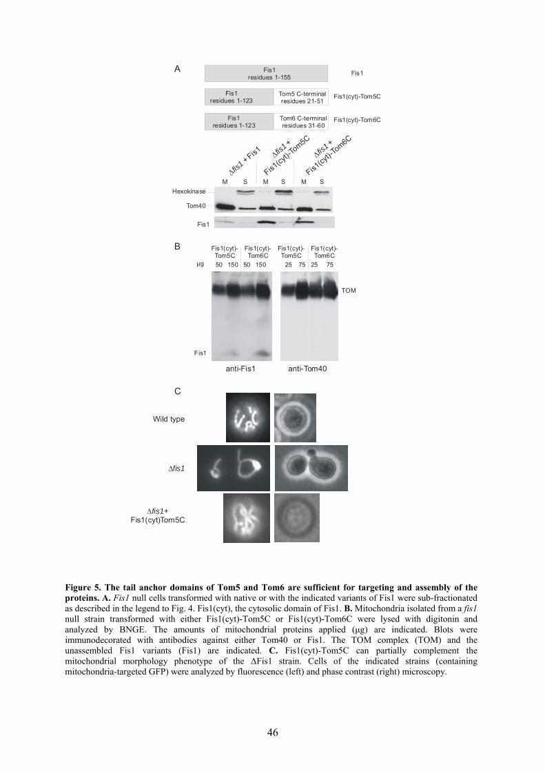
46
µg 50 150 50 150 25 75 25 75
anti-Fis1 anti-Tom40
Fis1(cyt)-Tom5C
Fis1(cyt)-Tom6C
Fis1(cyt)-Tom5C
Fis1(cyt)-Tom6C
Tom40
Hexokinase
Fis1
M S M S M S
∆fis1 +
Fis1(cy
t)-Tom5C
∆fis1 + Fis1 ∆fis1
+
Fis1(cy
t)-Tom6C
Fis1residues 1-123
Tom5 C-terminal residues 21-51
Fis1 residues 1-123
Tom6 C-terminal residues 31-60
A
B
C
Wild type
∆fis1
∆fis1+Fis1(cyt)Tom5C
Fis1
TOM
Fis1residues 1-155
Fis1
Fis1(cyt)-Tom5C
Fis1(cyt)-Tom6C
Figure 5. The tail anchor domains of Tom5 and Tom6 are sufficient for targeting and assembly of the proteins. A. Fis1 null cells transformed with native or with the indicated variants of Fis1 were sub-fractionated as described in the legend to Fig. 4. Fis1(cyt), the cytosolic domain of Fis1. B. Mitochondria isolated from a fis1 null strain transformed with either Fis1(cyt)-Tom5C or Fis1(cyt)-Tom6C were lysed with digitonin and analyzed by BNGE. The amounts of mitochondrial proteins applied (µg) are indicated. Blots were immunodecorated with antibodies against either Tom40 or Fis1. The TOM complex (TOM) and the unassembled Fis1 variants (Fis1) are indicated. C. Fis1(cyt)-Tom5C can partially complement the mitochondrial morphology phenotype of the ∆Fis1 strain. Cells of the indicated strains (containing mitochondria-targeted GFP) were analyzed by fluorescence (left) and phase contrast (right) microscopy.

47
3.1.3 The tail anchor domain of Tom6 plays a role in the stability of the TOM
complex
Tom6 was suggested to function as an assembly factor of the TOM core complex.
Deletion of Tom6 has a strong effect on the stability of the GIP complex. The interaction
between Tom40 and Tom22 is destabilized (Alconada et al., 1995; Dekker et al., 1998;
Dembowski et al., 2001), and the proteins are preferentially found in the lower-
molecular-mass range in blue native gel electrophoresis; Tom40 in an 100kDa
subcomplex and Tom22 below 60kDa. Tom5 and Tom7 remain associated with Tom40
in the 100kDa subcomplex (Dekker et al., 1998). Since Fis1(cyt)-Tom6C could assemble
into the TOM complex, we asked whether this hybrid protein could functionally replace
the native Tom6. To that end, Fis1(cyt)-Tom6C was transformed into a ∆tom6 strain and
the TOM complex was analyzed by BNGE. Western blotting and immunodecoration with
antibodies directed against Tom40 shows that most of the TOM core complexes regained
their stability (Fig. 6). Thus, the tail anchor domain of Tom6 is sufficient to fulfil the
stabilization role of the protein.
µg 25 75
∆tom6+Fis1(cyt)-Tom6C
25 75 25 75
Wild type∆tom6+pYX132
TOM
100K
anti-Tom40 Figure 6. The tail anchor domain of Tom6 is sufficient for the TOM-stabilizing function of the protein. Mitochondria isolated from a wild type strain and from a tom6 null strain transformed with either empty vector (∆tom6+pYX132) or vector encoding Fis1(cyt)-Tom6C (∆tom6+Fis1(cyt)-Tom6C) were lysed with digitonin and analyzed by BNGE. The amounts of mitochondrial proteins applied (µg) are indicated. The blot was immunodecorated with antibodies against Tom40. The native 410 kDa TOM core complex (TOM) and the 100 kDa form of Tom40 (100K) are indicated.
3.1.4 The tail-anchor domain of Tom5 plays an essential role in the function of the
protein
Deletion of the gene encoding Tom5 was reported to be lethal at 37C (Dietmeier et
al., 1997). Based on this observation, the importance of the TMD for the function of
Tom5 was examined. The fusion protein Fis1(cyt)–Tom5C was transformed into a
∆Tom5 strain, and its ability to complement the ts phenotype of this strain was tested.
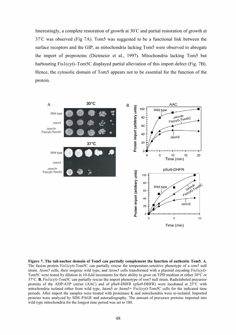
48
A
Wild type
∆tom5
∆tom5+Fis(cyt)-Tom5C
Wild type
∆tom5
∆tom5+Fis(cyt)-Tom5C
30°C
37°C
Interestingly, a complete restoration of growth at 30C and partial restoration of growth at
37C was observed (Fig 7A). Tom5 was suggested to be a functional link between the
surface receptors and the GIP, as mitochondria lacking Tom5 were observed to abrogate
the import of preproteins (Dietmeier et al., 1997). Mitochondria lacking Tom5 but
harbouring Fis1(cyt)–Tom5C displayed partial alleviation of this import defect (Fig. 7B).
Hence, the cytosolic domain of Tom5 appears not to be essential for the function of the
protein.
B
Figure 7. The tail-anchor domain of Tom5 can partially complement the function of authentic Tom5. A. The fusion protein Fis1(cyt)-Tom5C can partially rescue the temperature-sensitive phenotype of a tom5 null strain. ∆tom5 cells, their isogenic wild type, and ∆tom5 cells transformed with a plasmid encoding Fis1(cyt)-Tom5C were tested by dilution in 10-fold increments for their ability to grow on YPD medium at either 30°C or 37°C. B. Fis1(cyt)-Tom5C can partially rescue the import phenotype of tom5 null strain. Radiolabeled precursor proteins of the ADP/ATP carrier (AAC) and of pSu9-DHFR (pSu9-DHFR) were incubated at 25°C with mitochondria isolated either from wild type, ∆tom5 or ∆tom5+ Fis1(cyt)-Tom5C cells for the indicated time periods. After import the samples were treated with proteinase K and mitochondria were re-isolated. Imported proteins were analyzed by SDS–PAGE and autoradiography. The amount of precursor proteins imported into wild type mitochondria for the longest time period was set to 100.
Pro
tein
impo
rt (a
rbitr
ary
units
)Pr
otei
n im
port
(arb
itrar
y un
its)
Pro
tein
impo
rt (a
rbitr
ary
units
)Pr
otei
n im
port
(arb
itrar
y un
its)

49
3.2 Tob38, a novel essential component of the TOB complex
3.2.1 Identification of Tob38
The major aim of this thesis was to study the biogenesis of mitochondrial outer
membrane. This included identification and characterization of novel components of the
newly discovered TOB complex. This part of my work was carried out in collaboration
with Thomas Waizenegger. To find interacting partners of Tob55, mitochondria from
both a wild-type yeast and a strain carrying a His8-tag at the N terminus of Tob55
(Tob55His;(Paschen et al., 2003) were lysed with digitonin and Ni-NTA beads were
added. Proteins retained on the beads selectively with the Tob55His mitochondria were
identified by mass spectrometry (Protein analysis unit of Adolf-Butenandt Institute,
LMU, Munich). In addition to Mas37 and Tob55His, a protein of 38 kDa was identified
and it was termed Tob38 (Waizenegger et al., 2004). Tob38 is encoded by an essential
gene (ORF Yhr083w) (Niedenthal et al., 1999) and is predicted to consist of 330 amino
acid residues.
3.2.2 Tob38 is part of the TOB complex and essential for the biogenesis of β-barrel
proteins
Tob38 is a peripheral membrane protein of the TOB complex and is associated with
the cytosolic surface of the outer membrane. Combined with Tob55, Tob38 forms a
functional TOB core complex (Waizenegger et al., 2004). Due to the essential nature of
Tob38, a yeast strain was constructed in which the TOB38 gene was under the control of
the GAL10 promoter. In the presence of galactose, these cells grew at a similar rate to that
of the wild type cells. In contrast, in the presence of glucose, growth of the GAL10-
TOB38 strain was slowed down after 12 h and was strongly reduced after 19 h. Mas37
and Tob55 were present at strongly reduced levels as early as 12 h after the shift to
glucose. Depletion of Tob38 for 19 h resulted in even lower amounts of Mas37 and
Tob55 and in reduced amounts of the β-barrel proteins Tom40, Mdm10 and porin. In
contrast, other proteins of the various mitochondrial subcompartments were present at
roughly the same levels as those in the wild type.
Next, the involvement of Tob38 in the membrane insertion of β-barrel preproteins
was checked. Precursors of Tom40 and Mdm10 bind to the TOB complex, and thereby an
import intermediate of about 250 kDa is formed (Model et al., 2001; Paschen et al., 2003;
Wiedemann et al., 2003). In the case of mdm10, this import intermediate was strongly

50
reduced when Tob38-depleted mitochondria were used (Fig 8A). The import of another
β-barrel protein, porin, (Gentle et al., 2004; Kozjak et al., 2003; Wiedemann et al., 2003),
was analysed by the formation of assembled complexes . The formation of these
oligomeric species was inhibited in Tob38-depleted cells (Fig 8B). Import of porin,
however, shows less dependency of Tob38 than import of Tom40 and Mdm10. In
contrast to the β-barrel precursors, other proteins of the various mitochondrial
subcompartments, such as Tim23, (Fig 8C) were imported into mitochondria from
Tob38-depleted cells with similar efficiencies as into mitochondria from wild-type cells.
Taken together, Tob38 is a peripheral membrane protein of the TOB complex and has an
essential function in the biogenesis of β-barrel proteins.
Figure 8. Tob38 is required for the insertion and assembly of β-barrel precursor proteins. A, B. Mitochondria isolated from either wild-type or Tob38-depleted cells (Tob38↓) were incubated with radiolabelled precursors of Mdm10 (A) and porin (B) for the indicated time periods. Mitochondria were re-isolated and analysed by BNGE followed by autoradiography. The 250 kDa assembly intermediate of Mdm10 (250K) and the assembled complexes of porin are indicated. C. A radiolabeled precursor of Tim23 was imported into the indicated mitochondria. Samples were re-isolated, treated with PK and analysed by SDS–PAGE and autoradiography.
Mdm10 precursor
2 20 60 2 20 60
250 K
min
Wild type Tob38Tob38
A B Porin precursor
3 20 30 3 20 30minWild type Tob38Tob38
Ass
embl
edpo
rin
Tim23 precursor
min
Wild type3 201 8 3 201 8
Tob38Tob38C

51
3.3 Assembly of the TOB complex The TOB complex of mitochondria is composed of three identified components: the
major one, Tob55, is an essential β-barrel membrane-embedded protein. Tob55 mediates
the insertion of β-barrel proteins into the outer membrane. The other known components
of the TOB complex are the peripheral membrane proteins Mas37 and Tob38. Mas37
interacts with Tob55 and so far plays an unidentified role in the biogenesis of β-barrel
proteins. Tob38, as mentioned above, is essential for viability in yeast, and together with
Tob55 forms a functional TOB core complex. Despite the central role of the TOB
complex in the biogenesis of β-barrel proteins, little is known about the biogenesis of the
complex itself. In this part of the study, the mechanism by which Tob55 and Mas37 are
targeted to mitochondria and assembled into the TOB complex was elucidated.
3.3.1 The establishment of an in vitro import assay
To investigate the import and assembly pathways of Tob55 and Mas37, the precursors
were individually synthesized in rabbit reticulocyte lysate and labelled with [35S]
methionine. The precursors were then incubated with isolated mitochondria, and a
protease accessibility assay was used as a criterion for correct insertion into the outer
membrane. In the case of Tob55, treatment of mitochondria with proteinase K (PK)
resulted in the formation of a specific proteolytic fragment of 30 kDa (Paschen et al.,
2003). This fragment was also observed upon insertion of a radiolabeled Tob55 precursor
into the outer membrane of isolated mitochondria (Fig. 9A). A second fragment with an
apparent molecular mass of 20 kDa was also observed. These fragments were completely
degraded when mitochondria were solubilized with the detergent Triton X-100, and were
also absent when reticulocyte lysate containing the Tob55 precursor was treated with PK
(Fig. 9A). The two fragments are embedded in the outer membrane, as they could not be
extracted by alkaline solution (Fig. 9B). Thus, this protection assay can be employed to
investigate the mechanism of insertion of Tob55 into the mitochondrial outer membrane.
The intensity of the 30 kDa fragment served in further experiments to quantify membrane
insertion of Tob55. In order to study the association of Mas37 with the mitochondrial
outer membrane, the generation of two types of Mas37 fragment (14 and 20 kDa) upon
treatment of mitochondria with PK (Wiedemann et al., 2003) was utilized. The 14 kDa
fragment was recognized by antibodies against a carboxy terminal region of Mas37
(Wiedemann et al., 2003). The intensity of this band served as a measure for the
association of Mas37 with the outer membrane.
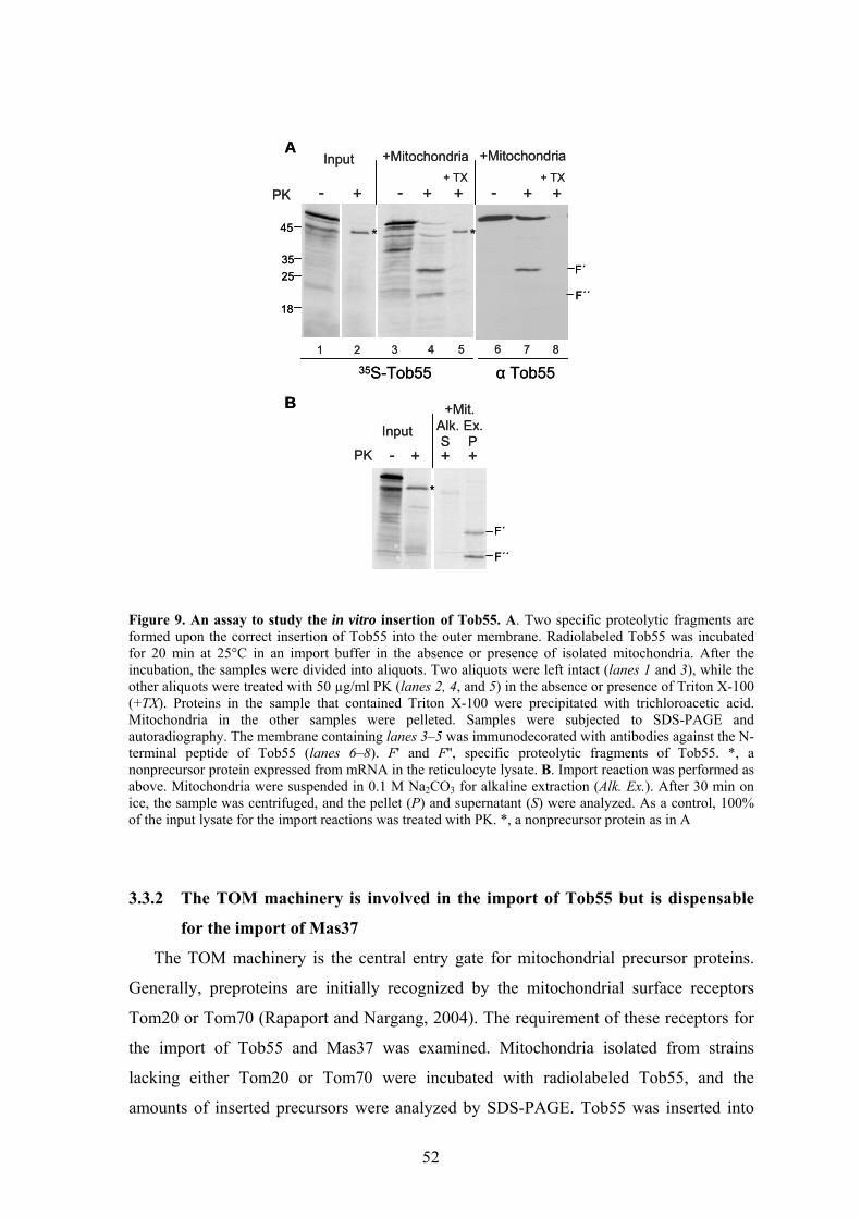
52
Figure 9. An assay to study the in vitro insertion of Tob55. A. Two specific proteolytic fragments are formed upon the correct insertion of Tob55 into the outer membrane. Radiolabeled Tob55 was incubated for 20 min at 25°C in an import buffer in the absence or presence of isolated mitochondria. After the incubation, the samples were divided into aliquots. Two aliquots were left intact (lanes 1 and 3), while the other aliquots were treated with 50 µg/ml PK (lanes 2, 4, and 5) in the absence or presence of Triton X-100 (+TX). Proteins in the sample that contained Triton X-100 were precipitated with trichloroacetic acid. Mitochondria in the other samples were pelleted. Samples were subjected to SDS-PAGE and autoradiography. The membrane containing lanes 3–5 was immunodecorated with antibodies against the N-terminal peptide of Tob55 (lanes 6–8). F' and F'', specific proteolytic fragments of Tob55. *, a nonprecursor protein expressed from mRNA in the reticulocyte lysate. B. Import reaction was performed as above. Mitochondria were suspended in 0.1 M Na2CO3 for alkaline extraction (Alk. Ex.). After 30 min on ice, the sample was centrifuged, and the pellet (P) and supernatant (S) were analyzed. As a control, 100% of the input lysate for the import reactions was treated with PK. *, a nonprecursor protein as in A
3.3.2 The TOM machinery is involved in the import of Tob55 but is dispensable
for the import of Mas37
The TOM machinery is the central entry gate for mitochondrial precursor proteins.
Generally, preproteins are initially recognized by the mitochondrial surface receptors
Tom20 or Tom70 (Rapaport and Nargang, 2004). The requirement of these receptors for
the import of Tob55 and Mas37 was examined. Mitochondria isolated from strains
lacking either Tom20 or Tom70 were incubated with radiolabeled Tob55, and the
amounts of inserted precursors were analyzed by SDS-PAGE. Tob55 was inserted into
45
3525
18
α Tob55
PK - +
+Mitochondria
- + +
* *
- + ++ TX
+MitochondriaInput
35S-Tob55
F´
F´´
B
A
PK - + + +
+Mit.Alk. Ex.InputS P
+ TX
F´
F´´
1 2 3 4 5 6 7 8
*
45
3525
18
α Tob55
PK - +
+Mitochondria
- + +
* *
- + ++ TX
+MitochondriaInput
35S-Tob55
F´
F´´
B
A
PK - + + +
+Mit.Alk. Ex.InputS P
+ TX
F´
F´´
1 2 3 4 5 6 7 8
*

53
these mutated mitochondria with a strongly reduced efficiency as compared to wild type
organelles (Fig. 10).
Figure 10. The insertion of Tob55 requires the import receptors. The radiolabeled precursor of Tob55 was incubated at 25°C for the indicated time periods with mitochondria isolated from either the wild type strain or from the strain lacking Tom20 (A) or Tom70 (B). After import, the samples were treated with proteinase K, and mitochondria were reisolated. Inserted proteins were analyzed by SDS-PAGE and autoradiography. The proteolytic fragments of Tob55 (F' and F'') are indicated. The insertion of Tob55 was quantified by analyzing the formation of the 30 kDa fragment (F'). The amount of precursor protein imported into wild type mitochondria for the longest time period was set to 100%.
In contrast, deletion of the import receptors did not affect the association of Mas37
with mitochondria (Fig. 11). Similar results were obtained when the import experiments
were performed at 15°C. Hence, although the import receptors Tom20 and Tom70 appear
to play only a minor role, if any, in the association of Mas37 with the outer membrane,
they have a crucial function in the recognition of Tob55 precursor.
min 103 20 103 20Wild type ∆Tom70
min 103 20 103 20Wild type ∆Tom20
0
20
40
60
80
100
0 5 10 15 20
Impo
rt (%
of c
ontro
l)
Time (min)
Wild type
∆Tom20
Impo
rt (%
of c
ontro
l)
Time (min)
Wild type
∆Tom70
0
20
40
60
80
100
0 5 10 15 20
BA
F´
F´´
F´
F´´
min 103 20 103 20Wild type ∆Tom70
min 103 20 103 20Wild type ∆Tom20
0
20
40
60
80
100
0 5 10 15 20
Impo
rt (%
of c
ontro
l)
Time (min)
Wild type
∆Tom20
Impo
rt (%
of c
ontro
l)
Time (min)
Wild type
∆Tom70
0
20
40
60
80
100
0 5 10 15 20
BA
F´
F´´
F´
F´´

54
0
20
40
60
80
100
0 5 10 15 20
Wild type
∆Tom70
100
Time (min)
Time (min)
Impo
rt (%
of c
ontro
l)Im
port
(% o
f con
trol)
0
20
40
60
80
0 5 10 15 20
Wild type∆Tom20
B
A
0
20
40
60
80
100
0 5 10 15 20
Wild type
∆Tom70
100
Time (min)
Time (min)
Impo
rt (%
of c
ontro
l)Im
port
(% o
f con
trol)
0
20
40
60
80
0 5 10 15 20
Wild type∆Tom20
0
20
40
60
80
0 5 10 15 20
Wild type∆Tom20
B
A Figure 11. The association of Mas37 with mitochondria is independent of import receptors. The radiolabeled precursor of Mas37 was incubated at 25°C for the indicated time periods with mitochondria isolated either from the wild type strain or from the strain lacking Tom20 (A) or Tom70 (B). After import, the samples were treated with proteinase K (15 µg/ml), and mitochondria were re-isolated. Associated proteins were analyzed by SDS-PAGE and autoradiography. The association of Mas37 was quantified by analyzing the formation of the 14 kDa fragment. The amount of precursor protein associated with wild type mitochondria for the longest time period was set to 100%.
Do Tob55 and Mas37 use the general import pore of the TOM complex? To shed
light on this question, the import pore was blocked by addition of an excess of
recombinant pSu9(1-69)-DHFR, a precursor consisting of the presequence of ATP
synthase subunit 9 fused in front of dihydrofolate reductase. A similar set of experiments
had shown previously that the import of radiolabeled matrix-destined precursors as well
as precursors of outer membrane β-barrel proteins was competed out by recombinant
pSu9(1–69)-DHFR (Krimmer et al., 2001; Paschen et al., 2003; Rapaport and Neupert,
1999). Consistent with these observations, the insertion of Tob55 was strongly reduced
upon the addition of excess preproteins (Fig. 12A). In contrast, excess recombinant
pSu9(1–69)-DHFR did not reduce the association of radiolabeled Mas37 with
mitochondrial outer membrane (Fig. 12A).
The involvement of Tom40 in the import of both precursor proteins was further
investigated. To that end, mitochondria isolated from strains harbouring temperature-
sensitive alleles of tom40 (tom40-3 and tom40-4) were used. It was shown previously that
the mutation in the tom40-3 strain did not affect the insertion of porin, whereas those in
tom40-4 resulted in lower efficiency of porin insertion into the membrane (Fig.
12B)(Krimmer et al., 2001). The insertion of Tob55 into the Tom40-4 mitochondria was
significantly less efficient in comparison to the insertion into wild-type mitochondria,
whereas no difference was observed with Tom40-3 mitochondria (Fig. 12B). The
association of Mas37 with mitochondria from the mutated strains was similar to that with
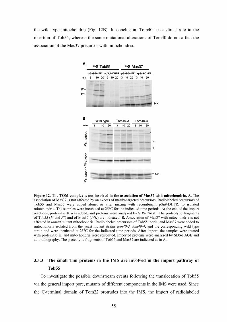
55
the wild type mitochondria (Fig. 12B). In conclusion, Tom40 has a direct role in the
insertion of Tob55, whereas the same mutational alterations of Tom40 do not affect the
association of the Mas37 precursor with mitochondria.
Figure 12. The TOM complex is not involved in the association of Mas37 with mitochondria. A. The association of Mas37 is not affected by an excess of matrix-targeted precursors. Radiolabeled precursors of Tob55 and Mas37 were added alone, or after mixing with recombinant pSu9-DHFR, to isolated mitochondria. The samples were incubated at 25°C for the indicated time periods. At the end of the import reactions, proteinase K was added, and proteins were analyzed by SDS-PAGE. The proteolytic fragments of Tob55 (F' and F'') and of Mas37 (14K) are indicated. B. Association of Mas37 with mitochondria is not affected in tom40 mutant mitochondria. Radiolabeled precursors of Tob55, porin, and Mas37 were added to mitochondria isolated from the yeast mutant strains tom40-3, tom40-4, and the corresponding wild type strain and were incubated at 25°C for the indicated time periods. After import, the samples were treated with proteinase K, and mitochondria were reisolated. Imported proteins were analyzed by SDS-PAGE and autoradiography. The proteolytic fragments of Tob55 and Mas37 are indicated as in A.
3.3.3 The small Tim proteins in the IMS are involved in the import pathway of
Tob55
To investigate the possible downstream events following the translocation of Tob55
via the general import pore, mutants of different components in the IMS were used. Since
the C-terminal domain of Tom22 protrudes into the IMS, the import of radiolabeled
min 3 10 20 3 10 20 3 10 20 3 10 20-pSu9-DHFR -pSu9-DHFR+pSu9-DHFR +pSu9-DHFR
35S-Tob55 35S-Mas37
min 103 20 103 20Wild type Tom40-4
103 20Tom40-3
35S
-Tob
5535
S-P
orin
B
A
F´
F´´
14K
F´
F´´
14K
35S
-Mas
37
min 3 10 20 3 10 20 3 10 20 3 10 20-pSu9-DHFR -pSu9-DHFR+pSu9-DHFR +pSu9-DHFR
35S-Tob55 35S-Mas37
min 103 20 103 20Wild type Tom40-4
103 20Tom40-3
35S
-Tob
5535
S-P
orin
B
A
F´
F´´
14K
F´
F´´
14K
35S
-Mas
37

56
min 103 20 103 20Wild type Tom22∆C
B
A
F´
F´´
min 103 20 103 20Wild type ∆Tim8/13
F´
F´´35S
-Tob
5535
S-T
ob55
Import(% of control) 7242 100 5235 69
Import(% of control) 7011 100 8236 103
min 103 20 103 20Wild type Tom22∆C
B
A
F´
F´´
min 103 20 103 20Wild type ∆Tim8/13
F´
F´´35S
-Tob
5535
S-T
ob55
Import(% of control) 7242 100 5235 69
Import(% of control) 7011 100 8236 103
Tob55 was analyzed in mitochondria isolated from strain-expressing Tom22 molecules
without this domain (Fig. 13A) (Moczko et al., 1997). The deletion had no effect on the
insertion efficiency. Thus, the C-terminal domain of Tom22 does not play an important
role in the insertion process of Tob55. Next, the involvement of the small Tim proteins
(Tim9/10 and Tim8/13 complexes) residing in the IMS was examined. The import of
radiolabeled Tob55 was carried out in the presence of mitochondria isolated from either
the ∆Tim8/13 strain or a strain with a mutated TIM10 allele (Fig. 13B and C). In both
cases, an impaired insertion as compared with the wild-type organelles was observed.
This reduction is similar to the previously observed one, when Tom40 was imported into
mitochondria containing the mutated allele of TIM10 (Wiedemann et al., 2004b).
Notably, the effect of the mutations in tim10 on the import of the ADP-ATP carrier
protein, a bona fide substrate of the Tim9/10 complex, was much more profound as
compared with the moderate effect on the insertion of Tob55 (Fig. 13C). In summary,
both complexes Tim8/13 and Tim9/10 appear to be involved in the import of Tob55.
Figure 13. The insertion of Tob55 into the outer membrane involves components in the intermembrane space. The radiolabeled precursor of Tob55 was incubated at 25°C for the indicated time periods with mitochondria isolated from either the wild type strain or from the indicated strains. A. A strain containing a Tom22 variant where the C-terminal domain is deleted (Tom22∆C). B. A strain lacking tim8 and tim13 (∆Tim8/13). C. A strain containing a mutated allele of tim10 (Tim10–1). The radiolabeled precursor of ADP-ATP carrier protein (35S-AAC) was also imported into the latter mitochondria for comparison. After import, the samples were treated with proteinase K, and mitochondria were re-isolated. The proteolytic fragments of Tob55 (F' and F'') are indicated. The bands corresponding to F' in panels B and C and the protected AAC precursor were quantified, and the amount of precursor protein imported into wild type mitochondria for 20 min was set to 100%.
Cmin 103 20 103 20
Wild type Tim10−1
35S
-AA
C35
S-T
ob55
F´
F´´
Import(% of control) 9420 100 7010 79
Import(% of control)
9747 100 84 16
Cmin 103 20 103 20
Wild type Tim10−1
35S
-AA
C35
S-T
ob55
F´
F´´
Import(% of control) 9420 100 7010 79
Import(% of control)
9747 100 84 16
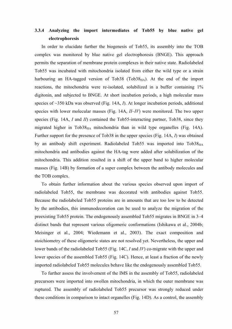
57
3.3.4 Analyzing the import intermediates of Tob55 by blue native gel
electrophoresis
In order to elucidate further the biogenesis of Tob55, its assembly into the TOB
complex was monitored by blue native gel electrophoresis (BNGE). This approach
permits the separation of membrane protein complexes in their native state. Radiolabeled
Tob55 was incubated with mitochondria isolated from either the wild type or a strain
harbouring an HA-tagged version of Tob38 (Tob38HA). At the end of the import
reactions, the mitochondria were re-isolated, solubilized in a buffer containing 1%
digitonin, and subjected to BNGE. At short incubation periods, a high molecular mass
species of ~350 kDa was observed (Fig. 14A, I). At longer incubation periods, additional
species with lower molecular masses (Fig. 14A, II–IV) were monitored. The two upper
species (Fig. 14A, I and II) contained the Tob55-interacting partner, Tob38, since they
migrated higher in Tob38HA mitochondria than in wild type organelles (Fig. 14A).
Further support for the presence of Tob38 in the upper species (Fig. 14A, I) was obtained
by an antibody shift experiment. Radiolabeled Tob55 was imported into Tob38HA
mitochondria and antibodies against the HA-tag were added after solubilization of the
mitochondria. This addition resulted in a shift of the upper band to higher molecular
masses (Fig. 14B) by formation of a super complex between the antibody molecules and
the TOB complex.
To obtain further information about the various species observed upon import of
radiolabeled Tob55, the membrane was decorated with antibodies against Tob55.
Because the radiolabeled Tob55 proteins are in amounts that are too low to be detected
by the antibodies, this immunodecoration can be used to analyze the migration of the
preexisting Tob55 protein. The endogenously assembled Tob55 migrates in BNGE in 3–4
distinct bands that represent various oligomeric conformations (Ishikawa et al., 2004b;
Meisinger et al., 2004; Wiedemann et al., 2003). The exact composition and
stoichiometry of these oligomeric states are not resolved yet. Nevertheless, the upper and
lower bands of the radiolabeled Tob55 (Fig. 14C, I and IV) co-migrate with the upper and
lower species of the assembled Tob55 (Fig. 14C). Hence, at least a fraction of the newly
imported radiolabeled Tob55 molecules behave like the endogenously assembled Tob55.
To further assess the involvement of the IMS in the assembly of Tob55, radiolabeled
precursors were imported into swollen mitochondria, in which the outer membrane was
ruptured. The assembly of radiolabeled Tob55 precursor was strongly reduced under
these conditions in comparison to intact organelles (Fig. 14D). As a control, the assembly

58
of Tom20 into the TOM complex was not affected by rupturing the outer membrane.
Hence, opening of the outer membrane interferes with the assembly of Tob55.
Figure 14. The assembly of Tob55 is inhibited upon opening of the outer membrane. A. The radiolabeled precursor of Tob55 was incubated with wild type and Tob38HA mitochondria at 25°C for the indicated time periods. Mitochondria were re-isolated and analyzed by BNGE followed by autoradiography. Species containing the radiolabeled Tob55 are indicated (I, II, III, and IV). B. Tob38 interacts with the precursor of Tob55. Wild type and Tob38HA mitochondria were incubated with the radiolabeled Tob55 precursor for 3 min at 25°C. Mitochondria were re-isolated and resuspended in a buffer containing 1% digitonin. One half of the samples was analyzed directly by BNGE, whereas the other half was incubated before analysis with antibodies against the HA-tag. The radiolabeled bands are indicated as in A. The unspecific band, which is often observed in BNGE, is indicated with an asterisk (Meisinger et al., 2004; Paschen, 2004). C. The radiolabeled precursor of Tob55 was incubated with wild type mitochondria at 25°C for 3 or 15 min. Mitochondria were re-isolated and analyzed by BNGE followed by autoradiography and immunodecoration with antibodies against Tob55. Species containing the radiolabeled Tob55 are indicated as in A. D. Opening of the outer membrane blocks the assembly of Tob55. Isolated mitochondria were preincubated in isotonic buffer (–SW) or in hypotonic buffer (+SW) for 30 min on ice. The mitochondria were re-isolated and incubated with radiolabeled precursors of Tob55 and Tom20 for the indicated time periods. At the end of the import reactions, mitochondria were analyzed by BNGE and autoradiography. The TOM complex and the assembly intermediates of Tob55 are indicated. *, unspecific band.
min 153 153
III
C
D
A
min 3 10 20 3 10 20 2 5 10 2 5 10- SW - SW+ SW + SW
35S-Tob55 35S-Tom20
III
TOM
Wild
type
Tob3
8 HA
α-HA - + - +
IV
*I
*
III
III
Wild type Tob38HA
B
35S-Tob55 α-Tob55
min 153 153
III
III
IV
IV
min 153 153
III
C
D
A
min 3 10 20 3 10 20 2 5 10 2 5 10- SW - SW+ SW + SW
35S-Tob55 35S-Tom20
III
TOM
Wild
type
Tob3
8 HA
α-HA - + - +
IV
*I
*
III
III
Wild type Tob38HA
B
35S-Tob55 α-Tob55
min 153 153
III
III
IV
IV

59
3.3.5 Assembly of Tob55 and Mas37 precursors into preexisting TOB complexes
The last stage in the import pathway of Tob55 and Mas37 is the assembly of these
precursors into preexisting TOB complexes. To assess this notion, radiolabeled Tob55 or
Mas37 were imported into mitochondria harbouring a His-tagged version of Tob38
(Waizenegger et al., 2004). After the import reaction was finished, mitochondria were
reisolated, solubilized in a buffer containing digitonin and incubated with Ni-NTA beads.
The beads were washed, bound material was eluted and analysed onto SDS-PAGE. Both
precursors were co-isolated with Tob38His, whereas no such interaction was observed
with the control precursor of Tom20 (Fig. 15A). To further investigate the role of
preexisting TOB machinery in the biogenesis of newly synthesized Tob55 and Mas37,
mitochondria harbouring mutated TOB complexes were used. In the absence of Tob38, or
upon depletion of preexisting Tob55, the insertion of Tob55 was strongly impaired (Fig.
15B and C). This reduction could be the outcome of the essential role of Tob55 in the
assembly of all β-barrel proteins in the outer membrane and/or the result of the
requirement of newly synthesized Tob55 molecules to assemble with preexisting Tob55
molecules. Only moderate reduction was observed when Tob55 was imported into
mitochondria lacking Mas37 (Fig. 15C). These results are consistent with the
observations that within the TOB complex, Tob55 interacts tightly with Tob38 and to a
lesser extent with Mas37 (Ishikawa et al., 2004b; Waizenegger et al., 2004).
To gain further insights into the interaction between Tob38 and Tob55, the BNGE
approach, in combination with carbonate extraction, was used. Tob55 is embedded in the
outer membrane, whereas its partner protein, Tob38, is only associated with the
membrane and can be extracted by an alkaline solution (Waizenegger et al., 2004).
Radiolabeled precursor of Tob55 was imported into mitochondria. The organelles were
reisolated, alkaline extraction was performed and the membrane pellet was analyzed by
BNGE. Interestingly, in mitochondria subjected to alkaline extraction no assembly
intermediates of Tob55 were observed (Fig. 15D). In addition, the endogenous TOB
complex was hardly detected upon immunostaining with antibodies against Tob55,
whereas the TOM complex and porin assemblies were not affected (Fig. 15E). Taken
together, these results lend further support to the importance of Tob38 in the assembly
and stability of the TOB complex. It might be that the extraction of Tob38 results in
destabilization and aggregation of its partner protein Tob55; therefore Tob55 cannot be
detected by BNGE.

60
Figure 15. Tob55 assembles into preexisting TOB complexes. A. wild-type and Tob38His mitochondria were incubated with the indicated radiolabeled precursors. Mitochondria were re-isolated and lysed in a buffer containing 1% digitonin. After lysis, a portion (total 20%) was removed. The rest was incubated with Ni-NTA beads. These were washed and eluted with an SDS-containing buffer (Bound). Fractions were analyzed by SDS-PAGE and autoradiography. B. mitochondria isolated from wild type and Tob38-depleted cells (Tob38↓) were incubated at 25°C with radiolabeled precursors of Tob55 for various time periods. After import, the samples were treated with proteinase K, and mitochondria were re-isolated. Inserted proteins were analyzed by SDS-PAGE and autoradiography. The proteolytic fragments of Tob55 (F' and F'') are indicated. C. mitochondria isolated from wild-type, ∆mas37, and Tob55↓ cells were incubated with radiolabeled Tob55 precursor. Further treatment and quantification were as described in the legend of Fig. 11. D. the assembly intermediates of Tob55 are not stable under alkaline conditions. Mitochondria were incubated with radiolabeled precursors of Tob55 for the indicated time periods. Samples were halved and re-isolated. One half was suspended in 100 µl of 20 mM Hepes-KOH, pH 7.4, while the other portion was suspended in 100 µl of 10 mM Hepes-KOH, 0.1 M Na2CO3 for alkaline extraction (Alk. Ex.). After 30 min on ice, the samples were centrifuged, and pellets were analyzed by BNGE. The radiolabeled bands are indicated as in Fig. 14A. E. The TOB complex is not stable under alkaline conditions. Mitochondria isolated from the wild type strain were analyzed directly by BNGE or were treated with alkaline solution as in D before such analysis. Immunodecoration with the indicated antibodies was performed.
Tob55
Mas37
Tom20
Wilde type Tob38His
Total (20%)
Bound
C
A
Wild type
∆Mas37
- Al. Ex. + Al. Ex.min 103 20 103 20 4040
D
0
20
40
60
80
100
0 5 10 15 20
Impo
rt (%
of c
ontro
l)
Time (min)
Tob55↓
III
III
Bmin 103 20 103 20
Wild type Tob38
F´F´´Total
(20%)Bound
EAlk.Ex. +- - -+ +
αPorin αTob55 αTom40
IV
Tob55
Mas37
Tom20
Wilde type Tob38His
Total (20%)
Bound
C
A
Wild type
∆Mas37
- Al. Ex. + Al. Ex.min 103 20 103 20 4040
D
0
20
40
60
80
100
0 5 10 15 20
Impo
rt (%
of c
ontro
l)
Time (min)
Tob55↓
III
III
Bmin 103 20 103 20
Wild type Tob38Tob38
F´F´´Total
(20%)Bound
EAlk.Ex. +- - -+ +
αPorin αTob55 αTom40
IV

61
Next, the association of Mas37 with the TOB complex and the requirement of each
component of this complex were investigated. Surprisingly, when mitochondria lacking
Mas37 were used, the association of Mas37 precursor molecules with mitochondria and
their assembly into the TOB complex were increased by at least 2-fold (Fig. 16A, B).
However, depletion of either Tob38 or Tob55 had an opposite effect. Mas37 precursor
was not assembled into high molecular mass complexes when either Tob38 or Tob55
were depleted (Fig. 16B). Thus, newly synthesized Mas37 molecules do not need
preexisting Mas37 molecules on mitochondria but rather require Tob55 and Tob38 for
their proper targeting and assembly. These results are consistent with the previous
observation that the endogenous levels of Mas37 in mitochondria depleted for either
Tob38 or Tob55 are clearly reduced (Waizenegger et al., 2004). It appears that precursors
of Mas37 assemble directly with the TOB core complex, which is composed of Tob38
and Tob55.
Figure 16. Mas37 associates directly with the TOB core complex. A. mitochondria isolated from wild-type and mas37 cells were incubated at 25 °C with radiolabeled Mas37 precursor for the indicated time periods. Further treatment and quantification were as described in the legend of Fig. 11. B. mitochondria isolated from wild-type (WT), ∆mas37, Tob38↓, and Tob55↓ cells were incubated at 25 °C for 20 min with radiolabeled Mas37 precursor. Mitochondria were re-isolated and analyzed by BNGE and autoradiography. The bands corresponding to the assembled Mas37 (Wiedemann et al., 2003) were quantified, and the amount of assembled material in the wild-type strain was set to 100%.
0
50
100
150
200
250
WT ∆Mas37 Tob38↓ Tob55↓
B
A
Ass
embl
ed M
as37
(%
of c
ontro
l)
0
50
100
150
200
250
0 5 10 15 20
Wild type
∆Mas37
min 3 10 20 3 10 20Wild Type ∆Mas37
Impo
rt (%
of c
ontro
l)
Time (min)
0
50
100
150
200
250
WT ∆Mas37 Tob38↓ Tob55↓
B
A
Ass
embl
ed M
as37
(%
of c
ontro
l)
0
50
100
150
200
250
0 5 10 15 20
Wild type
∆Mas37
min 3 10 20 3 10 20Wild Type ∆Mas37
Impo
rt (%
of c
ontro
l)
Time (min)

62
3.4 The N-terminal domain of Tob55 has a receptor-like function in the
biogenesis of mitochondrial β-barrel proteins
In the previous chapters, I described the various components that compose the TOB
complex, as well as the import and the assembly pathways of Tob55 and Mas37 into the
mitochondrial outer membrane. Despite some progress in understanding the assembly
and the structure of the TOB complex, many questions as to the function of its subunits
and their domains remain to be answered.
Tob55 is anchored in the outer membrane by the conserved C-terminal β-barrel
domain which most probably mediates the insertion function of this protein. The N-
terminal domain of Tob55, comprising about 100 amino acid residues, is predicted not to
be part of the β-barrel structure (http://www.imtech.res.in/cgibin/betatpred2/). It appears
to be exposed to the intermembrane space (Paschen et al., 2003). This suggests that this
domain might interact with precursors of β-barrel membrane proteins that are on their
way from the TOM to the TOB complex to become inserted into the outer membrane. In
this part of the study, I investigated the contribution of the N-terminal domain of Tob55
to the function of the protein.
As the location of this domain is crucial for such a working model, its topology was
analyzed. To this end, a Tob55 variant that carried a His8-tag at the N-terminus was
employed (Paschen et al., 2003) (Fig. 17A) and the proteolytic assay mentioned in 3.3.1
was used. Treatment of mitochondria carrying His8-tagged Tob55 with PK resulted, as
expected, in the formation of a 30 kDa fragment which could be immunodecorated with
antibodies against the His-tag (Fig. 17B). Formation of this fragment was abolished when
the outer membrane was ruptured by either osmotic shock or by solubilizing the
membrane with the detergent Triton X-100 (Fig. 17B). It is unlikely that the protection of
the His-tag against PK resulted from a stretch of eight Histidine residues buried within the
hydrophobic core of the membrane. Hence, these results imply that the N-terminus of
Tob55 is indeed exposed to the IMS.

63
Figure 17. The N-terminal domain of Tob55 is facing the intermembrane space. A. Schematic representation of cleavage of Tob55 by proteinase K (PK) resulting in two proteolytic fragments. B. Mitochondria from cells expressing His-Tob55 were isolated and were either left intact, subjected to osmotic swelling (SW) or solubilized with Triton X-100 (TX-100). Mitochondria were incubated for 15 min on ice in the absence or presence of 50 µg/ml PK. All samples were subjected to SDS-PAGE and immunoblotting with antibodies against His-tag (upper panel) or against the marker proteins DLD1 (IMS), Tom40 (embedded in the outer membrane) and Tom20 (an outer membrane protein with a large cytosolic domain). Full-length Tob55 and the N-terminal fragments are indicated.
3.4.1 Tob55 precursor devoid of its N-terminal domain is targeted to and
assembled into the outer membrane of mitochondria
To study the possible functions of the N-terminal domain of Tob55, constructs were
created in which 50, 80 or 102 of the N-terminal amino acid residues were deleted
resulting in Tob55∆50, Tob55∆80 and Tob55∆102, respectively. First, the targeting and
the insertion of these proteins into the outer membrane of mitochondria were checked in
vivo. Accordingly, Tob55 variants were cloned into a yeast expression vector, and wild
type cells were transformed with the resulting plasmids. Upon sub-cellular fractionation
all Tob55 variants were found in the mitochondrial fraction, like the mitochondrial
marker proteins Tom20 and porin (Fig. 18A), demonstrating the correct sub cellular
localization of these variants.
Mitochondrial targeting and membrane integration of Tob55 variants were further
studied using the in vitro import assay (see above and 3.3.1) with radiolabeled precursor
PK
N-His8-
C
A cytosol
OM
IMS
PK - + +- + -TX-100SW
His8-Tob55
His8-N-terminal fragment
B
Tom20
Tom40
DLD1
45
35
25
kDa

64
proteins and isolated mitochondria. Correct in vitro membrane insertion of the N-terminal
truncated variants was expected to result in smaller N-terminal fragments whereas the
fragments representing the C-terminal part of the protein were expected to remain
unchanged. Indeed, upon incubation of radiolabeled Tob55 truncated variants with
isolated mitochondria, smaller N-terminal proteolytic fragments were formed. Thus, all
the variants appear to be targeted to mitochondria and become inserted into the outer
membrane to reach a native topology (Fig. 18B).
Figure 18. The N-terminal domain of Tob55 precursor molecules is not required for targeting and membrane insertion. A. Wild type yeast cells were transformed with either empty yeast expression vector (pYX132) or with vector encoding the indicated variant of Tob55. Cells were ruptured by vortexing in the presence of glass beads. Crude mitochondria were pelleted by differential centrifugation (P) and were analyzed together with the supernatant fraction (S) by SDS-PAGE. The membrane was immunodecorated with polyclonal antibodies against Tob55 or with antibodies against the outer membrane marker proteins Tom20 and porin. B. Radiolabeled Tob55 variants were incubated at 25°C for the indicated time periods in the absence or presence of isolated mitochondria. The samples were divided into aliquots. Two of the aliquots were left untreated (-PK), the other aliquots were treated with 50 µg/ml PK at 0°C (+PK). Samples were subjected to SDS-PAGE and autoradiography. The migration of molecular mass markers is indicated on the left. Untreated radiolabeled precursors, N-terminal proteolytic fragments (arrowhead), and C-terminal proteolytic fragments (asterisk) are indicated.
min 20 103 20
Tob55∆80Tob55∆5050%input
+ mito.
PK - ++ +- +20 103 20
50%input
+ mito.
- ++ +- +
min 20 103 2050%input
+ mito.
PK - ++ +- +20 103 20
50%input
+ mito.
- ++ +- +
Tob55FLTob55∆102
45
35
25
18
45
35
2518
A
B
P PS S P PS S
Tob55
porin
Tom20
FL
pYX132Tob55-
FLTob55-
∆80Tob55-∆102
*
*

65
The import of the newly synthesized Tob55 precursors resembles the insertion route
of other β-barrel precursors. Accordingly, efficient insertion requires the import receptor
Tom20, the translocation pore of the TOM complex and the preexisting TOB complex.
Using the in vitro assay, the ability of the Tob55 N-terminal variants to follow this
common pathway was confirmed. First, the import efficiency of all three N-terminal
truncated variants was reduced upon deletion of Tom20 (Fig. 19A and B). Second,
blocking the TOM channel with a large molar excess of the recombinant matrix-destined
preprotein pSu9(1-69)-DHFR strongly inhibited the import and membrane insertion of
the variants of Tob55 precursors (Fig. 19C). Third, to study the involvement of
preexisting TOB complexes, the insertion of Tob55 variants into wild type mitochondria
and into mitochondria depleted of Tob55 was compared. All Tob55 radiolabeled variants
were inserted with strongly reduced efficiency into the Tob55-depleted mitochondria
(Fig. 19D). A similar inefficiency of insertion was observed for all Tob55 variants when
mitochondria depleted of Tob38 were used (not shown). These results demonstrate that
preexisting TOB complexes are essential for the membrane insertion of all Tob55
variants. Taken together, Tob55 precursors follow the insertion pathway of β-barrel
proteins even when lacking their N-terminal domain.

66
A C
B D
Figure 19. Truncated Tob55 variants use the common insertion pathway of β-barrel precursors. A. Efficient insertion of Tob55 variants is dependent on Tom20. Radiolabeled precursors of Tob55 variants were incubated at 25°C for the indicated time periods with mitochondria from either wild type strain or from strain lacking Tom20. After import the samples were treated with PK (50 µg/ml) and mitochondria were re-isolated. Inserted Tob55 was analyzed by SDS-PAGE and autoradiography. Proteolytic fragments are indicated as described in the legend to Fig. 18B. B. Insertion of Tob55 variants after 20 min incubation with mitochondria isolated from WT and ∆tom20 strain was quantified by monitoring the formation of the 30 kDa N-terminal fragment. The amount of precursor protein inserted into wild type mitochondria was set to 100%. C. The insertion of Tob55 variants is affected in the presence of excess matrix-targeted precursor. Radiolabeled precursors of Tob55 variants were added to isolated mitochondria in the absence or presence of recombinant pSu9-DHFR. The samples were incubated at 25°C for the indicated time periods. PK was added and proteins were analyzed by SDS-PAGE and autoradiography. Proteolytic fragments are indicated as described in the legend to Fig. 18B. Non-cleaved radiolabeled precursors are indicated by a circle. D. Insertion of Tob55 variants requires preexisting Tob55. Mitochondria isolated from either wild type or Tob55 depleted cells (Tob55↓) were incubated with radiolabeled precursors at 25°C for various time periods. Samples were treated with PK and mitochondria were re-isolated. Inserted proteins were analyzed by SDS-PAGE and autoradiography. Proteolytic fragments are indicated as described in the legend to Fig. 18B. Non-cleaved radiolabeled precursors are indicated by a circle.
Tob55∆50 Tob55∆102
min 103 20 103 20 103 20 103 20∆tom20WT WT ∆tom20
*
0
20
40
60
80
100
∆50 ∆80 ∆102
Inse
rtion
(% o
f con
trol)
WT ∆tom20
Tob55 variantsFL
min 103 20 103 20 103 20 103 20
Tob55∆50Tob55FL+ pSu9-DHFR
- pSu9-DHFR
+ pSu9-DHFR
- pSu9-DHFR
*
Tob55∆102Tob55∆80
*
° °
°°
min 103 20 103 20 103 20 103 20Tob55 WT
Tob5
5∆50
Tob5
5FL
Tob55WT
*
*
Tob5
5∆80
*
Tob5
5∆10
2
*
° °
°°

67
3.4.2 The truncated variants of Tob55 become assembled into pre-existing TOB
complexes
The assembly of Tob55 truncated variants into the pre-existing TOB complexes was
monitored by BNGE. As described above (see 3.3.4), the TOB complex migrates upon
BNGE as several species in which newly imported Tob55 becomes assembled (Habib et
al., 2005b; Ishikawa et al., 2004a; Meisinger et al., 2004). Initially, the radiolabeled
molecule of Tob55 migrate preferentially with the uppermost one (specie I) whereas upon
an extended incubation period (which may allow an assembly into the pre-existing TOB
complexes) the precursor molecules migrate mostly with another specie (specie II). This
specie migrates at an apparent molecular mass smaller than the intermediate of Tom40
precursor associated with the TOB complex. The three truncated variants assembled into
the same complex as that of the full-length protein (Fig. 20A). Moreover, the resistance
of this assembly to treatment with either high salt concentrations or 2 M urea was not
affected by the deletion of the N-terminal domain (not shown). Next, radiolabeled Tob55
variants were incubated with mitochondria isolated from either wild type or a strain
containing HA-tagged version of Tob38 (Tob38HA). The TOB complex from the latter
mitochondria migrates slower in BNGE than in the native complex (see 3.3.4). The
radioactive Tob55 species imported into the Tob38HA mitochondria showed the same
reduced electrophoretic mobility (Fig. 20B) and were in a complex which was recognized
by antibodies against the HA tag (Fig. 20C). This supports the notion that all studied
Tob55 variants become assembled into preexisting TOB complexes.
To obtain additional independent support for this conclusion antibody shift
experiments were performed (Paschen et al., 2003; Wiedemann et al., 2003). Antibodies
raised against a peptide comprising amino acid residues 1-15 of Tob55, which do not
recognize the two truncated variants, Tob55∆80 and Tob55∆102 were used.
Mitochondria with inserted radiolabeled Tob55 variants were lysed, incubated with
antibodies against the N-terminal peptide of Tob55 (or against Tim23 as control) and
then analyzed by BNGE. The addition of the anti-Tob55-N antibody (but not of the
control antibody) resulted in a shift of the upper precursor-containing band to a higher
apparent molecular mass by formation of a supercomplex with the antibodies (Fig. 20D).
Taken together, the absence of the N-terminal domain of the Tob55 precursor did not
impair its ability of Tob55 precursor to become inserted into the outer membrane and
assembled into preexisting TOB complexes.
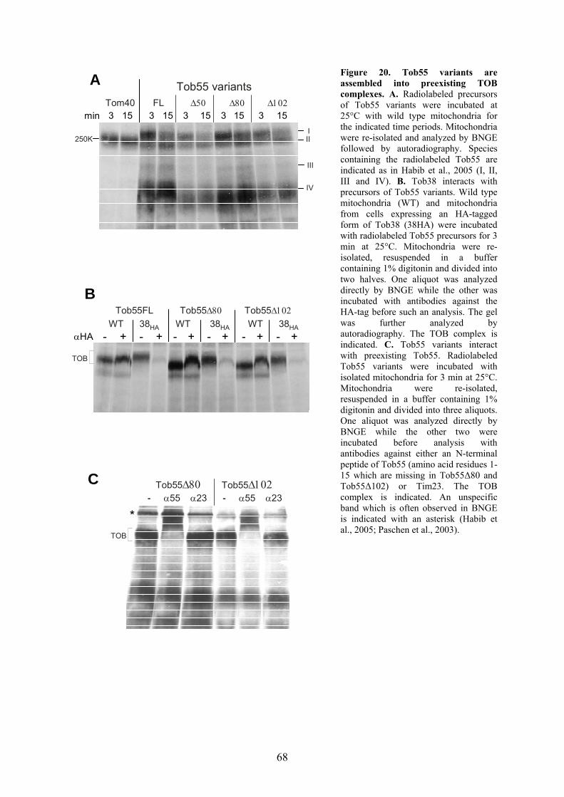
68
Figure 20. Tob55 variants are assembled into preexisting TOB complexes. A. Radiolabeled precursors of Tob55 variants were incubated at 25°C with wild type mitochondria for the indicated time periods. Mitochondria were re-isolated and analyzed by BNGE followed by autoradiography. Species containing the radiolabeled Tob55 are indicated as in Habib et al., 2005 (I, II, III and IV). B. Tob38 interacts with precursors of Tob55 variants. Wild type mitochondria (WT) and mitochondria from cells expressing an HA-tagged form of Tob38 (38HA) were incubated with radiolabeled Tob55 precursors for 3 min at 25°C. Mitochondria were re-isolated, resuspended in a buffer containing 1% digitonin and divided into two halves. One aliquot was analyzed directly by BNGE while the other was incubated with antibodies against the HA-tag before such an analysis. The gel was further analyzed by autoradiography. The TOB complex is indicated. C. Tob55 variants interact with preexisting Tob55. Radiolabeled Tob55 variants were incubated with isolated mitochondria for 3 min at 25°C. Mitochondria were re-isolated, resuspended in a buffer containing 1% digitonin and divided into three aliquots. One aliquot was analyzed directly by BNGE while the other two were incubated before analysis with antibodies against either an N-terminal peptide of Tob55 (amino acid residues 1-15 which are missing in Tob55∆80 and Tob55∆102) or Tim23. The TOB complex is indicated. An unspecific band which is often observed in BNGE is indicated with an asterisk (Habib et al., 2005; Paschen et al., 2003).
A
Tob55FL Tob55∆102WT 38HA
BTob55∆80
TOB
Tob55∆102Tob55∆80α55- α23 α55- α23
C
*
αHA - +WT 38HA WT 38HA
- + - + - + - + - +
TOB
minFL ∆102∆80∆50
IV
III
Tob55 variants
3 15 3 15 3 15 3 15 3 15Tom40
III250K

69
3.4.3 Deletion of the N-terminal domain of Tob55 results in a growth phenotype of
yeast cells
Does the absence of the N-terminal domain of Tob55 hinder the function of the
protein? In order to test the ability of the truncated variants to complement the function of
the endogenous molecules, the “plasmid shuffling” method was applied. Accordingly, an
URA3 plasmid expressing full-length Tob55 was transformed into a wild type haploid
strain. Next, the genomic TOB55 open reading frame in this strain was deleted resulting
in a strain where Tob55 is plasmid-encoded (strain YSH1). This strain was then
transformed with TRP1 plasmid expressing the full-length or truncated versions of
Tob55, and cells containing both plasmids were selected. Cells harboring both plasmids
were grown on plates containing 5-FOA to eliminate the wild type copy of Tob55 which
is encoded on the URA3-containing plasmid. Those strains that harbored a TRP1 plasmid
encoding either a full length or truncated form of Tob55 were tested for their ability to
grow on glycerol- and glucose-containing medium at various temperatures (Fig. 21A).
Growing the cells at 30°C resulted in only minor differences in the growth rates of the
various cells. In contrast, incubating the cells at 37°C or 24°C resulted in a slower growth
in the case of cells expressing Tob55∆80 and even more so in cells harboring
Tob55∆102. As expected, the growth phenotype was more conspicuous on the non-
fermentable carbon source where yeast cells are dependent on mitochondria for energy
production (Fig. 21A). Thus, already the first 80 amino acid residues of Tob55 are
required for optimal function of Tob55 and subsequently for normal growth of yeast
cells.
3.4.4 Deletion of the N-terminal domain of Tob55 results in impaired biogenesis of
β-barrel proteins
Due to the growth phenotype caused by the variants of Tob55, the biogenesis of β-
barrel proteins was investigated. Mitochondria were isolated from cells harboring
plasmid-encoded full-length Tob55 or its truncated variants and the amounts of expressed
proteins were controlled by immunodecoration. The levels of the β-barrel proteins
Tom40, Mdm10 and porin were strongly reduced in mitochondria containing the
truncated versions. Similarly, the levels of the truncated variants of Tob55 (β-barrel
proteins themselves) and the other two components of the TOB complex, Tob38 and
Mas37 were also reduced as compared to mitochondria containing full-length Tob55 as
analyzed by either SDS-PAGE or BNGE (Fig. 21B and 21C). In contrast, other proteins

70
of the various mitochondrial sub-compartments like Fis1 and Tom70 (OM), Tim70 and
AAC (IM) and Yah1 (matrix) were present at roughly control levels (Fig. 21B). Thus, the
N-terminal domain of Tob55 appears to have an important role in the biogenesis of β-
barrel proteins.
A
B C
Figure 21. The N-terminal domain of Tob55 is required for optimal growth. A. Cells harboring plasmid-encoded Tob55 variants were constructed as described in Materials and Methods. The cells were tested by dilution in 10-fold increments for their ability to grow at the indicated temperature on mediums containing either glycerol or glucose. B-C. Mitochondria were isolated from cells harboring plasmid-encoded full-length Tob55 (Tob55FL) or its truncated variants. The indicated amounts of mitochondria were analyzed by SDS-PAGE or BNGE and immunodecoration with antibodies against the indicated mitochondrial proteins.
Tob55FL
Tob55∆80
Tob55∆102
YP-glucose YP-glycerol24° C
37° C 37° C
24° C
30° C 30° CTob55FL
Tob55∆80
Tob55∆102
Tob55FL
Tob55∆80
Tob55∆102
µg 50 150 150 100 50 150 150 100
αTob55 αTob38
FL ∆80
∆TOB55 +WT∆102 FL ∆80
∆TOB55 +WT∆102
Tob55
Tom70
Tom40
Fis1
AAC
Tim17
µg 10 30 100 10 30 100
Yah1
Tob55FL Tob55∆8010 30 100
Tob55∆102
Porin
Tob38
Mdm10
Mas37

71
The assembly state of the TOB complex in the various mitochondria which harbor the
truncated version of Tob55 was analyzed by BNGE, a method which usually results in
several observed species of TOB complex. Due to the fact that mitochondria harboring
the truncated versions of Tob55 contain reduced amounts of this protein, larger amount of
these mitochondria were analyzed. The TOB complex from mitochondria harboring the
truncated versions migrated mainly as the higher molecular specie of the TOB complex.
Of note, all the Tob38 molecules in these mitochondria were assembled with Tob55 (Fig.
21C), excluding the possibility that due to the reduced levels of Tob55, Tob38 and Mas37
build partial complexes which exert a dominant negative effect. The full length Tob55
displayed a different migration pattern where a significant portion of it was found as low
molecular weight unassembled species (Fig. 21C). This behavior probably resulted from
the fact that, like the other Tob55 variants, it was expressed from an over-expression
plasmid whereas the interacting partners Tob38 and Mas37, are not over-expressed.
To emphasize the involvement of the N-terminal domain in biogenesis of β-
barrel proteins, in vitro protein import experiments were performed. Concomitantly to the
in vivo data, the import efficiencies of newly synthesized β-barrel precursors like Tom40
and porin were significantly reduced in mitochondria containing the truncated versions
(Fig. 22A). Other precursor proteins such as the inner membrane protein Tim23, and the
matrix-destined pSu9-DHFR were only moderately affected (Fig. 22A). This reduction is
probably due to the reduced level of the major subunit of the general import pore,
Tom40, in the mitochondria harboring the truncated variants. As mentioned above (see
3.3.5), the assembly of newly synthesized Tob55 and the association of the Mas37
precursor with mitochondria require preexisting Tob55 molecules. To check the
involvement of the N-terminal domain in the biogenesis of both proteins, in vitro proteins
import were performed. As expected, due to the reduced levels of Tob55 a moderate
reduction in the association of Mas37 was observed upon import into mitochondria with
truncated variants of Tob55 (Fig. 22B). A stronger reduction was observed upon import
of Tob55 precursor, a β-barrel protein itself (Fig. 22B). Thus, whereas the association of
Mas37 with mitochondria is probably reduced due to the lower levels of Tob55, the
insertion of Tob55 precursor (a β-barrel protein itself) is affected by both the reduced
levels of Tob55 and the absence of the N-terminal domain.
To exclude the possibility that the impaired biogenesis of β-barrel proteins could
be due to the reduced level of the endogenous truncated Tob55 variants in comparison to
the full length, in vitro control import experiments were performed. Radiolabeled

72
precursor proteins were incubated with either 50 µg of mitochondria harboring plasmid-
encoded full length Tob55, 150 µg of mitochondria harboring Tob55∆80 or with 100 µg
wild type (YPH499) mitochondria. Under these conditions equivalent amounts of TOB
and TOM complexes were present in import reactions with the two former types of
mitochondria (Fig. 22C). The matrix-destined protein, pSu9-DHFR, was imported with
similar efficiency in all reactions under these conditions. In contrast, the import of the β-
barrel precursor porin (Fig 22D) into mitochondria carrying the truncated Tob55 variant
was still impaired. Of note, although the samples with wild type mitochondria contain far
less Tob55 molecules than those containing Tob55∆80, the efficiency of porin import
into the former mitochondria was significantly higher (Fig. 22D). Furthermore, the
assembly of the β-barrel precursors Mdm10 (Fig. 22E) and Tom40 (Fig. 22F) was
strongly impaired in mitochondria harboring the truncated version of Tob55. Thus, the
reduced import of β-barrel precursors into mitochondria harboring truncated Tob55
variants is caused by the impaired function of the corresponding Tob55 molecules and is
not due to a reduced level of Tob55.
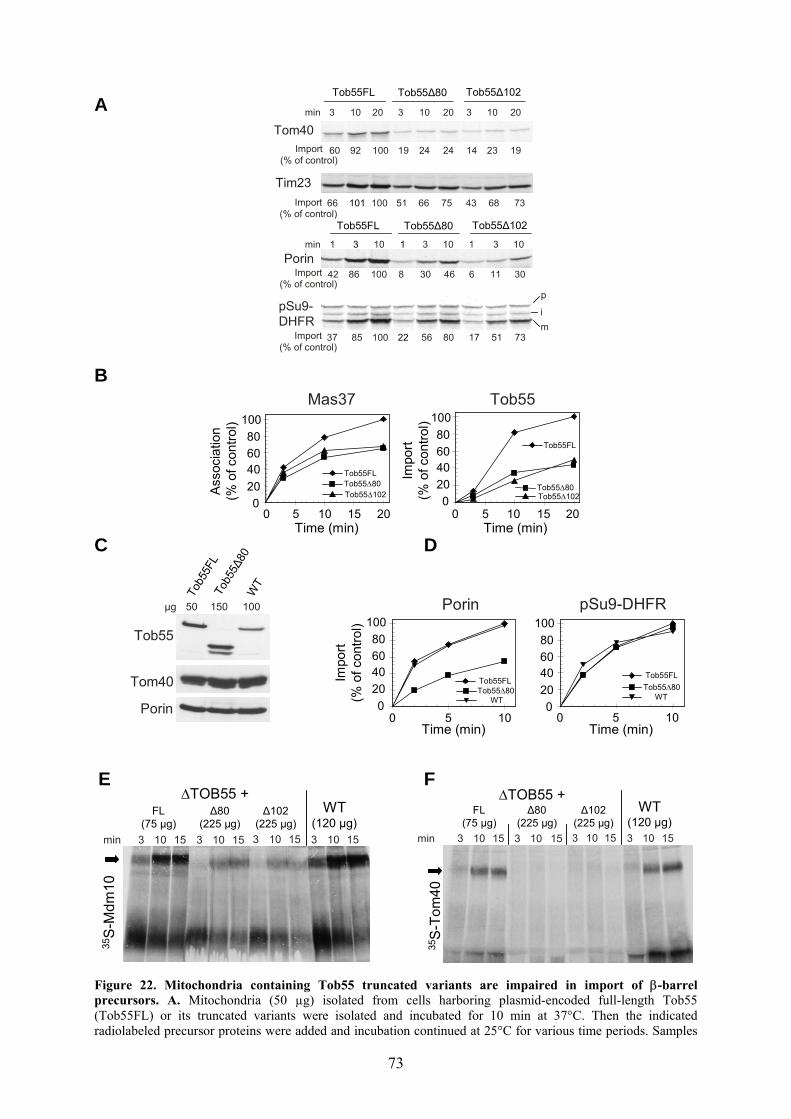
73
A
B
C D
E F
Figure 22. Mitochondria containing Tob55 truncated variants are impaired in import of β-barrel precursors. A. Mitochondria (50 µg) isolated from cells harboring plasmid-encoded full-length Tob55 (Tob55FL) or its truncated variants were isolated and incubated for 10 min at 37°C. Then the indicated radiolabeled precursor proteins were added and incubation continued at 25°C for various time periods. Samples
020406080
100
0 5 10 15 20Time (min)
Impo
rt (%
of c
ontro
l)
020406080
100
0 5 10 15 20Time (min)
Mas37 Tob55
Asso
ciat
ion
(% o
f con
trol)
Tob55FLTob55∆80Tob55∆102
Tob55FL
Tob55∆80Tob55∆102
Tob55FLTob55∆80
WT0
20406080
100
0 5 10Time (min)
pSu9-DHFR
0 5 10Time (min)
020406080
100Porin
Tob55FLTob55∆80
WT
Impo
rt (%
of c
ontro
l)
min 3 10 15
FL(75 µg)
∆80(225 µg)3 10 15 3 10 15 3 10 15
∆TOB55 +WT
(120 µg)∆102
(225 µg)
35S
-Tom
40
min 3 10 20 3 10 20
Tob55FL Tob55∆80
3 10 20
Tob55∆102
Tom40Import
(% of control)60 92 100 19 24 24 14 23 19
min 1 3 10 1 3 10
Tob55FL Tob55∆80
1 3 10
Tob55∆102
pSu9-DHFR
p
mi
Import(% of control)
37 85 100 22 56 80 17 51 73
PorinImport
(% of control)42 86 100 8 30 46 6 11 30
Tim23Import
(% of control)66 101 100 51 66 75 43 68 73
Tob5
5FL
Tob5
5∆80
WT
Porin
Tom40
Tob55
µg 50 150 100
min 3 10 15
FL(75 µg)
∆80(225 µg)3 10 15 3 10 15 3 10 15
∆TOB55 +WT
(120 µg)∆102
(225 µg)
35S-
Mdm
10

74
were treated with PK and mitochondria were re-isolated. Imported proteins were analyzed by SDS-PAGE and autoradiography. Membrane insertion of porin and Tom40 as well as translocation of the Tim23 precursor across the outer membrane were quantified by analyzing the protease protected precursors, whereas for pSu9-DHFR the bands corresponding to the mature protein were quantified. The amount of precursor proteins imported into mitochondria harboring full-length Tob55 for the longest time period was set to 100%. The precursor, intermediate, and mature forms of pSu9-DHFR are indicated as p, i and m, respectively. B. Radiolabeled precursors of Mas37 and Tob55 were incubated with mitochondria (50 µg) as in (A). The import (Tob55) and association (Mas37) of the precursor proteins were assayed by the formation of proteolytic fragments (Fig. 2 and (Habib et al., 2005b)). The amount of precursor proteins imported into or associated with mitochondria harboring full-length Tob55 for the longest time period was set to 100%. C. Mitochondria were isolated from cells harboring plasmid-encoded full-length Tob55 or Tob55∆80 or from wild type cells. The indicated amounts of mitochondria were analyzed by SDS-PAGE and immunodecoration with antibodies against the indicated outer membrane mitochondrial proteins. D. Mitochondria (50 µg Tob55FL, 150 µg Tob55∆80, and 100 µg WT) were incubated for 10 min at 37°C. Radiolabeled precursor proteins were added and incubation continued at 25°C for various time periods. The samples were then treated with PK and the mitochondria were re-isolated. Imported proteins were analyzed by SDS-PAGE and autoradiography. The insertion of porin was quantified by analyzing the protease protected precursor, whereas for pSu9-DHFR the bands corresponding to the mature protein were quantified. The amount of precursor proteins imported into mitochondria harboring plasmid-encoded Tob55FL for the longest time period was set to 100%. E, F. Radiolabeled precursor of Mdm10 or Tom40 (E and F, respectively) was incubated with the indicated mitochondria for various time periods. Imported proteins were analyzed by BNGE and autoradiography. The β-barrel precursor-TOB intermediate is indicated by an arrow.
3.4.5 Purified N-terminal domain of Tob55 binds β-barrel precursors
To gain insights into the function of the N-terminal domain of Tob55, its ability to
bind β-barrel precursors was tested. A fusion protein consisting of the N-terminal 120
amino acid residues of Tob55 and maltose binding protein, MBP-Tob55, was expressed in
and purified from E. coli. As controls, MBP alone and MBP-Fis1 (MBP fused to the
cytosolic domain of the mitochondrial outer membrane protein, Fis1) were expressed and
purified in parallel (Fig. 23A). The three fusion proteins were purified and bound to
amylose resin. Next, radiolabeled precursors were added and the interactions with the
recombinant proteins were analyzed. MBP-Tob55 but none of the control proteins, MBP
alone and MBP-Fis1, could bind significant amounts of radiolabeled porin or Mdm10
precursors (Fig. 23B). Only background levels of a matrix destined precursor, pSu9-DHFR,
were bound (Fig. 23B). To further verify that the binding to the N-terminal domain is
specific and saturable, increasing amounts of radiolabeled Mdm10 precursor were added to
equal small amounts of MBP-Tob55 or MBP alone as a control. In each added amount
several fold more Mdm10 molecules were bound to the MBP-Tob55 as compared to the
control protein. Notably, a saturation of binding was observed when large amounts of
precursor were used. Thus, the first 120 amino acid residues of Tob55 appear to be
sufficient to support specific interaction with β-barrel precursors.
To obtain further support for this proposal, the ability of the purified N-terminal
domain to compete out the import of porin and Mdm10 was tested. To that end,
radiolabeled precursors of porin or Mdm10 were incubated in a buffer in the absence or the
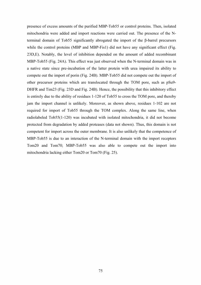
75
presence of excess amounts of the purified MBP-Tob55 or control proteins. Then, isolated
mitochondria were added and import reactions were carried out. The presence of the N-
terminal domain of Tob55 significantly abrogated the import of the β-barrel precursors
while the control proteins (MBP and MBP-Fis1) did not have any significant effect (Fig.
23D,E). Notably, the level of inhibition depended on the amount of added recombinant
MBP-Tob55 (Fig. 24A). This effect was just observed when the N-terminal domain was in
a native state since pre-incubation of the latter protein with urea impaired its ability to
compete out the import of porin (Fig. 24B). MBP-Tob55 did not compete out the import of
other precursor proteins which are translocated through the TOM pore, such as pSu9-
DHFR and Tim23 (Fig. 23D and Fig. 24B). Hence, the possibility that this inhibitory effect
is entirely due to the ability of residues 1-120 of Tob55 to cross the TOM pore, and thereby
jam the import channel is unlikely. Moreover, as shown above, residues 1-102 are not
required for import of Tob55 through the TOM complex. Along the same line, when
radiolabeled Tob55(1-120) was incubated with isolated mitochondria, it did not become
protected from degradation by added proteases (data not shown). Thus, this domain is not
competent for import across the outer membrane. It is also unlikely that the competence of
MBP-Tob55 is due to an interaction of the N-terminal domain with the import receptors
Tom20 and Tom70; MBP-Tob55 was also able to compete out the import into
mitochondria lacking either Tom20 or Tom70 (Fig. 25).

76
A B
C
D E
Figure 23. The N-terminal domain of Tob55 can bind β-barrel precursors in vitro. A. Maltose binding protein (MBP) alone or fused either to the cytosolic domain of Fis1 (MBP-Fis1) or to residues 1-120 of Tob55 (MBP-Tob55) were expressed in E. coli and purified. Proteins (5 µg each) were analyzed by SDS-PAGE and Coomassie staining. B. The indicated recombinant MBP-fusion proteins were bound to amylose resin and incubated with radiolabeled precursor proteins. The resins were washed, and bound proteins were eluted and analyzed by SDS-PAGE and autoradiography. C. Increasing amounts of radiolabeled Mdm10 in retic. lysate were incubated with MBP or MBP-Tob55 bound to amylose resin. The resins were washed and bound proteins were eluted and analyzed by SDS-PAGE and autoradiography. The highest amount of precursor protein bound to MBP-Tob55 was set to 100%. D. Purified N-terminal domain of Tob55 added to an import reaction inhibits the import of porin precursor. Radiolabeled precursors of porin or pSu9-DHFR were incubated in import buffer in the presence or absence of the indicated purified MBP-fusion proteins for 10 min at 0°C. Mitochondria were then added and the mixture was incubated for another 15 min at 25°C. At the end of the import reaction PK was added to degrade precursors that were not completely imported. Mitochondria were re-isolated and inserted proteins were analyzed by SDS-PAGE and autoradiography. The bands corresponding to protease-protected porin and to the mature form of pSu9-DHFR were quantified. The import without MBP-fusion protein was taken for each precursor as 100%. E. Radiolabeled precursor of Mdm10 was incubated for 10 min at 0°C in import buffer in the presence or absence of the indicated purified MBP-fusion proteins. Mitochondria were then added and the mixture was incubated for another 10 min at 25°C. The import reactions were analyzed by BNGE as in Fig. 22E and the bands representing Mdm10-TOB intermediate were quantified. The import without MBP-fusion protein was taken as 100%.
MBP
-Tob
55
MBP
MBP
-Fis1
3545
66kDa M
MBP
-Tob
55
MBP
MBP
-Fis1
Inpu
t (4%
)
Porin
pSu9-DHFR
Mdm10
0
20
40
60
80
100
0 50 100B
indi
ng (%
of m
ax.)
Radiolabeled Mdm10 (µl)
MBP
MBP-Tob55
min 5 20-
5 20 5 20 5 20
porin
pSu9
-D
HFR
MBPMBP-Fis1
MBP-Tob55
p
mi
Recombinant protein
Impo
rt (%
of c
ontro
l)
0
20
40
60
80
100
- MBP MBP-Fis1
MBP-Tob55
pSu9-DHFR - M
BP
MBP
-Fis1
MBP
-Tob
55
Recombinant protein
- MBPMBP-Fis1
MBP-Tob55
Impo
rt (%
of c
ontro
l)
0
20
40
60
80
100

77
A
B
Figure 24. The inhibition by MBP-Tob55 is concentration dependent and requires folded structure. A. Inhibition of porin import depends on the concentration of added recombinant MBP-Tob55. Radiolabeled precursors of porin or pSu9-DHFR were incubated in import buffer in the presence or absence of the indicated amounts of purified MBP-fusion proteins for 10 min at 0°C. Mitochondria were then added and the mixture was incubated for another 15 min at 25°C. At the end of the import reaction PK was added and further treatment was as described in the legend to Fig. 23D. B. Unfolded N-terminal domain of Tob55 cannot inhibit porin import. Mitochondria were incubated for 2 min on ice in import buffer with either native MBP-Tob55, MBP-Tob55 pretreated with urea or with diluted urea solution as a control. Radiolabeled precursors of porin, Tim23 or pSu9-DHFR were then added and the mixture was incubated further for the indicated time periods. At the end of the import reaction PK was added and further treatment was as described in the legend to Fig. 23D.
Recombinant protein (µg)
Impo
rt (%
of c
ontro
l)
Porin pSu9-DHFR
µg 0.5 10-
5MBPMBP-Fis1MBP-Tob55
20 0.5 1052 0.5 1052
Porin
pSu9
-D
HFR
p
m
020406080
100120
0 5 10
MBP-TOBMBP-Fis1MBP
0 5 10
MBP-TOBMBP-Fis1MBP
Recombinant protein (µg)
020406080
100120
i
min 103 20 103 20 103 20 103 20
MBP-Tob55
urea
MBP-Tob55+ urea
_
Porin
min 31 10 31 10 31 10 31 10m-Su9-DHFR
Tim23

78
A B
Figure 25. The absence of Tom receptors does not affect the inhibition of porin import caused by purified N-terminal domain of Tob55. Radiolabeled precursors of porin were incubated in import buffer in the presence or absence of the indicated purified MBP-fusion proteins for 10 min at 0°C. Mitochondria isolated from ∆Tom70 (A) or ∆Tom20 (B) and the corresponding wild type strains were then added and the mixture was incubated for 15 min at 25°C. At the end of the import reaction PK was added to degrade precursors that were not completely imported. Mitochondria were re-isolated and inserted proteins were analyzed by SDS-PAGE and autoradiography. The bands corresponding to protease protected porin were quantified. The amount of precursor protein imported into wild type mitochondria was set to 100%.
In order to further test the ability of the N-terminal domain to bind β-barrel precursors, its
capacity to bind to a water-soluble form of porin was analyzed (Pfaller et al., 1985). This
water-soluble porin, isolated from detergent-purified porin from N. crassa, has the properties
of the precursor form of porin and can be imported into the mitochondrial outer membrane
(Pfaller and Neupert, 1987; Pfaller et al., 1988). As all the experiments described so far were
conducted in the yeast S. cerevisiae, the ability of N. crassa porin to be imported into and
assembled in the outer membrane of yeast mitochondria was investigated. Indeed, the N.
crassa ortholog was imported into yeast mitochondria in the pathway that involved the
general insertion pore. Blocking this pathway with excess recombinant matrix-destined
preprotein, pSu9-DHFR, inhibited membrane integration (Fig. 26A) (Krimmer et al., 2001;
Paschen et al., 2003; Wiedemann et al., 2003). Furthermore, as was observed for yeast β-
barrel precursors, the import of N. crassa porin into yeast mitochondria was abrogated in
mitochondria lacking Tom20 (Fig. 26B) or harbouring reduced levels of Tob38 (Fig. 26C)
(Ishikawa et al., 2004a; Krimmer et al., 2001; Milenkovic et al., 2004; Waizenegger et al.,
2004). Porin is known to form several oligomeric structures which can be observed by
BNGE (Gentle et al., 2004; Krimmer et al., 2001; Waizenegger et al., 2004). The
0102030405060708090
100
- MBP Tob55 MBP Fis1
Impo
rt (%
of c
ontr
ol)
WT
∆Tom20
0102030405060708090
100
- MBP Tob55 MBP Fis1
Impo
rt (%
of c
ontr
ol)
WT
∆Tom20
0102030405060708090
100
- MBP Tob55 MBP Fis1
Impo
rt (%
of c
ontr
ol)
WT∆Tom70
0102030405060708090
100
- MBP Tob55 MBP Fis1
Impo
rt (%
of c
ontr
ol)
WT∆Tom70

79
radiolabeled N. crassa porin was assembled upon its import into yeast mitochondria into the
same oligomeric structures as the yeast protein (Fig. 26D). Taken together, N. crassa porin
can use the yeast machinery for biogenesis of β-barrel proteins and follows the same pathway
as its yeast ortholog.
A
B C
D E
Figure 26. Precursor of N. crassa porin is correctly inserted in vitro into yeast mitochondrial outer
membrane. A. Radiolabeled precursors of porin from either yeast or N. crassa were incubated with yeast mitochondria at 25°C for the indicated time periods in the presence or absence of excess recombinant matrix-destined precursor, pSu9-DHFR. At the end of the import reactions, PK (50 µg/ml) was added and inserted porin was analyzed by SDS-PAGE and autoradiography. B. Efficient insertion of N. crassa porin is dependent on Tom20. Radiolabeled precursors of N. crassa porin were incubated for the indicated time periods with mitochondria from either wild type strain or from strain lacking Tom20. Further treatment was as described in A. C. Efficient insertion of N. crassa porin is dependent on Tob38. Mitochondria isolated from either wild type or Tob55 depleted cells (Tob38↓) were incubated with radiolabeled precursors at 25°C for various time periods. Further treatment was as described in A. D. Precursor of N. crassa porin assembles into oligomeric structures in yeast mitochondria. Radiolabeled precursors of porin from either yeast or N. crassa were incubated with yeast mitochondria for the indicated time periods. At the end of the import reactions, mitochondria were lysed with digitonin and assembled porin was analyzed by BNGE and autoradiography. E. Water soluble porin can compete out the import of β-barrel precursors. Radiolabeled precursors of porin or Tom40 were incubated in import buffer in the presence or absence of the indicated proteins for 10 min at 0°C. Mitochondria were then added and the mixture was incubated for another 15 min at 25°C. At the end of the import reaction PK was added to degrade precursors that were not completely imported. Mitochondria were re-isolated and inserted proteins were analyzed by SDS-PAGE and autoradiography.
min 103 20103 20S.c. Porin N.c. Porin MBP
35S-Tom40
35S-Porin
WS-Porin1.8 3.6 3.6-µg
pSu9-DHFR
S.c. Porin N.c. Porin
min 41 7 41 7 41 7 41 7- + - +
3 10 20 3 10 20min
WT Tob38↓
Import (% of control)
92 112 100 43 46 48
3 10 20 3 10 20min
WT ∆Tom20
Import (% of control)
53 97 100 38 59 66

80
The overlay method described in Voulhoux et al. (Voulhoux and Tommassen, 2004) was
used to evaluate the binding of water-soluble porin with the N-terminal domain of Tob55.
Under semi-native gel electrophoresis, MBP-Tob55 migrated as two dominant bands (Fig.
27A). Various amounts of MBP-Tob55 and two control proteins were subjected to semi-
native SDS-PAGE and then they were transferred onto a nitrocellulose membrane. Water-
soluble porin was incubated with this membrane and the bound protein was detected by
immunodecoration. The N-terminal domain of Tob55 was found to specifically bind water-
soluble porin in a concentration-dependent manner (Fig. 27B). In conclusion, these results
demonstrate that the N-terminal domain of Tob55 is able to interact with precursors of β-
barrel proteins.
A B
Figure 27. The N-terminal domain of Tob55 can recognize water-soluble porin. A. MBP-Tob55 migrates on semi-native gel as multiple bands. MBP-fusion proteins (10 µg each) were analyzed by semi-native SDS-PAGE and further stained with Ponceau (left panel) or immunodecorated with antibodies against Tob55 (right panel). B. The N-terminal domain of Tob55 recognizes water-soluble porin. The indicated amounts of MBP-fusion proteins were analyzed by semi-native gel and blotted onto nitrocellulose membrane. Water-soluble porin (ws-Porin) isolated from N. crassa mitochondria (11 µg/ml) was incubated with this membrane for 2 h at 4°C, the membrane was washed and immunodecorated with antibodies against porin.
To further substantiate the capacity of the N-terminal domain to bind water-soluble porin
and to obtain more quantitative information on this binding, the interaction was examined in
solution. To that end, water-soluble porin was radiolabeled with 14C-formaldehyde by
reductive methylation using cyanoborohydride (NaBH3CN) as a reducing agent (Pfaller et
al., 1985). Increasing amounts of 14C-ws-porin were added to 2 µg of either MBP or MBP-
Tob55 bound to amylose beads. The binding was performed at low temperature (4°C) and in
the presence of BSA and salt. Under these conditions, the tendency of ws-porin to adhere to
surfaces and thus to cause unspecific binding was reduced. The beads were washed and
bound 14C-ws-porin was eluted and analyzed (Fig. 28A). To quantify the binding, increasing
amounts of 14C -ws-porin were loaded directly on a gel to serve as loading standards. Much
MBP
-Tob
55
MBP
MBP
-Fis1
MBP
-Tob
55
MBP
MBP
-Fis1
αTob55Ponceau stain
MBP
-Tob
55
MBP
MBP
-Fis1
MBP
-Tob
55
MBP
MBP
-Fis1
MBP
-Tob
55
MBP
MBP
-Fis1
2 µg 5 µg 10 µg
WS-Porin
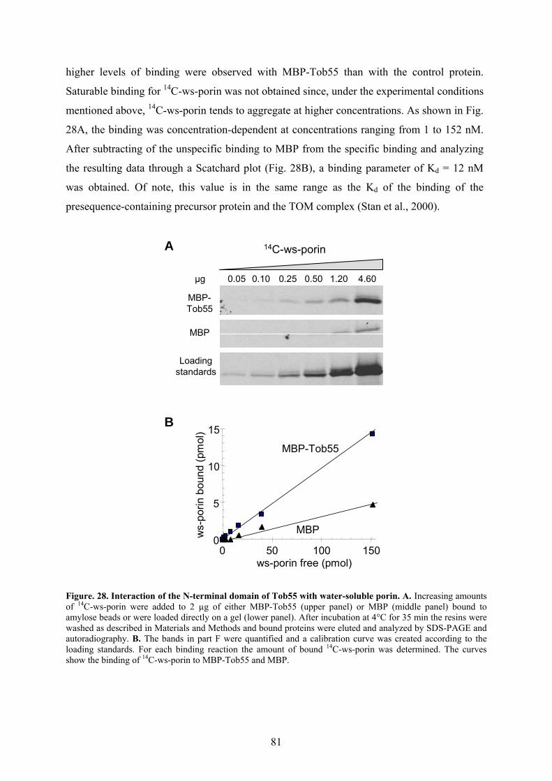
81
higher levels of binding were observed with MBP-Tob55 than with the control protein.
Saturable binding for 14C-ws-porin was not obtained since, under the experimental conditions
mentioned above, 14C-ws-porin tends to aggregate at higher concentrations. As shown in Fig.
28A, the binding was concentration-dependent at concentrations ranging from 1 to 152 nM.
After subtracting of the unspecific binding to MBP from the specific binding and analyzing
the resulting data through a Scatchard plot (Fig. 28B), a binding parameter of Kd = 12 nM
was obtained. Of note, this value is in the same range as the Kd of the binding of the
presequence-containing precursor protein and the TOM complex (Stan et al., 2000).
A
B
Figure. 28. Interaction of the N-terminal domain of Tob55 with water-soluble porin. A. Increasing amounts of 14C-ws-porin were added to 2 µg of either MBP-Tob55 (upper panel) or MBP (middle panel) bound to amylose beads or were loaded directly on a gel (lower panel). After incubation at 4°C for 35 min the resins were washed as described in Materials and Methods and bound proteins were eluted and analyzed by SDS-PAGE and autoradiography. B. The bands in part F were quantified and a calibration curve was created according to the loading standards. For each binding reaction the amount of bound 14C-ws-porin was determined. The curves show the binding of 14C-ws-porin to MBP-Tob55 and MBP.
0
5
10
15
0 50 100 150
MBP-Tob55
MBP
ws-porin free (pmol)
ws-
porin
boun
d (p
mol
)
14C-ws-porin
µg 0.05 0.10 0.25 0.50 1.20 4.60
MBP-Tob55
MBP
Loadingstandards

82
3.4.6 Translocation of porin precursor across the TOM complex is required for its
efficient insertion into the outer membrane
Is the translocation of β-barrel precursors through the TOM complex a prerequisite
for their subsequent recognition by The TOB complex? The observations on the import of β-
barrel precursors into mitochondria containing truncated Tob55 variants suggest that
functional Tob55 is required for translocation of the β-barrel precursor across the outer
membrane. This would in turn indicate that the passage across the TOM complex is tightly
coupled to interaction with the TOB complex. To further investigate this proposal the general
import pore of the TOM complex was blocked by an excess of matrix-destined precursor that
reduce both membrane insertion of β-barrel proteins and the import of matrix-destined
precursor proteins (Figs. 12A, 19C and (Hwang and Schatz, 1989; Krimmer et al., 2001;
Rapaport and Neupert, 1999; Stan et al., 2003)). As expected, rupturing the outer membrane
resulted in significant reduction in the insertion efficiency of porin precursors (Smith et al.,
1994). Surprisingly, rupturing the outer membrane and blocking the TOM complex strongly
impaired the membrane insertion of porin precursors (Fig. 29). This observation is different
from that of matrix-destined precursors, where rupturing the outer membrane can overcome
such blockage of the TOM channel (Hwang and Schatz, 1989). Accordingly, it seems that
efficient recognition and translocation of β-barrel precursors by the TOB complex require a
preceding translocation by the TOM complex.
A B
Figure 29. Translocation of porin precursor across the TOM complex is required for its efficient insertion into the outer membrane. A. Radiolabeled precursor of porin was incubated for the indicated time periods with isolated intact mitochondria or with mitochondria that had been subjected to osmotic swelling (Mitoplasts). The incubation was in the presence or absence of excess recombinant matrix-destined precursor, pSu9-DHFR. At the end of the import reaction, mitochondria were treated with PK and proteins were analyzed by SDS-PAGE and blotted to nitrocellulose membrane. Radiolabeled porin was detected by autoradiography whereas endogenous porin was detected by immunodecoration (α-Porin). B. To control the efficiency of rupturing the mitochondrial outer membrane and the protease treatment, samples from treated and untreated mitochondria were analyzed by SDS-PAGE and immunodecoration with antibodies against Tom70 (an outer membrane protein with large cytosolic domain), DLD1 (an IMS protein) and aconitase (matrix protein).
min 103 20 103 20 103 20 103 20
Mitochondria
+ pSu9-DHFR
- pSu9-DHFR
+ pSu9-DHFR
- pSu9-DHFR
Mitoplasts
35S-Porin
α-Porin
PK +- - +
Aconitase
DLD1
Tom70
SW ++ - -

83
4 Discussion Proteins residing in the mitochondrial outer membrane fulfil a variety of essential
functions for the biogenesis of the organelle and thereby for cell life. As of today, little is
known about their molecular structures, topogenesis and the mechanism by which these
proteins function. Here, I addressed several questions regarding the biogenesis of the
mitochondrial outer membrane proteins. At the inception of this study I investigated the
different roles that tail-anchored domains may play in the function of the proteins. In the
second and the major part, I studied in detail the biogenesis of the TOB complex, which
mediates the membrane insertion of β-barrel proteins. Several aspects pertaining to this
machinery were elucidated including the identification of the essential component Tob38,
and the exploration of the import routes of Tob55 and Mas37 and their assembly into the
outer membrane. Furthermore, analyzing the function of the N-terminal domain of Tob55
enabled me to gain new insights into the mechanism by which the TOB complex may
recognize and mediate the insertions of β-barrel proteins. In the following section I will
discuss these results together with new data gained from other studies.
4.1 Multiple functions of tail-anchor domains of mitochondrial outer
membrane Tail-anchored proteins constitute a class of integral membrane proteins that are held
in the phospholipid bilayer by a single stretch of hydrophobic amino acids close to the
carboxy terminus, while the entire amino-terminal portion faces the cytosol. Examples of
this class in the mitochondrial outer membrane include: Fis1, a protein of the
mitochondrial fission machinery, Tom5, Tom6 and Tom7, components of the TOM
complex. The results of my investigation suggest multiple functions of tail-anchor
domains. Initially, the domain is required for targeting of the respective protein to the
outer membrane of mitochondria. The importance of positive net charges in the C-
terminal segment for correct mitochondrial targeting has been demonstrated before in
mammalian mitochondria, but was questioned regarding targeting to yeast mitochondria
(Horie et al., 2002). An example in mammalian cells for the importance of positive
charges at the C-terminal segment is provided by cytochrome b5 and VAMP-1. Both of
these proteins exist in two homologous isoforms that are localized specifically either to
the mitochondrial outer membrane or to the endoplasmic reticulum. In both cases it has
been shown that the mitochondrial localization requires a lack of net negative charge at
the C-terminal segment (Borgese et al., 2003; Isenmann et al., 1998; Kuroda et al., 1998).

84
In the case of Fis1, I could show that such positive charges are crucial for targeting the
protein to mitochondria in yeast cells. During this part of the study Horie et al analyzed
in detail the targeting signal of Tom5 in yeast(Horie et al., 2003). They found that correct
targeting and proper assembly require the following: (1) an appropriate length of the
TMD rather than hydrophobicity (2) a proline residue located at the correct position
(proline 39) in the TMD and specific residues near the proline In particular, residues
localizing in the C-terminal vicinity of proline 39 are mainly important for targeting
efficiency, whereas those in the N-terminal vicinity are important for the functionality of
the protein. They further observed that correct targeting and functional integration of
Tom5 into the yeast TOM complex do not require the basic C-segment. However, and in
agreement with the results in this thesis, proteins dispersed in the outer membrane (Fis1
and the yeast Tom5 expressed in mammalian system (Horie et al., 2002)) do require the
basic C-segment for proper targeting.
My observation that the tail-anchor domain of Fis1 is functionally interchangeable
with that of Tom5 and Tom6 is quite surprising considering the lack of sequence
similarity in these regions. Although the tail-anchor domain has a crucial role in
topogenic signalling, it appears to play only a limited role in the specific function of Fis1.
Tom5 and Tom6 are unique in the sense that the tail-anchor domain can fulfil the
function of the protein even when the cytosolic domain is replaced by an unrelated
passenger domain. In the case of Tom6 this might have been expected considering the
proposed function of the protein in linking membrane-embedded Tom components
(Alconada et al., 1995; Dekker et al., 1998; Dembowski et al., 2001). For Tom5,
however, the ability of the tail-anchor domain to largely fulfil the function of the whole
protein is quite surprising. This interesting observation was also demonstrated by Horie et
al (Horie et al., 2003). In the latter study, they constructed green fluorescence protein
fusions with a C-terminal TMD in which a deduced sequence containing proline residue
(minimum Ser-Por-Met) was inserted at an appropriate positions within artificial Leu-Ala
repeats. These fusion proteins were not only targeted to mitochondria but also
complemented the ts phenotype of ∆tom5 strain. The cytosolic domain of Tom5 has a net
negative charge and was suggested to be part of an ‘acid chain’ that guides the sequential
transport of positively charged mitochondrial presequences (Dietmeier et al., 1997;
Schatz, 1997). As Tom5 was found to be in the vicinity of preproteins in transit
(Dietmeier et al., 1997), it is tempting to speculate that the tail-anchor domain of Tom5
builds part of the preprotein translocation pore. A recent study has demonstrated that the
85
deletion of Tom5 in N. crassa did not exhibit any growth defects and did not result in any
effect on proteins import in Neurospora mitochondria (Schmitt et al., 2005b). It was
demonstrated that yeast Tom5, but not N. crassa Tom5, is required for the stability of the
TOM complex. In agreement with the notion mentioned above, Schmitt et al also suggest
a structural role rather than a receptor–like function of Tom5.
In summary, this part of the study identified four distinct functions of the tail-anchor
domain. First, the domain mediates the targeting to mitochondria in a process that
probably requires a net positive charge at the C-terminally flanking segment. Second,
tail-anchor domains facilitate the insertion into the mitochondrial outer membrane. Third,
the tail-anchor is responsible for the assembly of the respective protein into functional
multi-subunit complexes; and fourth, tail-anchor domains can stabilize such complexes.
The mechanism(s) by which tail anchored proteins are recognized at the surface of
mitochondria and sequentially are inserted into the outer membrane is poorly understood.
The main reason for this is the difficulty in distinguishing between non specific binding
of precursor protein (due to the polar residues down stream the TMD) and proper
integration into the membrane in an in vitro import assay (Borgese et al., 2003; Rapaport,
2003). This problem might be the source of conflicting data regarding the involvement of
different elements, like the receptors, in the import pathway of tail-anchored proteins. For
example, while the targeting of Bcl-2 and Cytb5 has been suggested to be independent of
trypsin-sensitive components of the recipient membranes (Janiak et al., 1994), the
membrane targeting of VAMP1B to mitochondria was found to be dependent on surface
receptors (Lan et al., 2000). The import of the Bcl-2 precursor into yeast mitochondria
was also investigated. Motz et al showed that in comparison with wild type organelles,
the import of Bcl-2 precursor was reduced in mitochondria lacking Tom20 (Motz et al.,
2002). Furthermore, their study suggests that neither Tom70 nor components of the TOM
core complex are involved in the import pathway of Bcl-2.
Several studies on tail anchored proteins of the TOM core complex shed the light on
possible components involved in the import pathway of theses proteins. Accordingly,
membrane integration of full-length Tom22 has been found to be dependent on the
receptor proteins Tom20 and Tom70 (Keil and Pfanner, 1993). Similarly, the insertion of
Tom6 and Tom7 into the outer membrane of isolated mitochondria was inhibited by the
proteolytic removal of the cytosolic domains of surface receptors (Dembowski et al.,
2001).

86
The challenge for the future is to establish a reliable in vitro import assay as a tool to
generate more biochemical information. These data together with genetic approaches will
provide important insights into the molecular details of the mechanism by which tail-
anchored proteins are inserted into the outer membrane and assembled into functional
complexes.
4.2 Tob38, a novel component of the TOB complex Until recently, the biogenesis of β-barrel membrane proteins was an unresolved
process. An important breakthrough was achieved when a novel pathway for the
membrane insertion of β-barrel proteins was identified in both mitochondria and Gram-
negative bacteria. The key component of these pathways are Tob55 (Sam50) in
mitochondria and Omp85 in bacteria (Gentle et al., 2004; Kozjak et al., 2003; Paschen et
al., 2003; Voulhoux et al., 2003). Both proteins are embedded into the outer membrane
and form β-barrels of themselves. Tob55 was found to be part of the hetero-oligomeric
TOB (SAM) complex. The other known component of this complex is Mas37, a protein
associated to the cytosolic surface of the membrane. We have identified Tob38 (also
known as Sam35 (Milenkovic et al., 2004) or Tom38 (Ishikawa et al., 2004b)) as a new
component with an essential role in the biogenesis of mitochondrial β-barrel proteins
(Waizenegger et al., 2004). Tob38 appears to be present at the cytoplasmic surface of the
mitochondria as a peripheral membrane protein. It is tightly bound to Tob55 and the
levels of Tob38 are strongly reduced in Tob55-depleted cells. Thus, Tob38 is most
probably anchored to mitochondria via Tob55. In the present study I have shown that
depletion of Tob38 has a sever effect on the import of β-barrel precursors into isolated
mitochondria, but not on other preproteins destined to other mitochondrial sub-
compartments. Together with Tob55, Tob38 forms a functional TOB core complex even
in the absence of Mas37. A subcomplex of Mas37 with Tob55 on depletion of Tob38 was
not detected. Hence, Tob38 is required for the stability and assembly of the TOB
complex. Due to the location of Tob38 on the cytosolic surface of mitochondria several
additional functions could be suggested. Tob38 may promote interactions of the TOB
complex with cytosolic chaperones, helping to keep the β-barrel precursors in an import-
competent conformation before their interaction with the TOM complex. Tob38 could be
involved in mediating a transient association between these two complexes, thereby
facilitating the transfer of precursor proteins. Furthermore, as the pore structure formed
by Tob55 is probably involved in the membrane insertion of precursor proteins, Tob38

87
could also function as a regulator of this pore. In order to shed the light on these
possibilities, detailed dissection of the import pathway of β-barrel proteins is required.
4.3 Assembly of the TOB complex The TOB complex belongs to those protein machineries that are composed of two
different types of proteins, membrane embedded and membrane associated proteins. In
this part of the project I investigated how these different classes of proteins are targeted
to mitochondria and assembled into functional TOB complexes. I have shown that the
import pathway of Tob55 shares in principle the conserved route of other β-barrel
proteins. Initially, Tob55 is recognized by the mitochondrial surface receptors Tom70
and Tom20. Then, it is translocated via the general import pore of the TOM complex and
transferred to the TOB complex. The small Tim complexes (Tim8/13 and Tim9/10) seem
to be involved in this process. On the other hand, the assembly pathway of Tob55 differs
from the assembly pathways of other β-barrel precursors in two major aspects. First, all
β-barrel precursors are substrates of Tob38, but only Tob55 is an interacting partner of
Tob38 in the functional TOB complex. This difference is reflected by the requirement of
Tob38 for the stability of assembled Tob55. Although Tob38 is a peripheral membrane
protein, its extraction destabilizes the structure of the membrane-embedded Tob55. A
second difference in the assembly pathway of Tob55 in comparison with other β-barrel
precursors is the role played by Mas37. Although Mas37 was reported to have an
important role in the assembly of Tom40 and porin (Wiedemann et al., 2003), the
deletion of Mas37 had only a moderate effect on the insertion of Tob55 into the outer
membrane. These results are consistent with the previous observations that Tob55 and
Tob38 form the TOB core complex in the absence of Mas37 and that the levels of
expressed Tob55 are not affected by the deletion of Mas37 (Waizenegger et al., 2004).
The different roles that Mas37 plays in the topogenesis of various β-barrel proteins
may point to a function downstream of Tob55-Tob38 core complex. All β-barrel
precursors may interact first with Tob55-Tob38 before being transferred further to other
elements of the outer membrane in a process that requires Mas37. Since the precursor of
Tob55 does not have to leave the TOB complex, it may not require Mas37 for its
assembly. Recent findings on the role of Mdm10 in the assembly of the Tom40 precursor
support this proposal (Meisinger et al., 2004). It was suggested that Mdm10 is involved
in the assembly pathway of Tom40 precursor in a step downstream of the TOB complex.

88
The reported interaction of Mdm10 with Mas37 is compatible with a role of Mas37 in
mediating the release of β-barrel precursors from the TOB complex.
Up to date, the TOM complex was shown to be involved in the import of all
mitochondrial proteins analyzed so far. On the other hand, the import pathway of proteins
with similar topology to Mas37, namely membrane associated proteins, was not
investigated. Despite the previous report on a possible interaction between Mas37 and the
receptor Tom70 (Gratzer et al., 1995), the data provided here suggests that TOM complex
is dispensable for the association of Mas37 with mitochondria. The data propose that
Mas37 follows a unique association pathway, where it is directly recognized by and
assembled into the TOB core complex. Notably, the levels of Mas37 in mitochondria
depleted of either Tob38 or Tob55 were clearly reduced in comparison with those in
mitochondria from wild-type cells (Ishikawa et al., 2004b; Waizenegger et al., 2004). The
observed higher efficiencies of association of Mas37 precursor with mitochondria lacking
endogenous Mas37 molecules are consistent with such a pathway. In the wild type
organelles, the vast majority of the TOB core complex is in association with Mas37. In
such a situation, a docking site for newly synthesized Mas37 is occupied by the
preexisting molecules of Mas37. In the absence of endogenous Mas37, this site is free
and more newly synthesized Mas37 can dock on and assemble with the TOB core
complex. Further studies will have to analyze the targeting information within Mas37 and
the way this information is decoded by the mitochondrial outer membrane.
Due to a lack of proper in vitro import assay, the import pathway and the assembly of
Tob38 were not investigated. Notably, upon depletion of Tob55 form yeast cells, no
accumulation of Tob38 was observed in the cytosolic fraction (data not shown). This
observation might suggest that due to a lack of docking site on the outer membrane of
mitochondria, Tob38 most likely was degraded. Since Tob38 is also peripherally attached
to the TOB complex, it is assumable that Tob38 associates with the outer membrane in a
similar pathway to Mas37.

89
4.4 The N-terminal domain of Tob55 has a receptor-like function in the
biogenesis of mitochondrial β-barrel proteins Bacterial proteins from the family of Omp85/YaeT and eukaryotic proteins
homologues to Tob55 share a putative functional domain in their N-terminal part which
was named POTRA (for polypeptide-transport-associated domain) (Sanchez-Pulido et al.,
2003b). This domain has been suggested to be involved in interaction with precursor
proteins although experimental evidence for interaction of this domain with β-barrel
precursors is still missing. According to the prediction program
(http://www.pdg.cnb.uam.es/POTRA/cgCon.msf) the POTRA domain within Tob55
from yeast covers amino acid residues 29-108. This proposal is very appealing because
most of the N-terminal domain of Tob55 is predicted not to be part of the β-barrel
structure (http://www.imtech.res.in/cgibin/betatpred2/) and resides in the IMS (Paschen
et al., 2003). Thus, the N-terminal domain is ideally positioned to interact with β-barrel
precursors on their way between the TOM and the TOB complexes. This thesis presents
evidence that the N-terminal domain of Tob55 is indeed involved in recognition of β-
barrel precursors and thus in the transfer of precursors to the TOB complex. Notably,
chloroplast Toc75, another protein belonging to the β-barrel-type pores, also has a
POTRA-like region at its N-terminus. It was reported that the N-terminal domain of
Toc75 functions as a unit for recognition and complex assembly (Ertel et al., 2005). In
contrast to Tob55, Toc75 is involved in the translocation of precursor proteins with
chloroplast targeting signals across the outer membrane. It seems currently unclear
whether this protein is also involved in the insertion of β-barrel precursors into the
chloroplast outer membrane.
The reduced growth of cells harboring Tob55 that lacks the N-terminal domain
underscores the functional importance of this part of the protein. This domain seems to be
required neither for targeting to mitochondria nor for assembly of the protein into TOB
complexes. However, it might be that the N-terminal domain of Tob55 is also involved in
the stability and/or the integrity of the TOB complex. Tob55 interacts with the other two
components, Mas37 and Tob38, which are attached to the cytosolic side of the outer
membrane (Waizenegger et al., 2004; Wiedemann et al., 2003). Due to the location of the
N-terminal domain of Tob55 in the intermebrane space, an interaction with Mas37 and/or
Tob38 seems to be unlikely.
Which information within the sequence of mitochondrial β-barrel preproteins is
required for the recognition by the N-terminal domain of Tob55? The observation made
90
here with Tob55 and other reported results regarding porin and Tom40 (Court et al.,
1996; Rapaport et al., 2001) demonstrate that such an information is not found in the N-
terminal domain of these precursors. Currently, there are six putative β-barrel proteins in
yeast mitochondria: two isoforms of porin, Tom40, Tob55 itself and two proteins that
seem to be involved in maintenance of mitochondrial morphology, Mdm10 and Mmm2.
Despite their overall similar general structure these proteins present extensive divergence
of their primary sequences. Hence, a linear consensus sequence as a recognition and/or
sorting signal is unlikely.
The biogenesis of β-barrel proteins in gram-negative bacteria which are evolutionarily
related to the mitochondrial system provides some possibilities for features within
substrate sequence that can be used for selective substrate recognition. It was reported
that the conserved C-terminal phenylalanine residue in PhoE of E. coli is (part of) a
sorting signal, directing the protein efficiently to the outer membrane (de Cock et al.,
1997; Jansen et al., 2000). However, it is currently not clear whether this signal plays a
role in the insertion pathway mediated by Omp85/YaeT. Furthermore, it is unclear
whether similar features are used in the mitochondrial system, while most of
mitochondrial β-barrel proteins do not have Phe as their most C-terminal residues.
Irrespective of which signals are being recognized by the N-terminal domain of
Tob55 we can propose a working model for the biogenesis of mitochondrial β-barrel
membrane proteins. Precursors of these proteins are translocated across the outer
membrane to the IMS by the TOM complex. This way of delivery is required for the
subsequent productive recognition of precursor molecules by the TOB complex and
cannot be bypassed by opening the outer membrane. The N-terminal domain of Tob55 is
playing an important role in the initial interaction of the TOB complex with the precursor
proteins, most likely as soon as they emerge from the TOM complex. In the absence of
this domain the translocation across the TOM complex and subsequent membrane
integration of β-barrel precursors are impaired. Thus, it appears that the translocation of
β-barrel precursor proteins across the outer membrane and their recognition by the TOB
complex are coupled processes. Currently, the molecular mechanism of this coupling is
not clear.
One of the key proteins involved in the biogenesis of β-barrel proteins in bacteria is
SurA. SurA is a perplasmic chaperone that interacts with non native periplasmic porins-
folding intermediates and assists in their maturation from early to late outer membrane-
associated steps. Recent peptide libraries screen of β-barrel substrate proteins of SurA has

91
revealed two features which are critical for peptide binding by this protein: specific
patterns of aromatic residues and the orientation of their side chains (Hennecke et al.,
2005). Homologues of SurA in yeast were not identified yet and the small TIM
complexes which were suggested to be involved in biogenesis of β-barrel proteins were
not required for the observed interaction in our in vitro assays. Although the small Tims
might help to keep the β-barrel precursors in an insertion competent conformation, they
are not crucial for recognition by Tob55. Future screens with peptides related to
mitochondrial β-barrel proteins might shed light on the recognized pattern within these
substrates.
The crucial function of the N-terminal domain of Tob55, and its ability to recognize
β-barrel precursors raise interesting questions for future studies, such as: what is the
molecular structure of the POTRA-like domain and which signals in the β-barrel
precursors are recognized by this domain? Does the N-terminal domain of Tob55 play a
role in coupling the precursors to TOB complex? Furthermore, it will be very important
to understand how the precursor is passed on to the membrane, after being recognized by
the intermembrane space domain, to be folded and integrated into the outer membrane.
The answer for such questions will provide exciting insights into the molecular
mechanism by which Tob55 and TOB complex mediate the insertion of β-barrel
precursors.
At present, one can only speculate how Tob55 might function. Tob55 is predicted to
be 12-stranded β-barrel with an N-terminal extension in the IMS that is suggested to have
a chaperone-like qualities, namely the POTRA domain. The Tob55-POTRA domain is
involved in recognition and in binding the unfolded β-barrel precursors merged from the
TOM complex, whereupon folding could take place with the precursor still being
accessible to different folding catalysts in the IMS. Electron microscopy studies of
purified Tob55 complexes revealed the existence of channels with an inner diameter of 7
nm that should be large enough to accommodate a β-barrel of 16–22 β-strands. Hence,
one possibility could be that the insertion of newly synthesized precursor into the pore
and lateral release into the complex. However, such a mechanism would require major
structural rearrangements of the β-barrel and disruption of many hydrogen bonds (Gabriel
et al., 2001). The more likely mechanism suggests that the Tob55 molecules serve as a
scaffold for the β-barrel precursors. In such a mechanism, assembly can follow a
concerted partitioning of the bulky β-barrel into the membrane.
92
After the release of β-barrel precursors into the membrane, they adopt the proper
folding and assemble into relevant complexes. Further components in the outer
membrane might be involved in this process. For instance, it was recently demonstrated
that the outer membrane protein, Mim1 (also named Tom13) is involved, in a step after
the TOB complex, in the assembly of Tom40 (Ishikawa et al., 2004b; Waizenegger et al.,
2005). Further studies will be required to investigate in detail the final folding and
assembly of β-barrel proteins into the membrane and to identify the elements that assist
theses processes. Studies on the bacterial OMP insertion machineries will also, by
comparison, contribute to understanding the mitochondrial machineries. Theses studies
will also provide new insights into the evolutionary process that have occurred since
mitochondria developed from prokaryotic endosymbiont.
93
5 Summary The mitochondrial outer membrane harbors different sub-classes of proteins that
mediate numerous interactions between the mitochondrial metabolic and genetic system
and the rest of the eukaryotic cell. Two classes of these proteins were investigated in this
thesis. The first class, tail-anchored proteins, exposes a large domain to the cytosol and is
anchored to the membrane by a single hydrophobic segment close to the C-terminus. This
segment is usually flanked by positively–charged amino acids residues. In the first part of
my study I could identify that the tail anchor domain fulfills four distinct functions: First,
the tail anchor domain mediates the targeting to mitochondria in a process that probably
requires the positive charges down stream the transmembrane segment. Second, the
domain facilitates the insertion into the outer membrane. Third, the tail anchor domain is
required for assembly of the proteins into the relevant complexes. Finally, it can stabilize
such complexes.
In the second part of my study I investigated the biogenesis of β-barrel proteins.
These membrane proteins are unique for the outer membrane of mitochondria,
chloroplast and gram-negative bacteria. Recently a complex which mediates the
biogenesis of β-barrel proteins was identified and termed TOB (SAM) complex. At the
beginning of this work, the TOB complex was known to consist of the peripheral
associated membrane protein Mas37 and the essential membrane embedded component
Tob55. Initially, we could identify Tob38 as a new component of the TOB complex.
Tob38 is encoded by an essential gene and the protein associates with the TOB complex
at the cytosolic side of mitochondria. It interacts with Mas37 and Tob55 and is associated
with Tob55 even in the absence of Mas37. The Tob38–Tob55 core complex binds
precursors of β-barrel proteins and facilitates their insertion into the outer membrane. The
import of β-barrel precursors into Tob38-depleted mitochondria was demonstrated here to
be dramatically reduced in comparison to wild type organelles. Notably, such an effect
was not observed for other precursors of the outer membrane proteins and precursors
distained to the various sub-mitochondrial compartments. Taken together, we conclude
that Tob38 has a crucial function in the biogenesis of β-barrel proteins of mitochondria.
Next, the import and the assembly pathways of Mas37 and Tob55 were analyzed in
detail. Reduced insertion of the Tob55 precursor in the absence of Tom20 and Tom70
argues for initial recognition of the precursor of Tob55 by the import receptors. It is then
transferred through the import channel formed by Tom40. Variants of the latter protein
influenced the insertion of Tob55. Assembly of newly synthesized Tob55 into pre-

94
existing TOB complexes, as analyzed by blue native gel electrophoresis, depended on
pre-existing Tob55 and Tob38 but to a less extent on Mas37. In contrast, both the
association of Mas37 precursor with mitochondria and its assembly into the TOB
complex were not affected by mutation in the TOM complex. My results suggest that
Mas37 assembled directly with the TOB core complex. Hence, the biogenesis of Mas37
represents a novel import pathway of mitochondrial proteins.
Finally, the structure function relationships of Tob55 were investigated by
combination of biochemical and genetic approaches. Tob55 exposes an N-terminal
hydrophilic domain into the intermembrane space and is anchored in the outer membrane
by its C-terminal β-barrel domain. Deletion of various lengths at the N-terminal domain
did not affect the targeting of Tob55 precursor to mitochondria and its insertion into the
outer membrane. Replacement of wild type Tob55 by these deletion variants resulted in
reduced growth of cells. Mitochondria isolated from such cells contain reduced levels of
β-barrel proteins and are impaired in their capacity to import newly synthesized β-barrel
precursors. Finally, purified N-terminal domain of Tob55 was found to be able to bind β-
barrel precursors in a specific manner. Taken together, these results demonstrate that the
N-terminal domain of Tob55 has a function in recognizing precursors of β-barrel
proteins. Furthermore, the receptor-like function of the N-terminal domain of Tob55
seems to have a role in coupling the translocation of β-barrel precursors across the TOM
complex to their interaction with the TOB complex.

95
6 Abbreviation
AAC ADP/ATP carrier Ac acetate ADH alcohol dehydrogenase ADP adenosine diphosphate amp ampicillin APS ammonium peroxodisulfate ATP adenosine triphosphate ATPase adenosine triphosphatase β-ME β-mercaptoethanol BSA bovine serum albumin BNGE blue native gel electrophoresis Ci Curie CCCP carbonyl cyanide m-chlorphenylhydrazone CCHL cytochrome c haem lyase CV column volume DDM n-dodecyl-β-maltopyranosid DHFR dihydrofolate reductase DMSO dimethylsulfoxid dNTP deoxyribonucleoside triphosphate DNA desoxyribonucleic acid DTT dithiotreitol ∆ψ membrane potential E. coli Escherichia coli EDTA ethylendiamine tetraacetate ER endoplasmatic reticulum GIP general import pore HEPES N-2 hydroxyl piperazine-N`-2-ethane sulphonic acid 99 Hsp heat shock protein IgG immunoglobulin G IM inner membrane IMS intermembrane space IP immunoprecipitation IPTG isopropyl-β,D-thiogalactopyranoside KAN kanamycin kDa kilodalton LB Luria Bertani MBP maltose-binding protein MBS m-maleimidobenzoyl-N-hydroxysuccinimide ester MOPS N-morpholinopropane sulphonic acid MPP mitochondrial processing peptidase MSF mitochondrial import stimulating factor MTS matrix targeting signal MW molecular weight N- amino- N. crassa Neurospora crassa NADH nicotine amide adenine dinucleotide Ni-NTA nickel-nitrilotriacetic acid ODx optical density at x nm

96
OM outer membrane OMVs outer membrane vesicles PAGE polyacrylamide gel electrophoresis PCR polymerase chain reaction PEG polyethylene glycol PIS preimmune serum PK Proteinase K PMSF phenylmethylsulfonyfluoride PVDF polyvinylidene difluoride RNase ribonuclease RNasin ribonuclease inhibitor RT room temperature S. cerevisiae Saccharomyces cerevisiae SDS sodium dodecyl sulfate STI soybean trypsin inhibitor TBS Tris-buffered saline TCA trichloroacetic acid TCEP Tris-(2-carboxyethyl) phopshine TEMED N,N,N‘,N‘-tetramethylene diamine TIM translocase of the inner mitochondrial membrane TOM translocase of the outer mitochondrial membrane Tris tris-(hydroxymethyl)-aminomethane TX-100 Triton X-100 Vol. volumes v/v volume per volume w/v weight per volume ws-porin water soluble porin wt wild type

97
7 References Abe, Y., T. Shodai, T. Muto, K. Mihara, H. Torii, S. Nishikawa, T. Endo, and D. Kohda. 2000. Structural Basis of Presequence Recognition by the Mitochondrial Protein Import Receptor Tom20. Cell. 100:551-560. Ahting, U., M. Thieffry, H. Engelhardt, R. Hegerl, W. Neupert, and S. Nussberger. 2001. Tom40, the Pore-forming Component of the Protein-conducting TOM Channel in the Outer Membrane of Mitochondria. J. Cell Biol. 153:1151-1160. Ahting, U., C. Thun, R. Hegerl, D. Typke, F.E. Nargang, W. Neupert, and S. Nussberger. 1999. The TOM core complex: The general protein import pore of the outer membrane of mitochondria. J. Cell Biol. 147:959-968. Alconada, A., M. Kübrich, M. Moczko, A. Hönlinger, and N. Pfanner. 1995. The mitochondrial receptor complex: The small subunit Mom8b/Isp6 supports association of receptors with the general insertion pore and transfer of preproteins. Mol. Cell. Biol. 15:6196-6205. Alexander, C., M. Votruba, U.E. Pesch, D.L. Thiselton, S. Mayer, A. Moore, M. Rodriguez, U. Kellner, B. Leo-Kottler, G. Auburger, S.S. Bhattacharya, and B. Wissinger. 2000. OPA1, encoding a dynamin-related GTPase, is mutated in autosomal dominant optic atrophy linked to chromosome 3q28. Nat Genet. 26:211-5. Allen, R., B. Egan, K. Gabriel, T. Beilharz, and T. Lithgow. 2002. A conserved proline residue is present in the transmembrane-spanning domain of Tom7 and other tail-anchored protein subunits of the TOM translocase. FEBS Lett. 514:347-350. Bauer, M.F., S. Hofmann, W. Neupert, and M. Brunner. 2000. Protein translocation into mitochondria: role of TIM complexes. Trends Cell Biol. 10:25-31. Beddoe, T., and T. Lithgow. 2002. Delivery of nascent polypeptides to the mitochondrial surface. Biochim. Biophys. Acta. 1592:35-39. Berthold, J., M.F. Bauer, H.-C. Schneider, C. Klaus, K. Dietmeier, W. Neupert, and M. Brunner. 1995. The MIM complex mediates preprotein translocation across the mitochondrial inner membrane and couples it to the mt- Hsp70/ATP driving system. Cell. 81:1085-1093. Birnboim, H.C., and J. Doly. 1979. A rapid alkaline extraction procedure for screening recombinant plasmid DNA. Nucleic Acids Res. 7:1513-23 issn: 0301-5610. Bolliger, L., T. Junne, G. Schatz, and T. Lithgow. 1995. Acidic receptor domains on both sides of the outer membrane mediate translocation of precursor proteins into yeast mitochondria. Embo J. 14:6318-6326. Borgese, N., S. Colombo, and E. Pedrazzini. 2003. The tale of tail-anchored proteins: coming from the cytosol and looking for a membrane. J. Cell Biol. 161:1013-1019. Bos, M.P., and J. Tommassen. 2004. Biogenesis of the Gram-negative bacterial outer membrane. Curr. Opin. Microbiol. 7:610-616.

98
Brachmann, C.B., A. Davies, G.J. Cost, E. Caputo, J. Li, P. Hieter, and J.D. Boeke. 1998. Designer deletion strains derived from Saccharomyces cerevisiae S288C: a useful set of strains and plasmids for PCR-mediated gene disruption and other applications. Yeast. 14:115-132. Bradford, M.M. 1976. A rapid and sensitive method for quantitation of microgram quantities of protein utilising the principle of protein-dye binding. Anal. Biochem. 72:248-254. Braun, H.P., M. Emmermann, V. Kruft, and S.U. K. 1992. The general mitochondrial processing peptidase from potato is an integral part of cytochrome c reductase of the respiratory chain. EMBO J. 11:3219-3227. Brix, J., K. Dietmeier, and N. Pfanner. 1997. Differential Recognition of Preproteins by the Purified Cytosolic Domains of the Mitochondrial Import Receptors Tom20, Tom22 and Tom70. J. Biol. Chem. 272:20730-20735. Brix, J., S. Rudiger, B. Bukau, J. Schneider-Mergener, and N. Pfanner. 1999. Distribution of binding sequences for the mitochondrial import receptors Tom20, Tom22, and Tom70 in a presequence-carrying preprotein and a non-cleavable preprotein. J. Biol. Chem. 274:16522-16530. Caplan, A.J., D.M. Cyr, and M.G. Douglas. 1992a. YDJ1p facilitates polypeptide translocation across different intracellular membranes by a conserved mechanism. Cell. 71:1143-55. Caplan, A.J., J. Tsai, P.J. Casey, and M.G. Douglas. 1992b. Farnesylation of YDJ1p is required for function at elevated growth temperatures in Saccharomyces cerevisiae. J Biol Chem. 267:18890-5. Chacinska, A., M. Lind, A.E. Frazier, J. Dudek, C. Meisinger, A. Geissler, A. Sickmann, H.E. Meyer, K.N. Truscott, B. Guiard, N. Pfanner, and P. Rehling. 2005. Mitochondrial presequence translocase: switching between TOM tethering and motor recruitment involves Tim21 and Tim17. Cell. 120:817-29. Cheng, M.Y., F.U. Hartl, J. Martin, R.A. Pollock, F. Kalousek, W. Neupert, E.M. Hallberg, R.L. Hallberg, and A.L. Horwich. 1989. Mitochondrial heat-shock protein hsp60 is essential for assembly of proteins imported into yeast mitochondria. Nature. 337:620-5. Court, D.A., R. Kleene, W. Neupert, and R. Lill. 1996. Role of the N- and C-termini of porin in import into the outer membrane of Neurospora mitochondria. FEBS Lett. 390:73-77. Daum, G., P.C. Bohni, and G. Schatz. 1982. Import of proteins into mitochondria. Cytochrome b2 and cytochrome c peroxidase are located in the intermembrane space of yeast mitochondria. J Biol Chem. 257:13028-33.

99
Davis, A.J., N.B. Sepuri, J. Holder, A.E. Johnson, and R.E. Jensen. 2000. Two intermembrane space TIM complexes interact with different domains of Tim23p during its import into mitochondria. J. Cell Biol. 150:1271-1282. de Cock, H., M. Struyve, M. Kleerebezem, T. van der Krift, and J. Tommassen. 1997. Role of the carboxy-terminal phenylalanine in the biogenesis of outer membrane protein PhoE of Escherichia coli K-12. J Mol Biol. 269:473-8. Dekker, P.J.T., M.T. Ryan, J. Brix, H. Müller, A. Hönlinger, and N. Pfanner. 1998. Preprotein translocase of the outer mitochondrial membrane: Molecular dissection and assembly of the general import pore complex. Mol. Cell. Biol. 18:6515-6524. Dembowski, M., K.-P. Künkele, F.E. Nargang, W. Neupert, and D. Rapaport. 2001. Assembly of Tom6 and Tom7 into the TOM Core Complex of Neurospora crassa. J. Biol. Chem. 276:17679-17685. Diekert, K., G. Kispal, B. Guiard, and R. Lill. 1999. An internal targeting signal directing proteins into the mitochondrial intermembrane space. Proc. Natl. Acad. Sci. U S A. 96:11752-11757. Dietmeier, K., A. Hönlinger, U. Bömer, P.J.T. Dekker, C. Eckerskorn, F. Lottspeich, M. Kübrich, and N. Pfanner. 1997. Tom5 Functionally Links Mitochondrial Preprotein Receptors to the General Import Pore. Nature. 388:195-200. Doerrler, W.T., and C.R. Raetz. 2005. Loss of outer membrane proteins without inhibition of lipid export in an Escherichia coli YaeT mutant. J. Biol. Chem. 280:27679-87. Donzeau, M., K. Kaldi, A. Adam, S. Paschen, G. Wanner, B. Guiard, M.F. Bauer, W. Neupert, and M. Brunner. 2000. Tim23 links the inner and outer mitochondrial membranes. Cell. 101:401-412. Emtage, J.L., and R.E. Jensen. 1993. MAS6 encodes an essential inner membrane component of the yeast mitochondrial protein import pathway. J Cell Biol. 122:1003-12. Endo, T., and D. Kohdab. 2002. Functions of outer membrane receptors in mitochondrial protein import. Biochim. Biophsy. Acta. 1592:3-14. Endres, M., W. Neupert, and M. Brunner. 1999. Transport of the ADP/ATP carrier of mitochondria from the TOM complex to the TIM22.54 complex. EMBO J. 18:3214-3221. Epand, R.M., S.W. Hui, C. Argan, L.L. Gillespie, and G.C. Shore. 1986. Structural analysis and amphiphilic properties of a chemically synthesized mitochondrial signal peptide. J Biol Chem. 261:10017-20. Ertel, F., O. Mirus, R. Bredemeier, S. Moslavac, T. Becker, and E. Schleiff. 2005. The evolutionarily related beta-barrel polypeptide transporters from Pisum sativum and Nostoc PCC7120 contain two distinct functional domains. J Biol Chem. 280:28281-9. Esaki, M., T. Kanamori, S. Nishikawa, I. Shin, P.G. Schultz, and T. Endo. 2003. Tom40 protein import channel binds to non-native proteins and prevents their aggregation. Nat. Struct. Biol. 10:988-994.

100
Esaki, M., H. Shimizu, T. Ono, H. Yamamoto, T. Kanamori, S.I. Nishikawa, and T. Endo. 2004. Mitochondrial protein import: Requirement of the presequence elements and TOM components for precursor binding to the TOM complex. J. Biol. Chem. 279(44):45701-7 Fölsch, H., B. Gaume, M. Brunner, W. Neupert, and R.A. Stuart. 1998. C- to N-terminal translocation of preproteins into mitochondria. EMBO J. 17:6508-6515. Fölsch, H., B. Guiard, W. Neupert, and R.A. Stuart. 1996. Internal targeting signal of the BCS1 protein: a novel mechanism of import into mitochondria. EMBO J. 15:479-487. Fujiki, M., and K. Verner. 1991. Coupling of protein synthesis and mitochondrial protein import in a homologous yeast in vitro system. J. Biol. Chem. 266:6841-6847. Fujiki, M., and K. Verner. 1993. Coupling of cytosolic protein synthesis and mitochondrial protein import in yeast: Evidence for cotranslational import in vivo. J. Biol. Chem. 268:1914-1920. Gabriel, K., S.K. Buchanan, and T. Lithgow. 2001. The alpha and beta: protein translocation across mitochondrial and plastid outer membranes. Trends Biochem. Sci. 26:36-40. Gakh, O., P. Cavadini, and G. Isaya. 2002. Mitochondrial processing peptidases. Biochim. Biophys. Acta. 1592:63-77. Gärtner, F., U. Bomer, B. Guiard, and N. Pfanner. 1995. The sorting signal of cytochrome b(2) promotes early divergence from the general mitochondrial import pathway and restricts the unfoldase activity of matrix Hsp70. EMBO J. 14:6043-6057. Geissler, A., A. Chacinska, K.N. Truscott, N. Wiedemann, K. Brandner, A. Sickmann, H.E. Meyer, C. Meisinger, N. Pfanner, and P. Rehling. 2002. The mitochondrial presequence translocase: an essential role of Tim50 in directing preproteins to the import channel. Cell. 111:507-518. Genevrois, S., L. Steeghs, P. Roholl, J.J. Letesson, and P. van der Ley. 2003. The Omp85 protein of Neisseria meningitidis is required for lipid export to the outer membrane. EMBO J. 22:1780-1789. Gentle, I., K. Gabriel, P. Beech, R. Waller, and T. Lithgow. 2004. The Omp85 family of proteins is essential for outer membrane biogenesis in mitochondria and bacteria. J. Cell Biol. 164:19-24. Gentle, I.E., L. Burri, and T. Lithgow. 2005. Molecular architecture and function of the Omp85 family of proteins. Mol Microbiol. 58:1216-25. George, R., T. Beddoe, K. Landl, and T. Lithgow. 1998. The Yeast Nascent Polypeptide-associated Complex. Proc. Natl. Acad. Sci. USA. 95:2296-2301. Glick, B.S., E.M. Beasley, and G. Schatz. 1992a. Protein sorting in mitochondria. Trends Biochem. Sci. 17:453-459.

101
Glick, B.S., C. Wachter, and G. Schatz. 1992b. The energetics of protein import into mitochondria. Biochim Biophys Acta. 1101:249-51. Gratzer, S., T. Lithgow, R.E. Bauer, E. Lamping, F. Paltauf, S.D. Kohlwein, V. Haucke, T. Junne, G. Schatz, and M. Horst. 1995. Mas37p, a novel receptor subunit for protein import into mitochondria. J. Cell Biol. 129:25-34. Gray, M.W., G. Burger, and B.F. Lang. 1999. Mitochondrial evolution. Science. 283:1476-81. Gunter, T.E., D.I. Yule, K.K. Gunter, R.A. Eliseev, and J.D. Salter. 2004. Calcium and mitochondria. FEBS Lett. 567:96-102. Habib, S.J., A. Vasiljev, W. Neupert, and D. Rapaport. 2003. Multiple functions of tail-anchor domains of mitochondrial outer membrane proteins. FEBS Lett. 555:511-515. Habib, S.J., T. Waizenegger, M. Lech, W. Neupert, and D. Rapaport. 2005. Assembly of the TOB complex of mitochondria. J Biol Chem. 280:6434-6440. Hachiya, N., R. Alam, Y. Sakasegawa, M. Sakaguchi, K. Mihara, and T. Omura. 1993. A mitochondrial import factor purified from rat liver cytosol is an ATP-dependent conformational modulator for precursor proteins. EMBO J. 12:1579-1586. Hallermayer, G., R. Zimmermann, and W. Neupert. 1977. Kinetic studies on the transport of cytoplasmically synthesized proteins into the mitochondria in intact cells of Neurospora crassa. Eur J Biochem. 81:523-32. Hamajima, S., M. Sakaguchi, K. Mihara, S. Ono, and R. Sato. 1988. Both amino- and carboxy-terminal portions are required for insertion of yeast porin into the outer mitochondrial membrane. J Biochem (Tokyo). 104:362-7. Hammen, P.K., D.G. Gorenstein, and K. Weiner. 1996. Amphiphilicity determines binding properties of three mitochondrial presequences to lipid surface. Biochemistry. 35:3772-3781. Harkness, T.A., F.E. Nargang, I. van der Klei, W. Neupert, and R. Lill. 1994. A crucial role of the mitochondrial protein import receptor MOM19 for the biogenesis of mitochondria. J. Cell Biol. 124:637-648. Harmey, M.A., G. Hallermayer, H. Korb, and W. Neupert. 1977. Transport of cytoplasmically synthesized proteins into mitochondria in a cell-free system from Neurospora crassa. Eur. J. Biochem. 81:533-544. Harms, N., G. Koningstein, W. Dontje, M. Muller, B. Oudega, J. Luirink, and H. de Cock. 2001. The early interaction of the outer membrane protein phoe with the periplasmic chaperone Skp occurs at the cytoplasmic membrane. J Biol Chem. 276:18804-11. Hartl, F.-U., and W. Neupert. 1990. Membrane translocation and refolding of mitochondrial proteins. In Structural and Organizational Aspects of Metabolic Regulation,. Alan R. Liss, New York. 57-69.

102
Hartl, F.-U., J. Ostermann, B. Guiard, and W. Neupert. 1987. Successive translocation into and out of the mitochondrial matrix: targeting of proteins to the intermembrane space by a bipartite signal peptide. Cell. 51:1027-1037. Haucke, V., M. Horst, G. Schatz, and T. Lithgow. 1996. The Mas20p and Mas70p subunits of the protein import receptor of yeast mitochondria interact via the tetratricopeptide repeat motif in Mas20p: evidence for a single hetero-oligomeric receptor. EMBO J. 15:1231-1237. Haucke, V., T. Lithgow, S. Rospert, K. Hahne, and G. Schatz. 1995. The yeast mitochondrial protein import receptor Mas20p binds precursor proteins through electrostatic interaction with the positively charged presequence. J. Biol. Chem. 270:5565-5570. He, S., and T.D. Fox. 1997. Membrane translocation of mitochondrially coded Cox2p: distinct requirements for export of N and C termini and dependence on the conserved protein Oxa1p. Mol Biol Cell. 8:1449-1460. Hell, K., JM. Herrmann, E. Pratje, W, Neupert, RA. Stuart 1998. Oxa1p, an essential component of the N-tail protein export machinery in mitochondria. Proc Natl Acad Sci U S A 95:2250-5. Hell, K., W. Neupert, and R.A. Stuart. 2001. Oxa1p acts as a general membrane insertion machinery for proteins encoded by mitochondrial DNA. EMBO J. 20:1281-1288. Hennecke, G., J. Nolte, R. Volkmer-Engert, J. Schneider-Mergener, and S. Behrens. 2005. The periplasmic chaperone SurA exploits two features characteristic of integral outer membrane proteins for selective substrate recognition. J Biol Chem. 280:23540-8. Hill, K., K. Model, M.T. Ryan, K. Dietmeier, F. Martin, R. Wagner, and N. Pfanner. 1998. Tom40 forms the hydrophilic channel of the mitochondrial import pore for preproteins. Nature. 395:516-521. Honlinger, A., U. Bomer, A. Alconada, C. Eckerskorn, F. Lottspeich, K. Dietmeier, and N. Pfanner. 1996. Tom7 modulates the dynamics of the mitochondrial outer membrane translocase and plays a pathway-related role in protein import. Embo J. 15:2125-37. Hoppins, S.C., and F.E. Nargang. 2004. The Tim8-Tim13 complex of Neurospora crassa functions in the assembly of proteins into both mitochondrial membranes. J. Biol. Chem. 279:12396-12405. Horie, C., H. Suzuki, M. Sakaguchi, and K. Mihara. 2002. Characterization of Signal That Directs C-Tail-anchored Proteins to Mammalian Mitochondrial Outer Membrane. Mol. Biol. Cell. 13:1615-1625. Horie, C., H. Suzuki, M. Sakaguchi, and K. Mihara. 2003. Targeting and assembly of mitochondrial tail-anchored protein Tom5 to the TOM complex depend on a signal distinct from that of tail-anchored proteins dispersed in the membrane. J. Biol. Chem. 278:41462-41471.
103
Hwang, S., T. Jascur, D. Vestweber, L. Pon, and G. Schatz. 1989. Disrupted yeast mitochondria can import precursor proteins directly through their inner membrane. J. Cell. Biol. 109:487-493. Hwang, S.T., and G. Schatz. 1989. Translocation of proteins across the mitochondrial inner membrane, but not into the outer membrane, requires nucleoside triphosphates in the matrix. Proc. Natl. Acad. Sci. USA. 86:8432-8436. Isenmann, S., Y. Khew-Goodall, J. Gamble, M. Vadas, and B.W. Wattenberg. 1998. A splice-isoform of vesicle-associated membrane protein-1 (VAMP-1) contains a mitochondrial targeting signal. Mol. Biol. Cell. 9:1649-1660. Ishikawa, D., H. Yamamoto, Y. Tamura, K. Moritoh, and T. Endo. 2004. Two novel proteins in the mitochondrial outer membrane mediate beta-barrel protein assembly. J. Cell Biol. 166:621-627. Janiak, F., B. Leber, and D.W. Andrews. 1994. Assembly of Bcl-2 into microsomal and outer mitochondrial membranes. J. Biol. Chem. 269:9842-9849. Jansen, C., M. Heutink, J. Tommassen, and H. de Cock. 2000. The assembly pathway of outer membrane protein PhoE of Escherichia coli. Eur J Biochem. 267:3792-800. Jarvis, J.A., M.T. Ryan, N.J. Hoogenraad, D.J. Craik, and R.B. Hoj. 1995. Solution structure of the acetylated and noncleavable mitochondrial targeting signal of rat chaperonin 10. J Biol Chem. 270:1323-1331. Jentoft, N., and D.G. Dearborn. 1979. Labeling of proteins by reductive methylation using sodium cyanoborohydride. J Biol Chem. 254:4359-65. Jia, L., M. Dienhart, M. Schramp, M. McCauley, K. Hell, and R.A. Stuart. 2003. Yeast Oxa1 interacts with mitochondrial ribosomes: the importance of the C-terminal region of Oxa1. EMBO J. 22:6438-6447. Jiang, X., and X. Wang. 2004. Cytochrome C-mediated apoptosis. Annu Rev Biochem. 73:87-106. Káldi, K., M.F. Bauer, C. Sirrenberg, W. Neupert, and M. Brunner. 1998. Biogenesis of Tim23 and Tim17, integral components of the TIM machinery for matrix targeted preproteins. EMBO J. 17:1569-1576. Kanaji, S., J. Iwahashi, Y. Kida, M. Sakaguchi, and K. Mihara. 2000. Characterization of the signal that directs Tom20 to the mitochondrial outer membrane. J. Cell Biol. 151:277-288. Kanamori, T., S.-I. Nishikawa, M. Nakai, I. Shin, P.G. Schultz, and T. Endo. 1999. Uncoupling of transfer of the presequence and unfolding of the mature domain in precursor translocation across the mitochondrial outer membrane. Proc. Natl. Acad. Sci. USA. 96:3634-3639.

104
Kassenbrock, C.K., W. Cao, and M.G. Douglas. 1993. Genetic and biochemical characterization of ISP6, a small mitochondrial outer membrane protein associated with the protein translocation complex. EMBO J. 12:3023-3034. Kaufmann, T., S. Schlipf, J. Sanz, K. Neubert, R. Stein, and C. Borner. 2003. Characterization of the signal that directs Bcl-xL, but not Bcl-2, to the mitochondrial outer membrane. J. Cell Biol. 160:53-64. Keil, P., and N. Pfanner. 1993. Insertion of MOM22 into the mitochondrial outer membrane strictly depends on surface receptors. FEBS Lett. 321:197-200. Kellems, R.E., V.F. Allison, and R.A. Butow. 1975. Cytoplasmic type 80S ribosomes associated with yeast mitochondria. J. Cell Biol. 65:1-14. Kerscher, O., J. Holder, M. Srinivasan, R.S. Leung, and R.E. Jensen. 1997. The Tim54p-Tim22p complex mediates insertion of proteins into the mitochondrial inner membrane. J. Cell Biol. 139:1663-1675. Kerscher, O., N.B. Sepuri, and R.E. Jensen. 2000. Tim18p is a new component of the Tim54p-Tim22p translocon in the mitochondrial inner membrane. Mol. Biol. Cell. 11:103-116. Knox, C., E. Sass, W. Neupert, and O. Pines. 1998. Import into mitochondria, folding and retrograde movement of fumarase in yeast. J. Biol. Chem. 273:25587-25593. Knudsen, K.A. 1985. Proteins transferred to nitrocellulose for use as immunogens. Anal Biochem. 147:285-288. Koehler, C.M., S. Merchant, W. Oppliger, K. Schmid, E. Jarosch, L. Dolfini, T. Junne, G. Schatz, and K. Tokatlidis. 1998. Tim9p, an essential partner subunit of Tim10p for the import of mitochondrial carrier proteins. EMBO J. 17:6477-6486. Koehler, C.M., M.P. Murphy, N.A. Bally, D. Leuenberger, W. Oppliger, L. Dolfini, T. Junne, G. Schatz, and E. Or. 2000. Tim18p, a new subunit of the TIM22 complex that mediates insertion of imported proteins into the yeast mitochondrial inner membrane. Mol Biol Cell. 20:1187-1193. Kovermann, P., K.N. Truscott, B. Guiard, P. Rehling, N.B. Sepuri, H. Müller, R.E. Jensen, R. Wagner, and N. Pfanner. 2002. Tim22, the Essential Core of the Mitochondrial Protein Insertion Complex, Forms a Voltage-Activated and Signal-Gated Channel. Mol. Cell. 9:363-373. Kozjak, V., N. Wiedemann, D. Milenkovic, C. Lohaus, H.E. Meyer, B. Guiard, C. Meisinger, and N. Pfanner. 2003. An Essential Role of Sam50 in the Protein Sorting and Assembly Machinery of the Mitochondrial Outer Membrane. J. Biol. Chem. 278:48520-48523. Krimmer, T., D. Rapaport, M.T. Ryan, C. Meisinger, C.K. Kassenbrock, E. Blachly-Dyson, M. Forte, M.G. Douglas, W. Neupert, F.E. Nargang, and N. Pfanner. 2001. Biogenesis of the major mitochondrial outer membrane protein porin involves a complex import pathway via receptors and the general import pore. J. Cell Biol. 152:289-300.

105
Kübrich, M., P. Keil, J. Rassow, P.J. Dekker, J. Blom, M. Meijer, and N. Pfanner. 1994. The polytopic mitochondrial inner membrane proteins MIM17 and MIM23 operate at the same preprotein import site. Febs Lett. 349:222-8. Kuhn, A., R. Stuart, R. Henry, and R.E. Dalbey. 2003. The Alb3/Oxa1/YidC protein family: membrane-localized chaperones facilitating membrane protein insertion? Trends Cell Biol. 13:510-516. Künkele, K.-P., S. Heins, M. Dembowski, F.E. Nargang, R. Benz, M. Thieffry, J. Walz, R. Lill, S. Nussberger, and W. Neupert. 1998. The Preprotein Translocation Channel of the Outer Membrane of Mitochondria. Cell. 93:1009-1019. Kuroda, R., T. Ikenoue, M. Honsho, S. Tsujimoto, J. Mitoma, and A. Ito. 1998. Charged Amino Acids at the Carboxyl-Terminal Portions Determine the Intracellular Locations of Two Isoforms of Cytochrome b5. J. Biol. Chem. 273:31097-31102. Kyshe-Anderson, J. 1984. Electroblotting of multiple gels: a simple apparatus without buffer tank for rapid transfer of proteins from polyacrylamide to nitrocellulose. J. Biochem. Biophys. Methods. 10:203-207. Laemmli, U.K. 1970. Cleavage of structural proteins during the assembly of the head of bacteriophage T4. Nature. 227:680-685. Lan, L., S. Isenmann, and B.W. Wattenberg. 2000. Targeting and insertion of C-terminally anchored proteins to the mitochondrial outer membrane is specific and saturable but does not stricly require ATP or molecular chaperones. Biochem. J. 349:611-621. Lee, C.M., J. Sedman, W. Neupert, and R.A. Stuart. 1999. The DNA helicase, Hmi1p, is transported into mitochondria by a C-terminal cleavable targeting signal. J. Biol. Chem. 274:20937-20942. Lithgow, T. 2000. Targeting of proteins to mitochondria. FEBS Lett. 476:22-26. Lithgow, T., B.S. Glick, and G. Schatz. 1995. The protein import receptor of mitochondria. Trends Biochem. Sci. 20:98-101. Lithgow, T., T. Junne, C. Wachter, and G. Schatz. 1994. Yeast mitochondria lacking the two import receptors Mas20p and Mas70p can efficiently and specifically import precursor proteins. J. Biol. Chem. 269:15325-15330. Luciano, P., S. Geoffroy, A. Brandt, J.F. Hernandez, and V. Geli. 1997. Functional cooperation of the mitochondrial processing peptidase subunits. J. Mol. Bio. 272:213-225. Maarse, A.C., J. Blom, P. Keil, N. Pfanner, and M. Meijer. 1994. Identification of the essential yeast protein MIM17, an integral mitochondrial inner membrane protein involved in protein import. Febs Lett. 349:215-21. Marc, P., A. Margeot, F. Devaux, C. Blugeon, M. Corral-Debrinski, and C. Jacq. 2002. Genome-wide analysis of mRNAs targeted to yeast mitochondria. EMBO Rep. 3:159-164.
106
Matouschek, A., N. Pfanner, and W. Voos. 2000. Protein unfolding by mitochondria. The Hsp70 import motor. EMBO Rep. 1:404-410. Mayer, A., R. Lill, and W. Neupert. 1993. Translocation and insertion of precursor proteins into isolated outer membranes of mitochondria. J. Cell Biol. 121:1233-1243. Mayer, A., F.E. Nargang, W. Neupert, and R. Lill. 1995a. MOM22 is a receptor for mitochondrial targeting sequences and cooperates with MOM19. EMBO J. 14:4204-4211. Mayer, A., W. Neupert, and R. Lill. 1995b. Mitochondrial protein import: Reversible binding of the presequence at the trans side of the outer membrane drives partial translocation and unfolding. Cell. 80:127-137. McBride, H.M., D.G. Millar, J.M. Li, and G.C. Shore. 1992. A signal-anchor sequence selective for the mitochondrial outer membrane. J. Cell Biol. 119:1451-7. Meier, S., W. Neupert, and J.M. Herrmann. 2004. Conserved N-terminal negative charges in the Tim17 subunit of the TIM23 translocase play a critical role in the import of preproteins into mitochondria. J Biol Chem. 280:7777-7785. Meier, S., W. Neupert, and J.M. Herrmann. 2005. Proline residues of transmembrane domains determine the sorting of inner membrane proteins in mitochondria. J Cell Biol. 170:881-8. Meisinger, C., M. Rissler, A. Chacinska, L.K. Szklarz, D. Milenkovic, V. Kozjak, B. Schonfisch, C. Lohaus, H.E. Meyer, M.P. Yaffe, B. Guiard, N. Wiedemann, and N. Pfanner. 2004. The mitochondrial morphology protein Mdm10 functions in assembly of the preprotein translocase of the outer membrane. Dev. Cell. 7:61-71. Meisinger, C., M.T. Ryan, K. Hill, K. Model, J.H. Lim, A. Sickmann, H. Muller, H.E. Meyer, R. Wagner, and N. Pfanner. 2001. Protein import channel of the outer mitochondrial membrane: a highly stable Tom40-Tom22 core structure differentially interacts with preproteins, small tom proteins, and import receptors. Mol. Cell. Biol. 21:2337-2348. Mihara, K., and T. Omura. 1996. Cytoplasmic chaperones in precursor targeting to mitochondria - the role of MSF and hsp70. Trends Cell Biol. 6:104-108. Milenkovic, D., V. Kozjak, N. Wiedemann, C. Lohaus, H.E. Meyer, B. Guiard, N. Pfanner, and C. Meisinger. 2004. Sam35 of the mitochondrial protein sorting and assembly machinery is a peripheral outer membrane protein essential for cell viability. J. Biol. Chem. 279:22781-22785. Moczko, M., U. Bömer, M. Kübrich, N. Zufall, A. Hönlinger, and N. Pfanner. 1997. The intermembrane space domain of mitochondrial Tom22 functions as a trans binding site for preproteins with N-terminal targeting sequences. Mol. Cell. Biol. 17:6574-6584. Model, K., C. Meisinger, T. Prinz, N. Wiedemann, K.N. Truscott, N. Pfanner, and M.T. Ryan. 2001. Multistep assembly of the protein import channel of the mitochondrial outer membrane. Nat. Struct. Biol. 8:361-370.

107
Model, K., T. Prinz, T. Ruiz, M. Radermacher, T. Krimmer, W. Kuhlbrandt, N. Pfanner, and C. Meisinger. 2002. Protein translocase of the outer mitochondrial membrane: role of import receptors in the structural organization of the TOM complex. J. Mol. Biol. 316:657-666. Mokranjac, D., S.A. Paschen, C. Kozany, H. Prokisch, S.C. Hoppins, F.E. Nargang, W. Neupert, and K. Hell. 2003a. Tim50, a novel component of the TIM23 preprotein translocase of mitochondria. EMBO J. 22:816-825. Mokranjac, D., D. Popov-Celeketic, K. Hell, and W. Neupert. 2005a. Role of Tim21 in mitochondrial translocation contact sites. J Biol Chem. 280:23437-40. Mokranjac, D., M. Sichting, W. Neupert, and K. Hell. 2003b. Tim14, a novel key component of the import motor of the TIM23 protein translocase of mitochondria. EMBO J. 22:4945-4956. Mokranjac, D., M. Sichting, D.A. Popov-Celeketic, A. Berg, K. Hell, and W. Neupert. 2005b. The import motor of the yeast mitochondrial TIM23 preprotein translocase contains two different J proteins, Tim14 and Mdj2. J Biol Chem. Motz, C., H. Martin, T. Krimmer, and J. Rassow. 2002. Bcl-2 and Porin Follow Different Pathways of TOM-dependent Insertion into the Mitochondrial Outer Membrane. J. Mol. Biol. 323:729-738. Mozdy, A.D., J.M. McCaffery, and J.M. Shaw. 2000. Dnm1p GTPase-mediated Mitochondrial Fission is a Multi-step Process Requiring the Novel Integral Membrane Component Fis1p. J. Cell Biol. 151:367-379. Muhlenhoff, U., and R. Lill. 2000. Biogenesis of iron-sulfur proteins in eukaryotes: a novel task of mitochondria that is inherited from bacteria. Biochim Biophys Acta. 1459:370-82. Nargang, F.E., M. Preuss, W. Neupert, and J.M. Herrmann. 2002. The Oxa1 Protein Forms a Homooligomeric Complex and Is an Essential Part of the Mitochondrial Export Translocase in Neurospora crassa. J. Biol. Chem. 277:12846-12853. Neupert, W. 1997. Protein import into mitochondria. Annu. Rev. Biochem. 66:863-917. Neupert, W., and M. Brunner. 2002. The protein import motor of mitochondria. Nat Rev Mol Cell Biol. 3:555-565. Neupert, W., F.-U. Hartl, E.A. Craig, and N. Pfanner. 1990. How do polypeptides cross mitochondrial membranes? Cell. 63:447-450. Niedenthal, R., L. Riles, U. Guldener, S. Klein, M. Johnston, and J.H. Hegemann. 1999. Systematic analysis of S. cerevisiae chromosome VIII genes. Yeast. 15:1775-96. Ostermann, J., A.L. Horwich, W. Neupert, and F.-U. Hartl. 1989. Protein folding in mitochondria requires complex formation with hsp60 and ATP hydrolysis. Nature. 341:125-130.

108
Paschen, S.A. 2004. Biogenese von mitochondrialen b-Barrel-Membranproteinen - Identifizierung des TOB-Komplexes. Dissertation der Fakultät für Chemie und Pharmazie, Ludwig-Maximilians-Universität München. Paschen, S.A., and W. Neupert. 2001. Protein import into mitochondria. IUBMB Life. 52:101-12. Paschen, S.A., U. Rothbauer, K. Kaldi, M.F. Bauer, W. Neupert, and M. Brunner. 2000. The role of the TIM8-13 complex in the import of Tim23 into mitochondria. EMBO J. 19:6392-6400. Paschen, S.A., T. Waizenegger, T. Stan, M. Preuss, M. Cyrklaff, K. Hell, D. Rapaport, and W. Neupert. 2003. Evolutionary conservation of biogenesis of b-barrel membrane proteins. Nature. 426:862-866. Pfaller, R., H. Freitag, M. Harmey, R. Benz, and W. Neupert. 1985. A water-soluble form of porin from the mitochondrial outer membrane of Neurospora crassa. J. Biol. Chem. 260:8188-8193. Pfaller, R., and W. Neupert. 1987. High-affinity binding sites involved in the import of porin into mitochondria. EMBO J. 6:2635-2642. Pfaller, R., H.F. Steger, J. Rassow, N. Pfanner, and W. Neupert. 1988. Import pathways of precursor proteins into mitochondria: multiple receptor sites are followed by a common insertion site. J. Cell. Biol. 107:2483-2490. Pfanner, N., and A. Geissler. 2001. Versatility of the mitochondrial protein import machinery. Nat. Rev. Mol. Cell Biol. 2:339-349. Pfanner, N., and W. Neupert. 1987. Distinct steps in the import of the ADP/ATP carrier into mitochondria. J. Biol. Chem. 262:7528-7536. Pfanner, N., N. Wiedemann, C. Meisinger, and T. Lithgow. 2004. Assembling the mitochondrial outer membrane. Nat. Struct. Mol. Biol. 11:1044-1048. Puccio, H., and M. Koenig. 2000. Recent advances in the molecular pathogenesis of Friedreich ataxia. Hum Mol Genet. 9:887-892. Ramage, L., T. Junne, K. Hahne, T. Lithgow, and G. Schatz. 1993. Functional cooperation of mitochondrial protein import receptors in yeast. EMBO J. 12:4115-4123. Rapaport, D. 2002. Biogenesis of the mitochondrial TOM complex. Trends Biochem. Sci. 27:191-197. Rapaport, D. 2003. How to find the right organelle-Targeting signals in mitochondrial outer membrane proteins. EMBO Rep. 4:948-952. Rapaport, D., K.-P. Künkele, M. Dembowski, U. Ahting, F.E. Nargang, W. Neupert, and R. Lill. 1998a. Dynamics of the TOM Complex of Mitochondria during Binding and Translocation of Preproteins. Mol. Cell. Biol. 18:5256-5262.

109
Rapaport, D., A. Mayer, W. Neupert, and R. Lill. 1998b. Cis and trans sites of the TOM complex in unfolding and initial translocation of preproteins. J. Biol. Chem. 273:8806-8813. Rapaport, D., and W. Neupert. 1999. Biogenesis of Tom40, core component of the TOM complex of mitochondria. J. Cell Biol. 146:321-331. Rapaport, D., W. Neupert, and R. Lill. 1997. Mitochondrial protein import. Tom40 plays a major role in targeting and translocation of preproteins by forming a specific binding site for the presequence. J. Biol. Chem. 272:18725-18731. Rapaport, D., R.D. Taylor, M. Kaeser, T. Langer, W. Neupert, and F.E. Nargang. 2001. Structural requirements of Tom40 for assembly into preexisting TOM complexes of mitochondria. Mol. Biol. Cell. 12:1189-1198. Rassow, J., M.A. Harmey, H.A. Muller, W. Neupert, and M. Tropschug. 1990. Nucleotide sequence of a full-length cDNA coding for the mitochondrial precursor protein of the beta-subunit of F1-ATPase from Neurospora crassa. Nucleic Acids Res. 18. Rehling, P., K. Brandner, and N. Pfanner. 2004. Mitochondrial import and the twin-pore translocase. Nat. Rev. Mol. Cell Biol. 5:519-530. Rehling, P., K. Model, K. Brandner, P. Kovermann, A. Sickmann, H.E. Meyer, W. Kuhlbrandt, R. Wagner, K.N. Truscott, and N. Pfanner. 2003. Protein insertion into the mitochondrial inner membrane by a twin-pore translocase. Science. 299:1747-1751. Reichert, A.S., and W. Neupert. 2002. Contact sites between the outer and inner membrane of mitochondria-role in protein transport. Biochim. Biophys. Acta. 1592:41-49. Reid, G.A., and G. Schatz. 1982. Import of proteins into mitochondria: extramitochondrial pools and posttranslational import of mitochondrial protein precursors in vivo. J. Biol. Chem. 257:13062-13067. Rose, M.D., F. Winston, and P. Hieter. 1990. Methods in Yeast Genetics. Cold Spring Harbor Laboratory Press, New York. Rouviere, P.E., and C.A. Gross. 1996. SurA, a periplasmic protein with peptidyl-prolyl isomerase activity, participates in the assembly of outer membrane porins. Genes Dev. 10:3170-82. Ryan, K.R., and R.E. Jensen. 1993. Mas6p can be cross-linked to an arrested precursor and interacts with other proteins during mitochondrial protein import. J Biol Chem. 268:23743-23746. Sambrook, J., E.F. Fritsch, and T. Maniatis. 1989. Molecular cloning. CSH Laboratory Press, Cold Spring Harbor. Sanchez-Pulido, L., D. Devos, S. Genevrois, M. Vicente, and A. Valencia. 2003a. POTRA: a conserved domain in the FtsQ family and a class of beta-barrel outer membrane proteins. Trends Biochem. Sci. 28:523-526.

110
Schägger, H., W.A. Cramer, and G. von Jagow. 1994. Analysis of molecular masses and oligomeric states of protein complexes by blue native electrophoresis and isolation of membrane protein complexes by two-dimensional native electrophoresis. Anal. Biochem. 217:220-230. Schatz, G. 1997. Just follow the acid chain. Nature. 388:121-122. Schatz, G., and B. Dobberstein. 1996. Common principles of protein translocation across membranes. Science. 271:1519-1526. Schleiff, E., L.A. Eichacker, K. Eckart, T. Becker, O. Mirus, T. Stahl, and J. Soll. 2003. Prediction of the plant beta-barrel proteome: a case study of the chloroplast outer envelope. Protein Sci. 12:748-759. Schleiff, E., J.R. Silvius, and G.C. Shore. 1999. Direct membrane insertion of voltage-dependent anion-selective channel protein catalyzed by mitochondrial Tom20. J. Cell Biol. 145:973-978. Schlossmann, J., K. Dietmeier, N. Pfanner, and W. Neupert. 1994. Specific recognition of mitochondrial preproteins by the cytosolic domain of the import receptor MOM72. J. Biol. Chem. 269:11893-11901. Schlossmann, J., R. Lill, W. Neupert, and D.A. Court. 1996. Tom71, a novel homologue of the mitochondrial preprotein receptor Tom70. J. Biol. Chem. 271:17890-17895. Schmitt, S., U. Ahting, L. Eichacker, B. Granvogl, N.E. Go, F.E. Nargang, W. Neupert, and S. Nussberger. 2005a. Role of TOM5 in maintaining the structural stability of the TOM complex of mitochondria. J. Biol. Chem. 280:14499-14506. Schmitt, S., H. Prokisch, T. Schlunck, D.G. Camp, 2nd, U. Ahting, T. Waizenegger, C. Scharfe, T. Meitinger, A. Imhof, W. Neupert, P.J. Oefner, and D. Rapaport. 2006. Proteome analysis of mitochondrial outer membrane from Neurospora crassa. Proteomics. 6:72-80. Schwartz, M.P., and A. Matouschek. 1999. The dimensions of the protein import channels in the outer and inner mitochondrial membranes. Proc. Natl. Acad. Sci. USA. 96:13086-13090. Sherman, E.L., N.E. Go, and F.E. Nargang. 2005. Functions of the small proteins in the TOM complex of Neurospora crasssa. Mol Biol Cell. 16:4172-82. Shore, G.C., H.M. McBride, D.G. Millar, N.A.E. Steenaart, and M. Nguyen. 1995. Import and insertion of proteins into the mitochondrial outer membrane. Eur. J. Biochem. 227:9-18. Sikorski, R.S., and P. Hieter. 1989. A system of shuttle vectors and host strains designed for efficient manipulation of DNA in Saccharomyces cerevisiae. Genetics. 122:19-27. Sirrenberg, C., M.F. Bauer, B. Guiard, W. Neupert, and M. Brunner. 1996. Import of carrier proteins into the mitochondrial inner membrane mediated by Tim22. Nature. 384:582-585.

111
Smagula, C.S., and M.G. Douglas. 1988. ADP/ATP carrier of S. cerevisiae contains a mitochondrial import signal between amino acids 72 and 111. J. Cell. Biochem. 36:323-328. Smith, M., S. Hicks, K. Baker, and R. McCauley. 1994. Rupture of the mitochondrial outer membrane impairs porin assembly. J Biol Chem. 269:28460-4. Smith, M.D., M. Petrak, P.D. Bouchner, K.N. Barton, L. Carter, G. Reddy, E. Blanchy-Dyson, M. Forte, J. Price, K. Verner, and R.B. McCauley. 1995. Lysine Residues at Positions 234 and 236 in Yeast Porin are Involved in Its Assembly into the Mitochondrial Outer Membrane. J. Biol. Chem. 270:28331-28336. Stan, T., U. Ahting, M. Dembowski, K.-P. Künkele, S. Nussberger, S. Neupert, and D. Rapaport. 2000. Recognition of preproteins by the isolated TOM complex of mitochondria. EMBO J. 19:4895-4902. Stan, T., J. Brix, J. Schneider-Mergener, N. Pfanner, W. Neupert, and D. Rapaport. 2003. Mitochondrial protein import: recognition of internal import signals of BCS1 by the TOM complex. Mol. Cell. Biol. 23:2239-2250. Stein, I., Y. Peleg, S. Even-Ram, and O. Pines. 1994. The single translation product of the FUM1 gene (fumarase) is processed in mitochondria before being distributed between the cytosol and mitochondria in Saccharomyces cerevisiae. Mol. Cell. Biol. 14:4770-4778. Suissa, M., and G. Schatz. 1982. Import of proteins into mitochondria: translatable mRNAs for imported mitochondrial proteins are present in free as well as mitochondria-bound cytoplasmic polysomes. J. Biol. Chem. 257:13048-13055. Szyrach, G., M. Ott, N. Bonnefoy, W. Neupert, and J.M. Herrmann. 2003. Ribosome binding to the Oxa1 complex facilitates co-translational protein insertion in mitochondria. EMBO J. 22:6448-6457. Tamm, L.K., A. Arora, and J.H. Kleinschmidt. 2001. Structure and Assembly of b-barrel Membrane Proteins. J. Biol. Chem. 276:32399-32402. Tamm, L.K., H. Hong, and B. Liang. 2004. Folding and assembly of beta-barrel membrane proteins. Biochim. Biophys. Acta. 1666:250-263. Towbin, H., T. Staehelin, and J. Gordon. 1979. Electrophoretic transfer of proteins from polyacrylamide gels to nitrocellulose sheets: Procedure and some applications. Proc Natl Acad Sci U S A. 79:267-271. Trifunovic, A., A. Wredenberg, M. Falkenberg, J.N. Spelbrink, A.T. Rovio, C.E. Bruder, Y.M. Bohlooly, S. Gidlof, A. Oldfors, R. Wibom, J. Tornell, H.T. Jacobs, and N.G. Larsson. 2004. Premature ageing in mice expressing defective mitochondrial DNA polymerase. Nature. 429:417-23. Truscott, K.N., P. Kovermann, A. Geissler, A. Merlin, M. Meijer, A.J. Driessen, J. Rassow, N. Pfanner, and R. Wagner. 2001. A presequence- and voltage-sensitive channel of the mitochondrial preprotein translocase formed by Tim23. Nat. Struct. Biol. 8:1074-1082.

112
Truscott, K.N., W. Voos, A.E. Frazier, M. Lind, Y. Li, A. Geissler, J. Dudek, H. Müller, A. Sickmann, H.E. Meyer, C. Meisinger, B. Guiard, P. Rehling, and N. Pfanner. 2003. A J-protein is an essential subunit of the presequence translocase-associated protein import motor of mitochondria. J. Cell Biol. 163:707-713. van der Laan, M., A. Chacinska, M. Lind, I. Perschil, A. Sickmann, H.E. Meyer, B. Guiard, C. Meisinger, N. Pfanner, and P. Rehling. 2005. Pam17 is required for architecture and translocation activity of the mitochondrial protein import motor. Mol Cell Biol. 25:7449-58. Vasiljev, A., U. Ahting, F.E. Nargang, N.E. Go, S.J. Habib, C. Kozany, V. Panneels, I. Sinning, H. Prokisch, W. Neupert, S. Nussberger, and D. Rapaport. 2004. Reconstituted TOM core complex and Tim9/Tim10 complex of mitochondria are sufficient for translocation of the ADP/ATP carrier across membranes. Mol. Biol. Cell. 15:1445-1458. Verner, K. 1993. Co-translational protein import into mitochondria: an alternative view. Trends Biochem. Sci. 18:366-371. von Heijne, G. 1989. Control of topology and mode of assembly of a polytopic membrane protein by positively charged residues. Nature. 341:456-458. Voulhoux, R., M.P. Bos, J. Geurtsen, M. Mols, and J. Tommassen. 2003. Role of a highly conserved bacterial protein in outer membrane protein assembly. Science. 299:262-265. Voulhoux, R., and J. Tommassen. 2004. Omp85, an evolutionarily conserved bacterial protein involved in outer-membrane-protein assembly. Res. Microbiol. 155:129-135. Wach, A., A. Brachat, C. Alberti-Segui, C. Rebischung, and P. Philippsen. 1997. Heterologous HIS3 marker and GFP reporter modules for PCR-targeting in Saccharomyces cerevisiae. Yeast. 13:1065-1075. Waizenegger, T., S.J. Habib, M. Lech, D. Mokranjac, S.A. Paschen, K. Hell, W. Neupert, and D. Rapaport. 2004. Tob38, a novel essential component in the biogenesis of beta-barrel proteins of mitochondria. EMBO Rep. 5:704-709. Waizenegger, T., S. Schmitt, J. Zivkovic, W. Neupert, and D. Rapaport. 2005. Mim1, a protein required for the assembly of the TOM complex of mitochondria. EMBO Rep. 6:57-62. Waizenegger, T., T. Stan, W. Neupert, and D. Rapaport. 2003. Signal-anchor domains of proteins of the outer membrane of mitochondria: structural and functional characteristics. J. Biol. Chem. 278:42064-42071. Waltner, M., and H. Weiner. 1995. Conversion of a nonprocessed mitochondrial precursor protein into one that is processed by the mitochondrial processing peptidase. J Biol Chem. 270:26311-26317. Westermann, B., and W. Neupert. 2000. Mitochondria-targeted green fluorescent proteins: convenient tools for the study of organelle biogenesis in Saccharomyces cerevisiae. Yeast. 16:1421-1427.

113
Wiedemann, N., A.E. Frazier, and N. Pfanner. 2004a. The protein import machinery of mitochondria. J. Biol. Chem. 279:14473-14476. Wiedemann, N., V. Kozjak, A. Chacinska, B. Schönfish, S. Rospert, M.T. Ryan, N. Pfanner, and C. Meisinger. 2003. Machinery for protein sorting and assembly in the mitochondrial outer membrane. Nature. 424:565-571. Wiedemann, N., K.N. Truscott, S. Pfannschmidt, B. Guiard, C. Meisinger, and N. Pfanner. 2004b. Biogenesis of the protein import channel Tom40 of the mitochondrial outer membrane: intermembrane space components are involved in an early stage of the assembly pathway. J. Biol. Chem. 279:18188-18194. Wienhues, U., K. Becker, M. Schleyer, B. Guiard, M. Tropschug, A.L. Horwich, N. Pfanner, and W. Neupert. 1991. Protein folding causes an arrest of preprotein translocation into mitochondria in vivo. J Cell Biol. 115:1601-9. Wimley, W.C. 2003. The versatile beta-barrel membrane protein. Curr Opin Struct Biol. 13:404-411. Wu, T., J. Malinverni, N. Ruiz, S. Kim, T.J. Silhavy, and D. Kahne. 2005a. Identification of a multicomponent complex required for outer membrane biogenesis in Escherichia coli. Cell. 121:235-245. Wu, T., J. Malinverni, N. Ruiz, S. Kim, T.J. Silhavy, and D. Kahne. 2005b. Identification of a multicomponent complex required for outer membrane biogenesis in Escherichia coli. Cell. 121:235-45. Yamamoto, H., M. Esaki, T. Kanamori, Y. Tamura, S. Nishikawa, and T. Endo. 2002. Tim50 is a subunit of the TIM23 complex that links protein translocation across the outer and inner mitochondrial membranes. Cell. 111:519-528. Yano, M., K. Terada, and M. Mori. 2003. AIP is a mitochondrial import mediator that binds to both import receptor Tom20 and preproteins. J. Cell Biol. 163:45-56. Yoon, Y., E.W. Krueger, B.J. Oswald, and M.A. McNiven. 2003. The Mitochondrial Protein hFis1 Regulates Mitochondrial Fission in Mammalian Cells through an Interaction with the Dynamin-Like Protein DLP1. Mol. Cell. Biol. 23:5409-5420. Yoon, Y., and M.A. McNiven. 2001. Mitochondrial division: New partners in membrane pinching. Curr Biol. 11:R67-70. Young, J.C., N.J. Hoogenraad, and F.-U. Hartl. 2003. Molecular Chaperones Hsp90 and Hsp70 Deliver Preproteins to the Mitochondrial Import Receptor Tom70. Cell. 112:41-50. Zahedi, R.P., A. Sickmann, A.M. Boehm, C. Winkler, N. Zufall, B. Schonfisch, B. Guiard, N. Pfanner, and C. Meisinger. 2006. Proteomic Analysis of the Yeast Mitochondrial Outer Membrane Reveals Accumulation of a Subclass of Preproteins. Mol Biol Cell.

114
Publications resulting from this thesis: 1. Habib, S.J., A. Vasiljev, W. Neupert and D. Rapaport (2003) Multiple functions of tail-anchor domains of mitochondrial outer membrane proteins. FEBS Lett. 555, 511-515. 2. Vasiljev, A., U. Ahting, F.E. Nargang, N.E. Go, S.J. Habib, C. Kozany, V. Panneels, I. Sinning, H. Prokisch, W. Neupert, S. Nussberger and D. Rapaport (2004) Reconstituted TOM core complex and Tim9/Tim10 complex of mitochondria are sufficient for translocation of the ADP/ATP carrier across membranes. Mol. Biol. Cell, 15, 1445-1458. 3. Waizenegger, T., S.J. Habib, M. Lech, D. Mokranjac, S.A. Paschen, K.Hell, W. Neupert and D. Rapaport (2004) Tob38, a novel essential component in the biogenesis of β-barrel proteins of mitochondria. EMBO Rep. 5(7), 407-709. 4. Habib S.J., T. Waizenegger, M. Lech, W. Neupert and D. Rapaport (2004) Assembly of the TOB complex of mitochondria. J.Biol.Chem 280(8):6434-40 5. Habib S.J., W. Neupert and D. Rapaport (2006) Analysis and prediction of mitochondrial targeting signals. Methods Cell Biol. (in press) 6. Habib S.J., T. Waizenegger, A. Niewienda, S.A. Paschen, W. Neupert, and D. Rapaport (2006) The N-terminal domain of Tob55 has a receptor-like function in the biogenesis of mitochondrial β-barrel proteins. J. Cell.Biol (submitted).
115
Acknowledgments First and foremost, I would like to thank Prof. Walter Neupert for showing me the meaning of scientific creativity, and for support and guidance throughout my PhD studies. My sincere thanks to Prof. Doron Rapaport who made a special contribution to the completion of this thesis, make time to guide me and for detailed comments and suggestions on experiments. I would like to thank Agathe Niewienda for her technical assistance and Ms. Werner and Ms. Hauk-Otte for their continuous help. I would also like to thank my colleagues especially Dr. Tincuta S., Dr. Andreja V., Dr. Thomas W., Jelena PC., Dusan PC., Chrisitian Kö., Astrid K., Patrik S., Dr. Andreas R, Dr. Ljudmilo B, Chrisitan Ke., Gabi L., Marianne K., Zdenka S., and Marica M., for their help, advice and for the pleasant time. I would like to express deep appreciation to Shadia Slaieh for critical review and revision of this thesis, for being a good friend and for her continuous support. Finally, I would like to thank my family, my friends Hofit, Keren and Ira for all their continuously supporting.
116
Curriculum Vitae Shukry James Habib December 10, 1978 Born in Zefat Nationality Israeli Education 1984-1992 Primary school, Jish, Israel. 1992-1996 High school, Jish, Israel. 1997-2000 BSc in Biology. Technion Institute of Technology,
Israel. 2000-2002 MSc in Biochemistry. Tel Aviv University, Israel. Thesis topic: Purification and characterization of the mitochondrial protein Tim23. Supervisor: Dr. Azem Abdussalam. 2002-2006: PhD student in Adolf-Butenandt-Institut für
Physiologische Chemie, LMU, München, Germany. Thesis topic: Biogenesis and function of mitochondrail outer membrane proteins.
Supervisors: Prof. Walter Neupert and Dr. Doron Rapaport.