the integumentary system. skin – our largest organ accounts for 7% of body weight divided into...
TRANSCRIPT

The Integumentary System

The Integumentary System
Skin – our largest organAccounts for 7% of body weightDivided into two distinct layers
Epidermis Dermis
Hypodermis – lies deep to the dermis

The Integumentary System
Functions Protection & Defense Thermoregulation Energy storage & synthesis sensory reception Excretion & Secretion
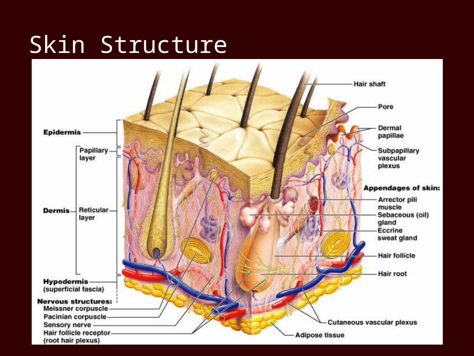
Skin Structure
Epidermis Contains four main cell types
Keratinocytes most abundant cell type in epidermis Arise from deepest layer of epidermis Produce keratin – a tough fibrous protein Produce antibodies and enzymes Keratinocytes are dead at skin's surface
Melanocytes – produce melaninMerkel cells – sensory Langerhans cells – defense cells
Layers of the Epidermis
Stratum basale (stratum geminativum) Stratum spinosum Stratum granulosum Stratum lucidum (only in thick skin, i.e. volar
surfaces) Stratum corneum
mnemonic device:Boys Spit Gross Luggies Constantly
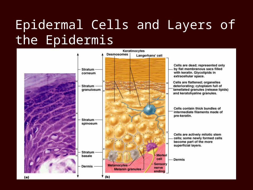
Epidermal Cells and Layers of the Epidermis
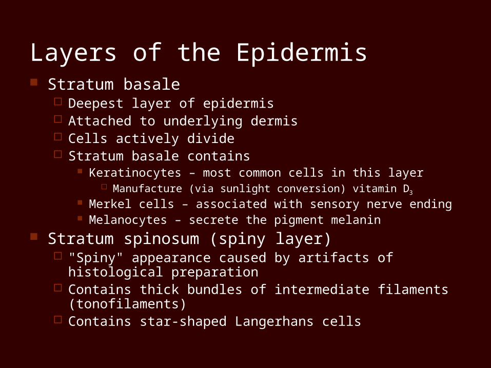
Layers of the Epidermis Stratum basale
Deepest layer of epidermis Attached to underlying dermis Cells actively divide Stratum basale contains
Keratinocytes – most common cells in this layer Manufacture (via sunlight conversion) vitamin D3
Merkel cells – associated with sensory nerve ending Melanocytes – secrete the pigment melanin
Stratum spinosum (spiny layer) "Spiny" appearance caused by artifacts of histological
preparation Contains thick bundles of intermediate filaments (tonofilaments) Contains star-shaped Langerhans cells
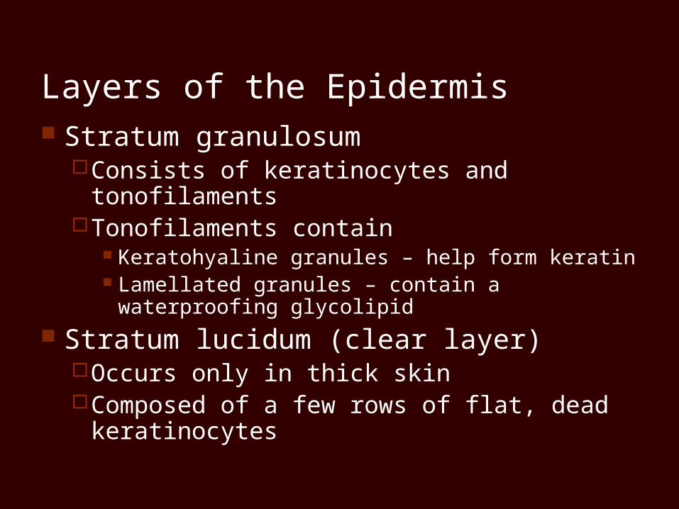
Layers of the Epidermis Stratum granulosum
Consists of keratinocytes and tonofilaments Tonofilaments contain
Keratohyaline granules – help form keratin Lamellated granules – contain a waterproofing
glycolipid
Stratum lucidum (clear layer)Occurs only in thick skinComposed of a few rows of flat, dead
keratinocytes
Layers of the Epidermis
Stratum corneum (horny layer)Thick layer of dead keratinocytes and
thickened plasma membranesProtects skin against abrasion and
penetration
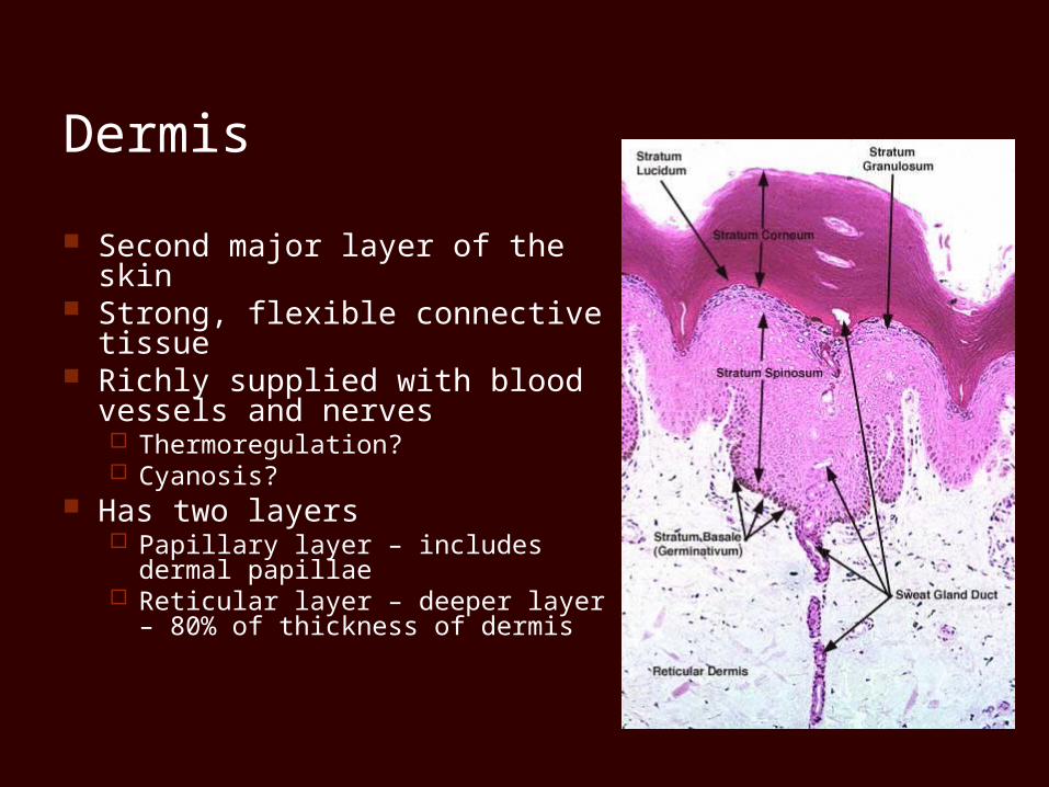
Dermis
Second major layer of the skin Strong, flexible connective
tissue Richly supplied with blood
vessels and nerves Thermoregulation? Cyanosis?
Has two layers Papillary layer – includes dermal
papillae Reticular layer – deeper layer –
80% of thickness of dermis
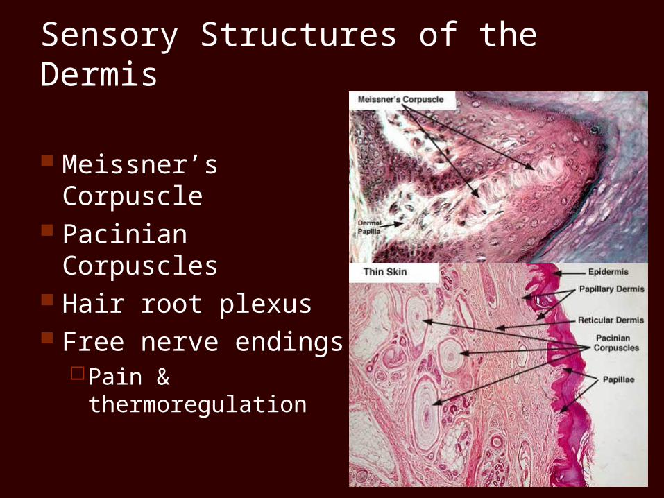
Sensory Structures of the Dermis
Meissner’s Corpuscle Pacinian Corpuscles Hair root plexus Free nerve endings
Pain & thermoregulation
Hypodermis
Deep to the skin – also called superficial fascia
Contains areolar and adipose connective tissues
Anchors skin to underlying structures Helps insulate the body
Skin Color
Three pigments contribute to skin colorMelanin – most important pigment – made
from tyrosineCarotene – yellowish pigment from carrots
and tomatoes Hemoglobin – Caucasian skin contains little
melanin Allows crimson color of blood to show through
Appendages of the Skin
HairFlexible strand of dead, keratinized cellsHard keratin – tough and durableChief parts of a hair
Root – imbedded in the skin Shaft – projects above skin's surface
Appendages of the Skin
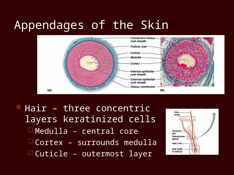
Hair – three concentric layers keratinized cells Medulla – central core Cortex – surrounds medulla Cuticle – outermost layer
Appendages of the Skin
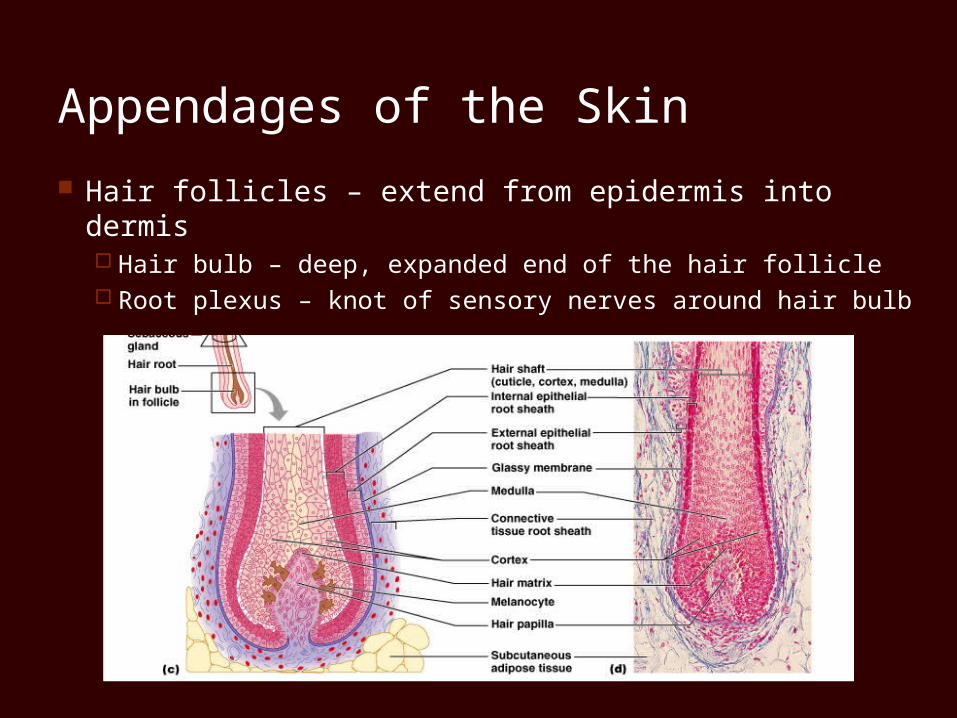
Hair follicles – extend from epidermis into dermis Hair bulb – deep, expanded end of the hair follicle Root plexus – knot of sensory nerves around hair bulb
Appendages of the Skin
Wall of hair follicleConnective tissue root sheath Epithelial root sheath
Arrector pili muscle – bundle of smooth muscleHair stands erect when arrector pili contracts
Types and Growth of Hair
Vellus hairs – body hairs of women and children
Terminal hairs – hair of scalp; axillary and pubic area (at puberty)
Hair thinning and baldnessDue to agingMale pattern baldness
Sebaceous Glands
Occur over entire body, except palms and soles Secrete sebum – an oily substance
Simple alveolar glands Holocrine secretion – entire cell breaks up to form
secretion
Most are associated with a hair follicle Functions of sebum
Collects dirt; softens and lubricates hair and skin
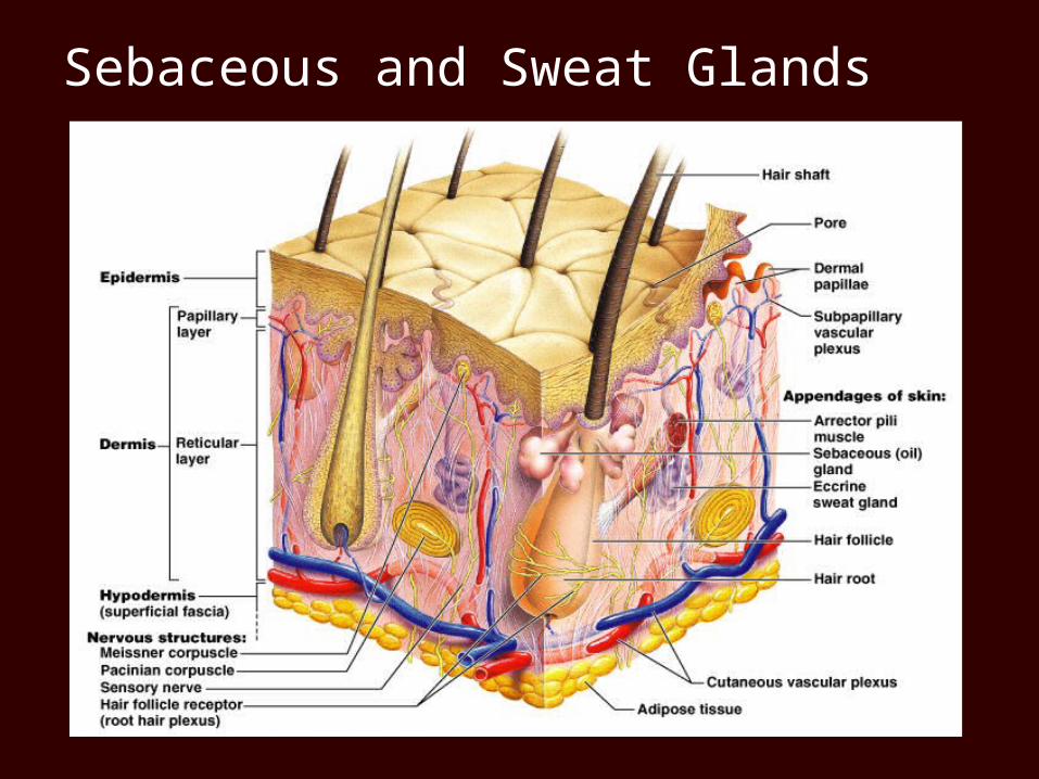
Sebaceous and Sweat Glands
Sweat Glands
Sweat glands (sudoriferous/eccrine glands) are widely distributed on body
Sweat – is a blood filtrate 99% water with some saltsContains traces of metabolic wastes
Sweat Glands
Two types of sweat gland Eccrine gland
Most numerous – produce true sweat (watery) Coiled tubular gland Controlled by the hypothalamus
Apocrine gland Confined to axillary, anal, and genital areas Produce a fatty secretion of sweat during periods of
stress/anxiety Even though they are called apocrine sweat glands they do
not secrete in an aprocrine fashion – rather in an eccrine or merocrine fashion as do the eccrine glands… the name has remained to avoid confusion of the two varieties of sweat glands!
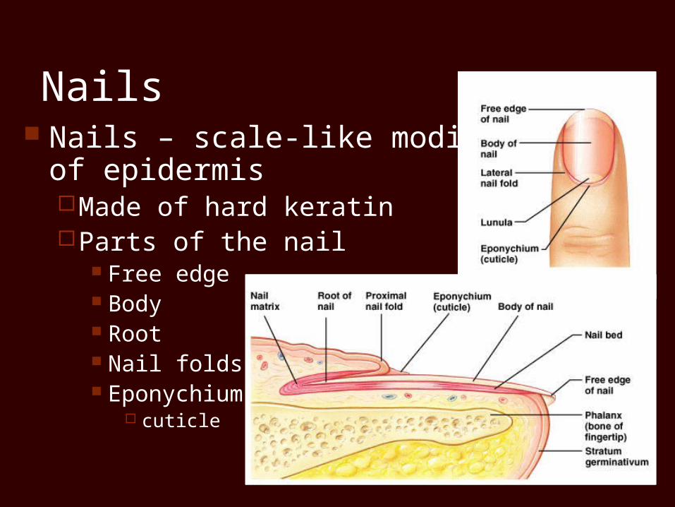
Nails Nails – scale-like modification
of epidermisMade of hard keratinParts of the nail
Free edge Body Root Nail folds Eponychium
cuticle
Burns
Classified by severityFirst degree burn – only epidermis is damagedSecond degree burn – upper part of dermis is
also damaged Blisters appear Skin heals with little scarring
Third degree burn – consume thickness of skin Burned area appears white, red, or blackened
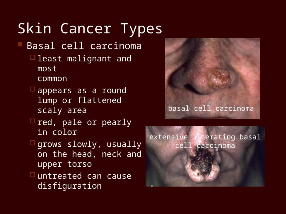
Skin Cancer Types Basal cell carcinoma
least malignant and most common
appears as a round lump or flattened scaly area
red, pale or pearly in color
grows slowly, usually on the head, neck and upper torso
untreated can cause disfiguration
basal cell carcinoma
extensive ulcerating basal cell carcinoma
Skin Cancer Types
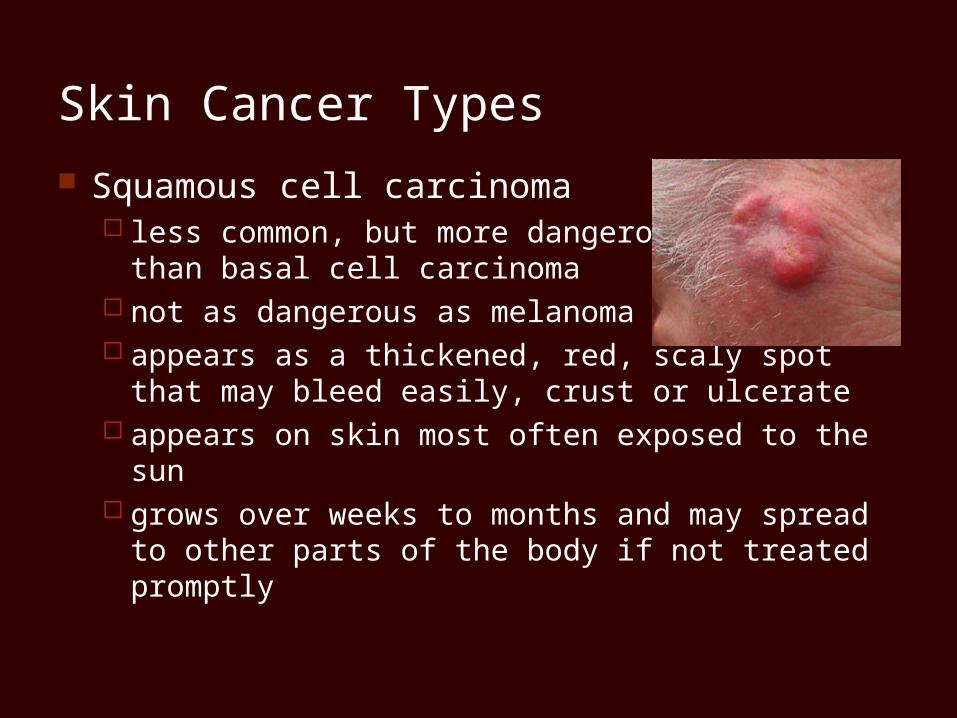
Squamous cell carcinoma less common, but more dangerous
than basal cell carcinoma not as dangerous as melanoma appears as a thickened, red, scaly spot
that may bleed easily, crust or ulcerate appears on skin most often exposed to the sun grows over weeks to months and may spread to other
parts of the body if not treated promptly
Skin Cancer Types
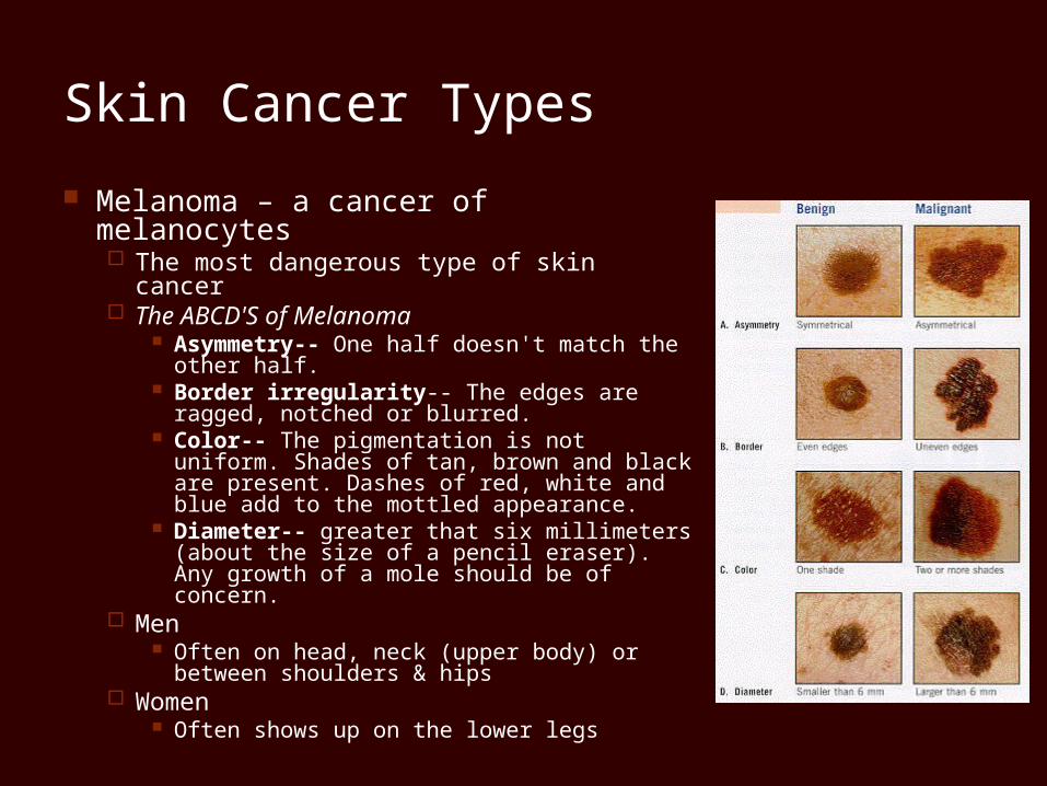
Melanoma – a cancer of melanocytes The most dangerous type of skin cancer The ABCD'S of Melanoma
Asymmetry-- One half doesn't match the other half.
Border irregularity-- The edges are ragged, notched or blurred.
Color-- The pigmentation is not uniform. Shades of tan, brown and black are present. Dashes of red, white and blue add to the mottled appearance.
Diameter-- greater that six millimeters (about the size of a pencil eraser). Any growth of a mole should be of concern.
Men Often on head, neck (upper body) or
between shoulders & hips Women
Often shows up on the lower legs
The Skin Throughout Life
At 5-6 months, the fetus is covered with lanugo (downy hairs)
In middle to old ageSkin thins and becomes less elasticShows harmful effects of environmental
damageSkin inflammations become more common